Hughes, Mansel & Webster''''s Benign Disorders and Diseases of the Breast pdf
Bạn đang xem bản rút gọn của tài liệu. Xem và tải ngay bản đầy đủ của tài liệu tại đây (22.36 MB, 349 trang )
an imprint of Elsevier Limited
© 2009, Elsevier Limited. All rights reserved.
First edition 1989 by Baillière Tindall
Second edition 2000 by Harcourt Publishers Limited
The right of Robert Mansel, David Webster and Helen Sweetland to be identified as authors of
this work has been asserted by them in accordance with the Copyright, Designs and Patents Act
1988.
No part of this publication may be reproduced or transmitted in any form or by any means,
electronic or mechanical, including photocopying, recording, or any information storage and
retrieval system, without permission in writing from the publisher. Permissions may be sought
directly from Elsevier’s Rights Department: phone: (+1) 215 239 3804 (US) or (+44) 1865
843830 (UK); fax: (+44) 1865 853333; e-mail: You may also
complete your request on-line via the Elsevier website at />ISBN 978-0-7020-2774-1
British Library Cataloguing in Publication Data
A catalogue record for this book is available from the British Library
Library of Congress Cataloging in Publication Data
A catalog record for this book is available from the Library of Congress
Notice
Medical knowledge is constantly changing. Standard safety precautions must be followed, but as
new research and clinical experience broaden our knowledge, changes in treatment and drug
therapy may become necessary or appropriate. Readers are advised to check the most current
product information provided by the manufacturer of each drug to be administered to verify the
recommended dose, the method and duration of administration, and contraindications. It is the
responsibility of the practitioner, relying on experience and knowledge of the patient, to determine
dosages and the best treatment for each individual patient. Neither the Publisher nor the authors
assume any liability for any injury and/or damage to persons or property arising from this
publication.
The Publisher
Working together to grow
libraries in developing countries
www.elsevier.com | www.bookaid.org | www.sabre.org
The
publisher’s
policy is to use
paper manufactured
from sustainable forests
Printed in China
Last digit is the print number: 9 8 7 6 5 4 3 2 1
One highlight of this edition is a remarkable chapter
‘History of benign breast disease’, which overviews the
lives and careers of six great figures (Sir Astley Cooper,
Alfred Velpeau, John Birkett, George Cheatle, Joseph
Bloodgood and Charles Geschickter), with particular
insight into the roles of mentorship, record keeping,
acceptance of new technologies, pathologic correlation
and the role of international travel and contacts. In an
age information technology and instantaneous commu-
nication, these elements are more important than ever.
The role of surgery in benign breast disease is chang-
ing. Mammography, ultrasound and (increasingly) MRI
offer the prospect of earlier cancer diagnosis but bring
with them a substantial burden of benign or equivocal
findings. Most are amenable to core biopsy but it remains
challenging to identify those that do or do not need
surgery. Surgical techniques for benign breast conditions
may seem simple in concept, but the experienced surgeon
will recognize that this simplicity is more apparent than
real and that pitfalls abound. In closing, let me enthusi-
astically recommend the chapter ‘Operations’. Here the
authors address core biopsy (with and without image
guidance) and the full range of surgical procedures for
benign breast diseases, presenting a set of ‘Important
principles’ for each. In these lists surgeons in training will
recognize a treasury of clinical pearls drawn from the
authors’ vast hands-on experience, and practising sur-
geons will recognize their own past surgical misadven-
tures which might have been avoided had these principles
been followed. This chapter is a small classic in its own
right and should be required reading for all surgeons who
treat breast disease, benign or malignant.
Benign breast disease comprises a wide range of condi-
tions which worry patients, which vex physicians, which
are vastly more common than breast cancer, and yet
which have to date received relatively little attention in
the medical literature. It is therefore a particular pleasure
for me to introduce the third edition of Hughes, Mansel
& Webster’s Benign Disorders and Diseases of the Breast, a
unique and classic work which fully succeeds in address-
ing this imbalance and builds on the substantial and
well-deserved success of the first (1989) and second
(2000) editions.
The authors correctly decry the term ‘fibrocystic disease’,
proposing instead that benign breast conditions are not
‘disease’ per se, but are instead minor aberrations of normal
development and involution (‘ANDI’). The ANDI frame-
work, for the first time, puts the study of benign breast
disease on a scientific basis which correlates pathogene-
sis, histology and clinical features. This model is, in my
opinion, a robust foundation for further progress in the
understanding and treatment of benign breast disease,
and deserves much wider recognition, particularly in the
US, where it is relatively unknown.
Professor Mansel and his colleagues comprehensively
address every aspect of benign breast disease following
a format in which all elements (graphics, tables and
photographs) work harmoniously to create a whole larger
than the sum of its parts. Each chapter heading includes
‘key points and new developments’ for a quick summary
of the contents. As in a Victorian novel, these chapter
headings are irresistible and one cannot resist delving
into the contents. Throughout, one benefits in equal
measure from the authors’ scholarship, from their long
first-hand experience and from their refreshing practical-
ity in managing benign breast disease.
xi
Foreword
Hiram S. Cody III
Q1
xii
a few cases with atypical epithelial hyperplasia, benign
change is not of itself an important determinant. Clinics,
however, are dominated by the concern to exclude cancer
and to determine future risk.
The imaging chapter has been extensively revised by
Kate Gower-Thomas and the xeromammograms have
been replaced with modern digital mammograms.
Plastic surgery for both augmentation and reduction is
now so well detailed in the plastic surgery literature that
we have omitted this chapter; similarly, the chapter on
geographical variation has been subsumed into the chap-
ters about individual problems.
Professor Leslie Hughes has provided a fascinating
chapter on the lives and influences of some of the great
names in the development of our understanding of the
changes in the breast.
The ANDI concept provides a framework to enable
clinicians to explain to patients the nature of their problem
in an easily assimilated way. It is important to emphasise
that ANDI is not a diagnosis in itself.
REM, DJTW, HS
January 2009
Preface
It is now 20 years since the first edition of this book and
9 since the second edition. The intervening years have
seen advances in imaging technology, understanding of
the molecular events leading to disease and drug develop-
ments. While most of the focus has been on breast cancer,
there have been benefits to an understanding of the
changes occurring in the breast from physiology through
disorders to diseases.
One of the consequences of an improved under-
standing of what is happening in the breast and confi-
dence in the ability to diagnose the problem actively
has been the disappearance of open surgical diagnostic
biopsy and, except for a few areas, surgery for benign
conditions. The diagnostic pathway using triple assess-
ment with core needle biopsy is now the standard in
most breast clinics; it gives a 99% sensitivity for cancer
and dramatically reduces operations for true benign
disease.
Much work has been done in identifying patients with
an increased risk of developing breast cancer and we have
addressed this by including a new chapter on risk of
breast cancer written by Professor Gareth Evans of Man-
chester. Family history is important here but apart from
and Dr Kathleen Lyons, and of Pathology – especially Drs
Winsor Fortt and Tony Douglas-Jones. This book could
not have been produced without the exceptional service
given by the Department of Medical Illustration under
Professor R. Marshall and now Professor R. Morton.
The secretarial staff of the University Department of
Surgery, both clinical and academic, have facilitated all
aspects of the clinical and research work and documenta-
tion behind the book, and Mrs Edna Lewis has given
many years of voluntary service to the Mastalgia Clinic.
Above all we are grateful to our families who have
foregone so much over many years in the cause of research
and the writing of this book.
xiii
Acknowledgements
We owe a debt of gratitude to many people who have
contributed to work on which this book is based. Fore-
most are those research fellows who have been responsi-
ble for the day-to-day conduct of many studies and
clinical trials in this department over the last 30 years:
Paul Preece, John Wisbey, Nigel Pashby, Jonathan Pye,
Sandeep Kumar, Anurag Srivastava, Barney Harrison,
Paul Maddox, Graham Pritchard, Stephen Courtney, Glyn
Neades, Richard Cochrane, Eleri Lloyd-Davies, Chris
Gateley, Anup Sharma, Eifion Williams, Sumit Goyal,
Amit Goyal, Kelvin Gomez, Alok Chaabra and Bedanta
Baruah.
We are much indebted to co-operation from the
Departments of Radiology – especially Dr Huw Gravelle
THIS BOOK IS DEDICATED TO
CD Haagensen
Surgeon Pathologist
JD Azzopardi
Surgical Pathologist
Whose meticulous studies have cast so much light on breast disorders, and
whose monographs are quoted so freely in this book
IH Gravelle
Radiologist
Friend, colleague, an imaging pioneer, who enthusiastically joined us in this project to
integrate structure and function in benign disorders of the breast.
Problems of concept and nomenclature of
benign disorders of the breast
Key points and new developments
1. Only by taking a historical view of benign disorders of the breast can the confusion persisting until recent decades be
understood.
2.
In the past, benign conditions (and the patients carrying them) have been regarded as requiring exclusion of cancer or cancer
risk, rather than entities requiring management in their own right.
3.
Clinical conditions, such as painful nodularity, have been equated with and confused with histological conditions, such as
fibrosis or hyperplasia.
4.
Most accept that the concepts and terminology of ‘fibrocystic disease’ and ‘fibroadenosis’ cannot be justified, but this
recognition has so far been matched by masterly inactivity.
5.
Accurate and meaningful terminology will be achieved only if those in the field agree on one and accept it and use it. The
aberrations of normal development and involution (ANDI) concept and terminology provides a means of achieving this.
giving a definition, this author, like many before him,
states that the term fibrocystic disease has no real meaning
and should probably be abandoned. Nevertheless, he
also lists the histological features, fibroadenomas, macro-
cysts, fibrosis, duct dilatation and stasis, periductal round
cell infiltrate, fat necrosis, papillomatosis, apocrine meta-
plasia, sclerosing adenosis and hyperplastic lesions of
duct and lobule. This covers the whole range of benign
conditions of the breast, and it is clearly inappropriate to
equate this histological panorama with a mild, or even
severe, degree of painful nodularity.
With such a loose equivalence between clinical and
histological detail, it is not surprising that Foote and
Stewart wrote in 1945: ‘chronic cystic mastitis is so
ingrained in the minds of some pathologists that this
diagnosis of a locally excised portion of the breast almost
amounts to a surgico-pathological reflex’.
2
What is sur-
prising is that pathologists are still the most insistent
The source of the problem
The condition commonly called fibrocystic disease, or
fibroadenosis of the breast, has been a clinical problem
for centuries, as reflected in writings as early as those of
Astley Cooper at the beginning of the nineteenth century.
For patients, it causes discomfort and anxiety which varies
from nuisance value to serious interference with their
quality of life. For clinicians, the condition causes a range
of problems of diagnosis, assessment and management
which are not always clearly recognized.
Although all clinicians have a concept of what fibro-
cystic disease represents, it is difficult to define, and none
of its protagonists has given a meaningful differentiation
between it and normality. One definition
1
is ‘palpable
lumps in the breast, usually associated with pain and
tenderness that fluctuate with the menstrual cycle and
become progressively worse until the menopause’. Despite
C H A P T E R
1
1
Benign disorders and diseases of the breast
2
single group to maintain the use of the term, despite this
stinging remark from eminent members of their own
discipline.
Greater interest in benign breast disorders in recent
years has led to a more precise understanding of the clini-
cal pictures associated with individual elements, and the
histological changes of cyclical nodularity are increas-
ingly recognized as lying within the range of histological
appearance in the normal breast. Many authors have tried
to determine and assess premalignant potential of fibro-
cystic disease but most attempts have resulted in confu-
sion and frustration. Recent workers, especially Page and
co-workers,
3,4
have shown that only a few specific histo-
logical patterns have an association with cancer and these
show no consistent correlation with the clinical picture
which in the past has been ascribed to fibrocystic disease.
This poor correlation between histology and clinical
symptoms led Love and her co-authors
5
to conclude that
fibrocystic disease of the breast is a ‘non-disease’. Their
arguments are cogent in a histological context by denying
the loosely defined cancer risk, but a concept of non-
disease does little to help the many women who suffer
from a variety of physical symptoms – sometimes of dis-
tressing severity. Disorder is a better term than disease
because so many of the symptomatic conditions lie within
the spectrum of normality. The magnitude of the problem
is escalating with the wider concern of women about
breast disease and the wider introduction of breast screen-
ing programmes.
Benign conditions of the breast have always been
neglected in comparison to cancer, despite the fact that
only one out of ten patients presenting to a breast clinic
suffers from cancer. This is not surprising in view of the
emotional implications of breast cancer and its treatment,
but it has meant that the study of the benign breast has
been undeservedly neglected. Until the 1970s, reported
studies were directed largely towards a possible relation-
ship to cancer, rather than towards the basic processes
underlying benign conditions.
There has been a noticeable and welcome correction
to this neglect in recent years, but already the interest in
benign disorders evident for two decades is again on the
wane, at a time when advances in molecular biology give
promise of understanding the basic physiology of human
breast development, function and involution.
This neglect is most evident in standard textbooks (the
most recent comprehensive texts on breast disease devote
less than 5% of their material to benign conditions)
because interest in benign processes can be found when
studying historical reference material. Great names in
surgery such as Hunter, Astley Cooper, Billroth, Cheatle,
Semb, Bloodgood and Atkins appear in the literature. But
whereas breast cancer has stimulated a continuous,
ongoing body of research – each new project building on
the work preceding it – benign disease has been the
subject of a relatively small number of isolated and
unconnected projects, earlier related work having often
been ignored. The sporadic nature of these investigations
and the insularity of the resulting publications had led to
much confusion which has had more serious conse-
quences than neglect alone.
Consideration of benign breast disorders from a his-
torical point of view provides a clearer understanding of
how the present problems have arisen.
History
Sir Astley Cooper was an important early worker in this
field. He described many aspects of benign breast disor-
ders as well as malignant disease in his monograph, Illus-
trations of Diseases of the Breast,
6
published in 1829. Among
the conditions discussed are cystic disease, pain and
fibroadenoma. He distinguished two main groups of
patients with mastalgia – those with and those without a
palpable tumour, which we might now better define as
painful nodularity and non-cyclical breast pain. He also
laid much of the basis of the macroscopic anatomy of the
breast in his book on the anatomy of diseases of the
breast published in 1845. The French surgeon Reclus gave
an excellent description of the clinical and pathological
aspects of cystic disease in 1893, recognizing both the
multiplicity and bilaterality of the cysts.
7
Many of the current problems in terminology and
understanding derive from the publications of German
surgeons in the late nineteenth century. Koenig
8
called
the disease ‘chronic cystic mastitis’, because he believed
it had an inflammatory basis. At the same time, Schim-
melbusch
9
described the same condition, compounding
the problem by calling it ‘cystadenoma’. Both authors
gave the disease inexact names, and both gave incomplete
descriptions of the pathology. Certainly they did not
recognize the wide range of histological appearances
found in these breasts, and they failed to recognize
these as merely variants of normal processes within the
breast.
Problems of concept and nomenclature of benign disorders of the breast
1
3
There was an early reaction to this confusion. Cabot
10
questioned the inflammatory connotation of the term
chronic cystic mastitis and urged more precise terminol-
ogy, but unfortunately his pleas fell on stony ground. In
the 1920s there were major studies by Semb
11
in Norway
and Cheatle and Cutler
12
in the UK and their disease
descriptions and data are still worth serious study.
However, Cheatle and Cutler gave the name ‘cystipho-
rous desquamative epithelial hyperplasia’ to the clinical
spectrum we have termed aberrations of normal develop-
ment and involution in Chapter 3 and this can hardly be
regarded as helpful. The tendency of the Scandinavians
to use Semb’s term ‘fibroadenomatosis’ also caused diffi-
culty because of its confusion with the term fibroade-
noma.
11
In spite of detailed investigations, Cheatle and
Cutler confused changes of cyclical nodularity with both
duct ectasia and fibroadenomas
12
and the term they
finally chose – ‘mazoplasia’ – is hardly evocative in a
descriptive sense.
While most workers concentrated on the clinical prob-
lems of fibrocystic disease, some gave accurate descrip-
tions of other benign breast conditions. The paper on ‘the
varicocele tumour’ by Bloodgood is a striking account of
the clinical and macropathological aspects of duct ectasia
and its clinical variants.
13
The accuracy and detail of the
observations come as a surprise to those who believe
advances in medical understanding are recent.
Special clinics for breast disease set up by Atkins in
London and Geschickter in the USA concentrated experi-
ence and allowed adequate documentation and assess-
ment of the results of treatment for the first time during
the 1940s. Both authors made many contributions to
benign breast disorders,
14,15
but suffered equally from the
limited knowledge at that time of basic pathology and
endocrinology of the breast. They both unfortunately
continued the use of the term chronic mastitis. The 50
years since their contributions has seen an increasing
momentum in investigation of benign breast conditions.
Great benefit has derived from histological study of the
normal breast and the development of hormonal estima-
tions using radioimmunoassay. In particular, the autopsy
study of Sandison
16
showed that most of the changes
previously regarded as disease are so common as to be
within the spectrum of normality, and his work stimu-
lated others to define the wide range of histological
appearances of the normal breast. For example, Parks
17
studied both surgical and autopsy specimens and showed
a gradation between normal lobules and fibroadenomas,
and between involuting lobules and cyst formation. He
also showed that papillary epithelial hyperplasia of the
terminal ducts is so common in the premenopausal
period as to be regarded as normal, and that these lesions
regress without treatment after the menopause. In 1961,
Oberman and French
18
also stressed the concept of a
continuum between normality and benign conditions:
‘adenofibromas, fibrocystic disease and intraductal papil-
lomas do not appear to represent distinct entities, but
rather form a spectrum of conditions having their basis
in an abnormality between hormonal stimulus to the
breast, principally estrogen, and stromal and epithelial
response’.
These writers have had a profound insight into the
concepts discussed in this book, and it is salutary to go
back even further. In 1922, McFarland
19
wrote: ‘The so-
called chronic mastitis is not inflammatory, and is not a
pathological entity; it is nothing but a result – or at most
a perversion – of involution. The only difficulty lies in
clearly defining when the process of involution can be
said to become abnormal, when it is so diversified.’ The
seed scattered by these workers has largely fallen on stony
ground.
The present and the future
In the past, each worker has tended to introduce their
own terminology for a condition, either to stress a par-
ticular aspect they have noted, or through ignorance of
work that has gone on perhaps many years before. As an
illustration of this, Table 1.1 shows the large number of
names that have been associated with just three condi-
tions: so-called fibrocystic disease, duct ectasia and giant
fibroadenomas.
This list is by no means comprehensive; some 40
names have been used to describe the variety of condi-
tions covered by the old term, chronic fibrocystic disease,
none of which can be considered satisfactory.
Because of their multiplicity and lack of specificity, past
terms are better replaced by the use of clinical or histo-
logical terms which are specific and accurate in relation
to the clinical and/or histological condition to which they
refer. Examples of appropriate clinical terms are mastalgia
and cyclical nodularity. Examples of appropriate histo-
logical terms that have evolved over recent years are
sclerosing adenosis and atypical ductal hyperplasia.
Terms that accurately reflect both clinical and histological
Benign disorders and diseases of the breast
4
counterparts are fibroadenoma, duct papilloma and mac-
rocyst, for example.
When it is desirable to cover the whole range of
(unspecified) benign breast disorders, it is appropriate to
use a term which, unlike fibrocystic disease, does not
imply a disease state, but acknowledges the spectrum of
change extending from normality and recognizes that
most of the spectrum does not represent disease. We
suggest that ‘aberrations of normal development and
involution’ (ANDI) is a term which meets these criteria;
it is comprehensive, and meaningful and descriptive in
terms of pathogenesis.
Why has it taken so long to reach a reasonable under-
standing of the processes involved in benign breast condi-
tions? The main stumbling block has been the failure to
appreciate the range of basic physiological and structural
changes within the normal breast – an organ dynamic
throughout the reproductive period of life as it first devel-
ops, then undergoes repeated cyclical change and finally
involutes. Because it is an organ under systemic hormo-
nal influence, one would expect the breast to be uniform
throughout in its appearance and behaviour, but this is
not so. Like other endocrine target organs such as the
thyroid, it varies greatly from one part to another, and
end-organ response must be a factor in this variability. It
has been usual practice to concentrate on the local find-
ings as shown by biopsy, at one point in time when the
patient presents with a clinical problem, assuming that
the particular clinical condition at that time is directly
associated with the local radiological and biopsy findings.
It is tempting to ignore the findings of Parks and Sandi-
son and others that all these apparently specific findings
are frequently found in asymptomatic breasts. So a par-
ticular clinical event that leads a patient to biopsy must
be assessed against the background of this almost random
variation in histological appearance which is a part of
normality.
A further source of confusion has arisen from the asso-
ciation of radiological appearances with pathological
descriptions, without adequate correlative studies to
establish a relationship. An example from recent decades
has been the description of radiological density as ‘dys-
plasia’ in relation to Wolfe patterns – when detailed study
can show that density is unrelated to epithelial dyspla-
sia.
20
The situation was then compounded by using the
term ‘dysplastic breast’ for a radiological pattern, without
histological correlation or confirmation. The welfare of
the patient with benign breast problems will be best
served by abandoning terminology that implies disease,
and substituting terminology which reflects the normality
of many of the underlying processes, reserving ‘disease’
for those conditions where clinical morbidity or histo-
logical significance warrants such a term. The terminol-
ogy should come from consideration of the basic
physiological and pathological processes that lead a
patient to present to a breast clinic.
Perhaps the reason for persisting and increasing confu-
sion is an unwillingness to be sufficiently radical in
moving away from ideas that do not fit in with present
knowledge. Not only must the concept of fibrocystic
disease as a clinical concept or a histopathological entity
be done away with, it must be replaced by an accurate
terminology consistent with present knowledge. Many
breast physicians accept the first half of this statement,
but are unwilling to accept the corollary inherent in the
second half.
These basic aspects of the non-malignant breast, and
the arguments for the aberrations of normal develop-
ment and involution terminology, are considered in
Chapter 4.
Table 1.1 Some of the names used for common benign breast
disorders
CYCLICAL NODULARITY
Fibrocystic disease
Fibroadenosis
Cystic
hyperplasia
Hyperplastic cystic disease
Schimmelbusch’s disease
Chronic cystic mastitis
Cystic mastopathy
DUCT ECTASIA/PERIDUCTAL MASTITIS
Plasma cell mastitis
Varicocele tumour
Comedo mastitis
Mastitis obliterans
Secretory disease
GIANT FIBROADENOMATOUS TUMOURS
Giant fibroadenoma
Cystosarcoma phyllodes
Phyllodes tumour
Juvenile fibroadenoma
Serocystic disease of Brodie
Problems of concept and nomenclature of benign disorders of the breast
1
5
REFERENCES
1. Scanlon EF. The early diagnosis of breast cancer. Cancer
1981; 48: 523–526.
2.
Foote FW & Stewart FW. Comparative study of cancerous
versus noncancerous breast. II. The role of so-called
chronic cystic mastitis in mammary carcinogenesis.
Annals of Surgery 1945; 121: 197–222.
3.
Page DL, Vander-Zwag R, Rogers LW et al. Relationship
between component parts of fibrocystic disease complex
and breast cancer. Journal of the National Cancer Institute
1978; 61: 1055–1063.
4.
Page DL & Dupont WD. Anatomic indications (histologic
and cytologic) of increased breast cancer risk. Breast
Cancer Research and Treatment 1993; 28: 157–162.
5.
Love SM, Gelman RS & Silen W. Fibrocystic ‘disease’ of
the breast. A non disease. New England Journal of Medicine
1982; 307: 1010–1014.
6.
Cooper A. Illustrations of Diseases of the Breast. London:
Longmans; 1829.
7.
Reclus P. Maladie Kystique De La Mammelle. La Semaine
Medicale 1893; 13: 353–354.
8.
Koenig P. Mastitis chronica cystica. Centralblatt für
Chirurgie 1893; 20: 49–53.
9.
Schimmelbusch C. Das Fibroadenom der Mamma. Archiv
für Klinische Chirurgie 1892; 64: 102–116.
10.
Cabot RC. Irritable breasts, or chronic lobular mastitis.
Boston Medical and Surgical Journal 1900; CXLIII: 555–557.
11.
Semb C. Pathologico-anatomical and clinical
investigations of fibroadenomatosis cystica mammae.
Acta Chirurgica Scandinavica Supplementum 1928; 64(10):
1–484.
12.
Cheatle GL & Cutler M. Tumours of the Breast. London:
Edward Arnold, 1931.
13.
Bloodgood JC. The clinical picture of dilated ducts
beneath the nipple frequently to be palpated as a
doughy, worm-like mass – the varicocele tumour of the
breast. Surgery, Gynecology and Obstetrics 1923; 26: 486–
495.
14.
Atkins HJB. Chronic mastitis. Lancet 1938; i: 707–712.
15.
Geschickter CF. Diseases of the Breast, 2nd edn.
Philadelphia: JB Lippincott & Co.; 1945.
16.
Sandison AT. An autopsy study of the human breast.
National Cancer Institute Monograph No. 8, US Dept
Health, Education and Welfare, 1962.
17.
Parks AG. The microanatomy of the breast. Annals of
the Royal College of Surgeons of England 1959; 25:
295–311.
18.
Oberman HA & French AJ. Chronic fibrocystic disease of
the breast. Surgery, Gynecology and Obstetrics 1961; 112:
647–652.
19.
McFarland J. Residual lactation acini in the female
breasts. Their relationship to chronic cystic mastitis
and malignant breasts. Archives of Surgery 1922; 5:
1–64.
20.
Mansel RE, Gravelle IH & Hughes LE. The interpretation
of mammographic ductal enlargement in cancerous
breasts. British Journal of Surgery 1979; 66: 701–702.
History of benign breast disease
Leslie E. Hughes
frequency and significance. The inadequate attention gen-
erally given to benign conditions is shown by Lisfranc,
who as late as the 1840s was still arguing at the Academie
de Medicin in Paris that all breast lumps became
malignant.
Evidence that Lisfranc’s view was wrong, and details of
differentiation of benign from malignant, was first clearly
presented by Cooper. Furthermore, he stressed the impor-
tance of the non-malignant by devoting Part 1 of his
Introduction
The century and a half from 1800 to 1950 saw a remark-
able expansion in the understanding and management of
benign breast conditions. Many contributed to this expan-
sion, but six workers have been chosen for this chapter,
based on the degree of innovation and the breadth and
influence of their work. Of course many others made
major contributions, though of less depth and impact.
Brodie and Paget of the UK, Semb of Norway, Reclus
of France and Schimmelbusch and Billroth of Austro-
Germany are examples.
Two other outstanding contributors of the second half
of the twentieth century certainly match our chosen six,
Cushman D. Haagensen, surgeon pathologist of the USA,
and John Azzopardi, surgical pathologist of the UK. As
their work overlaps the professional span of many of the
present generation of breast specialists, they have been
left to future study.
This chapter is not the history of benign conditions of
the breast; this is dealt with elsewhere. It is a biographical
examination of six great men, with some attempt to
discern the social and professional background leading
to such major contributions.
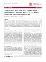
Sir Astley Paston Cooper,
Bt. F
RS DCL GCH. 1768–1841
Cancer of the breast has been recognized and its treat-
ment discussed for many centuries. On the other hand,
except perhaps for lactational abscess, benign conditions
received little attention, and received no detailed consid-
eration in textbooks until Astley Cooper realized their
C H A P T E R
2
7
Fig. 2.1 Sir Astley Paston Cooper.
Benign disorders and diseases of the breast
8
intended two-part book on breast disease to benign con-
ditions. Thus, he presented the first monograph devoted
to benign breast disorders in 1829, and this was probably
the only such one until the 1980s.
1
Early life
Astley Cooper enjoyed a good genetic inheritance; his
father, a Norfolk vicar, and his mother, a descendant of
Isaac Newton, both had considerable literary output,
while one uncle and his grandfather were surgeons. Born
in 1768, he was one of a family of 10 children, but all
five sisters eventually died of tuberculosis.
Educated at home, he was a poor student, showing
little interest in study and preferring to roam the coun-
tryside and get involved in wild escapades with local
youths. In this regard he was remarkably similar to his
teacher and guru, John Hunter, and in later life he also
resembled Hunter in his passion for research and hard
work. Whether these latter attributes were inherent or
the result of a direct influence of Hunter, it is difficult
to say.
Two incidents helped arouse his interest in surgery.
First, his stepbrother was run over by a wagon and died
of haemorrhage because no local doctor was willing to
come to the accident scene. Second, he observed an oper-
ation for stone, performed in a masterly manner in the
Norfolk and Norwich Hospital, which ‘inspired me with
a strong impression of the utility of surgery’.
This led to his apprenticeship at the age of 16 to his
uncle, William Cooper, a senior surgeon at Guy’s Hospi-
tal in London for the usual period of seven years. But
Astley Cooper soon transferred his apprenticeship to
Henry Cline, a young (34-years-old) surgeon at the closely
linked St Thomas’s Hospital, with a reputation as an
excellent operator and one of the few London surgeons
who appreciated John Hunter’s teachings. In contrast,
William Cooper said he could never understand Hunter’s
lectures, and usually went to sleep during them. Astley
Cooper became a frequent and attentive attender.
2
He soon exhibited Hunter’s passion for acquiring per-
sonal knowledge rather than following textbooks, and for
experiment and hard work, taking anatomical and pathol-
ogy specimens to Cline’s house for dissection, and was
(like Velpeau later) quite heavily involved in the body
snatching trade. He used his considerable wealth to
placate the municipal worthies unhappy at this practice,
as well as supporting the families of some of those impris-
oned for the activity.
He soon stood out above his colleagues, and showed
an early interest in breast disease from student days. A
long convalescence from an attack of typhus gave an
opportunity to spend a session in Edinburgh, where his
brilliance was recognized and coupled with great popu-
larity. So much did he impress in these ways that an offer
was made to make him President of the Royal Medical
Society should he return to Edinburgh. At this stage of his
life he showed strong support for the revolutionary politi-
cal developments across the channel in France, tenden-
cies which had an adverse effect when he applied for the
vacant consultant post at conservative Guy’s when his
uncle retired. He was appointed after he renounced all
political activity.
With his apprenticeship completed, he married the
daughter of a wealthy merchant, so that he never had to
work to earn a living. But nevertheless, work he did with
a vengeance. With a typical day he would rise at 6 a.m.,
dissect in his private laboratory for research and to have
prepared specimens for his lecture, see non-paying
patients before breakfast, then to his consulting rooms
(in 1815 his professional income was an incredible
£21
000). He would then proceed to Guy’s for a ward
round with students, seeing every interesting patient and
making notes on them, to St Thomas’s to lecture, teaching
in the dissecting room, followed by private operations,
home for dinner followed by 3 hours work in the evening.
As his daily activity involved producing dissections for his
anatomical lectures and selecting patients for his clinical
lectures from those of all surgeons, he had access to a
huge body of clinical material, and was able to observe
the results of different methods of treatment by different
surgeons. This, together with the detailed observation
and documentation of his own patients, provided the
basis for his teaching and publications.
He was an outstanding operating surgeon, a quality
not enjoyed by his two senior surgeon colleagues, who
would not operate unless he was available to help.
Cooper’s surgical contributions, from advocating
catgut 50 years before Lister, to pioneering vascular
surgery, are so well known that they need no further
recounting. Likewise, his success as a teacher was legen-
dary, with his lectures and ward rounds always crowded
with students.
Professional career
Cooper moved rapidly up the professional ladder, and
particularly within the Royal College of Surgeons hierar-
History of benign breast disease
2
9
chy, first as anatomy lecturer, then Hunterian Professor
of Comparative Anatomy and later President for two
terms. Perhaps it was in the organization of the very out-
moded College that he was a breath of fresh air and made
an outstanding contribution. The younger Fellows of the
College were particularly frustrated by outdated attitudes;
while senior Council members could enter through the
front door; ordinary members had to come through a
small back door. Examinations were antiquated and pro-
vincial hospitals were not recognized for training. Once
elected to Council, the position was held for life.
When some younger fellows were elected to Council,
they found Cooper a strong supporter. He was made
chairman of a committee which was set up ‘to consider
the present state of the College’, essentially to look at
modernization of the College and he was notably success-
ful in introducing many improvements. Placating the
elderly College ‘establishment’ was undoubtedly associ-
ated with his popular persona, his high professional
standing and his respected judgement. The younger
fellows were delighted. The committee was responsible
for much modernization: reforming and liberalizing the
examination system, ensuring that all members were kept
in touch with Council decisions and extending training
to provincial hospitals.
Astley Cooper and breast disease
All Cooper’s work – lectures, lecture notes and mono-
graphs – were based on personal investigation of anatomy,
physiology and pathology, followed by personal observa-
tion of clinical patients and the results of his treatment.
In 1825, he retired from his position as surgeon to Guy’s
and this gave the time and opportunity to produce his
book on breast disease – Part 1 on benign conditions – in
1829 (as well as holding the presidency of the College in
1827). It is a remarkable work for its time, recognizing
and giving clear description of much benign pathology
and differentiating it from cancer. Likewise, it gives
detailed management recommendations, some reflecting
the practice of the time, others having a remarkably
modern flavour, such as using a lancet to confirm the
diagnosis of a simple cyst, a forerunner of the quite recent
acceptance of needle aspiration as satisfactory treatment.
His description of fibroadenoma and its differentiation
from cancer could not be bettered: younger woman,
mobile, lobulated, slow growth leading to a stationary
phase and finally regression. This appreciation of the
limited growth pattern with the possibility of regression
has only been brought back into prominence in the last
20 years of the twentieth century. His illustrations are
remarkably accurate – that of cystic disease shows multi-
ple blue domed cysts of varying sizes, preceding Blood-
good by almost 100 years, while his plate of a fibroadenoma
shows faithfully the typical lobulation.
Unfortunately, his attention was diverted from Part 2
of his book on breast disease (dealing with carcinoma)
to diseases of the testicle and thymus. When he came
to take up the subject of breast disease again he
realized the fundamental importance of anatomy and
physiology, and produced his book Anatomy of the Breast
in 1840 at the age of 72, dedicated charmingly as
follows:
To members of the medical Profession.
I dedicate this work to you for two reasons. First. To
express the delight I feel at observing your increased love
for the Science of the Profession, and your earnest desire
to found your Practice on an intimate knowledge of
Anatomy, Physiology and Pathology. Secondly to thank
you for your unmeasured kindness and attention to myself
during a period of 50 years.
3
The book contains an amazingly detailed and accurate
account of every aspect of anatomy and physiology of the
breast at all stages of life, including pregnancy and lacta-
tion, and in different races, together with chemical analy-
sis of milk, and injection studies of the mammary glands
of a wide variety of animals. Once again, the detail and
accuracy of the text and illustrations is amazing. Regret-
tably, his intention to follow this with Part 2 of his work
on (malignant) breast disease was frustrated by failing
health and he died a year later, thus depriving surgery of
what would have been a remarkable trilogy. This was
obviously a disappointment to him, since following a
false report in 1835 that he had died of apoplexy, he
wrote to his nephew stating that he was still very much
alive, that he intended to continue work for a further 13
years (taking him to 80) and then enjoy 20 years of retire-
ment. In fact, he continued operating in spite of severe
dyspnoea, so that patients had to be carried downstairs if
he was to see them. He performed his last operation on
Lady Jersey 2 months before he died.
It is easy to see the basis of his ability, an outstanding
intellect, contact with outstanding role models – Hunter
in research and Cline in clinical surgery – devotion to
personal analysis and recording at experimental and clini-
cal levels, and keeping to his motto, ‘first observe and
then think’.
Benign disorders and diseases of the breast
10
John Hunter and Joseph Lister have always been
regarded as the giants of surgery and rightly so. But con-
sidered analysis of Astley Cooper’s contributions, experi-
mental, clinical and professional, puts him on a similar
level – certainly a charismatic prince among British sur-
geons, and a pre-eminent investigator of breast disease.
Alfred Velpeau. 1785–1867
Early life
Despite being brought up in a poor, rural environment,
Velpeau was blessed with the forenames Alfred Armand
Louis Marie. His father was a farrier, and he was expected
to take up the same trade. He was given some basic educa-
tion by the village priest, and became interested in medi-
cine. He fed this interest by buying medical textbooks
with the money accumulated from collecting and selling
chestnuts. He used the knowledge gained from these
books to attempt the treatment of a sad, depressed young
girl with hellebore, a species of Ranunculus widespread in
southern Europe, used in medicine for its stimulating
properties but poisonous in large quantities. He suc-
ceeded only in poisoning her.
This proved a turning point in his life; the local physi-
cian called in to treat her was so impressed by his medical
knowledge and obvious intelligence that he arranged for
Velpeau to join in lessons with the children of a local
aristocrat. In turn, the two introduced him to the surgeon
at the nearby city of Tours. Thus, when Velpeau was 21
years old he came under the influence of Pierre-Fidele
Bretonneau, who had recently been appointed as the
Head Physician of the hospital.
4
Bretonneau, although he moved from Paris to the pro-
vincial city of Tours, was the outstanding French physi-
cian of his time, deeply engrossed in research and study
of his patients, as well as research using animals and
corpses. He was more interested in these than in publiciz-
ing his achievements, which included the recognition and
naming of diphtheria, (probably) the first successful tra-
cheostomy for diphtheria and the separation of typhus
and typhoid as distinct entities. Indeed, his promulgation
of the ‘specificity of disease’, that different clinical pic-
tures were the end result of different aetiological agents,
was a revolutionary concept which was to be fulfilled by
the work of Pasteur. He proved to be an outstanding
physician and teacher (Trousseau was another of his
pupils), and played a pivotal role in training Velpeau in
medicine and pathology. Learning pathology necessitated
dissection of corpses obtained by body snatching from
cemeteries; Velpeau later recounted obtaining 36 necrop-
sies in a few months. As was the case with Astley Cooper,
there was some local recognition and tolerance – although
Velpeau later said that he still carried lead in his body
from having been fired at during these escapades.
At the age of 24 Velpeau was ‘Officier de Santé’ (surgeon)
at the hospital, but Bretonneau was keen to see him
undertake formal medical training. So a year later he trav-
elled to Paris and through the support of Bretonneau was
given a post at St Louis Hospital, where he earned a small
amount teaching younger medical students. He lived
under conditions of frugality almost amounting to starva-
tion, yet obtained the anatomy and physiology prizes as
well as learning Latin. After 4 years, he was able to gradu-
ate with honours, writing his thesis (on chronic and inti-
mate fevers) in Latin under the supervision of Laennec.
Fig. 2.2 Alfred Velpeau.
History of benign breast disease
2
11
Velpeau, the mature surgeon
At 33 he obtained the ‘Chirurgical’, higher surgical degree,
and was appointed surgeon to La Pitié. At 38 he was
appointed to the University Chair of Surgery at La Charité
which he held for 33 years. On appointment, he wrote to
Bretonneau, expressing his gratitude to his patron.
He soon had the largest consulting practice in Paris,
and attracted a huge entourage of students and foreign
visitors. William Osler describes in detail the experiences
of Dr John Bassett, a young Alabama doctor who travelled
to Europe in 1836 and spent 3 years in Velpeau’s clinic.
His work covered every area of medical practice, and
he produced six textbooks, on surgical anatomy, obstet-
rics, operative medicine, embryology, diseases of the
uterus and diseases of the breast. It has been claimed that
his publications covered 340 titles and 10 000 pages.
Perhaps the very profuseness and breadth of his output
may have had a bearing on his work in breast diseases.
At the age of 72, while still totally immersed in his
work (he saw his wife, daughter and grandchildren at
their country house south of Paris only at the weekend)
he caught influenza but refused to lessen his activities. He
died a few days after performing his last operation.
Contribution to breast diseases
There can be no doubt that Velpeau had a huge experi-
ence of breast disease, that his management commanded
much respect amongst his onlookers, and that his publi-
cations came to be quoted more than most if not all
others, in the literature of the next 50 years, and later in
the literature of the history of breast disease. But closer
examination suggests part of this may have been more
the result of his flamboyance than of making major new
contributions.
His book
5
consists of a very large series of case reports,
more than 2000 patients treated under his care, put under
individual headings and without much in the way of
comprehensive classification. In this way it contrasts with
the book of our next subject, Birkett. But he does report
large numbers of patients, 177 patients with breast abscess
for example, and described cases of fistula, both in lactat-
ing and non-lactating patients. Perhaps the lesser quality
of his treatise may be the result of his wide range of
interests and busy lifestyles as hinted at in the preface
of his book:
The majority of the cases made use of in this work have
been collected under my eyes and by my directions, rather
than by me. Four or six young gentlemen have been
entrusted with this work year by year; consequently more
than 100 medical men have taken part in it. I ought to
mention two younger pupils, Messieurs Barberau and
Roby, for the compilation of my statistical tables.
He did not lack confidence, continuing in the
preface:
A treatise on diseases of the mamma did not exist in the
French language and the articles of Boyer (an 11-volume
treatise on surgery by this French surgeon published 40
years earlier) and A. Cooper found in our dictionaries
and consecrated to this group of affections could no longer
be held to supply the want. The work I now present to the
public has as its object to fill up in part this deficiency. It
was commenced 30 years ago. It is not the lack of
materials which has influenced me (that is to delay
writing this book for 30 years) no one I believe has such
a mass of material on which to base his opinions.
Without neglecting the opinions of my predecessors, I
have occasion to remain contented with my own.
It is interesting that the book came out relatively late
in his career at the age of 59, and just 4 years after that
of Birkett. Could Birkett’s publication have stimulated
this sudden, rushed book by Velpeau? Could Velpeau
have been miffed at losing precedence after this 30 years’
gestation period? Some aspects of his preface suggest
more than an inkling of this.
I admit that in many parts this work is but a sketch.
Engagements of every kind, and the requirements of
numerous duties, have prevented my consecrating to its
composition all the time necessary.
He was aware of Birkett’s book, quoting it a couple of
times, but does not give any indication of the ground-
breaking nature of the book, nor include it beside the
desultory mention of Astley Cooper and Boyer in his
preface.
It is clear that the translator of the English edition did
not hold Velpeau in the same light as he himself or pos-
terity; he is quite critical in the translator’s preface:
It is not for me to express any opinion as to the value of
this treatise, but, as a key to certain peculiarities that may
strike the reader, it may be observed that M. Velpeau is a
great clinical teacher, and as such he appears to exercise
a licence in his writings which could pass unnoticed in
the lecture theatre, although sure to attract attention in a
written document. It will be seen that upon many points
of importance I have considered it my duty to express
Benign disorders and diseases of the breast
12
dissent from the claims of priority, which, if allowed
would pluck a leaf from the chaplet that adorns the
illustrious dead, for the purpose of adding to the
reputation, already great, of the author himself. I think
the deliberate judgement of any impartial person must be
that Sir A. Cooper is not open to the criticisms advanced
against him, but that he is fairly entitled to the honours
that have usually been accorded to him.
6
Perhaps this relates particularly to Velpeau’s claim to
be the first to differentiate benign lumps from cancer: ‘I
seldom happen to be deceived on this point, as witnessed
by many thousands of students and young medical men.’
In fact, Astley Cooper had given a much clearer descrip-
tion many years earlier.
Velpeau and the surgical profession
It is perhaps not surprising that Velpeau lacked universal
admiration from his contemporaries, and he missed the
boat with some other major advances of his time. He
remained strongly opposed to anaesthesia throughout his
life. ‘Avoiding pain is a will-of-the-wisp that is no longer
pursued. We must accept that sharp instruments and
pain during surgery are two things which will always be
linked.’
When Paris surgeon Charles Margault, speaking on
diphtheria at the Royal Academy of Medicine in 1830,
stressed the importance of early tracheostomy at the time
obstruction was first apparent, Velpeau opposed him on
the grounds that it might subsequently prove unneces-
sary, even though Trousseau stated in 1835 that Velpeau
had never had a survival from tracheostomy. He took a
similar head-in-the-sand attitude to the high rate of
wound infection and surgical deaths in Paris hospitals
and, when a member of a committee in the 1860s, ruled
against the use of alcohol in wounds, despite excellent
results reported in relation to compound fractures.
He was equally opposed to the use of the microscope
(which he regarded with disdain) in tumour diagnosis,
stating that young professionals in Paris, using micros-
copy, failed to differentiate between two types of
tumours ‘as different as lipoma and hypertrophy of the
tongue’.
Thus, Velpeau was an outstanding, hardworking
surgeon of great intellect, but certainly not without fault,
and whose lasting reputation for an authoritative contri-
bution to the knowledge of breast disease may have been
too highly regarded by posterity. Certainly his work does
not show the innovative element so obvious in that of
the other five surgeons discussed here.
John Birkett FRCS Fellow of the Linnean
Society. 1815–1904
John Birkett, whose surgical career overlapped that of
Velpeau although born 30 years earlier, comes down to
us as the author of a largely forgotten book on breast
disease written in the mid-nineteenth century, and before
Velpeau produced a parallel book. It was remarkable, for
this time, for the range of conditions covered and the
detail in which they are described. In addition, his book
is the first to present the varied range of benign condi-
tions in a structured way, all of which is much in advance
of his time and of his contemporaries. Yet Birkett has
been largely forgotten in the context of breast disease,
and also in historical works relating to the College of
Surgeons, and receives no mention in Wilks and
Bettany’s Biographical History of Guy’s Hospital.
Early life
Born near London in 1815, he received a very wide educa-
tion at several private schools; among his masters were
a Frenchman, a mathematician/astrologer and a Greek
scholar. Hence it is not surprising that he moved effort-
lessly within European surgical societies and translated
surgical works from German into English.
At the age of 16 he was apprenticed to Bransby Cooper,
the nephew of Astley Cooper and also a surgeon to Guy’s
Hospital. Birkett was probably one of the last people
to follow the tradition of paying an apprenticeship fee
of £500 to his master, who expected such a fee in order
to enhance his chances of an appointment as surgeon
to the hospital when one became vacant. Having been
elected assistant surgeon in 1849, he achieved his
objective in 1853 when Bransby Cooper retired. During
his student training he had attended a course in Paris,
and in view of Velpeau’s reputation, it seems likely he
may have fallen under his influence; if so, we do not
know if he was impressed or went away determined to
do better!
He early took an interest in histology, and introduced
the teaching of histology in Guy’s Hospital in 1845. Not
surprisingly, he extended this interest to histopathology,
and advocated its use in diagnosing cancer at a time when
History of benign breast disease
2
13
Velpeau and most other surgeons were disinterested or
directly opposed to it.
7
Birkett and breast disease
In 1848, at the age of 34, he was awarded the Jacksonian
Prize of the Royal College of Surgeons for his dissertation
on diseases of the mammary gland, and this was pub-
lished as a monograph entitled Diseases of the Breast and
their Treatment in 1850.
8
The appearance of his book
quickly made him one of the leading authorities on breast
disease in Britain. It stood out because of the quality and
comprehensiveness of the material and its presentation.
For the first time, the dominance of benign conditions in
clinical practice, often ignored in favour of cancer, is
reflected in 215 pages devoted to benign conditions, and
just 42 to cancer. The novelty of these proportions is
shown in the extensive bibliography he gives, of 88 pub-
lications quoted, almost all relate to cancer. None of the
authors discussed in this chapter is now associated with
breast disease except Cooper and Brodie.
He states in the preface: ‘Opportunities on a large scale
have occurred to me through the kindness of many friends
and my connection with Guy’s Hospital.’ He clearly
studied clinical aspects in detail and combined this with
histological study. He is almost apologetic about the
detail given: ‘and if I have been rather prolix in my
description of their own minute anatomy I trust that the
fault may be forgiven’. This detailed personal study con-
trasts with Velpeau, who used many young surgeons to
record his cases, and scorned the use of the microscope.
In fact, it seems likely that the publication of his book
irked Velpeau by its precedence since Velpeau hurriedly
published his own book in 1854, stating that it had been
in gestation for 30 years. Although much better known,
Velpeau’s book compares unfavourably with that of
Birkett, who introduced a simple but logical classification
which stands out in contrast to previous and contempo-
rary publications:
1. Diseases before puberty
2. Diseases during the establishment of puberty
3. Diseases after the establishment of puberty
A. During pregnancy, puerperium and lactation
B. At any period or age after puberty.
Each condition is related to relevant anatomy and
physiology, and an accurate clinical description provided,
together with useful (if now outmoded) management.
His detailed description of duct ectasia (including
museum specimens and his own observations) predates
Bloodgood’s varicocele tumour by half a century, while a
typical mammary fistula and the treatment of fistulae by
seton is described.
The plates, for example of duct ectasia and fibroade-
noma, show accurate macroscopic and microscopic illus-
trations ahead of their time. The caption of a duct ectasia
illustration is: ‘Delineation of a tumour depending on a
diseased condition of the ducts – containing solid mater-
ial consisting of epithelium and oily matter.’
He describes breast cysts in great detail (perhaps not
surprising, as one who attended Astley Cooper’s lectures)
and allocates remarkably prescient significance to the
interstitial connective tissue extending right to surround
the terminal vesicles, believing it to carry the ‘nutrient’
serum. Mastalgia and galactorrhea are described in accu-
rate detail.
Birkett’s surgical career
He moved up through the Royal College of Surgeons, as
lecturer, Hunterian Professor of Anatomy and Pathology,
member of Council, member of the Court of Examiners,
Vice-President (1875–76) and President 1877.
He is recorded as being a reliable and meticulous
surgeon rather than brilliant, and as a slow and uninspir-
ing teacher. Working in pre-Listerian days, he avoided
dangerous surgery, abdominal and joint surgery was
abhorrent to him, although the results of his breast
surgery in particular were regarded as being extremely
satisfactory. His patients did well because he did not go
to the anatomy room before operating; he kept his hands
and his clothes clean and was meticulous in his washing
and preparation of the patient both before leaving the
ward and in theatre. As he retired in 1875 when aseptic
surgery was still in its infancy, it is not surprising that he
remained cautious of the serious complications which
occurred so often with abdominal surgery.
Like all great men, he had his faults – while President
of the College, he spoke out strongly against the admis-
sion of women surgeons!
Why was he so successful?
Undoubtedly he was an astute observer; he always made
very detailed clinical observations and examinations, and
kept meticulous notes of all his patients. His care of
patients was equally meticulous, to a degree that caused
Benign disorders and diseases of the breast
14
his students to complain, so he was very much aware of
the longer-term outcome of the treatment of the condi-
tions he observed. He was involved in the wider advances
in medicine, particularly the application of histology to
surgical disease, being a founder member of, frequent
contributor to and Vice-President (1860–62) of the Path-
ological Society of London and the Royal Medical and
Surgical Society, and a frequent associate of European
surgical societies, including French, German and Danish.
His use of the primitive histology available at that time
undoubtedly increased his understanding of breast
pathology, although microscopy would be taken to a
much higher level by the time of Bloodgood, and with
the use of whole breast sections by Cheatle. Birkett at this
time constituted a pinnacle of accurate clinical observa-
tion, analysis and hypothesis; it is unfortunate that much
of his pioneering work was subsequently forgotten. In
his obituary in the Lancet, however, it is stated that ‘his
success would probably have been greater had he not
been of a shy and reserved disposition, totally lacking in
the push and go which would have rendered conspicu-
ous, men of far less ability’.
Despite his wide interests in surgery and medical
science, he did not confine his interest to these subjects.
Other interests included the Worshipful Company of
Ironmongers, of which he became Master, expertise in
botany and horticulture with frequent visits to Kew and
the Alpine region of Switzerland and an enthusiastic
walker and map reader, an aspect of his career drawing
comment in all his obituaries. He often castigated his
younger colleagues for being too ready to use a carriage,
and until he reached his eighties, he would frequently
walk from home in the West End to Guy’s Hospital. He
must have passed this on to his children, since two of his
sons represented England in international football.
He died following a stroke in his ninetieth year. Four
sons and a daughter from his 10 children survived him.
George Lenthal Cheatle. 1865–1951
George Lenthal Cheatle was born on the 13 June 1865,
the son of a solicitor, and had an advantaged education
typical of many London surgeons. His education at
Merchant Taylor’s School led on to the medical course
at King’s College and King’s College Hospital. Again,
like many London consultants, he pursued his career at
the one institution, King’s College and the ‘old’ King’s
College Hospital in the Strand – anatomy demonstrator,
house surgeon, surgical registrar, demonstrator in surgical
pathology and assistant surgeon, this last vacancy arising
on the retiral of Lord Lister in 1893 – and finally full
surgeon in 1900.
His relationship to Lord Lister was close; he was Lister’s
last surgical registrar and assistant at Lister’s last opera-
tion. Cheatle was profoundly influenced by the ‘Chief’,
not only in regard to Lister’s surgical knowledge and
operative technique, but also by Lister’s devotion to
research and attention to the most minute of detail. This
carried over with Cheatle as nothing less than an obses-
sion. With it went other facets of Lister, his aphorisms,
his dress – Cheatle continued to wear morning suit and
topcoat long after most of his colleagues had given them
up – and his mannerisms; he had Lister’s characteristic
habit of sighing deeply before answering a question.
It is not surprising that sepsis was the subject of a deep
research interest, but although Cheatle was a great advo-
cate of Lister’s antiseptic methods, he was flexible in his
approach, being the first surgeon at King’s to move
towards the use of aseptic principles.
Cheatle and breast disease
However, it was in the area of breast disease that he made
his greatest contributions – from a combination of insa-
tiable curiosity, hard work to the point of obsession and
above all the application of new technology. The tech-
nique was whole-organ sections of the breast, cut by his
technician on a very large microtome designed by Cheatle
himself and capable of cutting sections 10 inches square.
His 35-year devotion to this study led to a huge collection
of sections of every type of normal breast and breast
disease, from which he could readily select examples to
support any point he was making.
In this way he was the first to demonstrate conclusively
the continuity between Paget’s disease and underlying
cancer. He also argued conclusively that cells of the lesion
now regarded as carcinoma in situ were not precursors of
neoplasm, but were malignant cells already. ‘From this
point of view they are not “pre-cancerous” or “potentially
carcinomatous” they are actually in a state of carcinoma.’
9
Equally, he showed that simple hyperplasia and papil-
lomas were benign, contrary to most views of that time.
Whereas many authors equated cysts with dilated ducts,
he was convinced they derived from acini. He also recog-
nized the different types of connective tissue related to
lobules and periductal tissue – very relevant to present-
day understanding of breast pathology – and showed
History of benign breast disease
2
15
that unsuspected fibroadenomata were present in 25% of
‘normal’ breasts.
From his studies of serial sections of the whole breast
of patients he had examined and followed up, he was
able to classify clinical breast disorders in terms of pathol-
ogy, and correlate pathology with clinical management.
This unique work has led to his book with Cutler being
described as ‘the first modern textbook of mammary
pathology’.
9
Perhaps the one downside to all Cheatle’s
work was the use of very convoluted terminology,
such as ‘cystipherous degenerative epithelial hyperplasia’
which probably inhibited the full recognition of his
contributions.
Cheatle’s research was interrupted by service in the
Boer War and First World War (when he held the rank of
Surgeon Rear-Admiral), in both of which he served at
home and in the active war front with great distinction.
It was also held back by the immense amount of patho-
logical material awaiting analysis, competing with his
very onerous duties in the hospital and a very large private
practice. His practice was immense; performing 10 radical
mastectomies in a week was not unusual, while he put
much effort into the planning of the new King’s College
Hospital and Medical School on Denmark Hill. Some
relief came with retiral from his hospital post in 1930, at
which time he was able to bring his research work to
fruition. This occurred with the publication in 1931, in
collaboration with his American radiotherapist colleague
Max Cutler (the originator of transillumination as a diag-
nostic aid in breast disease) of Tumours of the Breast. Their
pathology, symptoms, diagnosis and treatment.
10
Cheatle vis-à-vis Bloodgood
It is interesting to see the parallels and the differences
between Cheatle’s and Bloodgood’s work, carried out
more or less contemporaneously on opposite sides of the
Atlantic. Bloodgood worked in a huge, vibrant, gener-
ously funded interactive academic milieu, while Cheatle
was a relative loner in terms of his research work, toiling
away in a smallish institute, with meagre facilities and
little academic buzz. While equally dedicated to breast
pathology and disease process, Bloodgood concentrated
on frozen sections of small tissue samples to give imme-
diate confirmation or otherwise of his macroscopic diag-
nosis, and to provide documentary evidence to allow
later analysis and correlation with long-term clinical
outcome, as well as providing a balm for his itching to
know the diagnosis immediately. In contrast, Cheatle
concentrated on the overall picture of the pathological
process evolving in the breast, allowing him to trace con-
tinuity from normal, through noninvasive cancer cells,
to frank malignancy, and also differentiate truly benign
lesions from those of greater pathological significance.
Yet each in his own way was able to make great contribu-
tions to the benefit of women with breast disease. Blood-
good concentrated on the wider picture from immense
numbers of cases with long-term follow-up, and took his
crusades to the wider medical community, and even more
to the public. Cheatle concentrated on much more
detailed analysis of pathological processes, and sent his
message largely to the medical profession involved with
breast disease, although he by no means lacked wider
recognition; he received high honours from the govern-
ments of France and Italy as well as Britain and the
USA.
Cheatle the teacher
Tall, slender and upright, with a winning smile, Cheatle
was always popular, but most of all with his students, for
he preferred discussing patients or his histological sec-
tions with small groups rather than formal lectures. There
are many reminiscences of this work from his students
and registrars. He had two small laboratories, one at
King’s and one in his Harley Street home.
He was always happy when his ward round was over, so
that he could rush away to the little room in the hospital
where was housed the giant microtome of his invention.
There his technician would be cutting and staining
sections of the whole breast removed at operation. The
sections that were ready for examination would be
wrapped up in a brown-paper parcel for Cheatle to take
home to Harley Street, where in a little room on the first
floor, he used to keep them in a state of apparent
disarray. There seemed to be thousands of them littering
this room, huge plates of glass, 10 inches square. It was
fascinating to spend an hour or two with him there, and
none would enjoy it more than Cheatle himself.
11
He was critical of work with which he didn’t agree, and
took an uncompromising attitude towards his critics.
When Geoffrey Keynes gave a Hunterian lecture on
chronic mastitis and published the same material simul-
taneously in two journals, he deflected anticipated criti-
cism with a statement: ‘I am aware that at the present time
it is considered in some quarters that the only satisfactory
way of examining a breast is by means of large scale or
“window-frame” sections of the whole gland, and the
Benign disorders and diseases of the breast
16
method I have used has been somewhat contemptuously
designated the “cheese-tasting” method.’ When one looks
at the superficial nature of Keyne’s work, with its multiple
publications, there is little doubt as to who was contemp-
tuous of his work, and there is no doubt that Cheatle held
the high moral ground.
Cheatle’s eminence culminated in a prolonged tour of
the USA in 1936, lasting 2 years. One surprising feature
was the granting of honorary American citizenship for 1
week, to allow him to lecture and operate at the Hines
Hospital, in Chicago, an appointment normally allowed
only to American citizens. This was possibly an unprece-
dented concession. How did it come about? Perhaps a
clue comes from his book, dedicated to ‘Our generous
friend the Honourable Lucius Littauer’. Littauer was the
son of a Jewish immigrant who joined his father’s glove-
making business after graduating from Harvard. (He is
also reputed to have been the first ever coach in American
college football history when he coached the Harvard
team.) He grew the leather glove business into the largest
in the USA, and became one of the great American phi-
lanthropists. Later a Republican member of Senate, he
was one of the most valued and trusted personal advisers
of Franklin D. Roosevelt – probably the route to Cheatle’s
award.
Cheatle’s wife was equally welcome as she travelled
with him, a tireless charity worker and an excellent speaker
with a mastery of prose similar to that of her cousin,
Robert Louis Stevenson; it is recorded that her ‘histrionic
gifts’ were well known in both the UK and the USA.
Cheatle died on 2 January 1951.
Joseph Colt Bloodgood. 1867–1935
If Astley Cooper had a profound effect on the practice of
the whole subject of surgery in the UK, Bloodgood was
to have a profound effect in the USA on two particular
aspects, the interaction of surgery with pathology (par-
ticularly the relation of benign and malignant breast con-
ditions) and the interaction of cancer surgery with public
health. Along with Cheatle, Bloodgood stands at a turning
point in surgical history, because the development of
microscopy meant they could combine expertise in the
cellular understanding of disease and the macroscopic
understanding of disease which comes from the practice
of surgery.
He had outstanding mentors, first Osler then Halsted
in clinical surgery and Halsted and Welch in histopathol-
ogy. Again, he was extremely hard working, and a meticu-
lous recorder of patient detail. In addition, he was very
popular with everyone, especially students, who called
him ‘old bloody’. Paradoxically, it is also claimed that he
was well known for his lack of organization!
Early life and formative years
Joseph Colt Bloodgood was born into a distinguished
Milwaukee law family in 1867, and took a science degree
in histology and embryology, during which he learned to
make histological sections. He took his medical degree at
the University of Philadelphia and, caught in the fire of
enthusiasm about the opening of the new hospital in
Baltimore so richly endowed by the Quaker wholesale
grocer Johns Hopkins, joined Halsted’s resident staff (his
fourth and youngest resident) at Johns Hopkins in 1892.
Halsted was not initially very impressed with Bloodgood,
and appointed him as resident only after the intervention
of William Osler. Both Halsted and Bloodgood had
worked with Osler, the latter when resident at the Phila-
Fig. 2.3 Joseph Colt Bloodgood. (From the Alan Mason Chesney
Archives of the Johns Hopkins Medical Institutions, with
permission.)
History of benign breast disease
2
17
delphia Children’s Hospital. He obviously impressed, for
after 6 months Halsted sent him on a years’ tour of
Europe. He visited widely, to see all the major European
surgical centres, as well as centres with an interest in
pathology, visiting von Recklinghausen and spending
time in Vienna where Billroth was one of the great surgi-
cal pathologists. He returned home with a frozen section
microtome, ‘which allowed us to see the sections more
quickly after the operation to satisfy our curiosity’. After
moving through the residency programme he became
Halsted’s chief assistant in 1897 with special responsibil-
ity for organizing a Department of Surgical Pathology and
the teaching of the subject. He also played a major role
in collecting and collating material for Halsted’s studies,
who wrote: ‘It affords me the greatest pleasure to express
anew my obligation to Dr. Bloodgood for his efficiency
and inexhaustible zeal in collating facts year after year.’
His early studies included a review of Halsted’s inguinal
hernia cases (459) and radical mastectomies (232).
Bloodgood was assisting Halsted during a particularly
difficult operation when Halsted said, ‘You know
Bloodgood, you will never be as good a surgeon as I.’
Bloodgood, visibly shaken, asked why. ‘Because, dear sir,’
replied Halsted, ‘you do not have a Bloodgood.’
12
Although he could be a speedy and skilful operator,
operations tended to be slow and tedious, because Blood-
good would take numerous tissues for frozen section, and
leave the theatre in the middle of the operation to review
the prepared slides, as well as leaving an operation to
take part in another operation proceeding in an adjacent
theatre.
He was passionate about maintaining the highest
standards in surgery, and was the first surgeon to insist
that rubber gloves be worn by all members of the operat-
ing team at all operations.
Surgical pathology was initiated and practised within
Departments of Surgery in most institutions at this time,
academic pathologists on the whole being interested only
in research based on material from autopsy studies. This
practice had continued from the birth of pathology in
renaissance Italy in the fifteenth century, when physicians
started performing autopsies on their patients who died
without obvious cause. The surgical pathology depart-
ment was the first speciality initiated by Halsted within
his Department of Surgery. Halsted was himself a surgical
pathologist, having worked with Welch, the first Professor
of Pathology at Johns Hopkins. Halsted described in
detail the techniques of fixation, etcetera when making
slides. He insisted all specimens should be kept complete
with orientating ligature. ‘One person should be respon-
sible for the preservation of breast material from first to
last’ – and it was obvious that this should be the surgeon.
Shortly after Bloodgood’s appointment as resident,
Halsted suggested he undertake the pathological study of
all tumours and other tissues removed at operation.
Perhaps Halsted was influenced by Howard Kelly’s adja-
cent Department of Gynaecology, which was prominent
in gynaecological pathology and already studied all surgi-
cal specimens.
Deliberately or fortuitously, Halsted arranged for
the laboratory to be set up across the hall from Welch’s
laboratory. Welch and Bloodgood became close friends
and informally exchanged information on problem
cases.
In 1906, Bloodgood became Chief Surgeon to St. Agnes
Hospital, Baltimore, while maintaining his role as Clini-
cal Professor of Surgery in charge of Surgical Pathology
at Johns Hopkins.
Only at the age of 41 did Bloodgood have time to
marry, Edith Holt, daughter of a publishing magnate, a
perfect hostess noted for her philanthropy and charitable
work, particularly on behalf of the blind.
Bloodgood died of heart disease on 2 October 1935.
Bloodgood and breast surgery
His interests gradually concentrated on breast disease
(and on bone tumours).
He soon began to make good use of the massive data-
bases he had accumulated on behalf of Halsted and in
relation to his own practice, correlating clinical features
with macroscopic and histological findings and long-
term outcome. By 1923, he could refer to 33 000 patients
with these data recorded in the surgical pathological labo-
ratory. A detailed, systematic, correlative study on this
scale was unique for that time, and hence a great advance
on the much more limited contributions of Cooper,
Birkett and Velpeau. One incident underlines the value
of this collection of cases. When William Osler left Johns
Hopkins to take up the Regius Chair of Medicine in
Oxford, he was asked to write an article for Keen’s ‘New
System of Surgery’ on abdominal tumours. He wrote to
his colleague C.P. Howard, in Baltimore, ‘ask Bloodgood
if you could not look over his list’.
In breast disease he was the first to give a credible
account of the malignant potential of benign breast con-
ditions and stress that mastectomy was not necessary in
most. Before him, many surgeons regarded ‘chronic cystic
Benign disorders and diseases of the breast
18
disease’ as premalignant and hence as requiring mastec-
tomy, particularly in young women, presumably because
of their long life expectancy. In a stunning 97-page paper
in the Archives of Surgery in 1921, he set out in great detail
the clinical, macroscopic and histological features of
‘chronic cystic disease’, based on 350 cases personally
studied in his laboratory. A majority of these had under-
gone mastectomy by other surgeons, so he was able to
study individual benign conditions in relation to the total
breast histology.
13
He recognized the problem of borderline conditions
(a term he used – and probably introduced – for lesions
about which ‘both the surgeon and pathologist are in
doubt’), submitting 60 such lesions to a group of pathol-
ogists and showing how they were unable to agree on
whether the lesions were benign or malignant.
He emphasized the benign nature of duct papilloma,
something pathologists and surgeons contested for
another 50 years, and gave a comprehensive account,
both clinical and pathological, of duct ectasia and peri-
ductal mastitis based on 41 cases. However, he quotes no
previous literature on the subject and doesn’t mention
Birkett’s excellent clinicopathological description based
on a smaller number of cases.
Whether or not he knew of Cooper’s and Birkett’s
work, he expanded and built on their more limited clini-
cal and pathological accounts by adding greater numbers
and detailed histological correlations. So comprehensive
were his clinical descriptions, for example of duct ectasia,
that he was called the ‘Hippocrates’ of benign breast
disease. One interesting feature of Bloodgood’s publica-
tions on breast disease is the lack of reference to relevant
work by other authors. He does not seem to mention
Birkett’s book anywhere, although he does cite Velpeau’s
book occasionally, describing him as a good macroscopic
surgical pathologist but an inexperienced histologist.
Perhaps he considered that his combination of macro-
scopic, histological and clinical data with prolonged
follow-up eclipsed all previous work. In his most seminal
papers, the only references given are to his own publica-
tions, and these are freely given! Perhaps this is why
not everyone could resist taking a gentle ‘dig’ at him. Sir
Lenthal Cheatle wrote in a letter to Sir Harold Stiles in
Edinburgh in 1932, ‘I expect Bloodgood will annex your
letter, I have noticed he collects a great deal of informa-
tion of which he makes no particular use.’
It is not clear whether he visited the UK during his
year-long European tour of ‘the surgical clinics of most of
the countries in Europe’, although in view of his admira-
tion of Lister and the Edinburgh school, it is likely he
would have done so. He had a penchant for descriptive
names that stuck; as well as the blue domed cyst (although
this had been described by Astley Cooper) and the varic-
ocele tumour, he was the first to use the term comedo
cancer for obvious reason.
He published some 80 papers on breast disease, while
the index of his publications, including those in the
lay press and public education pamphlets, runs to 50
pages.
Bloodgood as a surgical oncologist
The value of his papers owes much to his attention to
detail. Even when his records exceeded 30 000 cases, he
insisted on annual or semiannual letters to both patient
and referring physician, funded by a research fund he set
up in his own name.
He dictated elaborate operative notes to his secretary
at St. Agnes Hospital and then telephoned equally detailed
notes to Johns Hopkins. Five copies had to be made, two
of which remained in the Surgical Pathology Laboratory
at Johns Hopkins. Likewise, duplicates were kept of all
correspondence.
Bloodgood became an excellent microscopist, and was
also known as ‘the doctor with a microscope’. When other
surgeons had doubt as to the nature of the pathology on
their slides, they always said, ‘send it to Bloodgood’. He
was convinced that cancer developed in abnormal tissue
rather than ab initio – and thus laid the basis for diagno-
sis, assessment and management of hyperplasias and car-
cinoma in situ. Perhaps he got some of his ideas from
Cheatle, who was demonstrating these concepts so clearly
with his whole-organ sections.
He was an advocate of biopsy of clinical lesions before
malignancy became obvious, and as a skilled microscopist
he appreciated the presence of borderline lesions and the
difficulties of interpretation. But his careful study of so
many specimens, and prolonged follow-up, allowed him
to make much progress in defining benign, premalignant
and malignant processes. Thus, his insistence in his later
years on biopsy before radical surgery, and diagnosing
and treating premalignant lesions, and forceful advocacy
to the surgical profession, was pivotal in allowing preven-
tive surgery for many, while avoiding unnecessary mas-
tectomies in young women.
He was the first consistent advocate of the use of frozen
section routinely in surgical diagnosis, although earlier
he was reluctant to rely on a frozen section diagnosis,
History of benign breast disease
2
19
proclaiming in 1904 ‘Bloodgood’s Law’ in relation to
tumours ‘the lynch law is a far better procedure than due
process’, implying it is better to risk an unnecessary oper-
ation than miss a malignancy. At this time he used frozen
section mainly for investigation, teaching and to get a
quick satisfaction of his curiosity. But his attitude was to
undergo total transformation, by 1927 becoming a fervent
advocate, and recommending that every surgical theatre
in the country should have frozen section facilities avail-
able. He was very effective in popularizing the procedure,
not only because of his surgical stature, but because of
his previous opposition. His change of heart is not alto-
gether surprising; there were many frozen section misdi-
agnoses in 1904, and by 1927 women were presenting
earlier with less obvious lesions.
He was one of the first surgeons to see the benefit of
irradiation for cancer, trying to decide whether to give it
pre- or postoperatively for breast cancer.
As a surgical oncologist, Bloodgood’s contribution to
bone tumours, his second great interest, was also great.
He was a key figure in setting up the first bone tumour
registry, and made a great advance in the management of
giant cell tumour of bone. His was the first scientific
analysis to show giant cell tumours to be benign, and
showed that they could be adequately managed by curet-
tage. He advocated at least 6 years’ follow-up to define
efficacy of treatment, leading to a management pro-
gramme which could be confidently recommended, and
which in many ways remains unchanged today.
In 1929, Francis Garvan, a chemical industrialist, gave
$60 000 to enlarge the Surgical Pathology Laboratory and
train young surgical pathologists, setting up the Garvan
Research Institute. In return, Bloodgood was to experi-
ment with new chemical dyes for use in frozen section
diagnosis. This institute was to provide the milieu for the
next progressive step in the investigation of breast disease
under Geschickter.
Bloodgood the public educator
Bloodgood believed passionately that better cancer
control would come from public education. He believed
his greatest contribution was his conclusion that cancer
usually developed in a focus of abnormal tissue already
having undergone a still noninvasive change, thus
opening the possibility of detection and pre-empting
frank malignant change. He took this message to the
public, speaking at meetings for lay people, and advocat-
ing (often in newspapers) periodic examinations of
apparently normal individuals to detect precancerous
lesions, such as of the uterine cervix. This caused great
antipathy among some of his younger colleagues, who
felt he was only trying to increase his private practice; they
even tried unsuccessfully to have him expelled from the
local medical and surgical society. Both Bloodgood and
Howard Kelly (the eminent gynaecologist) received harsh
treatment at Johns Hopkins in their later years, and this
is now considered a very dark blemish on the otherwise
outstanding record of a great medical institution.
His zealousness for communicating with the public led
him to be the first physician to give radio talks on cancer
prevention sponsored by the Federal Government, and
led to a major role in establishing the American Society
for the Prevention of Cancer.
Some of his newspaper headlines were:
Wants tax to push medical research (NY Times 1928)
Education saves lives (The Democrat 1929)
The use of tobacco may induce cancer (NY Times 1930)
Says people need women physicians (NY Times 1934)
Bloodgood the teacher
We have already seen that Bloodgood had a very great
influence as an educator of the surgery and pathology
worlds and the public. Equally profound was his influ-
ence on medical students and surgical residents. Blood-
good saw the problem of limited exposure for medical
students to less common conditions when depending
on out-patient clinic teaching, so in 1903 presented his
answer in a paper to the American Surgical Association.
He described his practice of giving systematic instruction
in surgical diseases using museum specimens with associ-
ated pamphlets setting out the clinical and histological
features relating to the specimen. He further pre-empted
by a century the current ‘fashion’ of surgeons (and plastic
surgeons in particular!) to use simultaneous projectors,
but not just two projectors for Bloodgood! He would use
four lantern projectors and screens to show the patient,
X-ray, gross specimen and histology simultaneously.
Soon he began courses of study in surgical pathology for
medical students and residents, as well as outside sur-
geons, which he pursued until his death.
His entire team had to present themselves at his
laboratory on Sunday mornings, when they would go
over histories and specimens of cases being prepared for
publication, with his technician cutting further frozen
sections from formalin-fixed specimens to confirm the
Benign disorders and diseases of the breast
20
conclusions. Such sessions often lasted from 10 a.m. to
4 p.m. On Sunday evenings he would dictate publications
to his secretary, reputedly while Mrs Bloodgood sat quietly
by mending socks. ‘One of us (a resident) had to be
present with the histories and tabulations from the labo-
ratory records.’
14
He kept abreast of surgical literature, not only of the
English-speaking world but French and German as well.
This was possible because his secretary, Herman Shapiro,
was fluent in both. Shapiro would collect articles from
the library, shut himself with Bloodgood in the labora-
tory, and translate line-by-line as Bloodgood made notes.
He spent every working hour in his laboratory, teaching
undergraduates or postgraduates, and analysing and
recording material. He scorned wasting time driving, so
used his wife as a chauffeur, with his personal secretary
in the back seat of the car taking notes or dictation while
travelling between hospital and consulting rooms or
clinic.
While in the early years he was said to be a tyrant, like
most of his colleagues at Johns Hopkins including Harvey
Cushing, Geschickter, who worked with him for 10 years
in his later life, said, he ‘never heard him utter a harsh or
profane word, and certainly in later life he was exception-
ally kind, hospitable and generous to a fault’.
Charles F. Geschickter. 1901– ?
Charles Freeman Geschickter holds an interesting place
in the history of benign breast disease. He appears to be
the first investigator to pursue large and integrated studies
into the physiological basis and hormonal therapy of
benign breast conditions, particularly mastodynia. His
life story is of interest, too, in that he fades from a posi-
tion of considerable prominence in its first half to a state
of virtual oblivion in the second. A biographical sketch
by his oncology colleague Dr Murray Copeland,
15
cover-
ing the first section of his life, appeared in 1959, along
with many important contributions to the medical litera-
ture up to that time. Thereafter, he virtually disappears
from website search engines, apart from a monograph on
the kidney in 1973, and many references to a 1977 Senate
enquiry into postwar covert research for the CIA.
Early life
He was born on 8 January 1901 in Washington DC of a
father who had a wide variety of interests including
cabinet making and the fur trade, with an entrepreneurial
trait suggested by his penchant toward amateur inven-
tions and mechanical devices. Geschickter also showed
early entrepreneurial activity, partly financing his educa-
tion by his own endeavours, starting with delivery of
baseball scores to cigar stores at the age of 10. Raising
money was something he did throughout his life, for the
Geschickter Foundation was a successful private charita-
ble fund set up to support his work at the Georgetown
University and was still in existence in the 1970s.
His achievements in early adult life already marked
him out as a person of exceptional ability. He worked as
an engineer while at college, but moved to postgraduate
study in educational psychology, a field in which he was
very successful, being awarded MA and MS degrees. This
lead to a scholarship in the subject in a prestigious
unit at Columbia University. Although after this he
was diverted into medicine, psychology was presumably
an influence carried on into later life in his CIA
connections.
His move to medicine came via an interest in zoology,
and a special letter of recommendation from the Profes-
sor of Zoology at George Washington University led to
his later admission as an extra student to an already full
class at Johns Hopkins in 1923. Here Bloodgood noted
Geschickter’s enthusiasm and analytical mind during the
surgical pathology element in the third year of the medical
course. He invited Geschickter to work on multiple
myeloma, fitting in with Bloodgood’s second major inter-
est – bone tumours. Geschickter in turn invited a class-
mate, Murray Copeland, to work with him. Later they
were to cooperate extensively in the Departments of
Pathology and Oncology at Georgetown University. They
were obviously a powerful team, for this led to their
widely acclaimed book on tumours of bone published in
1931.
After internship, Bloodgood invited both to return to
work in his surgical pathology laboratory studying bone
tumours, where Dean Lewis, head of surgery at Johns
Hopkins, was also impressed by them and arranged surgi-
cal fellowships for both at the Mayo Clinic in 1929.
After only a few months at the Mayo, Bloodgood sent
an urgent call to Geschickter to come back and work in
the recently created Garvan Cancer Research Laboratory,
to which he acceded. He was first sent to Europe, where
he visited many of the leading pathology centres includ-
ing Warburg’s biochemistry unit in Berlin. Henceforward,
his interest would lie more in pathology and basic cancer
research than surgery, yet his publications show that he