Báo cáo khoa học: Control of nuclear receptor function by local chromatin structure doc
Bạn đang xem bản rút gọn của tài liệu. Xem và tải ngay bản đầy đủ của tài liệu tại đây (671.49 KB, 20 trang )
MINIREVIEW
Control of nuclear receptor function by local chromatin
structure
Malgorzata Wiench, Tina B. Miranda and Gordon L. Hager
Laboratory of Receptor Biology and Gene Expression, National Cancer Institute, Bethesda, MD, USA
Introduction
Steroid hormone receptors (SHRs) are transcription
factors (TFs) that become activated after binding to
steroid hormones. Upon activation, SHRs regulate
specific target genes in order to accomplish an appro-
priate physiological response. The transcriptional
response is highly cell-specific and can be achieved on
multiple levels with chromatin structure and accessi-
bility implicated as a key step. Although many
advances have been made in recent years, the role
that chromatin structure plays in the regulation of
genes by nuclear receptors (NRs) is only beginning to
be understood.
Stimulation with ligand leads to a series of rapidly
occurring steps. First, hormone binding to the receptor
takes place either in the cytoplasm or in the nucleus
and is followed by ligand-specific changes in receptor
conformation. These changes are accompanied by dis-
sociation of the receptor from heat shock factors (e.g.
heat shock protein 90, Hsp90). If initial localization of
the receptor is cytoplasmic, translocation to the
Keywords
chromatin remodeling; DNA methylation;
DNase I hypersensitivity; enhancer; histone
modifications; nuclear receptors;
nucleosome positioning; promoter
Correspondence
G. L. Hager, Laboratory of Receptor Biology
and Gene Expression, National Cancer
Institute, NIH, Building 41, B602, 41 Library
Drive, Bethesda, MD 20892-5055, USA
Fax: +1 301 496 4951
Tel: +1 301 496 9867
E-mail:
(Received 11 November 2010, revised 1
February 2011, accepted 17 February 2011)
doi:10.1111/j.1742-4658.2011.08126.x
Steroid hormone receptors regulate gene transcription in a highly tissue-
specific manner. The local chromatin structure underlying promoters and
hormone response elements is a major component involved in controlling
these highly restricted expression patterns. Chromatin remodeling com-
plexes, as well as histone and DNA modifying enzymes, are directed to
gene-specific regions and create permissive or repressive chromatin environ-
ments. These structures further enable proper communication between
transcription factors, co-regulators and basic transcription machinery. The
regulatory elements active at target genes can be either constitutively acces-
sible to receptors or subject to rapid receptor-dependent modification. The
chromatin states responsible for these processes are in turn determined dur-
ing development and differentiation. Thus access of regulatory factors to
elements in chromatin provides a major level of cell selective regulation.
Abbreviations
AF, activation function; 5-Aza-dC, 5-aza-2¢-deoxycytidine; AR, androgen receptor; ARE, androgen response element; BAF, BRG1-associated
factor; ChIP, chromatin immunoprecipitation; DBD, DNA binding domain; DHS, DNase I hypersensitive site; ER, estrogen receptor; ERE,
estrogen response element; GFP, green fluorescent protein; GR, glucocorticoid receptor; GRE, glucocorticoid response element; GRU,
glucocorticoid responsive unit; HAT, histone acetyltransferase; HDAC, histone deacetylase; HRE, hormone response element; LBD, ligand
binding domain; LTR, long terminal repeat; MBD, methyl-CpG binding domain; MMTV, mouse mammary tumor virus; NF1, nuclear factor 1;
NLS, nuclear localization signal; NR, nuclear receptor; PR, progesterone receptor; SHR, steroid hormone receptor; TF, transcription factor;
TSS, transcription start site.
FEBS Journal 278 (2011) 2211–2230 Journal compilation ª 2011 FEBS. No claim to original US government works 2211
nucleus follows. While in the nucleus, hormone–recep-
tor complexes are recruited, usually as dimers, to
defined DNA sequences termed hormone response ele-
ments (HREs) [1]. HREs either are located in close
proximity to transcription start sites (TSSs) of target
genes or function as enhancers and control transcrip-
tion from distal loci. Sequence specificity of an HRE
serves as a precise docking element for an appropriate
NR to bind. However, it is chromatin, not naked
DNA, that makes up an environment for SHRs and
other TFs to regulate gene transcription. Herein, we
discuss the mechanisms by which DNA sequence and
local chromatin structure control the NR response in a
cell-specific and promoter-specific manner. The main
emphasis will be put on the formation and detection of
chromatin structures due to the nucleosome reorgani-
zation and the role of chromatin remodeling complexes
in this process (see also accompanying review [2,3]).
Additionally, the spatial organization of the genome in
the nucleus and the role it plays in directing the physi-
cal association of enhancers and promoters are also
important components of hormone signaling. An
increasing effort is being placed on explaining how dis-
tant regulatory elements are brought together in a
functional manner. This subject has been addressed
elsewhere, however, and will not be brought up in the
current review [4].
The basic building block of chromatin architecture
in eukaryotic cells is a nucleosome, which consists of
147 bp of DNA wrapped 1.65 times around a histone
octamer (two molecules each of H2A, H2B, H3 and
H4) [5,6]. This complex is stabilized by strong interac-
tions between the DNA phosphate backbone and
lysine and arginine residues on the surface of the oct-
amer, while the unstructured N-terminal histone
tails protrude outside the nucleosome core and are
the subjects of numerous modifications [7]. Histones
are known to be the most evolutionary conserved pro-
teins since histone equivalents and a simplified chro-
matin structure have been observed in Archaeabacteria
[8]. It has been suggested that the primary ⁄ ancestral
function of these prototype proteins is regulatory
rather than structural and the function of DNA com-
paction evolved much later either as a result of or as a
necessary precondition for increasing sizes of the
genomes [8].
Eukaryotic chromatin can be divided into two
extreme groups: an active (inducible) form called
euchromatin and an inactive (silent) form known as
heterochromatin [9]. Although a gene resides in a
euchromatin compartment it does not mean that it is
actively transcribed. In fact euchromatin can have a
highly repressive effect on gene transcription and plays
an important role in buffering the transcriptional
noise. In inducible gene expression (i.e. by hormone),
chromatin provides an environment for suppression of
the gene before the stimulus and fast activation of the
same after the stimulus.
SHRs and their model systems
All SHRs are modular proteins composed of six
domains (A–F) [1,10]. The divergent A ⁄ B region con-
tains the transcription activation function domain 1
(AF1) and is followed by two domains with high degree
of sequence conservation: the DNA binding domain
(DBD, region C) and the ligand binding domain (LBD,
region E). DBD and LBD are separated by a flexible
hinge region (region D) encompassing a nuclear locali-
zation signal (NLS). The multifunctional carboxyl ter-
minus domain is a less conserved region which takes
part in ligand-dependent activation (AF2). Both AFs
act cooperatively to link receptor with basal TFs and
co-regulators.
Approximately 50 NRs have been identified in mam-
mals; however, most of them still lack a designated
ligand. Glucocorticoid (GR), androgen (AR), proges-
terone (PR) and mineralocorticoid receptors (MR)
form a subgroup with high homology within the DBD.
As a result, all four receptors bind to similar sequence
motifs, originally described as glucocorticoid response
elements (GREs) [11]. GREs are composed of palin-
dromic repeats of a hexanucleotide sequence separated
by three non-conserved base pairs with each HRE
half-site being bound by one receptor monomer
[12,13].
Out of the multitude of potential binding sites, the
receptor occupies only a small subset of them in a
given cell type. Similarly, the observed overlap between
glucocorticoid-mediated expression profiles between
cell lines is modest [14–16]. Since the DNA sequence is
identical in every cell, the mechanism of tissue-specific
regulation must lie beyond the genetic composition of
regulatory elements. Possible mechanisms by which tis-
sue-specific regulation is dictated include differential
expression of receptors (and receptors’ isoforms) and
other co-factors, metabolism of ligands, and expression
of selective modulators [17,18]. In addition, chromatin
structure can play a role in the tissue-specific regula-
tion of genes [10,17,19,20]. Specific structural altera-
tions to the chromatin permit the binding of the
receptor and Pol II transcriptional machinery. The
process involves a variety of chromatin remodeling
activities, all of them dependent on energy stored in
ATP [19,21,22]. Once remodeled, these sites become
‘open’ and can be measured by their accessibility to
Nuclear receptor regulation by chromatin M. Wiench et al.
2212 FEBS Journal 278 (2011) 2211–2230 Journal compilation ª 2011 FEBS. No claim to original US government works
DNase I digestion [23]. Two other activities, histone
modifications and DNA methylation, have also been
described as participating in the generation of open or
closed chromatin structures.
It is logical to assume that hormone-dependent
genes are placed in less permissive chromatin. This
would allow for fine-tuned regulation and would pre-
vent constitutive activation. In fact, studies have
shown that transcription from chromatin templates,
but not from transiently transfected DNA, is properly
regulated by hormone in both GR- and ER-mediated
response [24–26]. Furthermore, localization of the
otherwise inducible pS2 promoter in a highly active
chromatin compartment causes its constitutive and
hormone-independent activation [27]. This proves that
permissive chromatin is, at least in some cases, para-
mount over the TF requirements.
Current understanding of GR-regulated gene
expression is based on extensive analysis of two gene
model systems: the long terminal repeat of the
mouse mammary tumor virus (MMTV-LTR) [28–30]
and the glucocorticoid responsive unit of the rat
tyrosine aminotransferase gene (Tat-GRU) [31]. The
MMTV-LTR serves as a proximal promoter GRE
whereas the GRU of the Tat gene is an enhancer
located )2.5 kb from the TSS. Nevertheless, both of
them show a similar reliance on ATP-dependent
remodeling activity upon hormone activation which
results in increased accessibility to DNase I and
other nucleases and leads to the recruitment of several
TFs [31,32]. The MMTV-LTR, when assembled into
chromatin, forms a well described nucleosomal structure
with six (A–F) positioned nucleosomes and binding sites
for GR, nuclear factor 1 (NF1), octamer transcription
factor (OTF) and TATA binding protein [28,33,34].
Activation of MMTV by hormone results in the
receptor binding to GREs within the nucleosomes B–C
followed by a chromatin transition within this region
[35–37].
However, in order to examine the role of chromatin
and chromatin remodeling in hormone-regulated gene
expression it remains crucial to sample the biological
processes as they happen within a higher order chro-
matin organization. To accomplish this, a tandem
array of MMTV-LTR repeats has been integrated near
the centromere of chromosome 4 in 3134 (murine
mammary epithelial adenocarcinoma) cells forming a
system with well defined nucleosome positioning and
localization of TF binding sites. The array consists of
2 Mb of 200 MMTV-LTR copies encompassing 800–
1200 GR binding sites and can be visualized in living
cells by the binding of green fluorescent protein (GFP)
tagged versions of steroid receptors or associated
factors [38]. This system is an excellent model for
studying NR binding in vivo [38–40].
These described model systems are indispensable for
examining chromatin dynamics and the results
obtained by using them are cited throughout the
review. However, they represent only a small subset
of possible regulatory processes. Thus genome-wide
studies are necessary in order to research the complex-
ity of DNA sequences and protein components of
chromatin.
DNA sequence as a factor in
nucleosomal positioning and tissue-
specific recognition by NRs
Contrary to previous assumptions, most NR binding
events are not proximal to TSSs but are found at con-
siderable distances from the promoter, and are distrib-
uted almost evenly between upstream and downstream
sequences [41]. Sixty-three percent of GREs are found
further than 10 kb from the TSS and only 9% of
GREs [41], 4% of estrogen response elements (EREs)
[42] and a similar number of androgen response ele-
ments (AREs) [43,44] have been mapped within )800
to +200 bp from TSSs of known genes.
NRs recognize short specific motifs but their binding
certainly takes much more than simple sequence recog-
nition. In the genome there are numerous sequences
which could potentially be recognizable by each of the
receptors. For example, in the murine genome we esti-
mate the number of potential binding sites for the GR
to be approximately 4 · 10
6
. The vast majority of
these sites are never occupied by a receptor, some are
recognized only in a tissue-specific manner and a small
number seem to be bound and activated ubiquitously
across different cell lines (Fig. 1). Similarly, only 14%
of computationally predicted EREs show genuine ER
binding [45] and only a fraction of AREs are observed
to be functional [43]. One factor in determining the
occupancy of a specific site by a receptor might be
the neighboring sequence. It has been proposed that
the native GREs as well as AREs are in fact composite
elements composed of multiple factor binding sites (i.e.
GR and AP-1, ETS, SP1, C ⁄ EBP, HNF4) [41,46]. The
individual loci that feature the GRE binding site and
GRE composite architecture (up to 1 kb) remain evo-
lutionarily conserved even if the sequences of GRE
motifs themselves have been shown to be quite diverse.
This allows the conservation of loci to serve as a good
predictor of occupancy by the receptor in vivo [47].
Furthermore, the variety of GRE sequences provides
another level of selective regulation. It has been
suggested that the core GR binding sequence might,
M. Wiench et al. Nuclear receptor regulation by chromatin
FEBS Journal 278 (2011) 2211–2230 Journal compilation ª 2011 FEBS. No claim to original US government works 2213
similarly to the effect of different ligands, impose
unique allosteric restrictions on the receptor itself. This
in turn could alter the types of co-regulators associated
with NRs uniquely based on DNA sequence [48–50].
Within the chromatin architecture TF binding sites
tend to cluster in linker DNA. However, there is still a
large fraction of the regulatory elements that are bur-
ied inside the nucleosome [51]. Some factors are able
to recognize and interact with their cognate elements
even if they are placed within a nucleosome, but for
many of them the affinity decreases by 10- to 100-fold
[52–58]. Therefore, nucleosome positioning can play an
important role in regulating TF access to specific
DNA sequences.
The first observation that nucleosome position can
be determined by sequence-dependent modulations of
DNA structure was made more than 30 years ago [59].
Thanks to the most recent genome-wide analyses of
nucleosome positioning an increasing number of
reports followed suggesting strongly that the informa-
tion about the nucleosome positioning might be
embedded within the DNA sequence itself [60–64].
Evolution may have selected for specific arrangements
of nucleosomes and indeed it is observed that a large
fraction of nucleosomes are well positioned in vivo
[51,65,66]. Nucleosomes within promoter regions often
show reproducible, non-random organization which
could potentially serve as another level of regulation
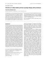
Fig. 1. Tissue-specific chromatin architecture revealed in localization of DHSs. A schematic representation of DHSs before and after hor-
mone stimulation in two cell types. The majority of hormone-responsive genes have a TSS that is embedded within a localized region of
DNase I hypersensitivity. These promoter regions are generally hypersensitive across multiple cell types, and usually correlate with CpG
islands (A). Common and preprogrammed DHSs present at distal regulatory elements often overlap with insulators (B). Hormone receptors
recognize short DNA motifs (HREs), but only a small percentage of them are occupied by a receptor in a given cell type. NR binding occurs
usually at distal enhancers and is highly correlated with the presence of accessible chromatin regions (C, D, E). Only a small fraction of
enhancer-related DHSs are universally utilized in multiple cell lines and they usually represent hormone-independent chromatin structures
(pre-programmed DHSs) (C). Most distal DHSs are tissue-specific and can be either hormone-independent (D) or appear only after hormone
stimulation (inducible DHSs) (E). Thus the presence of a DHS and subsequent receptor ⁄ transcription factor binding results in a hormone-
dependent and tissue-specific transcriptional regulation of a particular gene (gene II). A gene can be activated by the same hormone receptor
in different tissues, although through different regulatory elements (gene I; elements C and E).
Nuclear receptor regulation by chromatin M. Wiench et al.
2214 FEBS Journal 278 (2011) 2211–2230 Journal compilation ª 2011 FEBS. No claim to original US government works
for TF binding. Six nucleosomes within the earlier
described MMTV-LTR promoter tend to occupy
exactly the same positions in vivo as they do after they
are assembled in vitro. Similar observations are made
when the osteocalcin promoter is reconstituted in vitro
using SWI ⁄ SNF complexes as remodelers [67].
The published yeast-based models predict that nucle-
osome occupancy at promoters and functional TF
binding sites is low (termed nucleosome-free regions or
nucleosome-depleted regions) and that there are more
stable nucleosomes at nonfunctional sites [57,60]. One
can imagine that sequences evolved to encode unstable
nucleosomes and thus facilitate their accessibility for
TFs and transcription machinery. Indeed DNase I
hypersensitive sites (DHSs) are found to be enriched in
nucleosome-excluding sequences, including short
repeats of adenine (A16), long CCG triplet repeats and
TGGA repeats [61]. In contrast to yeast, the analysis
of human regulatory sequences predicts that there is a
higher nucleosome occupancy in chromatin in vivo
[68]. Thus, the preference for high nucleosome occu-
pancy at the regulatory elements can be amenable for
the restricted and tissue-specific regulation observed in
higher eukaryotes but not in yeast.
In agreement with that, the inducible genes in yeast
are also characterized by promoters that have been
described as ‘covered’ with nucleosomes that are able
to compete efficiently with TFs’ binding [69]. These
kinds of promoters tend to contain a TATA box and
numerous binding sites for different TFs and are
highly dependent on chromatin remodeling (Fig. 2).
They also display higher plasticity and noisy expres-
sion and are more sensitive to genetic perturbations,
and thus are more prone to change their expression
under evolutionary pressure [70,71]. In contrast, the
chromatin architecture for yeast genes which are con-
stitutively active is characterized by an open promoter
structure, the presence of a nucleosome-depleted region
with well positioned nucleosomes further upstream,
and H2A.Z histone variants at the +1 and )1 nucleo-
somes [69].
The differences in nucleosome positioning between
active and silenced genes in human cells have also been
examined recently [66]. The promoters of expressed
genes are characterized by several well positioned
nucleosomes, whereas only one nucleosome down-
stream from the TSS (+1) is phased when silenced
genes are considered. The position of the first nucleo-
some upstream from the TSS ()1) in inactive promot-
ers is replaced in active genes by Pol II binding and
this results in a shift of the +1 nucleosome 30 bp
towards the 3¢ end. Also, within the functional enhanc-
ers, nucleosomes become more localized after activation
in a way such that potential binding sites are moved to
more accessible positions within the linker regions [66].
Specifically, androgen treatment dismisses a central
nucleosome present at AREs allowing for ARs to
bind. After remodeling the AR binding site is also
found to be flanked by a pair of well positioned nucle-
osomes marked with H3-K4me2 or H3-K9,14ac
[72,73].
As mentioned before, the studies based on yeast
models suggest that intrinsic DNA sequence features
have a dominant role in nucleosome organization
in vivo [60,64]. However, discrepancies exist between
nucleosome positions observed in vivo and computa-
tional predictions based on thermodynamic properties
of DNA–histone interactions. One would expect these
differences to be an integral part of inducible or cell-
type-specific gene regulation with nucleosome location
further modulated by the presence of specific features
such as histone variants, DNA methylation and, to a
lesser extent, histone modifications. In the last two
cases, however, it is more difficult to differentiate
between the direct effect of a modification on his-
tone–DNA interactions and the indirect influence it
has on another factor’s binding, which could conse-
quently affect nucleosome positioning. Nevertheless, a
strong link between CpG methylation and nucleosome
positioning has been suggested based on observations
that the presence of a methyl group can directly influ-
ence DNA bendability (dependent on the specific
DNA sequence and extent of DNA methylation)
[60,74]. On the other hand, nucleosome positioning
has been observed to influence genome-wide methyla-
tion patterns by preferentially targeting DNA meth-
yltransferases to nucleosome-bound DNA than to
linker regions [75]. Further discrepancies between pre-
dicted versus observed nucleosome locations are
believed to come from the competition between nucle-
osomes and TFs for access to DNA and the activity
of chromatin remodelers. It has recently been
reported that in yeast cells the depletion of the
remodeling complex RISC caused the nucleosome-free
regions to shrink and in vivo nucleosome occupancy
to obtain positions reflecting the theoretical predic-
tions more closely [76].
Overall, the results show that genomes encode and
preserve both the sequences recognized by NRs and
the positioning and stability of nucleosomes in regions
that are critical for gene regulation. Those regions can
be further rearranged which is accompanied by
changes in DNA sensitivity to nucleases such as
DNase I and restriction enzymes. Sites within the
DNA which are accessible to DNase I are termed
hypersensitive (DNase I hypersensitive site, DHS).
M. Wiench et al. Nuclear receptor regulation by chromatin
FEBS Journal 278 (2011) 2211–2230 Journal compilation ª 2011 FEBS. No claim to original US government works 2215
DNase I hypersensitivity as a marker of
many different regulatory elements
Mapping DHSs is believed to be an effective method
for determining the localization of the functional reg-
ulatory elements including promoters, enhancers,
silencers, insulators and locus control regions [77].
DHSs have been identified in six cell lines within
1% of the genome as a part of an ENCODE project
[78] and across the whole genome for CD4+ T cells
[79]. Only 16–22% of sites are consistently present in
all cell lines proving that the majority of gene regu-
latory elements are cell-type-specific. These shared
sites have been further characterized by close
(< 2 kb) proximity to TSSs, high CpG content, and
binding of basal transcription machinery or CTCF
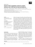
Fig. 2. Dynamics of chromatin structures at inducible genes. (A) Inducible genes are regulated by a ‘covered’ class of promoters character-
ized by the presence of a TATA box and nucleosomes competing efficiently with TFs for access to DNA. Both promoters and enhancers are
marked as chromatin structures staged for remodeling by the H2A.Z histone variant. In addition, enhancers available for subsequent receptor
binding have a decreased level of DNA methylation. (B) Induction (i.e. hormone stimulation) leads to localized incorporation of H3.3 and for-
mation of very labile H2A.Z ⁄ H3.3 nucleosomes at both the promoter and enhancer. These nucleosomes are very dynamic and can be easily
ejected thus enabling TF binding. At enhancers, the receptor binding leads to nucleosome reorganization where two stable nucleosomes
flank the receptor binding sites. Additionally, the +1 nucleosome at the promoter has been reported to move 30 bp downstream leaving
space for RNA Pol II and the basic transcriptional machinery to dock at the TSS. Mediator complexes hold the promoter and enhancer
together and changes in DNA methylation (red dots) are observed in at least a subset of enhancers. (C) Full transcriptional response is
achieved due to synchronized binding of hormone receptor and other TFs, as well as to additional receptor binding events at neighboring
HREs.
Nuclear receptor regulation by chromatin M. Wiench et al.
2216 FEBS Journal 278 (2011) 2211–2230 Journal compilation ª 2011 FEBS. No claim to original US government works
(Fig. 1). Overall, the results indicate that the com-
mon DHSs belong to housekeeping promoters or,
when distal, to insulators but not to enhancers. Cell-
type-specific DHSs, on the other hand, are more dis-
tal, and are found to be enriched for binding sites
of proteins known for their enhancer function
(p300), sequence motifs for TFs, and cell-type-specific
histone modifications [78,79]. Recent studies have
suggested that proximal DHSs which do not overlap
with promoters are associated with activating histone
marks (H3-K4me3, AcH3) usually found at promot-
ers [78,79]. Distal sites, however, are more enriched
in H3-K27me3, H3-K9me2 and H3-K9me3 marks,
while those found in the transcribed regions have
higher levels of H3-K27me1 and H3-K9me1 (Fig. 3)
[79]. Chromatin immunoprecipitation (ChIP) analyses
have shown that the hypersensitive sites, both proxi-
mal and distant, are enriched for the H2A.Z histone
variant, which has been reported to be a subject of
exchange upon hormone treatment [80]. Therefore, it
is argued that H2A.Z is associated with chromatin
sites that are staged for remodeling and TF binding
(Fig. 2A).
Correlation of DHSs with gene expression has
shown that all expressed genes are marked by a DHS
at the TSS [79]. However, although the presence of a
DHS might be necessary for gene expression, it is
clearly not sufficient. Inactive genes that are character-
ized by the presence of a DHS may be in a transcrip-
tionally poised state. This is supported by an
observation that activating histone marks and Pol II
binding are also present at these genes. In contrast,
Fig. 3. Characteristics of local chromatin structures within promoters, enhancers and coding regions. The non-random positioning of a nucle-
osome is dictated by DNA sequence, activity of remodeling complexes (like SWI ⁄ SNF) and competition of the nucleosome with TFs for
access to specific DNA sequences. The regulatory regions are characterized by high turnover of histone proteins (depicted by purple nucleo-
somes). The histone marks identified at the promoters and enhancers of active (red) and silent (blue) genes are indicated. The gradients
reflect changes of histone marks across the coding region. Contradictory observations about the presence of H3-K9me3 and H3-K4me3
within enhancer regions have been reported. Both promoters and enhancers are marked by DHSs and H2A.Z histone variants. Most promot-
ers are characterized by increased density of CpG dinucleotides (CpG islands) which are usually unmethylated (open circles). Enhancers also
show highly localized CpG enrichment with DNA methylation status correlating with their activity. The CpG dinucleotides are under-repre-
sented within coding regions and contain high methylation levels (filled circles) in order to prevent spurious transcription.
M. Wiench et al. Nuclear receptor regulation by chromatin
FEBS Journal 278 (2011) 2211–2230 Journal compilation ª 2011 FEBS. No claim to original US government works 2217
promoter regions near silenced genes with no DHSs
showed no evidence of these marks [79].
We have found that GR binding invariably occurs
at nuclease-accessible sites [80,81] (Fig. 1). When pro-
files are compared between two cell lines, the lack of
response to GR regulation is consistently correlated
with the lack of GR binding and the absence of chro-
matin transition at the corresponding sites. Interest-
ingly, the hypersensitive sites either pre-exist in
chromatin (pre-programmed), or appear only after
stimulation with hormone (de novo) [80,81]. The find-
ing that GR interacts with the pre-existing DHSs is
surprising as GR has classically been considered to be
a pioneer factor which triggers the initiation of chro-
matin remodeling processes.
Steroid receptors have been frequently shown to
induce DNase I hypersensitivity within the region of
their binding sites [82–84]. Although there is strong
evidence for histone loss after hormone induction, the
remodeled site is not completely nucleosome-free and
the question about the nature of DNA hypersensitivity
to nucleolytic attack stays open. Two possibilities are
taken into account: the nucleosome can either be repo-
sitioned to the neighboring regions or be temporarily
unfolded from the template. The meticulous study per-
formed at the Tat-GRU speaks in favor of the latter
possibility [85]. No modification of the distribution of
nucleosome frames has been observed while H1 and
H3 interaction is clearly lost upon remodeling. The sig-
nificance of H1 loss for transcription activation has
also been shown using MMTV as a model [86,87].
Once a nucleosome’s binding becomes weaker and
DNA becomes accessible, synergistic binding between
receptors and other TFs is observed (Fig. 2C). On the
MMTV promoter PR binding to the exposed element
enables NF1 access to DNA. This in turn facilitates
more PR binding to the remaining elements resulting
in a full transcriptional response. The transcription is
significantly compromised by the NF1 depletion or
mutations in NF1 binding site [88]. Importantly, the
synergistic binding between receptor and NF1 to
MMTV is strongly dependent on the nucleosomal
structure and is not observed for naked DNA [33].
Furthermore, we suggest that the vast majority of
localized reorganization events are not stable but in
fact represent a highly dynamic process. We have pro-
posed that the rapid exchange observed for TFs and
response elements in chromatin [38,39,89,90] has a
direct correlation to chromatin remodeling
[37,39,91,92]. The nucleosomes at promoter regions are
also characterized by a high turnover rate independent
of whether they are in active or repressed state [71,93].
This constant movement, assembly and disassembly of
nucleosomes is a product of ATP-dependent remodel-
ing activity.
Chromatin remodeling activity
and achieving an open
⁄
accessible
chromatin structure
Chromatin remodeling appears to be the first step in
an ordered sequence of events required for hormone-
regulated transcription. During the remodeling
reaction DNA can be transiently unwound from a
nucleosome or a nucleosome can be moved to a neigh-
boring position (sliding) [94]. These reactions are
energy-dependent and are executed by protein com-
plexes that were first identified in yeast-based screens
as mutations that control gene transcription triggered
by extracellular signals [95–97]. The ATP-dependent
remodeling engines can exist in multiple forms, usually
as large ( 2 MDa) multiprotein complexes with a
core catalytic ATPase subunit and a team of auxiliary
factors. The nature of an ATPase subunit underlies the
current classification of remodeling complexes into
four major classes: SWI ⁄ SNF, ISWI, Mi-2 ⁄ NuRD and
INO80 [94,98]. They also differ in the mechanism by
which chromatin remodeling is executed (sliding, loop-
ing etc.). Both SWI ⁄ SNF and ISWI can slide nucleo-
somes along DNA; however, SWI ⁄ SNF may
additionally be able to create stable DNA loops within
nucleosome structures and remove ⁄ exchange histone
dimers or octamers [94,99,100]. At the level of pro-
moter activity regulation it translates into an ability to
generate nucleosome-free regions (when coupled to his-
tone chaperones), exchange canonical histone dimers
for histone variants or, if there is not enough space for
repositioning, expose specific DNA sequences as loops.
Therefore, the SWI ⁄ SNF complexes are perceived as
the most potent in rearranging promoter structures
during transcriptional activation and as such are
the best exploited in the studies of SHR-regulated
transcription [101–105]. The ATPase subunit in human
SWI ⁄ SNF complexes is either BRG1 or BRM, and is
associated with up to a dozen additional factors
including BRG1-associated factors (BAFs) [106–108].
It is worth mentioning that both the ATPase core and
a composition of BAFs can be responsible for
promoter-specific and tissue-specific regulation [107,
109,110].
Extensive studies based on the MMTV model have
shown that SWI ⁄ SNF-dependent chromatin remodel-
ing is a necessary prerequisite for optimal hormone-
dependent transcription and in this case GR can utilize
both BRG1- and BRM- containing complexes [36,105].
GR does not contact BRG1 directly but rather
Nuclear receptor regulation by chromatin M. Wiench et al.
2218 FEBS Journal 278 (2011) 2211–2230 Journal compilation ª 2011 FEBS. No claim to original US government works
through the associated factors BAF57 and BAF60a
which are common for both BRG1 and BRM com-
plexes [111]. Transfection experiments with dominant
negative forms of either BRG1 or BRM have resulted
in an inhibition of transcription, lack of both Pol II
loading and chromatin transition, as well as compro-
mised decondensation of the MMTV array [105]. Fur-
thermore, using a UV laser crosslinking approach it
has been possible to establish highly transient and peri-
odic interactions of GR with the MMTV template dur-
ing the remodeling reaction. This is further reflected by
periodic binding of SWI ⁄ SNF, H2A and H2B [112].
The suggested model requires that receptor binding is
aided during the early phase of the nucleosome remod-
eling reaction, but when the remodeling reaction is
completed and nucleosomes return to the basal state,
receptors are actively removed from the promoter. In
human cells lacking BRG1 and BRM (i.e. SW-13)
transactivation by GR is weak and can be selectively
enhanced by the ectopic expression of either BRG1 or
BRM [102]. However, it cannot be substituted by the
activity of ISWI or Mi-2 complexes, both present in
SW-13 [19]. BRG1 remodeling action is also specifi-
cally required for PR- and AR-dependent activation of
MMTV-LTR chromatin [113,114] as well as for ER-
regulated genes [103,104,115]. These results suggest
that SWI ⁄ SNF complexes are commonly utilized by
NRs for creating chromatin transition states during
hormone induction. On the other hand, the transcrip-
tion profile obtained after overexpression of a domi-
nant negative form of BRG1 shows significant
reduction in only 40% of glucocorticoid-activated
genes [80]. An even smaller effect (11%) is observed
when glucocorticoid-repressed genes are analyzed.
Consistent with that, only a subset of DHSs, both pre-
existing and hormone-inducible, are dependent on
BRG1 action in these cells. Hence, the contribution of
other remodeling complexes seems to be an obvious
possibility.
The picture that emerges is that receptor-based gene
regulation is always dependent on the presence of
remodeled chromatin. However, this feature can either
be formed during cell development, and continuously
present, or be triggered by the receptor itself only after
hormone stimulation (Fig. 1). In addition, chromatin
remodeling alone is not sufficient for transcriptional
activation and depends on the context of the DNA
and histone modifications. In some cases opening the
chromatin structure by chromatin remodeling enzymes
is necessary for subsequent acetylation of histones
[116–118]. In other cases, the recruitment of chromatin
remodeling complexes must be preceded by RNA Pol
II binding and histone acetylation which in turn
creates binding sites for bromodomain-containing pro-
teins, i.e. BAFs [85,119,120].
Thus the action of remodeling complexes should not
be separated from the action of histone and DNA
modifying enzymes, as they operate simultaneously on
the same sequences and influence each other. In fact,
large multifunctional complexes have been found
in vivo where the chromatin remodelers are associated
with histone-modifying enzymes including histone de-
acetylases (HDACs, NCoR complex), histone meth-
yltransferases, such as CARM1 (nucleosomal
methylation activation complex, NUMAC), as well as
other proteins with co-regulatory functions (mSin3a,
BRCA1, TOPO II, actin). Furthermore, Mi2 ⁄ NURD,
part of the NCoR complex, can repress NR-dependent
transcription [121,122] and is targeted to specific areas
of chromatin through recruitment by transcription
repressors or by factors that recognize methylated
DNA.
Histone modifications and histone
variants as a part of gene architecture
and transcription regulation
Over 60 different residues within histone tails have
been identified as targets for post-translational modifi-
cations (reviewed in [7,55]). The most common histone
modifications are acetylation or ubiquination of lysine
residues, methylation of arginine and lysine residues,
and phosphorylation of serine and threonine residues.
Acetylation usually occurs cumulatively on multiple
lysine residues and utilizes different histone acety-
ltransferases (HATs) in a seemingly non-specific man-
ner. In contrast, other histone marks are deposited by
a specific enzyme on a defined residue. Furthermore,
methylation can exist as monomethylation, dimethyla-
tion or trimethylation with different methyltransferases
being active at each step. All modifications can affect
one another and many of them are positively or nega-
tively correlated [7,55,123].
The mechanisms by which histone modifications
exert their function include alterations in DNA–nucle-
osome and nucleosome–nucleosome interactions as
well as in the recruitment of non-histone regulatory
proteins (reviewed in [7,55]). The internucleosomal and
intranucleosomal interactions can become relaxed sim-
ply due to the change in the net charge of nucleosomes
caused by most (methylation is an exception) modifica-
tions. Among them, lysine acetylation is believed to
be the most potent due to both its ability to neutralize
the basic charge and its abundance. This idea is
supported by the experimental observation that acety-
lated histones are easier to displace from DNA both
M. Wiench et al. Nuclear receptor regulation by chromatin
FEBS Journal 278 (2011) 2211–2230 Journal compilation ª 2011 FEBS. No claim to original US government works 2219
in vivo [124] and in vitro [120,125]. Recently, acetyla-
tion of H3-K14 has been shown to be essential for
nucleosome eviction [126]. The effect of histone modifi-
cations can also go beyond local contacts and directly
influence higher-order chromatin structure. For exam-
ple, acetylation of H4-K16 is known to inhibit the for-
mation of 30 nm fibers [127] as discussed in detail in
an accompanying review [2]. The alternative function
of histone modifications is to create a ‘code’ which can
be recognized and read by other proteins. Proteins
with chromo-like domains can bind to methylated his-
tone residues, whereas acetylation is recognized by
bromodomains. These proteins, in turn, provide enzy-
matic activities which further influence chromatin
dynamics and function.
Globally, active euchromatin and inactive hetero-
chromatin are marked by different histone modifica-
tions. Acetylation of H3 and H4 and methylation of
H3-K4, H3-K36 and H3-K79 are characteristic of
active chromatin whereas low levels of acetylation and
high levels of H3-K9me3, H3-K27me3 and H4-K20
methylation are associated with inactive chromatin [7].
These modifications frequently spread along extended
chromosomal regions and are sharply separated from
each other by boundary elements associated with the
insulator binding protein CTCF [128,129].
Within euchromatin, actively transcribed genes are
further characterized by a set of features that show a
more complex and localized pattern within enhancers,
the core promoter, coding regions and the 3¢ end of
the gene [7,128,130] (Fig. 3). Multiple studies have
proved that H3 and H4 histones within the TSSs are
generally acetylated [128,131–134]. As far as other
modifications are concerned, high levels of all three
states of H3-K4 methylation and H2A.Z form a
peak within the promoter and TSS regions whereas H3-
K27me1, H3-K79me, H2B-K5me1, H4-K20me1 and
H3-K9me1 are associated with the entire transcribed
region. Unlike other marks, H3-K36me1 tends to accu-
mulate towards the 3¢ end of the gene. Interestingly, the
signatures of both promoters and insulators appear to
be invariant across different cell lines [129,135] and sev-
eral studies have shown that both active and inactive
promoters are associated with histone acetylation and
H3-K4me3 [128,136–138]. Chromatin modification
patterns at inducible genes have also proved to be rela-
tively stable during activation of resting T cells with
active modifications being already in place [139]. In
contrast to that, enhancers are believed to be the most
variable elements and display a highly cell-type-specific
pattern of histone modifications.
Identification of enhancer elements has not been an
easy undertaking because of their distant localization
from regulated genes and the lack of specific sequence
elements. Attempts made thus far to identify enhancer
regions have been based on sequence conservation, the
position of DHSs [78,79,140] or p300 binding to DNA
outside the promoter regions [135,136,141]. Based on
the latter, over 55 000 enhancers have been identified
in only two cell lines (K562 and HeLa), thus leading
to the prediction of 10
5
–10
6
enhancers existing in total
[135].
Once enhancer elements have been recognized their
chromatin characteristics can be described (Fig. 3). Sim-
ilar to promoters, enhancer elements are marked by
H3 acetylation, H3-K4 monomethylation, H2A.Z and
H3-K9me1, but lack other promoter-specific modifications
[128,132,134,135,142,143]. Surprisingly, H3-K27me3, pre-
viously ascribed to the repressive chromatin, has also
been identified within enhancer elements. The combina-
tion of H3-K4me1 and H3-K4me3 has been proposed as
the strongest discriminator between enhancers and pro-
moters with enhancers being deprived of trimethylation
[140,142,144]. However, this might not be the universal
feature since it has recently been shown that H3-K4me3
is also present at the enhancers when a DHS-based
approach is applied to identify these regions [140].
Furthermore, each of the modifications including
H3-K4me1, 2 and 3, H3-K9me1 and H2A.Z have been
detected at only 20–40% of putative enhancers sug-
gesting that they are found only in unique subgroups
[128,140]. No significant correlation between specific
modification patterns at the enhancer regions and gene
expression has been observed [140].
Even if current literature lacks the global overview
of histone marks specifically in terms of regulation by
steroid receptors there is no reason to assume that
their common pattern would be different from that
mentioned above. Arginine methylation of both H3R2
and H4R3 has been previously suggested to play a role
in NR-mediated transcription activation [145]; how-
ever, none of these marks showed any characteristic
patterns in a genome-wide analysis [128]. It is still
unknown how many enhancers can be identified based
on their characteristics before hormone induction and
to what extent the chromatin marks within enhancer
elements can change after induction and receptor bind-
ing. When HeLa cells are treated with interferon-c
only 25% of STAT1 binding is observed within pre-
dicted enhancers [135]. Analysis of GR binding sites,
however, shows that most fall into DHS regions exist-
ing before hormone stimulation and only 15% of
binding events are followed by a chromatin transition
and possibly by changes in at least some of the chro-
matin signatures [80,81]. Dynamic changes of histone
modifications after hormone induction have not been
Nuclear receptor regulation by chromatin M. Wiench et al.
2220 FEBS Journal 278 (2011) 2211–2230 Journal compilation ª 2011 FEBS. No claim to original US government works
globally analyzed yet but have been observed on
selected steroid-hormone-regulated promoters. The
changes are shown to be limited to a few or even a sin-
gle nucleosome. At the MMTV promoter, GRE-con-
taining nucleosomes B and C, but not F, undergo
rapid (30 min after stimulation) deacetylation at H3
and H4, both of which show a high level of acetylation
in the 3134 cell line before induction [146]. Similar
experiments in T47D breast cancer cells have shown
that H4 deacetylation is preceded by initial acetylation
which precisely coincides with the time of recruitment
of progesterone receptor, Pol II and p300 [147].
However, the most detailed data concerning
dynamic recruitment of co-regulators and histone mod-
ification changes have been collected for the ER-regu-
lated pS2 gene. In the initial experiments the increase
of acetylation of histone H3 and H4 at ER-regulated
cathepsin D and pS2 promoters was observed within
10 min of estrogen treatment, peaking at 1 h post-
treatment and decreasing to near-basal levels within
6 h. The transient increase in histone acetylation coin-
cided with a transient increase in the association of
SRC-3, p300, CBP and RNA Pol II, as well as a tran-
sient increase in the transcription level [148,149]. Fur-
ther experiments that included a more detailed time
course and using a-amanitin-synchronized cells demon-
strated in fact cyclic behavior of histone acetylation
with peaks every 40–60 min corresponding to produc-
tive transcription cycles [150,151].
Core histones not only undergo covalent post-trans-
lational modifications but can also be exchanged with
histone variants (reviewed in [55,152]). Differences
between variants and canonical histones can be as
small as a few amino acids, as in the case of H3.3, or
can apply to larger domains within the histone tails
(MacroH2A) or in the histone fold domains
(H2ABdb). In contrast to canonical histones, histone
variants are expressed mainly outside of S phase and
are thought to be deposited into nucleosomes in a rep-
lication-independent manner by means of specific pro-
tein complexes. H2A.Z can be incorporated into a
nucleosome either by Swr1 through ATP-dependent
histone exchange reactions [153] or via the help of rep-
lication-independent chaperones like Nap1 [154], and
H3.3 is assembled by histone regulator A (HIRA).
Incorporation of histone variants into chromatin
impacts its structure in various ways [155]. For exam-
ple, histone H2A.Z replaces histone H2A at the
promoter sites, insulators and enhancers and when
co-assembled with H3.3 forms a very unstable struc-
ture that can be ejected following gene activation
[156,157] (Fig. 2B). This observation sheds a new light
on the previously observed nucleosome-free regions at
active promoters and enhancers. These nucleosome-
free regions may now be explained by the presence of
highly labile H2A.Z ⁄ H3.3-containing nucleosomes that
are easily displaced by TFs. This fact has been over-
looked before because of H2A.Z ⁄ H3.3 disruption in
the moderate salt concentrations usually used for chro-
matin purification [156]. It has been suggested that
transcription of as many as 30% of genes can be regu-
lated through incorporation of H2A.Z [158]. Taken
together, histone modifications and histone variants
play a role in defining the chromatin architecture of
active or potentially active gene transcription units and
undergo dynamic changes during ligand stimulation.
DNA methylation and MeCP2 – link to
chromatin architecture and remodeling
DNA methylation of cytosine is the most abundant
covalent DNA modification. In differentiated cells
DNA methylation appears almost exclusively in the
CpG context and is deposited there by one of the
DNA methyltransferases (Dnmt1, Dnmt3a or Dnmt3b)
[159]. In mammalian genomes the distribution of a
methyl mark is described as a global methylation pat-
tern [160]. Approximately 98% of CpG dinucleotides
are located within the CpG-poor regions and 80% of
them are methylated. The observed high level of DNA
methylation within gene bodies and non-coding regions
is believed to serve as a suppressor of transcriptional
noise by preventing the spurious transcription initia-
tion from cryptic promoters [160,161]. The remaining
2% of genomic CpGs are densely grouped in short
stretches located mostly at the 5¢ end of the genes [160]
(Fig. 3). These patches are referred to as CpG islands
and typically stay unmethylated independent of the
gene expression [162,163].
High CpG density promoters are associated with
two classes of genes, commonly expressed housekeep-
ing genes and highly regulated key developmental
genes, whereas low CpG density promoters are gener-
ally linked to tissue-specific genes [164]. In contrast to
CpG-rich sequences, CpG-poor regulatory elements
are more prone to active and de novo methylation and
demethylation, which might provide yet another level
of gene regulation. As mentioned before, cis regulatory
elements active in a particular cell type are often
associated with marks of open chromatin such as
H3-K4me2 or H3-K4me1 [131,142]. It has been shown
that CpGs found at H3-K4me2-enriched sites (outside
of promoters and CpG islands) have significantly lower
DNA methylation levels than those at H3-K4me2-
depleted sites, and this relationship is particularly
strong for CpGs located within highly conserved
M. Wiench et al. Nuclear receptor regulation by chromatin
FEBS Journal 278 (2011) 2211–2230 Journal compilation ª 2011 FEBS. No claim to original US government works 2221
non-coding elements [164]. We have observed a high
correlation between CpG methylation status and activ-
ity of enhancer elements at GR binding sites with
demethylation strongly linked to chromatin accessibil-
ity and GR binding (Wiench M, John S, Baek S, John-
son TA, Sung M-H, Escobar T, Simmons CA, Pearce
KH, Biddie SC, Sabo PJ, Thurman RE, Stamatoyan-
nopoulos JA & Hager GL, unpublished results). The
lack of DNA methylation, often restricted to a single
CpG site, has been shown to be the remnant of inter-
actions with pioneer TFs (such as PU.1 or C ⁄ EBPb)in
early embryonic development [165]. These interactions
might be necessary to prevent the enhancers from
assembling into repressive chromatin structures during
development and to make them amenable for subse-
quent activation of tissue-specific genes. Thus activa-
tion of these genes may be possible only after specific
interactions and changes in chromatin structure have
occurred at earlier stages in development.
DNA methylation leads to transcriptional repression
through several mechanisms. It can directly affect
the TF’s binding to its DNA recognition element as
well as the positioning and stability of a nucleosome.
However, it primarily acts indirectly through the
action of proteins like MeCP2 and MBD1-4 that selec-
tively bind methylated CpGs through their methyl-
CpG binding domain (MBD) [166]. Various members
of the MBD family display different DNA binding
specificities. They can recognize and bind to more
complex sequences than a single methylated CpG. For
example, high affinity binding by MeCP2 requires four
or more A ⁄ T base pairs adjacent to the methylated
CpG [167].
Even if methylation marks a DNA molecule directly,
the silencing effect is observed only after the DNA
template is assembled into chromatin [168,169]. More-
over, MeCP2 has been shown to bind methylated
DNA only in a nucleosome context [170]. Hence, the
mechanism through which DNA methylation and
MBDs accomplish the silencing effect employs both
chromatin modification and remodeling activities. Dif-
ferent sets of proteins have been identified to bind a
nucleosome when DNA methylation is combined with
different histone modifications [170]. Furthermore,
MeCP2 has been found to interact with histone deacet-
ylases (HDAC2, Sin3A) [171,172], histone meth-
yltransferases (SUV39H1) [173] and remodeling
complexes [174]. Both MeCP2 and BRM were shown
to be associated with each other on the same sequences
within hypermethylated promoters, and treatment with
inhibitors of DNA methylation (5-aza-2¢-deoxycyti-
dine, 5Aza-dC) results in a loss of methylation, loss of
BRM and MeCP2 binding and reactivation of tran-
scription [174]. In cancer cells, changes in DNA meth-
ylation promoted by SWI ⁄ SNF complexes induce
transcriptional activation and rescue the transcription
of CD44 and E-cadherin [175]. Although MeCP2 as
well as MBD2 are likely to be responsible for initial
recruitment of chromatin remodelers, studies in vitro
and in vivo suggest that chromatin remodeling activi-
ties further facilitate binding of MBD proteins to those
methylated sites that are not initially accessible on nu-
cleosomal templates and by doing so further stimulate
MBD-mediated gene repression [174].
In many vertebrates site-specific demethylation
affects tissue-specific genes [176]. Unlike plants, meth-
ylation of DNA in mammals has long been considered
to be a stable mark that is removed only during a pas-
sive process which involves replication. Recently, evi-
dence showing that the demethylation process might
be active and dynamic is accumulating. Demethylation
is often restricted to regulatory elements outside the
core promoter where transcription factors bind and
where chromatin is hypersensitive to DNase I [164]. It
has been reported that the Bdnf regulatory region is
demethylated by 25–45% and MeCP2 is redistributed
2–3 days after depolarization of neuron cells which is
accompanied by increase in Bdnf synthesis [177]. In
another study, prolonged glucocorticoid treatment
caused demethylation of all four CpGs located within
the remodeled area of a Tat-GRU while neighboring
CpGs located outside the remodeled area remained
methylated. In this case the demethylation is com-
pleted in 3 days and allows for the recruitment of
additional transcription factors [178].
A kinetics profile of 2–3 days does not point to an
active and efficient process. However, very fast and
cyclical changes (with a periodicity of about 100 min)
of DNA methylation and demethylation have also
been observed within the promoters of pS2 and several
other ER-regulated genes [179]. It is not clear yet what
kind of mechanism is primarily involved in the deme-
thylation but it has been suggested previously that it
most probably involves the creation of nicks in DNA
3¢ to the methylcytidine [176,180]. A recent report con-
firms this observation and suggests that deamination
paired with glycosylation enzymatic activities
(AID ⁄ MBD4) and a base excision repair process are
involved [181]. Rapid demethylation after activation
seems to be a common event at hormone-inducible ele-
ments since it has been observed during ER [179,182],
vitamin D receptor [183] and GR (Wiench et al., sub-
mitted) regulation even though the mechanism is
poorly understood (Fig. 2).
In addition to its role in gene silencing, MeCP2 has
been described as a chromosomal architecture element.
Nuclear receptor regulation by chromatin M. Wiench et al.
2222 FEBS Journal 278 (2011) 2211–2230 Journal compilation ª 2011 FEBS. No claim to original US government works
MeCP2 has been shown to mediate the formation of
complex chromatin structure by promoting chromatin
looping at the Dlx5 gene with the loop itself marked by
an H3-K9me2 repressive mark [184]. A very recent
report describes MeCP2 in neuronal cells as a highly
abundant nuclear protein that might replace H1 bind-
ing and globally alter chromatin state [185]. The
described action of MeCP2 is dependent on cytosine
methylation. However, another study reported that the
MeCP2 protein can organize chromatin independently
of DNA methylation and in the absence of a functional
MBD domain. The addition of MeCP2 to unmethylat-
ed nucleosomal arrays leads to significant chromatin
compaction, greater than that achieved by histone H1
[87]. At the ratio of one MeCP2 molecule per nucleo-
some, electron microscopy revealed formation of a
novel 60S ellipsoidal structure. Also, DNA when
methylated is able to adopt a distinctive chromatin
structure after being assembled into chromatin which
results in a loss of DNase I hypersensitivity [186,187].
This is in agreement with the fact that demethylated
CpGs are more often found at regions where chromatin
remodeling creates DHSs (Figs 2 and 3). Regions of
DNA methylation have also been found to be deficient
in H2A.Z, a marker of inducible chromatin, and in fact
these two chromatin features seem to be mutually
exclusive in both plants [188] and mammals [158].
To summarize, DNA methylation appears to be a
more dynamic feature than previously thought espe-
cially within distal regulatory elements where it teams
up with other chromatin signatures defined by the
presence of DHSs and characteristic histone modifica-
tions. The specificity can be achieved (a) by utilizing
different DNA methyltransferases to deposit the
methyl mark and (b) by recognition of the mark by
different DNA methyl binding proteins. Methyl-CpG
binding proteins have been shown recently to provide
a functional link between DNA methylation, chroma-
tin remodeling and histone modifications, as well as to
serve as structural proteins of chromatin.
Conclusions
During the last two decades model systems have been
developed and well exploited to study the mechanisms
governing steroid hormone gene regulation in the chro-
matin environment. Those systems have made it possi-
ble to explore the effects of nucleosome positioning,
the changes in the nucleosome structure in response to
ligand stimulation, and the role chromatin remodeling
complexes and histone modifying enzymes have during
promoter progression. However, it is apparent that
they represent unique examples rather than a universal
type of regulated promoter. Thus, to overcome this
limitation a new set of methods has been developed to
study the epigenome on a high throughput basis. The
necessary precondition is to reliably identify promot-
ers, enhancers and other regulatory elements within
the genome. Recent reports discussed in this review
show that this step can be achieved. The next step is
to prove their functionality and to characterize them
by the presence of specific chromatin signatures. The
results of the first attempts have been published during
the last several months proving the complexity of the
system and revealing even more questions. Thanks to
ChIP-chip, ChIP-seq, DNase I-seq, MeDIP-seq and
even genome-wide bisulfite sequencing becoming more
available and affordable, a big leap forward can be
made in understanding how local chromatin architec-
ture affects tissue-specific gene regulation and regula-
tion by NRs.
Acknowledgements
This research was supported by the Intramural
Research Program of the NIH, National Cancer Insti-
tute, Center for Cancer Research. T.B. Miranda is
funded in part by the NIGMS Pharmacology Research
Associate Fellowship.
References
1 Beato M & Klug J (2000) Steroid hormone receptors:
an update. Hum Reprod Update 6, 225–236.
2 Cockerill PN (2011) Structure and function of active
chromatin and DNase I hypersensitive sites. FEBS J
278, 2182–2210.
3 Bednar J & Dimitrov S (2011) Chromatin under
mechanical stress: from single 30 nm fibers to single
nucleosomes. FEBS J 278, 2231–2243.
4 Hakim O, Sung MH & Hager GL (2010) 3D shortcuts
to gene regulation. Curr Opin Cell Biol 22, 305–313.
5 Richmond TJ, Finch JT, Rushton B, Rhodes D &
Klug A (1984) Structure of the nucleosome core parti-
cle at 7 A
˚
resolution. Nature 311, 532–537.
6 Luger K, Mader AW, Richmond RK, Sargent DF &
Richmond TJ (1997) Crystal structure of the nucleo-
some core particle at 2.8 A
˚
resolution. Nature 389,
251–260.
7 Kouzarides T (2007) Chromatin modifications and
their function. Cell 128, 693–705.
8 Felsenfeld G & Groudine M (2003) Controlling the
double helix. Nature 421, 448–453.
9 Horn PJ & Peterson CL (2006) Heterochromatin
assembly: a new twist on an old model. Chromosome
Res 14, 83–94.
M. Wiench et al. Nuclear receptor regulation by chromatin
FEBS Journal 278 (2011) 2211–2230 Journal compilation ª 2011 FEBS. No claim to original US government works 2223
10 Kinyamu HK & Archer TK (2004) Modifying chroma-
tin to permit steroid hormone receptor-dependent tran-
scription. Biochim Biophys Acta 1677, 30–45.
11 Scheidereit C, Geisse S, Westphal HM & Beato M
(1983) The glucocorticoid receptor binds to defined
nucleotide sequences near the promoter of mouse
mammary tumour virus. Nature 304, 749–752.
12 Beato M (1989) Gene regulation by steroid hormones.
Cell 56, 335–344.
13 Luisi BF, Xu WX, Otwinowski Z, Freedman LP,
Yamamoto KR & Sigler PB (1991) Crystallographic
analysis of the interaction of the glucocorticoid recep-
tor with DNA. Nature 352, 497–505.
14 Rogatsky I, Wang JC, Derynck MK, Nonaka DF,
Khodabakhsh DB, Haqq CM, Darimont BD, Garabe-
dian MJ & Yamamoto KR (2003) Target-specific utili-
zation of transcriptional regulatory surfaces by the
glucocorticoid receptor. Proc Natl Acad Sci USA 100,
13845–13850.
15 John S, Johnson TA, Sung MH, Koch-Paiz CA, Davis
SR, Walker R, Meltzer P & Hager GL (2009) Kinetic
complexity of the global response to glucocorticoid
receptor action. Endocrinology 150, 1766–1774.
16 Wang JC, Derynck MK, Nonaka DF, Khodabakhsh
DB, Haqq C & Yamamoto KR (2004) Chromatin
immunoprecipitation (ChIP) scanning identifies pri-
mary glucocorticoid receptor target genes. Proc Natl
Acad Sci USA 101, 15603–15608.
17 Collingwood TN, Urnov FD & Wolffe AP (1999)
Nuclear receptors: coactivators, corepressors and
chromatin remodeling in the control of transcription.
J Mol Endocrinol 23, 255–275.
18 Hsiao PW, Deroo BJ & Archer TK (2002) Chromatin
remodeling and tissue-selective responses of nuclear
hormone receptors. Biochem Cell Biol 80, 343–351.
19 Trotter KW & Archer TK (2007) Nuclear receptors
and chromatin remodeling machinery. Mol Cell Endo-
crinol 265–266, 162–167.
20 Aoyagi S, Trotter KW & Archer TK (2005) ATP-
dependent chromatin remodeling complexes and their
role in nuclear receptor-dependent transcription in
vivo. Vitam Horm 70, 281–307.
21 Elbi C, Walker DA, Romero G, Sullivan WP, Toft
DO, Hager GL & DeFranco DB (2004) Molecular
chaperones function as steroid receptor nuclear
mobility factors. Proc Natl Acad Sci USA 101,
2876–2881.
22 Agresti A, Scaffidi P, Riva A, Caiolfa VR & Bianchi
ME (2005) GR and HMGB1 interact only within
chromatin and influence each other’s residence time.
Mol Cell 18, 109–121.
23 Sabo PJ, Humbert R, Hawrylycz M, Wallace JC,
Dorschner MO, McArthur M & Stamatoyannopoulos
JA (2004) Genome-wide identification of
DNaseI hypersensitive sites using active chromatin
sequence libraries. Proc Natl Acad Sci USA 101, 4537–
4542.
24 Bresnick EH, Rories C & Hager GL (1992) Evidence
that nucleosomes on the mouse mammary tumor virus
promoter adopt specific translational positions. Nucleic
Acids Res 20, 865–870.
25 Lee H-L & Archer TK (1994) Nucleosome mediated
disruption of transcription factor:chromatin initiation
complexes at the mouse mammary tumour virus long
terminal repeat in vivo. Mol Cell Biol 14, 32–41.
26 Kraus WL & Kadonaga JT (1998) p300 and estrogen
receptor cooperatively activate transcription via differ-
ential enhancement of initiation and reinitiation. Genes
Dev 12, 331–342.
27 Oduro AK, Fritsch MK & Murdoch FE (2008) Chro-
matin context dominates estrogen regulation of pS2
gene expression. Exp Cell Res 314, 2796–2810.
28 Richard-Foy H & Hager GL (1987) Sequence specific
positioning of nucleosomes over the steroid-inducible
MMTV promoter. EMBO J 6, 2321–2328.
29 Archer TK, Cordingley MG, Wolford RG & Hager GL
(1991) Transcription factor access is mediated by accu-
rately positioned nucleosomes on the mouse mammary
tumor virus promoter. Mol Cell Biol 11, 688–698.
30 Truss M, Bartsch J, Hache RJ & Beato M (1993)
Chromatin structure modulates transcription factor
binding to the mouse mammary tumor virus (MMTV)
promoter. J Steroid Biochem Mol Biol 47, 1–10.
31 Grange T, Roux J, Rigaud G & Pictet R (1991) Cell-
type specific activity of two glucocorticoid responsive
units of rat tyrosine aminotransferase gene is associ-
ated with multiple binding sites for C ⁄ EBP and a
novel liver-specific nuclear factor. Nucleic Acids Res
19, 131–139.
32 Trotter KW & Archer TK (2004) Reconstitution of
glucocorticoid receptor-dependent transcription in vivo.
Mol Cell Biol 24, 3347–3358.
33 Vicent GP, Zaurin R, Ballare C, Nacht AS & Beato
M (2009) Erk signaling and chromatin remodeling in
MMTV promoter activation by progestins. Nucl
Recept Signal 7, e008.
34 Bresnick EH, John S, Berard DS, Lefebvre P & Hager
GL (1990) Glucocorticoid receptor-dependent disrup-
tion of a specific nucleosome on the mouse mammary
tumor virus promoter is prevented by sodium butyrate.
Proc Natl Acad Sci USA 87, 3977–3981.
35 Fragoso G, Pennie WD, John S & Hager GL (1998)
The position and length of the steroid-dependent
hypersensitive region in the mouse mammary tumor
virus long terminal repeat are invariant despite
multiple nucleosome B frames. Mol Cell Biol 18,
3633–3644.
36 Fryer CJ & Archer TK (1998) Chromatin remodeling
by the glucocorticoid receptor requires the BRG1 com-
plex. Nature 393, 88–91.
Nuclear receptor regulation by chromatin M. Wiench et al.
2224 FEBS Journal 278 (2011) 2211–2230 Journal compilation ª 2011 FEBS. No claim to original US government works
37 Fletcher TM, Xiao N, Mautino G, Baumann CT,
Wolford RG, Warren BS & Hager GL (2002) ATP-
dependent mobilization of the glucocorticoid receptor
during chromatin remodeling. Mol Cell Biol 22, 3255–
3263.
38 McNally JG, Mueller WG, Walker D, Wolford RG &
Hager GL (2000) The glucocorticoid receptor: rapid
exchange with regulatory sites in living cells. Science
287, 1262–1265.
39 Rayasam GV, Elbi C, Walker DA, Wolford RG,
Fletcher TM, Edwards DP & Hager GL (2005) Ligand
specific dynamics of the progesterone receptor in living
cells and during chromatin remodeling in vitro. Mol
Cell Biol 25, 2406–2418.
40 Mueller WG, Walker D, Hager GL & McNally JG
(2001) Large scale chromatin decondensation and
recondensation in living cells and the role of transcrip-
tion. J Cell Biol 154, 33–48.
41 So AY, Chaivorapol C, Bolton EC, Li H & Yamamot-
o KR (2007) Determinants of cell- and gene-specific
transcriptional regulation by the glucocorticoid recep-
tor. PLoS Genet 3, e94.
42 Carroll JS, Meyer CA, Song J, Li W, Geistlinger TR,
Eeckhoute J, Brodsky AS, Keeton EK, Fertuck KC,
Hall GF et al. (2006) Genome-wide analysis of estro-
gen receptor binding sites. Nat Genet 38, 1289–1297.
43 Jia L, Berman BP, Jariwala U, Yan X, Cogan JP,
Walters A, Chen T, Buchanan G, Frenkel B & Coetzee
GA (2008) Genomic androgen receptor-occupied
regions with different functions, defined by histone
acetylation, coregulators and transcriptional capacity.
PLoS ONE 3, e3645.
44 Wang Q, Li W, Liu XS, Carroll JS, Janne OA, Keeton
EK, Chinnaiyan AM, Pienta KJ & Brown M (2007) A
hierarchical network of transcription factors governs
androgen receptor-dependent prostate cancer growth.
Mol Cell 27, 380–392.
45 Vega VB, Lin CY, Lai KS, Kong SL, Xie M, Su X,
Teh HF, Thomsen JS, Yeo AL, Sung WK et al. (2006)
Multiplatform genome-wide identification and model-
ing of functional human estrogen receptor binding
sites. Genome Biol 7, R82.
46 Bolton EC, So AY, Chaivorapol C, Haqq CM, Li H
& Yamamoto KR (2007) Cell- and gene-specific regu-
lation of primary target genes by the androgen recep-
tor. Genes Dev 21, 2005–2017.
47 So AY, Cooper SB, Feldman BJ, Manuchehri M &
Yamamoto KR (2008) Conservation analysis predicts
in vivo occupancy of glucocorticoid receptor-binding
sequences at glucocorticoid-induced genes. Proc Natl
Acad Sci USA 105, 5745–5749.
48 Meijsing SH, Pufall MA, So AY, Bates DL, Chen L &
Yamamoto KR (2009) DNA binding site sequence
directs glucocorticoid receptor structure and activity.
Science 324, 407–410.
49 Lefstin JA & Yamamoto KR (1998) Allosteric effects
of DNA on transcriptional regulators. Nature 392,
885–888.
50 Lefstin JA, Thomas JR & Yamamoto KR (1994)
Influence of a steroid receptor DNA-binding domain
on transcriptional regulatory functions. Genes Dev 8,
2842–2856.
51 Schnitzler GR (2008) Control of nucleosome positions
by DNA sequence and remodeling machines. Cell Bio-
chem Biophys 51, 67–80.
52 Beato M & Eisfeld K (1997) Transcription factor
access to chromatin. Nucleic Acids Res 25, 3559–
3563.
53 Pina B, Bru
¨
ggemeier U & Beato M (1990) Nucleosome
positioning modulates accessibility of regulatory pro-
teins to the mouse mammary tumor virus promoter.
Cell 60, 719–731.
54 Pina B, Barettino D, Truss M & Beato M (1990)
Structural features of a regulatory nucleosome. J Mol
Biol 216, 975–990.
55 Li B, Carey M & Workman JL (2007) The role of
chromatin during transcription. Cell 128, 707–719.
56 Bernstein BE, Liu CL, Humphrey EL, Perlstein EO &
Schreiber SL (2004) Global nucleosome occupancy in
yeast. Genome Biol 5, R62.
57 Lee CK, Shibata Y, Rao B, Strahl BD & Lieb JD
(2004) Evidence for nucleosome depletion at active
regulatory regions genome-wide. Nat Genet 36, 900–
905.
58 Sekinger EA, Moqtaderi Z & Struhl K (2005) Intrinsic
histone–DNA interactions and low nucleosome density
are important for preferential accessibility of promoter
regions in yeast. Mol Cell 18, 735–748.
59 Satchwell SC, Drew HR & Travers AA (1986)
Sequence periodicities in chicken nucleosome core
DNA. J Mol Biol 191, 659–675.
60 Segal E & Widom J (2009) What controls nucleosome
positions? Trends Genet 25, 335–343.
61 Montecino M, Stein JL, Stein GS, Lian JB, Van Wij-
nen AJ, Cruzat F, Gutierrez S, Olate J, Marcellini S &
Gutierrez JL (2007) Nucleosome organization and tar-
geting of SWI ⁄ SNF chromatin-remodeling complexes:
contributions of the DNA sequence. Biochem Cell Biol
85, 419–425.
62 Segal E, Fondufe-Mittendorf Y, Chen L, Thastrom A,
Field Y, Moore IK, Wang JP & Widom J (2006)
A genomic code for nucleosome positioning. Nature
442, 772–778.
63 Richmond TJ (2006) Genomics: predictable packaging.
Nature 442, 750–752.
64 Kaplan N, Moore IK, Fondufe-Mittendorf Y, Gossett
AJ, Tillo D, Field Y, Leproust EM, Hughes TR, Lieb
JD, Widom J et al. (2009) The DNA-encoded nucleo-
some organization of a eukaryotic genome. Nature
458, 362–366.
M. Wiench et al. Nuclear receptor regulation by chromatin
FEBS Journal 278 (2011) 2211–2230 Journal compilation ª 2011 FEBS. No claim to original US government works 2225
65 Ozsolak F, Song JS, Liu XS & Fisher DE (2007)
High-throughput mapping of the chromatin structure
of human promoters. Nat Biotechnol 25, 244–248.
66 Schones DE, Cui K, Cuddapah S, Roh TY, Barski A,
Wang Z, Wei G & Zhao K (2008) Dynamic regulation
of nucleosome positioning in the human genome. Cell
132, 887–898.
67 Gutierrez J, Paredes R, Cruzat F, Hill DA, Van Wij-
nen AJ, Lian JB, Stein GS, Stein JL, Imbalzano AN &
Montecino M (2007) Chromatin remodeling by
SWI ⁄ SNF results in nucleosome mobilization to pref-
erential positions in the rat osteocalcin gene promoter.
J Biol Chem 282, 9445–9457.
68 Tillo D, Kaplan N, Moore IK, Fondufe-Mittendorf Y,
Gossett AJ, Field Y, Lieb JD, Widom J, Segal E &
Hughes TR (2010) High nucleosome occupancy is
encoded at human regulatory sequences. PLoS ONE 5,
e9129.
69 Cairns BR (2009) The logic of chromatin architecture
and remodelling at promoters. Nature 461, 193–198.
70 Tirosh I, Barkai N & Verstrepen KJ (2009) Promoter
architecture and the evolvability of gene expression.
J Biol 8, 95.
71 Tirosh I & Barkai N (2008) Two strategies for gene
regulation by promoter nucleosomes. Genome Res 18,
1084–1091.
72 Berman BP, Frenkel B, Coetzee GA & Jia L (2010)
Androgen receptor responsive enhancers are flanked
by consistently-positioned H3-acetylated nucleosomes.
Cell Cycle 9, 2249–2250.
73 He HH, Meyer CA, Shin H, Bailey ST, Wei G, Wang
Q, Zhang Y, Xu K, Ni M, Lupien M et al. (2010)
Nucleosome dynamics define transcriptional enhancers.
Nat Genet 42, 343–347.
74 Pennings S, Allan J & Davey CS (2005) DNA methyl-
ation, nucleosome formation and positioning. Brief
Funct Genomic Proteomic 3, 351–361.
75 Chodavarapu RK, Feng S, Bernatavichute YV, Chen
PY, Stroud H, Yu Y, Hetzel JA, Kuo F, Kim J, Cok-
us SJ et al. (2010) Relationship between nucleosome
positioning and DNA methylation. Nature 466,
388–392.
76 Hartley PD & Madhani HD (2009) Mechanisms that
specify promoter nucleosome location and identity.
Cell 137, 445–458.
77 Heintzman ND & Ren B (2009) Finding distal regula-
tory elements in the human genome. Curr Opin Genet
Dev 19, 541–549.
78 Xi H, Shulha HP, Lin JM, Vales TR, Fu Y, Bodine
DM, McKay RD, Chenoweth JG, Tesar PJ, Furey TS
et al. (2007) Identification and characterization of cell
type-specific and ubiquitous chromatin regulatory
structures in the human genome. PLoS Genet 3, e136.
79 Boyle AP, Davis S, Shulha HP, Meltzer P, Margulies
EH, Weng Z, Furey TS & Crawford GE (2008) High-
resolution mapping and characterization of open chro-
matin across the genome. Cell 132, 311–322.
80 John S, Sabo PJ, Johnson TA, Sung MH, Biddie SC,
Lightman SL, Voss TC, Davis SR, Meltzer PS, Stama-
toyannopoulos JA et al. (2008) Interaction of the glu-
cocorticoid receptor with the global chromatin
landscape. Mol Cell
29, 611–624.
81 John S, Sabo PJ, Thurman RE, Sung MH, Biddie SC,
Johnson TA, Hager GL & Stamatoyannopoulos JA
(2011) Chromatin accessibility pre-determines gluco-
corticoid receptor binding patterns. Nat Genet 43,
264–268.
82 Kaye JS, Pratt-Kaye S, Bellard M, Dretzen G, Bellard
F & Chambon P (1986) Steroid hormone dependence
of four DNase I-hypersensitive regions located within
the 7000-bp 5¢-flanking segment of the ovalbumin
gene. EMBO J 5, 277–285.
83 Jantzen K, Fritton HP, Igo-Kemenes T, Espel E, Ja-
nich S, Cato AC, Mugele K & Beato M (1987) Partial
overlapping of binding sequences for steroid hormone
receptors and DNaseI hypersensitive sites in the rabbit
uteroglobin gene region. Nucleic Acids Res 15, 4535–
4552.
84 Becker PB, Renkawitz R & Schutz G (1984) Tissue-
specific DNaseI hypersensitive sites in the 5¢-flanking
sequences of the tryptophan oxygenase and the tyro-
sine aminotransferase genes. EMBO J 3, 2015–2020.
85 Flavin M, Cappabianca L, Kress C, Thomassin H &
Grange T (2004) Nature of the accessible chromatin at
a glucocorticoid-responsive enhancer. Mol Cell Biol 24,
7891–7901.
86 Bresnick EH, Bustin M, Marsaud V, Richard-Foy H
& Hager GL (1992) The transcriptionally-active
MMTV promoter is depleted of histone H1. Nucleic
Acids Res 20, 273–278.
87 Georgel PT, Fletcher TM, Hager GL & Hansen JC
(2003) Formation of higher-order secondary and
tertiary chromatin structures by genomic mouse
mammary tumor virus promoters. Genes Dev 17,
1617–1629.
88 Vicent GP, Zaurin R, Nacht AS, Font-Mateu J, Le
Dily F & Beato M (2010) Nuclear factor 1 synergizes
with progesterone receptor on the mouse mammary
tumor virus promoter wrapped around a histone
H3 ⁄ H4 tetramer by facilitating access to the central
hormone-responsive elements. J Biol Chem 285,
2622–2631.
89 Klokk TI, Kurys P, Elbi C, Nagaich AK, Hendarwan-
to A, Slagsvold T, Chang CY, Hager GL & Saatcioglu
F (2007) Ligand-specific dynamics of the androgen
receptor at its response element in living cells. Mol Cell
Biol 27, 1823–1843.
90 Sharp ZD, Mancini MG, Hinojos CA, Dai F, Berno
V, Szafran AT, Smith KP, Lele TT, Ingber DE &
Mancini MA (2006) Estrogen-receptor-alpha exchange
Nuclear receptor regulation by chromatin M. Wiench et al.
2226 FEBS Journal 278 (2011) 2211–2230 Journal compilation ª 2011 FEBS. No claim to original US government works
and chromatin dynamics are ligand- and domain-
dependent. J Cell Sci 119, 4101–4116.
91 Nagaich AK, Walker DA, Wolford RG & Hager GL
(2004) Rapid periodic binding and displacement of the
glucocorticoid receptor during chromatin remodeling.
Mol Cell 14, 163–174.
92 Fletcher TM, Ryu B-W, Baumann CT, Warren BS,
Fragoso G, John S & Hager GL (2000) Structure and
dynamic properties of the glucocorticoid receptor-
induced chromatin transition at the MMTV promoter.
Mol Cell Biol 20, 6466–6475.
93 Dion MF, Kaplan T, Kim M, Buratowski S, Friedman
N & Rando OJ (2007) Dynamics of replication-inde-
pendent histone turnover in budding yeast. Science
315, 1405–1408.
94 Narlikar GJ, Fan HY & Kingston RE (2002) Cooper-
ation between complexes that regulate chromatin
structure and transcription. Cell 108, 475–487.
95 Winston F & Carlson M (1992) Yeast SNF ⁄ SWI tran-
scriptional activators and the SPT ⁄ SIN chromatin con-
nection. Trends Genet 8, 387–391.
96 Roberts SM & Winston F (1997) Essential functional
interactions of SAGA, a Saccharomyces cerevisiae
complex of Spt, Ada, and Gcn5 proteins, with the
Snf ⁄ Swi and Srb ⁄ mediator complexes. Genetics 147,
451–465.
97 Sudarsanam P & Winston F (2000) The Swi ⁄ Snf fam-
ily nucleosome-remodeling complexes and transcrip-
tional control. Trends Genet 16, 345–351.
98 Eberharter A & Becker PB (2004) ATP-dependent
nucleosome remodelling: factors and functions. J Cell
Sci 117, 3707–3711.
99 Racki LR & Narlikar GJ (2008) ATP-dependent chro-
matin remodeling enzymes: two heads are not better,
just different. Curr Opin Genet Dev 18, 137–144.
100 Bouazoune K, Miranda TB, Jones PA & Kingston RE
(2009) Analysis of individual remodeled nucleosomes
reveals decreased histone-DNA contacts created by
hSWI ⁄ SNF. Nucleic Acids Res 37, 5279–5294.
101 Yoshinaga SK, Peterson CL, Herskowitz I & Yamam-
oto KR (1992) Roles of SWI1, SWI2, and SWI3 pro-
teins for transcriptional enhancement by steroid
receptors. Science 258, 1598–1604.
102 Muchardt C & Yaniv M (1993) A human homologue
of Saccharomyces cerevisiae SNF2 ⁄ SWI2 and Drosoph-
ila brm genes potentiates transcriptional activation by
the glucocorticoid receptor. EMBO J 12, 4279–4290.
103 Chiba H, Muramatsu M, Nomoto A & Kato H (1994)
Two human homologues of Saccharomyces cerevisiae
SWI2 ⁄ SNF2 and Drosophila brahma are transcrip-
tional coactivators cooperating with the estrogen
receptor and the retinoic acid receptor. Nucleic Acids
Res
22, 1815–1820.
104 Ichinose H, Garnier JM, Chambon P & Losson R
(1997) Ligand-dependent interaction between the estro-
gen receptor and the human homologues of
SWI2 ⁄ SNF2. Gene 188, 95–100.
105 Johnson TA, Elbi C, Parekh BS, Hager GL & John S
(2008) Chromatin remodeling complexes interact
dynamically with a glucocorticoid receptor regulated
promoter. Mol Biol Cell 19, 3308–3322.
106 Wang W, Xue Y, Zhou S, Kuo A, Cairns BR & Crab-
tree GR (1996) Diversity and specialization of mam-
malian SWI ⁄ SNF complexes. Genes Dev 10, 2117–
2130.
107 Wang W, Cote J, Xue Y, Zhou S, Khavari PA, Biggar
SR, Muchardt C, Kalpana GV, Goff SP, Yaniv M
et al. (1996) Purification and biochemical heterogeneity
of the mammalian SWI–SNF complex. EMBO J 15,
5370–5382.
108 Yan Z, Cui K, Murray DM, Ling C, Xue Y, Ger-
stein A, Parsons R, Zhao K & Wang W (2005)
PBAF chromatin-remodeling complex requires a
novel specificity subunit, BAF200, to regulate expres-
sion of selective interferon-responsive genes. Genes
Dev 19, 1662–1667.
109 Lemon B, Inouye C, King DS & Tjian R (2001) Selec-
tivity of chromatin-remodelling cofactors for ligand-
activated transcription. Nature 414, 924–928.
110 Decristofaro MF, Betz BL, Rorie CJ, Reisman DN,
Wang W & Weissman BE (2001) Characterization of
SWI ⁄ SNF protein expression in human breast cancer
cell lines and other malignancies. J Cell Physiol 186,
136–145.
111 Hsiao PW, Fryer CJ, Trotter KW, Wang W & Archer
TK (2003) BAF60a mediates critical interactions
between nuclear receptors and the BRG1 chromatin-
remodeling complex for transactivation. Mol Cell Biol
23, 6210–6220.
112 Nagaich AK & Hager GL (2004) UV laser cross-link-
ing: a real-time assay to study dynamic protein ⁄ DNA
interactions during chromatin remodeling. Sci STKE
256, PL13.
113 Vicent GP, Zaurin R, Nacht AS, Li A, Font-Mateu J,
Le DilyF, Vermeulen M, Mann M & Beato M (2009)
Two chromatin remodeling activities cooperate during
activation of hormone responsive promoters. PLoS
Genet 5, e1000567.
114 Fryer CJ, Nordeen SK & Archer TK (1998) Antipro-
gestins mediate differential effects on glucocorticoid
receptor remodeling of chromatin structure. J Biol
Chem 273, 1175–1183.
115 DiRenzo J, Shang Y, Phelan M, Sif S, Myers M,
Kingston RE & Brown M (2000) BRG-1 is recruited
to estrogen-responsive promoters and cooperates with
factors involved in histone acetylation. Mol Cell Biol
20, 7541–7549.
116 Li Q, Imhof A, Collingwood TN, Urnov FD & Wolffe
AP (1999) p300 stimulates transcription instigated by
ligand-bound thyroid hormone receptor at a step sub-
M. Wiench et al. Nuclear receptor regulation by chromatin
FEBS Journal 278 (2011) 2211–2230 Journal compilation ª 2011 FEBS. No claim to original US government works 2227
sequent to chromatin disruption. EMBO J 18, 5634–
5652.
117 Dilworth FJ, Fromental-Ramain C, Yamamoto K &
Chambon P (2000) ATP-driven chromatin remodeling
activity and histone acetyltransferases act sequentially
during transactivation by RAR ⁄ RXR in vitro. Mol
Cell 6, 1049–1058.
118 Ito T, Ikehara T, Nakagawa T, Kraus WL & Mura-
matsu M (2000) p300-mediated acetylation facilitates
the transfer of histone H2A-H2B dimers from nucleo-
somes to a histone chaperone. Genes Dev 14, 1899–
1907.
119 Agalioti T, Lomvardas S, Parekh B, Yie J, Maniatis T
& Thanos D (2000) Ordered recruitment of chromatin
modifying and general transcription factors to the
IFN-beta promoter. Cell 103, 667–678.
120 Hassan AH, Neely KE & Workman JL (2001) Histone
acetyltransferase complexes stabilize swi ⁄ snf binding to
promoter nucleosomes. Cell 104, 817–827.
121 Underhill C, Qutob MS, Yee SP & Torchia J (2000) A
novel nuclear receptor corepressor complex, N-CoR,
contains components of the mammalian SWI ⁄ SNF
complex and the corepressor KAP-1. J Biol Chem 275,
40463–40470.
122 Kehle J, Beuchle D, Treuheit S, Christen B, Kennison
JA, Bienz M & Muller J (1998) dMi-2, a hunchback-
interacting protein that functions in polycomb repres-
sion. Science 282, 1897–1900.
123 Suganuma T & Workman JL (2008) Crosstalk among
histone modifications. Cell 135, 604–607.
124 Reinke H & Horz W (2003) Histones are first hyper-
acetylated and then lose contact with the activated
PHO5 promoter. Mol Cell 11, 1599–1607.
125 Chandy M, Gutierrez JL, Prochasson P & Workman
JL (2006) SWI ⁄ SNF displaces SAGA-acetylated nucle-
osomes. Eukaryot Cell 5, 1738–1747.
126 Luebben WR, Sharma N & Nyborg JK (2010) Nucleo-
some eviction and activated transcription require p300
acetylation. Proc Natl Acad Sci USA 107, 19254–
19259.
127 Shogren-Knaak M, Ishii H, Sun JM, Pazin MJ, Davie
JR & Peterson CL (2006) Histone H4-K16 acetylation
controls chromatin structure and protein interactions.
Science 311, 844–847.
128 Barski A, Cuddapah S, Cui K, Roh TY, Schones DE,
Wang Z, Wei G, Chepelev I & Zhao K (2007) High-
resolution profiling of histone methylations in the
human genome. Cell 129, 823–837.
129 Cuddapah S, Jothi R, Schones DE, Roh TY, Cui K &
Zhao K (2009) Global analysis of the insulator binding
protein CTCF in chromatin barrier regions reveals
demarcation of active and repressive domains. Genome
Res 19, 24–32.
130 Bernstein BE, Meissner A & Lander ES (2007) The
mammalian epigenome. Cell 128, 669–681.
131 Bernstein BE, Kamal M, Lindblad-Toh K, Bekiranov
S, Bailey DK, Huebert DJ, McMahon S, Karlsson
EK, Kulbokas EJ III, Gingeras TR et al. (2005) Geno-
mic maps and comparative analysis of histone modifi-
cations in human and mouse. Cell
120, 169–181.
132 Roh TY, Cuddapah S & Zhao K (2005) Active chro-
matin domains are defined by acetylation islands
revealed by genome-wide mapping. Genes Dev 19, 542–
552.
133 Roh TY, Ngau WC, Cui K, Landsman D & Zhao K
(2004) High-resolution genome-wide mapping of his-
tone modifications. Nat Biotechnol 22, 1013–1016.
134 Koch CM, Andrews RM, Flicek P, Dillon SC, Karaoz
U, Clelland GK, Wilcox S, Beare DM, Fowler JC,
Couttet P et al. (2007) The landscape of histone modi-
fications across 1% of the human genome in five
human cell lines. Genome Res 17, 691–707.
135 Heintzman ND, Hon GC, Hawkins RD, Kheradpour
P, Stark A, Harp LF, Ye Z, Lee LK, Stuart RK,
Ching CW et al. (2009) Histone modifications at
human enhancers reflect global cell-type-specific gene
expression. Nature 459, 108–112.
136 Heintzman ND, Stuart RK, Hon G, Fu Y, Ching
CW, Hawkins RD, Barrera LO, Van Calcar S, Qu C,
Ching KA et al. (2007) Distinct and predictive chro-
matin signatures of transcriptional promoters and enh-
ancers in the human genome. Nat Genet 39, 311–318.
137 Guenther MG, Levine SS, Boyer LA, Jaenisch R &
Young RA (2007) A chromatin landmark and tran-
scription initiation at most promoters in human cells.
Cell 130, 77–88.
138 Ng HH, Ciccone DN, Morshead KB, Oettinger MA &
Struhl K (2003) Lysine-79 of histone H3 is hypomethy-
lated at silenced loci in yeast and mammalian cells: a
potential mechanism for position-effect variegation.
Proc Natl Acad Sci USA 100, 1820–1825.
139 Barski A, Jothi R, Cuddapah S, Cui K, Roh TY,
Schones DE & Zhao K (2009) Chromatin poises miR-
NA- and protein-coding genes for expression. Genome
Res 19, 1742–1751.
140 Wang Z, Zang C, Rosenfeld JA, Schones DE, Barski
A, Cuddapah S, Cui K, Roh TY, Peng W, Zhang MQ
et al. (2008) Combinatorial patterns of histone acetyla-
tions and methylations in the human genome. Nat
Genet 40, 897–903.
141 Visel A, Blow MJ, Li Z, Zhang T, Akiyama JA, Holt
A, Plajzer-Frick I, Shoukry M, Wright C, Chen F
et al. (2009) ChIP-seq accurately predicts tissue-specific
activity of enhancers. Nature 457, 854–858.
142 Birney E, Stamatoyannopoulos JA, Dutta A, Guigo
R, Gingeras TR, Margulies EH, Weng Z, Snyder M,
Dermitzakis ET, Thurman RE et al. (2007) Identifica-
tion and analysis of functional elements in 1% of the
human genome by the ENCODE pilot project. Nature
447, 799–816.
Nuclear receptor regulation by chromatin M. Wiench et al.
2228 FEBS Journal 278 (2011) 2211–2230 Journal compilation ª 2011 FEBS. No claim to original US government works
143 Roh TY, Wei G, Farrell CM & Zhao K (2007) Gen-
ome-wide prediction of conserved and nonconserved
enhancers by histone acetylation patterns. Genome Res
17, 74–81.
144 Won KJ, Chepelev I, Ren B & Wang W (2008) Predic-
tion of regulatory elements in mammalian genomes
using chromatin signatures. BMC Bioinformatics 9,
547.
145 Lee J & Bedford MT (2002) PABP1 identified as an
arginine methyltransferase substrate using high-density
protein arrays. EMBO Rep 3, 268–273.
146 Sheldon LA, Becker M & Smith CL (2001) Steroid
hormone receptor-mediated histone deacetylation and
transcription at the mouse mammary tumor virus pro-
moter. J Biol Chem 276, 32423–32426.
147 Aoyagi S & Archer TK (2007) Dynamic histone acety-
lation ⁄ deacetylation with progesterone receptor-medi-
ated transcription. Mol Endocrinol 21, 843–856.
148 Chen H, Lin RJ, Xie W, Wilpitz D & Evans RM
(1999) Regulation of hormone-induced histone hyper-
acetylation and gene activation via acetylation of an
acetylase. Cell 98, 675–686.
149 Shang Y, Hu X, DiRenzo J, Lazar MA & Brown M
(2000) Cofactor dynamics and sufficiency in estrogen
receptor-regulated transcription. Cell 103, 843–852.
150 Reid G, Hubner MR, Metivier R, Brand H, Denger S,
Manu D, Beaudouin J, Ellenberg J & Gannon F
(2003) Cyclic, proteasome-mediated turnover of unli-
ganded and liganded ERalpha on responsive promot-
ers is an integral feature of estrogen signaling. Mol
Cell 11, 695–707.
151 Metivier R, Penot G, Hubner MR, Reid G, Brand H,
Kos M & Gannon F (2003) Estrogen receptor-alpha
directs ordered, cyclical, and combinatorial recruitment
of cofactors on a natural target promoter. Cell 115,
751–763.
152 Talbert PB & Henikoff S (2010) Histone variants –
ancient wrap artists of the epigenome. Nat Rev Mol
Cell Biol 11, 264–275.
153 Mizuguchi G, Shen X, Landry J, Wu WH, Sen S &
Wu C (2004) ATP-driven exchange of histone H2AZ
variant catalyzed by SWR1 chromatin remodeling
complex. Science 303, 343–348.
154 Park YJ & Luger K (2006) Structure and function of
nucleosome assembly proteins. Biochem Cell Biol 84,
549–558.
155 Altaf M, Auger A, Covic M & Cote J (2009) Connec-
tion between histone H2A variants and chromatin
remodeling complexes. Biochem Cell Biol 87, 35–50.
156 Jin C, Zang C, Wei G, Cui K, Peng W, Zhao K &
Felsenfeld G (2009) H3.3 ⁄ H2A.Z double variant-con-
taining nucleosomes mark ‘nucleosome-free regions’ of
active promoters and other regulatory regions. Nat
Genet 41, 941–945.
157 Jin C & Felsenfeld G (2007) Nucleosome stability
mediated by histone variants H3.3 and H2A.Z. Genes
Dev 21, 1519–1529.
158 Conerly ML, Teves SS, Diolaiti D, Ulrich M, Eisen-
man RN & Henikoff S (2010) Changes in H2A.Z
occupancy and DNA methylation during B-cell lym-
phomagenesis. Genome Res 20, 1383–1390.
159 Goll MG & Bestor TH (2005) Eukaryotic cytosine
methyltransferases. Annu Rev Biochem
74, 481–514.
160 Suzuki MM & Bird A (2008) DNA methylation land-
scapes: provocative insights from epigenomics. Nat
Rev Genet 9, 465–476.
161 Suzuki MM, Kerr AR, de Sousa D & Bird A (2007)
CpG methylation is targeted to transcription units in
an invertebrate genome. Genome Res 17, 625–631.
162 Weber M, Hellmann I, Stadler MB, Ramos L, Paabo
S, Rebhan M & Schubeler D (2007) Distribution,
silencing potential and evolutionary impact of pro-
moter DNA methylation in the human genome. Nat
Genet 39, 457–466.
163 Illingworth R, Kerr A, Desousa D, Jorgensen H, Ellis
P, Stalker J, Jackson D, Clee C, Plumb R, Rogers J
et al. (2008) A novel CpG island set identifies tissue-
specific methylation at developmental gene loci. PLoS
Biol 6, e22.
164 Meissner A, Mikkelsen TS, Gu H, Wernig M, Hanna
J, Sivachenko A, Zhang X, Bernstein BE, Nusbaum C,
Jaffe DB et al. (2008) Genome-scale DNA methylation
maps of pluripotent and differentiated cells. Nature
454, 766–770.
165 Smale ST (2010) Pioneer factors in embryonic stem
cells and differentiation. Curr Opin Genet Dev 20, 519–
526.
166 Clouaire T & Stancheva I (2008) Methyl-CpG binding
proteins: specialized transcriptional repressors or struc-
tural components of chromatin? Cell Mol Life Sci 65,
1509–1522.
167 Klose RJ, Sarraf SA, Schmiedeberg L, McDermott
SM, Stancheva I & Bird AP (2005) DNA binding
selectivity of MeCP2 due to a requirement for A ⁄ T
sequences adjacent to methyl-CpG. Mol Cell 19, 667–
678.
168 Buschhausen G, Wittig B, Graessmann M & Graess-
mann A (1987) Chromatin structure is required to
block transcription of the methylated herpes simplex
virus thymidine kinase gene. Proc Natl Acad Sci USA
84, 1177–1181.
169 Kass SU, Landsberger N & Wolffe AP (1997) DNA
methylation directs a time-dependent repression of
transcription initiation. Curr Biol 7, 157–165.
170 Bartke T, Vermeulen M, Xhemalce B, Robson SC,
Mann M & Kouzarides T (2010) Nucleosome-interact-
ing proteins regulated by DNA and histone methyla-
tion. Cell 143, 470–484.
M. Wiench et al. Nuclear receptor regulation by chromatin
FEBS Journal 278 (2011) 2211–2230 Journal compilation ª 2011 FEBS. No claim to original US government works 2229
171 Jones PL, Veenstra GJ, Wade PA, Vermaak D, Kass
SU, Landsberger N, Strouboulis J & Wolffe AP (1998)
Methylated DNA and MeCP2 recruit histone deacety-
lase to repress transcription. Nat Genet 19, 187–191.
172 Nan X, Ng HH, Johnson CA, Laherty CD, Turner
BM, Eisenman RN & Bird A (1998) Transcriptional
repression by the methyl-CpG-binding protein MeCP2
involves a histone deacetylase complex. Nature 393,
386–389.
173 Fuks F, Hurd PJ, Deplus R & Kouzarides T (2003)
The DNA methyltransferases associate with HP1 and
the SUV39H1 histone methyltransferase. Nucleic Acids
Res 31, 2305–2312.
174 Harikrishnan KN, Chow MZ, Baker EK, Pal S, Bassal
S, Brasacchio D, Wang L, Craig JM, Jones PL, Sif S
et al. (2005) Brahma links the SWI ⁄ SNF chromatin-
remodeling complex with MeCP2-dependent transcrip-
tional silencing. Nat Genet 37, 254–264.
175 Banine F, Bartlett C, Gunawardena R, Muchardt C,
Yaniv M, Knudsen ES, Weissman BE & Sherman LS
(2005) SWI ⁄ SNF chromatin-remodeling factors induce
changes in DNA methylation to promote transcrip-
tional activation. Cancer Res 65, 3542–3547.
176 Kress C, Thomassin H & Grange T (2001)
Local DNA demethylation in vertebrates: how could
it be performed and targeted? FEBS Lett 494, 135–
140.
177 Martinowich K, Hattori D, Wu H, Fouse S, He F, Hu
Y, Fan G & Sun YE (2003) DNA methylation-related
chromatin remodeling in activity-dependent BDNF
gene regulation. Science 302, 890–893.
178 Thomassin H, Flavin M, Espinas ML & Grange T
(2001) Glucocorticoid-induced DNA demethylation
and gene memory during development. EMBO J 20,
1974–1983.
179 Kangaspeska S, Stride B, Metivier R, Polycarpou-Sch-
warz M, Ibberson D, Carmouche RP, Benes V, Gan-
non F & Reid G (2008) Transient cyclical methylation
of promoter DNA. Nature 452, 112–115.
180 Kress C, Thomassin H & Grange T (2006) Active
cytosine demethylation triggered by a nuclear receptor
involves DNA strand breaks. Proc Natl Acad Sci USA
103, 11112–11117.
181 Rai K, Huggins IJ, James SR, Karpf AR, Jones DA &
Cairns BR (2008) DNA demethylation in zebrafish
involves the coupling of a deaminase, a glycosylase,
and gadd45. Cell 135, 1201–1212.
182 Metivier R, Gallais R, Tiffoche C, Le Peron C, Jur-
kowska RZ, Carmouche RP, Ibberson D, Barath P,
Demay F, Reid G et al. (2008) Cyclical DNA methyla-
tion of a transcriptionally active promoter. Nature 452,
45–50.
183 Kim MS, Kondo T, Takada I, Youn MY, Yamamoto
Y, Takahashi S, Matsumoto T, Fujiyama S, Shirode
Y, Yamaoka I et al. (2009) DNA demethylation in
hormone-induced transcriptional derepression. Nature
461, 1007–1012.
184 Horike S, Cai S, Miyano M, Cheng JF & Kohwi-Shig-
ematsu T (2005) Loss of silent-chromatin looping and
impaired imprinting of DLX5 in Rett syndrome. Nat
Genet 37, 31–40.
185 Skene PJ, Illingworth RS, Webb S, Kerr AR, James
KD, Turner DJ, Andrews R & Bird AP (2010) Neuro-
nal MeCP2 is expressed at near histone-octamer levels
and globally alters the chromatin state. Mol Cell 37,
457–468.
186 Nguyen CT, Gonzales FA & Jones PA (2001) Altered
chromatin structure associated with methylation-
induced gene silencing in cancer cells: correlation of
accessibility, methylation, MeCP2 binding and acetyla-
tion. Nucleic Acids Res 29, 4598–4606.
187 Keshet I, Lieman-Hurwitz J & Cedar H (1986) DNA
methylation affects the formation of active chromatin.
Cell 44 , 535–543.
188 Zilberman D, Coleman-Derr D, Ballinger T & Henik-
off S (2008) Histone H2A.Z and DNA methylation are
mutually antagonistic chromatin marks. Nature 456 ,
125–129.
Nuclear receptor regulation by chromatin M. Wiench et al.
2230 FEBS Journal 278 (2011) 2211–2230 Journal compilation ª 2011 FEBS. No claim to original US government works