Báo cáo khoa học: Gene cloning, expression and characterization of avian cathelicidin orthologs, Cc-CATHs, fromCoturnix coturnix pdf
Bạn đang xem bản rút gọn của tài liệu. Xem và tải ngay bản đầy đủ của tài liệu tại đây (819.85 KB, 12 trang )
Gene cloning, expression and characterization of avian
cathelicidin orthologs, Cc-CATHs, from Coturnix coturnix
Feifei Feng
1,2,
*, Chen Chen
3,
*, Wenjuan Zhu
2
, Weiyu He
1
, Huijuan Guang
2
, Zheng Li
2
, Duo Wang
1
,
Jingze Liu
1
, Ming Chen
5
, Yipeng Wang
4
and Haining Yu
1,2
1 College of Life Sciences, Hebei Normal University, Shijiazhuang, Hebei, China
2 School of Life Science and Biotechnology, Dalian University of Technology, Dalian, Liaoning, China
3 College of Biological Science and Engineering, Shaanxi University of Technology, Hanzhong, Shaanxi, China
4 Biological Resources Laboratory, Yantai Institute of Coastal Zone Research, Chinese Academy of Sciences, Yantai, Shandong, China
5 Department of Nephrology, Teaching Hospital of Chengdu University of Traditional Chinese Medical, Chengdu, China
Introduction
A large group of gene-encoded antimicrobial peptides
has been discovered in almost all species of organism,
forming a first line of host defense against environmen-
tal microorganisms [1–3]. This group is classified into
several families, including cathelicidin, liver-expressed
antimicrobial peptide or hepcidin, histatin and defensin
[4–8]. At the chemical level, the defensins and hepci-
dins comprise small peptides that are usually rich in
cysteine [5–7], whereas histatins and cathelicidin-
derived antimicrobial peptides are mostly linear mole-
cules without disulfide bridges [8].
Cathelicidins represent a relatively young family of
endogenous antibiotics first discovered in bovine neu-
trophils [9]. Subsequently, numerous cathelicidins have
Keywords
cathelicidin; Coturnix coturnix; expression;
molecular cloning; structure and function
Correspondence
H. Yu or Y. Wang, College of Life Sciences,
Hebei Normal University, Shijiazhuang,
Hebei 050016, China; Biological Resources
Laboratory, Yantai Institute of Coastal Zone
Research, Chinese Academy of Sciences,
Yantai, Shandong 264003, China
Fax: +86 311 86268842
Tel: +86 311 86268842
E-mail: ;
*These authors contributed equally to this
work
(Received 7 November 2010, revised 20
February 2011, accepted 23 February 2011)
doi:10.1111/j.1742-4658.2011.08080.x
Cathelicidins comprise a family of antimicrobial peptides sharing a highly
conserved cathelin domain, which play a central role in the early innate
host defense against infection. In the present study, we report three novel
avian cathelicidin orthologs cloned from a constructed spleen cDNA
library of Coturnix coturnix, using a nested-PCR-based cloning strategy.
Three coding sequences containing ORFs of 447, 465 and 456 bp encode
three mature antimicrobial peptides (named Cc-CATH1, 2 and 3) of 26,
32 and 29 amino acid residues, respectively. Phylogenetic analysis indi-
cated that precursors of Cc-CATHs are significantly conserved with
known avian cathelicidins. Synthetic Cc-CATH2 and 3 displayed broad
and potent antimicrobial activity against most of the 41 strains of bacte-
ria and fungi tested, especially the clinically isolated drug-resistant strains,
with minimum inhibitory concentration values in the range 0.3–2.5 l
M for
most strains with or without the presence of 100 m
M NaCl. Cc-CATH2
and 3 showed considerable reduction of cytotoxic activity compared to
other avian cathelicidins, with average IC
50
values of 20.18 and 17.16 lM,
respectively. They also exerted a negligible hemolytic activity against
human erythrocytes, lysing only 3.6% of erythrocytes at a dose up to
100 lgÆmL
)1
. As expected, the recombinant Cc-CATH2 (rCc-CATH2)
also showed potent bactericidal activity. All these features of Cc-CATHs
encourage further studies aiming to estimate their therapeutic potential as
drug leads, as well as coping with current widespread antibiotic resis-
tance, especially the new prevalent and dangerous ‘superbug’ that is resis-
tant to almost all antibiotics.
Abbreviations
IPTG, isopropyl thio-b-
D-galactoside; MH, Mueller–Hinton; MIC, minimum inhibitory concentration; rCc-CATH2, recombinant Cc-CATH2.
FEBS Journal 278 (2011) 1573–1584 ª 2011 The Authors Journal compilation ª 2011 FEBS 1573
been identified from mammals, including humans,
monkey, mouse, rat, rabbit, guinea pig, pig, cattle,
sheep, goat and horse [9–14]. Cathelicidins have also
been reported in bird and fish species, such as fowlici-
din-1, -2, -3, B1 and myeloid antimicrobial peptide 27
from chicken [15,16], as well as Atlantic hagfish (Myx-
ine glutinosa), rainbow trout (Oncorhynchus mykiss)
and Atlantic salmon (Salmo salar). Hagfish cathelici-
dins were considered as ancient members of the cath-
elicidin family [17–19]. Recently, cathelicidin sequences
from reptile species such as Naja atra, Bungarus fascia-
tus and Ophiophagus hannah were also obtained
[20,21]. Generally, cathelicidins are characterized by a
highly conserved N-terminal signal peptide (approxi-
mately 30 residues) and cathelin domain (99–114
residues long), followed by a highly heterogeneous
C-terminal mature peptide (12–100 residues) [4,22,23].
In addition to their primary antimicrobial activities,
cathelicidins are also found to be actively involved in
various phases of host defense, such as the induction
of angiogenesis, the promotion of wound healing, and
chemotaxis for neutrophils, monocytes, mast cells and
T cells, as well as the inhibition of apoptosis [1,24,25].
Consistent with their critical role in the host innate
immune system, the aberrant expression of cathelici-
dins is often associated with various disease processes
[26,27].
In the present study, the gene cloning and character-
ization of three avian cathelicidin orthologs, namely
Cc-CATH precursors from Coturnix coturnix,is
reported, and the relationship between quail cathelici-
dins and other known vertebrate cathelicidins is ana-
lyzed. Two of the three cathelicidin-derived
antimicrobial peptides, Cc-CATH2 and 3, were chemi-
cally synthesized and their antimicrobial activities were
examined. They were found to kill Gram-positive and
-negative bacteria, as well as fungi, in a salt-indepen-
dent manner, with almost no hemolytic activity and
cytotoxicity. Moreover, recombinant Cc-CATH2
(rCc-CATH2) was produced in Escherichia coli. The
purified rCc-CATH2 maintained its broad and potent
bactericidal activity. The present study may represent
the probation experiment for future industrial, large-
scale production.
Results
Identification and characterization of quail
cathelicidins
Total RNA was extracted from the quail spleen. On
the basis of the end of the 5¢-UTR and the first 20 bp
of the fowlicidin signal peptide cDNA sequence, a set
of primers was designed. Several positive clones con-
taining inserts of 545, 530 and 555 bp were identified
and isolated. The complete nucleotide and translated
amino acid sequences of the three quail cathelicidins
(GenBank accession numbers: GU232858, GU171373
and GU171374 for Cc-CATH1, 2 and 3, respectively)
are shown in Figs 1 and 2. Alignment of three Cc-
CATHs revealed that they share high sequence similar-
ity with each other (Fig. 1) and that Cc-CATH1 and 3
are more closely related, with 93% identity throughout
the entire sequence. Using a blast search, and
unlike the highly divergent mammal cathelicidins even
within the same genus, Cc-CATHs (C. coturnix) were
found to share a high degree of similarity with previ-
ously characterized Pc-CATHs from pheasant [28] and
fowlicidins from chicken (Gallus gallus) [16], particu-
larly in the prosequence region (Figs 1 and 2). The
avian cathelicidins all include a predicted signal
peptide, a conserved cathelin domain and a cationic
C-terminal mature antimicrobial peptide (Fig. 2).
Computational predication with signalp 3.0 software
(http: ⁄⁄www.cbs.dtu.dk ⁄ services ⁄ SignalP ⁄ ) indicates a
17 amino acid signal peptide located at the N-termi-
nus. Noticeably, four cysteines that are conserved in
the cathelin domain of all cathelicidins identified to
date are also invariantly spaced in Cc-CATHs precur-
sor [11] (Fig. 3).
The processing of cathelicidin to generate mature
antimicrobial peptides has been studied both in vitro
and in vivo [29–31]. The valine of the three prepropep-
tides is assumed to comprise the processing site for
elastase-like protease to generate Cc-CATH1, 2 and 3.
Further assisted by alignment with chicken fowlicidins
and pheasant Pc-CATHs, three mature antimicrobial
peptides were predicted (Fig. 2): Cc-CATH1 (26 amino
acids), RVKRVLPLVIRTVIAGYNLYRAIKRK; Cc-
CATH2 (32 amino acids), LVQRGRFGRFLKKVRR
FIPKVIIAAQIGSRFG; and Cc-CATH3 (29 amino
acids), RVRRFWPLVPVAINTVAAGINLYKAIR
RK. Analysis using the protparam tool (-
asy.org/tools/protparam.html) showed a theoretical
pI ⁄ Mw for Cc-CATH1, 2 and 3 of 11.85 ⁄ 3096.85,
12.70 ⁄ 3715.54 and 12.18 ⁄ 3379.11, respectively. Similar
to classic cathelicidins, Cc-CATHs are highly basic at
the C-terminus as a result of the presence of cationic
residues (Arg and Lys), which implies that they would
be readily attracted by and adhere to the negative-
charged bacterial surface, thus explaining its high anti-
microbial potency.
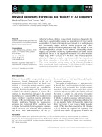
The avian multisequence alignments were performed
on basis of the proregion and mature domain each.
Two condensed multifurcating trees were constructed,
emphasizing the reliable portion of pattern branches
Characterization of cathelicidins from C. coturnix F. Feng et al.
1574 FEBS Journal 278 (2011) 1573–1584 ª 2011 The Authors Journal compilation ª 2011 FEBS
(Fig. 4). Fig. 4A reveals that there is very little differ-
ence in the proregion segment of CATH1 and CATH3;
thus, they are considered to show evolutionary ‘close-
ness’ because there has been insufficient time for many
mutations to accumulate in their proregion. For
CATH2 (fowlicidin-2, Pc-CATH2 and Cc-CATH2),
the more different proregions from CATH1 and 3 were
observed (the less homology shown) (Fig. 3B), indicat-
ing the further evolutionary distance of CATH2 from
CATH1 and 3, as well as the greater length of time
CATH2 since they shared a common ancestor. In
addition, CATH1 and 3 from C. coturnix fall into one
branch, and CATH1 and 3 from Phasianus colchicus
and chicken fowlicidins are in another branch, suggest-
ing that cathelicidins in the C. coturnix-specific cluster
arose earlier from a common ancestor than the other
two species. Unlike the highly distinct mammalian
cathelicidins resulting from repeating gene duplication
events and subsequent divergence, phylogenetic analy-
sis of the mature peptide segment revealed significant
similarity of avian cathelicidin-derived antimicrobial
peptides, as supported by bootstrap values of up to
100% (Fig. 4B). One possible explanation might be
that the much stronger activity of Aves cathelicidin
(compared with Reptilia and Mammalia) is a result of
it having undergone much less gene evolution [28].
Cc-CATH1 ATGCTGAGCTGCTGGGTGCTGGTGCTGGCGCTGCTGGGGGGGGCCTGTGCCCTCCCGGCC 60
Cc-CATH2 T 60
Cc-CATH3 60
Cc-CATH1 CCCCTGGATTACAACCAGGCTCTGGCCCAGGCTGTGGACTCCTACAACCAACGGCCCGAG 120
Cc-CATH2 T AGC CC G AT A 120
Cc-CATH3 C 120
Cc-CATH1 GTGCAGAATGCCTTCAGGCTGCTCAGCGCCGACCCCGAACCCGGCCCAAACGTCCAGCTC 180
Cc-CATH2 -C T GG-A-TG-T G 180
Cc-CATH3 180
Cc-CATH1 AGCTCCCTGCACAACCTCAACTTCACCATCATGGAGACGCGGTGCCAGGCGCGTTCGGGT 240
Cc-CATH2 -A-A-G GGG-G CGA GTCC-CA-CG-AC-G 240
Cc-CATH3 240
Cc-CATH1 GCCCAGCTTGAAAGCTGCGACTTCAAGGAGGACGGGCTCGTCAAGGACTGCGCTGCGCCC 300
Cc-CATH2 A-A-GCA-C TGA A GC-A T-G-G A 300
Cc-CATH3 300
Cc-CATH1 GTGGTGCTGCAAGGCGGCCGCGCCGTGCTCGATGTCACCTGCGTGGACTCCATGGCTGAT 360
Cc-CATH2 ACCA-C-TGCAG-A-GCAC-T-A-A AGCC-G-A AGA G TC-T-G 360
Cc-CATH3 360
Cc-CATH1 CCTGTCCGTGTCAAGCGCGTCTTGCCGCTGGT CATCAGGACTGTGATTGCA 411
Cc-CATH2 C TC C G G G-TTGGCC GC-T-C 397
Cc-CATH3 G T G GCCGGTGGC AC G GC G 420
Cc-CATH1 GGATACAACCTCTACCGGGCAATCAAGAGGAAGTGAgccgtccccagagctgctgtcacc 471
Cc-CATH2 T AGA-GGTC-G GCTT TC-CTA TCA-C-T-GCCG T-G CA-G-T 457
Cc-CATH3 CAT AAA C G ATGA acg-t c 480
Cc-CATH1 actgtcccctcgctgccttccatccaataaa
ggtctttgctggtaaaaaaaaaaaaaaaa 531
Cc-CATH2 TTG-CTGAg-gaataaa
-ggggc gtgtg c-accaagc-a 517
Cc-CATH3 g tc a cc c aataaa
-c-g ttca-gct 540
C c- CATH 1 aa a a aa a a aa aa a a 5 45
C c -C AT H 2 - - - - 5 31
C c -C AT H 3 - - - - a 55 5
Fig. 1. Alignment of the cDNA sequences
of three Cc-CATHs. The stop codons (‘TGA’)
are shown in bold. Dashes represent similar
sequences. The 3¢-UTR is shown in lower-
case letters. The potential polyadentlation
signal (aataaa) is underlined. Gaps are
inserted to maximize the similarity.
F. Feng et al. Characterization of cathelicidins from C. coturnix
FEBS Journal 278 (2011) 1573–1584 ª 2011 The Authors Journal compilation ª 2011 FEBS 1575
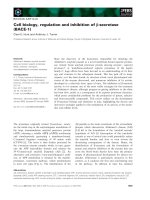
Cc-CATH2 expression and purification
In the Escherichia coli BL21 and pET-32a(+) plasmid
protein expression system, the deduced mature Cc-
CATH2 was expressed directly as a His-tagged fusion
protein. After induction with 1 mm isopropyl thio-b-d-
galactoside (IPTG) for 4 h, a high expression level of
fusion protein was noted in E. coli BL21 (Fig. 5A).
However, the fusion protein was primarily produced as
the inclusion body (Fig. 5B). After denaturation and
His-tag affinity chromatography, the fusion protein
was renatured and examined by SDS ⁄ PAGE gel
(Fig. 5C), indicating a clear and unique protein band
of 21.7 kDa, which matched well with the theoretical
mass of the fusion protein.
After formic acid cleavage for almost 24 h at 50 °C,
the fusion protein was cleaved into two parts: rCc-
CATH2 ( 3.8 kDa) and carrier protein ( 16.9 kDa).
The reaction mixture was lyophilized to remove formic
acid and then the rCc-CATH2 was subjected to further
purification by RP-HPLC. The antibacterial activity of
rCc-CATH2 toward Staphylococcus aureus ATCC2592
was examined by an inhibition zone assay, and a clear
inhibition zone was observed around the spot of the
peptide, indicating that the recombinant Cc-CATH2
retained antimicrobial activity.
Antimicrobial activity of Cc-CATHs
Cc-CATH2 and Cc-CATH3 were commercially synthe-
sized by the standard solid phase synthesis method
and purified to > 95% purity. LL-37 characterized
from humans and the antibiotics, ampicillin and kana-
mycin, were used as positive controls. Essentially,
Cc-CATH2 and Cc-CATH3 showed strong and broad-
spectrum antimicrobial activities against most of the
tested microorganisms, especially a number of clinical
drug-resistant strains (Table 1). For most strains, the
minimum inhibitory concentration (MICs) are within
the range 1.3–2.5 lm, with and without the presence of
100 mm NaCl, whereas ampicillin, kanamycin and
LL-37 often did not show detectable activity in an
inhibition zone assay at dose of up to 2 mgÆmL
)1
. The
lowest MICs of Cc-CATH2 and 3 were detected both
for S. aureus ATCC25922, 0.3 and 0.2 lm, respec-
tively. With respect ot several Gram-positive S. aureus
clinical strains, Cc-CATH3 showed an almost ten-fold
higher activity than Cc-CATH2. However, for most of
Gram-negative bacteria tested, the result was opposite
(i.e. Cc-CATH2 was much more active than Cc-
CATH3). For example, the MIC of Cc-CATH2 to
E. coli ATCC25922 was as low as 2.5 lm, although no
detectable activity was observed for Cc-CATH3 at
2mgÆmL
)1
. By contrast to LL-37 and EA-CATH1
(cathelicidin-derived antimicrobial peptides from
Equus asinus), which have weak Gram-negative bacte-
ricidal activities [32], Cc-CATH2 exerted comparable
antimicrobial activity upon most of the E. coli, with
MICs in the range 1.3–2.5 lm.
The effect of sodium upon the antimicrobial
activities of Cc-CATH2 and 3 was also examined
(Table 1). Unlike many antimicrobial peptides for
which activities are inhibited by sodium at physiologi-
cal concentrations [33–37], Cc-CATH2 and 3 showed
salt-independent activities with or without the presence
of 100 mm NaCl (Table 1), suggesting their suitability
for both local and systemic therapeutic applications.
Cytotoxicity, hemolysis of Cc-CATHs
The 3-(4,5-dimethylthiazol-2-yl)-2,5-diphenyltetrazolium
bromide method was exploited to evaluate the cytotox-
icity of Cc-CATHs toward two mammalian cell lines,
HUVEC (human umbilical vein endothelial cells) and
Raw 264.7. The results obtained revealed average IC
50
values of 75 lgÆmL
)1
(20.18 lm) for Cc-CATH2 and
58 lgÆmL
)1
(17.16 lm) for Cc-CATH3 toward both
cell lines, which is almost ten-fold higher than their
corresponding MICs, suggesting the potential for thera-
peutic application.
Cc-CATH1 MLSCWVLVLALLGGACALPAPLDYNQALAQAVDSYNQRPEVQNAFRLLSADPEPGPNVQL 60
Cc-CATH2 V S-P I T A GID- 60
Cc-CATH3 60
Cc-CATH1 SSLHNLNFTIMETRCQARSGAQLESCDFKEDGLVKDCAAPVVLQGGRAVLDVTCVDSMAD 120
Cc-CATH2 NT-RE E-VPSARTRIDD N-AI SG TILQDAPEISLN-R-ASS- 120
Cc-CATH3 120
Cc-CATH1 PVRVKRVLPLV IRTVIAGYNLYRAIKRK 148
Cc-CATH2 L-Q-GRFGRFLKKVRRFIPKVIIA-QIGSRFG 154
Cc-CATH3 RVR-FW PVA-N A I K R 151
Fig. 2. Alignment of the predicted precursor
amino acid sequences of the Cc-CATHs.
Gaps are inserted to optimize the alignment.
Identical residues are indicated by dashes.
Characterization of cathelicidins from C. coturnix F. Feng et al.
1576 FEBS Journal 278 (2011) 1573–1584 ª 2011 The Authors Journal compilation ª 2011 FEBS
A possible limitation to the clinical application of
antimicrobial peptides as antibiotics is their potential to
cause injury to mammalian cell membranes. In the
present study, the hemolytic activities of Cc-CATHs
were also examined using freshly prepared human
erythrocytes. As shown in Table 2, Cc-CATH2 and 3
both displayed negligible hemolytic activities, lysing only
3.6% and 4.1% of erythrocytes at concentrations up to
26.9 lm (100 lgÆmL
)1
) and 29.6 lm (100 lgÆmL
)1
),
respectively. The hemolysis concentrations are much
62Pc-CATH1
62Pc-CATH2
62Pc-CATH3
62Cc-CATH1
62Cc-CATH2
62Cc-CATH3
62Fowlicidin1
62Fowlicidin2
62Fowlicidin3
72Ea CATH1
72Ec CATH1
72Ec CATH2
72Ec CATH3
73Hs LL37
72Ss PR39
72Bt CATHL1
73Oa SMAP29
72Ch BAC5
73Cp CAP11
70Mm CRAMP
72Oc CAP18
72Clf K9CATH
68Bf cath
MLSCWVLVLALLGGACALPAP LGYSQALAQAVDSYNQRPEVQ.NAFRLLSADPEPGPN.VQLGS
MLSCWVLVLALLGGVCALPAP LSYPQALTQAVDSYNQRPELQ.NAFRLLSADPEPGPG.VDLST
MLSCWVLVLALLGGACALPAP LGYSQALAQAVDSYNQRPEVQ.NAFRLLSADPEPGPN.VQLGS
MLSCWVLVLALLGGACALPAP LDYNQALAQAVDSYNQRPEVQ.NAFRLLSADPEPGPN.VQLSS
MLSCWVLVLALLGGVCALPAP LSYPQALIQAVDTYNQRPEAQ.NAFRLLSADPEPGPG.IDLNT
MLSCWVLVLALLGGACALPAP LDYNQALAQAVDSYNQRPEVQ.NAFRLLSADPEPGPN.VQLSS
MLSCWVLLLALLGGACALPAP LGYSQALAQAVDSYNQRPEVQ.NAFRLLSADPEPGPN.VQLSS
MLSCWVLLLALLGGVCALPAP LSYPQALIQAVDSYNQRPEVQ.NAFRLLSADPEPGPG.VDLST
MLSCWVLLLALLGGACALPAP LGYSQALAQAVDSYNQRPEVQ.NAFRLLSADPEPGPN.VQLSS
METQRDSCSLGWWSLLLLLLGLMIPLATT.QALSYKEAVLRAVDGLNQWSSDE.NLYRLLELDPLPKGD.EAPDT
METQRNTRCLGRWSPLLLLLGLVIPPATT.QALSYKEAVLRAVDGLNQRSSDE.NLYRLLELDPLPKGD.KDSDT
METQRDSCSLGRWSLLLLLLGLVIPLATT.QTLSYKEAVLRAVDGLNQRSSDE.NLYRLLELDPLPKED.EDPDT
METQRNTRCLGRWSPLLLLLGLVIPPATT.QALSYKEAVLRAVDGLNQRSSDE.NLYRLLELDPLPKGD.KDSDT
MKTQRDGHSLGRWSLVLLLLGLVMPLAIIAQVLSYKEAVLRAIDGINQRSSDA.NLYRLLDLDPRPTMD.GDPDT
METQRASLCLGRWSLWLLLLGLVVPSAST.QALSYREAVLRAVDRLNEQSSEA.NLYRLLELDQPPKAD.EDPGT
METPRASLSLGRWSLWLLLLGLALPSASA.QALSYREAVLRAVDQLNEQSSEP.NIYRLLELDQPP.QDDEDPDS
METQRASLSLGRRSLWLLLLGLVLASARA.QALSYREAVLRAVDQLNEKSSEA.NLYRLLELDPPPKQDDENSNI
METQGASLSLGRWSLWLLLLGLVVPLASA.QALSYREAVLRAVGQLNERSSEA.NLYRLLELDPAPNDE.VDPGT
MGTPRDAASGGPRLLLPLLLLLLLTPATA.WVLSYQQAVQRAVDGINKNLADNENLFRLLSLDTQPPGD.NDPYS
MQFQRDVPSLWLWRSLSLLLLLGL GFS.QTPSYRDAVLRAVDDFNQQSLDT.NLYRLLDLDPEPQGD.EDPDT
METHKHGPSLAWWSLLLLLLGLLMPPAIA.QDLTYREAVLRAVDAFNQQSSEA.NLYRLLSMDPQQLED.AKPYT
METQKDSPSLGRWSLLLLLLGLVITPAAS.RALSYREAVLRAVNGFNQRSSEE.NLYRLLQLNSQPKGD.EDPNI
MEGFFWKTLLVVGALAIAGTSSLPH.KPLIYEEAVDLAVSIYNSKSGEDS.LYRLLEAVSPPKWD.PLSES
L
L
L
L
L
L
L
L
L
L
L
L
L
L
L
L
L
L
L
L
L
L
L
Y
Y
Y
Y
Y
Y
Y
Y
Y
Y
Y
Y
Y
Y
Y
Y
Y
Y
Y
Y
Y
Y
Y
A
A
A
A
A
A
A
A
A
A
A
A
A
A
A
A
A
A
A
A
A
A
A
A
A
A
A
A
A
A
A
A
A
A
A
A
A
A
A
A
A
A
A
A
A
A
N
N
N
N
N
N
N
N
N
N
N
N
N
N
N
N
N
N
N
N
N
N
N
R
R
R
R
R
R
R
R
R
R
R
R
R
R
R
R
R
R
R
R
R
R
R
L
L
L
L
L
L
L
L
L
L
L
L
L
L
L
L
L
L
L
L
L
L
L
L
L
L
L
L
L
L
L
L
L
L
L
L
L
L
L
L
L
L
L
L
L
L
122Pc-CATH1
122Pc-CATH2
122Pc-CATH3
122Cc-CATH1
122Cc-CATH2
122Cc-CATH3
122Fowlicidin1
122Fowlicidin2
122Fowlicidin3
130Ea CATH1
130Ec CATH1
130Ec CATH2
130Ec CATH3
133Hs LL37
130Ss PR39
143Bt CATHL1
131Oa SMAP29
130Ch BAC5
131Cp CAP11
139Mm CRAMP
134Oc CAP18
134Clf K9CATH
143Bf cath
LHNLNFTIIETRCQARSGAQLDSCEFKEDGLVKDCAAPVVLQGGRATFDVTCVESVADPV
LRTLNFTIMETECVPRAQTPIDDCDFKENGVIRDCSGPVTILQDTPEINLRCRDASSDPV
LHNLNFTIMETRCQARSGAQLDSCEFKEDGLVKDCAAPVVLQGGRATFDVTCVDSMADPV
LHNLNFTIMETRCQARSGAQLESCDFKEDGLVKDCAAPVVLQGGRAVLDVTCVDSMADPV
LRELNFTIMETECVPSARTRIDDCDFKENGAIKDCSGPVTILQDAPEISLNCRDASSDPV
LHNLNFTIMETRCQARSGAQLESCDFKEDGLVKDCAAPVVLQGGRAVLDVTCVDSMADPV
LHNLNFTIMETRCQARSGAQLDSCEFKEDGLVKDCAAPVVLQGGRAVLDVTCVDSMADPV
LRALNFTIMETECTPSARLPVDDCDFKENGVIRDCSGPVSVLQDTPEINLRCRDASSDPV
LHNLNFTIMETRCQARSGAQLDSCEFKEDGLVKDCAAPVVLQGGRAVLDVTCVDSMADPV
PKPVSFTVKETVCPRTTQQPLEQCDFKENGLVKQCVGTVILDPVKASVDIGCDEPQRV
PKPVSFMVKETVCPRIMKQTPEQCDFKENGLVKQCVGTVILGPVKDHFDVSCGEPQRV
PKPVSFTVKETVCPRTTQQPLEECDFKENGLVKQCVGTVVLDPAKDYFDISCDKPQPI
PKPVSFMVKETVCPRIMKQTPEQCDFKENGLVKQCVGTVILDPVKDYFDASCDEPQRV
PKPVSFTVKETVCPRTTQQSPEDCDFKKDGLVKRCMGTVTLNQARGSFDISCDKDNKRFA
PKPVSFTVKETVCPRPTQRPPELCDFKENGRVKQCVGTVTLNPSNDPLDISCNEIQSV
PKRVSFRVKETVCSRTTQQPPEQCDFKENGLLKRCEGTVTLDQVRGNFDITCNNHQSIRITKQPWAPPQAA
PKPVSFRVKETVCPRTSQQPAEQCDFKENGLLKECVGTVTLDQVGNNFDITCAEPQSV
RKPVSFTVKETVCPRTTQQPPEECDFKENGLVKQCVGTVTLDPSNDQFDINCNELQSV
PKPVSFTIKETVCTKMLQRPLEQCDFKENGLVQRCTGTVTLDSAFNVSSLSCLGGRRF
PKSVRFRVKETVCGKAERQLPEQCAFKEQGVVKQCMGAVTLNPAADSFDISCNEPGAQPFRFKKISRLA
PQPVSFTVKETECPRTTWKLPEQCDFKEDGLVKRCVGTVTRYQAWDSFDIRCNRAQESPEPT
PKPVSFTVKETVCPKTTQQPLEQCGFKDNGLVKQCEGTVILDEDTGYFDLNCDSILQVKKID
NQELNFTMKETVCLVAEERSLEECDFQEDGVVMGCTGYYFFGESPPVVVLTCKPVGEEGEQKQEEGNEEEKEVEE
F
F
F
F
F
F
F
F
F
F
F
F
F
F
F
F
F
F
F
F
F
F
F
E
E
E
E
E
E
E
E
E
E
E
E
E
E
E
E
E
E
E
E
E
E
E
T
T
T
T
T
T
T
T
T
T
T
T
T
T
T
T
T
T
T
T
T
T
T
C
C
C
C
C
C
C
C
C
C
C
C
C
C
C
C
C
C
C
C
C
C
C
C
C
C
C
C
C
C
C
C
C
C
C
C
C
C
C
C
C
C
C
C
C
C
F
F
F
F
F
F
F
F
F
F
F
F
F
F
F
F
F
F
F
F
F
F
F
G
G
G
G
G
G
G
G
G
G
G
G
G
G
G
G
G
G
G
G
G
G
G
C
C
C
C
C
C
C
C
C
C
C
C
C
C
C
C
C
C
C
C
C
C
C
C
C
C
C
C
C
C
C
C
C
C
C
C
C
C
C
C
C
C
C
C
C
C
148Pc-CATH1
154Pc-CATH2
151Pc-CATH3
148Cc-CATH1
154Cc-CATH2
151Cc-CATH3
148Fowlicidin1
154Fowlicidin2
151Fowlicidin3
155Ea CATH1
156Ec CATH1
157Ec CATH2
170Ec CATH3
170Hs LL37
172Ss PR39
155Bt CATHL1
160Oa SMAP29
176Ch BAC5
177Cp CAP11
172Mm CRAMP
171Oc CAP18
172Clf K9CATH
191Bf cath
RIKRFWPVVIRTVVAGYNLYRAIKKK
LVQRGRFGRFLSKIRRFRPKFTITIQGSGRFG
RIKRFWPLVPVAINTVAAGINLYKAIKRK
RVKRVLPLVIRTVIAGYNLYRAIKRK
LVQRGRFGRFLKKVRRFIPKVIIAAQIGSRFG
RVRRFWPLVPVAINTVAAGINLYKAIRRK
RVKRVWPLVIRTVIAGYNLYRAIKKK
LVQRGRFGRFLRKIRRFRPKVTITIQGSARFG
RVKRFWPLVPVAINTVAAGINLYKAIRRK
KRRGSVTTRYQFLMIHLLRPKKLFA
KRFGRLAKSFLRMRILLPRRKILLAS
KRRHWFPLSFQEFLEQLRRFRDQLPFP
KRFHSVGSLIQRHQQMIRDKSEATRHGIRIITRPKLLLAS
LLGDFFRKSKEKIGKEFKRIVQRIKDFLRNLVPRTES
RRRPRPPYLPRPRPPPFFPPRLPPRIPPGFPPRFPPRFPGKR
RLCRIVVIRVCR
RGLRRLGRKIAHGVKKYGPTVLRIIRIAG
RFRPPIRRPPIRPPFNPPFRPPVRPPFRPPFRPPFRPPIGPFPGRR
RRMVGLRKKFRKTRKRIQKLGRKIGKTGRKVWKAWREYGQIPYPCR
GLLRKGGEKIGEKLKKIGQKIKNFFQKLVPQPE
GLRKRLRKFRNKIKEKLKKIGQKIQGFVPKLAPRTDY
RLKELITTGGQKIGEKIRRIGQRIKDFFKNLQPREEKS
EEQEEDEKDQPRRV KRFKKFFRKLKKSVKKRAKEFFKKPRVIGVSIPF
A
Fig. 3. (A) Multiple sequence alignment of Cc-CATHs with classic cathelicidins from different species. The conserved amino acid residues in
cathelin domain are shaded, including the typical four conserved cysteine residues. Each mature cathelicidin is aligned in the third line. Pc,
P. colchicus (ring necked pheasant); Fowlicidin (chicken); Hs, Homo sapiens (human); Ss, Sus scrofa (pig); Bt, Bos taurus (cattle); Oa, Ovis
aries (sheep); Ch, Capra hircus (goat); Cp, Cavia porcellus (guinea pig); Ec, Equus caballus (horse); Mm, Mus musculus (mouse); Oc, Oryctol-
agus cuniculus (rabbit); Clf, Canis lupus familiars (dog); Bf, Bungarus fasciatus (snake). Dots are inserted to maximize the similarity. (B) Align-
ment of Cc-CATHs with avian cathelicidins. Each mature cathelicidin is boxed.
F. Feng et al. Characterization of cathelicidins from C. coturnix
FEBS Journal 278 (2011) 1573–1584 ª 2011 The Authors Journal compilation ª 2011 FEBS 1577
higher than the corresponding MICs, except for two of
the S. aureus clinical isolated strains (Table 1), suggest-
ing the considerable selectivity of Cc-CATH2 and 3 for
microorganisms over mammalian cells in vitro.
Discussion
The emergence of widespread antibiotic resistance in
numerous commonly encountered bacteria requires the
discovery of new bactericidal agents with therapeutic
potential. Currently, a new superbug is being reported
that is resistant to even the most powerful antibiotics,
and has produced dangerous infections in countries
such as the USA, Canada, Australia and the Nether-
lands [38]. The bacteria synthesizes an enzyme called
NDM-1 that can exist inside different bacteria, such as
E. coli, making them resistant to one of the most pow-
erful groups of antibiotics (i.e. carbapenems). There-
fore, tight surveillance and new drugs are needed to
manage this threat. The cathelicidin family of endoge-
nous antimicrobial peptides serves a critical role in
mammalian innate immune defense against invasive
bacterial infection [39]. The cathelicidin-derived antimi-
crobial peptides have recently received attention
because of their much stronger bactericidal activities
compared to chemical drugs, as well as their unique
killing mechanism, as a result of which drug resistance
is difficult to develop. They kill microorganisms by cre-
ating pores or holes in pathogen membranes, unlike
the conventional b-lactam antibiotics, which kill most
bacteria by inhibiting the synthesis of one of their cell
wall layers [40,41]. Cathelicidins can kill both Gram-
positive and -negative bacteria, enveloped viruses
including HIV, and fungi including Candida and Cryp-
tococcus [3]. As antibiotics, cathelicidins are also effec-
tive against resistant staphylococcus, enterococcus and
pseudomonas in animal models [34,42,43]. They are
also found to bind lipopolysaccharide or recruit the
immune system, and to inhibit reactive oxygen species
created by neutrophils, thus mitigating excess tissue
damage [44–46].
In the present study, three cathelicidins were identi-
fied from a C. coturnix cDNA library. The cDNAs of
Cc-CATHs demonstrate the same conserved cathelici-
din family gene organization, including the signal
B
Fig. 3. (Continued).
A
Fowlicidin 1
Fowlicidin 3
PC-CATH1
PC-CATH3
CC-CATH-1
CC-CATH-3
Fowlicidin 2
PC-CATH2
CC-CATH-2
98
95
94
70
100
B
CC-CATH-3
Fowlicidin 3
PC-CATH3
CC-CATH-1
Fowlicidin 1
PC-CATH1
CC-CATH-2
PC-CATH2
Fowlicidin 2
84
81
58
53
79
100
Fig. 4. Phylogenetic analyses of Cc-CATHs and avian cathelicidins
on the basis of the proregion (A) and mature domain (B). The phylo-
genetic dendrogram was constructed by the Neighbor-joining
method based on the proportion difference of aligned amino acid
sites of the sequence. Only branches supported by a bootstrap
value of at least 50% (expressed as percentage of 1000 bootstrap
samples supporting the branch) are shown at the branching points.
Characterization of cathelicidins from C. coturnix F. Feng et al.
1578 FEBS Journal 278 (2011) 1573–1584 ª 2011 The Authors Journal compilation ª 2011 FEBS
peptide, the cathelin domain, and the deduced mature
antimicrobial peptide of 26, 32 and 29 amino acid resi-
dues, respectively. Moreover, the four highly conserved
cysteines were also maintained in the pro-region
sequences. Cc-CATH1-3 is markedly conserved with
chicken fowlicidin-1–3. However, the data obtained
from antimicrobial testing indicated that Cc-CATH2
was not as strongly active as its pair fowlicidin-2, and
Cc-CATH3 was also less active compared to fowlici-
din-1 and -2 [16]. The MICs of fowlicidin-1 and -2 are
in the range 0.4–2.0 lm for most strains [16]. Another
cathelicidin-derived peptide, Pc-CATH1 (pairs with
fowlicidin1 and Cc-CATH1), which was identified
from P. colchicus in a previous study [28], also pos-
sesses potent antimicrobial activity, with most MICs
in the range 0.09–2.95 lm. To explain the different
bactericidal performances of these peptides that have
great sequence similarity, their secondary structures
were predicted online using gor iv (http://npsa-pbil.
ibcp.fr/cgi-bin/npsa_automat.pl?page=npsa_gor4.html).
The results obtained demonstrated that the a-helical
content for the ‘strong group’, including fowlicidin 1-2
and Pc-CATH1, is 38.46%, 38.71% and 38.46%,
respectively. For the ‘weak group’, Cc-CATH2 and 3,
the a-helical content is 62.50% and 65.52%, respec-
tively, which is almost two-fold higher than the ‘strong
group’. Although the a-helical structure is considered
to be responsible for the formation of pores in the
membranes of target microorganisms [47], the results
of the present study indicate that the percentage of the
a-helix must be within an optimal range for the pep-
tide to achieve its best activity.
Although the antimicrobial activities of Cc-CATHs
are not as potent as those of Pc-CATH1 and fowlici-
din-1 and -2, the hemolytic activities of Cc-CATHs are
significantly lower. The considerable reduction of cyto-
toxic activity, as well as potent and broad-spectrum
antimicrobial activity, even against clinical drug-resis-
tant strains, offers a marked improvement in terms of
the application of Cc-CATHs for the treatment of bac-
terial and fungal infections.
Materials and methods
Collection of tissues
Two adult female quails were captured from Zhengding,
Hebei Province of China. One quail was killed and the
spleen was dissected immediately and frozen in liquid nitro-
gen until use.
Total RNA extraction and SMART cDNA
synthesis
Total RNA was extracted from the spleen of quail using
RNeasy Mini Kit (Qiagen, Hildenberg, Germany) in accor-
dance with the manufacturer’s instructions. cDNA synthesis
was carried out by a PCR-based method using a CreatorÔ
kDa
21.7 kDa
kDa 1 2 3 4 0 5 6 7 8
01 2 3 4
10kDa
100
80
60
50
40
30
20
12
234
170
A
B
C
130
100
70
55
40
35
25
15
170
130
100
70
55
40
35
25
15
Fig. 5. (A) Expression and purification of
Cc-CATH2 fusion protein (indicted by an
arrow) followed by SDS ⁄ PAGE (15%). Lane
1, the whole lysate without IPTG; lanes 2–4,
the whole lysate with 1 m
M IPTG for 4 h;
lane 0, protein standards (kDa). (B) The
results of SDS ⁄ PAGE (15%) for supernatant
and precipitation at the same time. Lanes
1–3, precipitation with IPTG; lane 4, precipi-
tation without IPTG; lane 5, supernatant
without IPTG; lanes 6–8, supernatant with
IPTG; lane 0, protein standards. (C) Protein
bands after affinity chromatography and
renaturing process. Lanes 1 and 2, protein
bands after separation by affinity column;
lanes 3 and 4, protein bands after renaturing
process: lane 0, protein standards.
F. Feng et al. Characterization of cathelicidins from C. coturnix
FEBS Journal 278 (2011) 1573–1584 ª 2011 The Authors Journal compilation ª 2011 FEBS 1579
Table 1. Antimicrobial activity of Cc-CATHs. MIC, minimal inhibitory concentration (these concentrations represent the mean values of three
independent experiments performed in duplicate); Amp, ampicillin; Kana, kanamycin; ND, no detectable activity in inhibition zone assay at a
dose of 2 mgÆmL
)1
; IS, clinically isolated strain; Dra, drug resistance for ceftazidime, cefoperazone and aztreonam; DRb, drug resistance for
compound sulfamethoxazole, erythromycin, ciprofloxacin and penicillin.
Microorganism
MIC (l
M)
Cc-CATH2
(0 m
M NaCl)
Cc-CATH2
(100 mM NaCl)
Cc-CATH3
(0 mM NaCl)
Cc-CATH3
(100 mM NaCl)
LL-37
(0 mM NaCl) Amp Kana
Gram-positive
Staphylococcus aureus
ATCC2592
0.3 0.3 0.2 0.4 1.0 0.79 8.05
S. aureus 1.3 1.3 0.7 0.7 4.2 6.31 128.74
S. aureus (IS 1303) 20.2 20.2 2.8 2.8 ND 100.97 ND
S. aureus (IS 1307) > 26.9 > 26.9 2.8 2.8 > 22.3 6.31 ND
S. aureus (IS 1348) 20.2 20.2 2.8 2.8 > 22.3 50.48 ND
S. aureus (IS 1349) 20.2 20.2 2.8 2.8 > 22.3 100.97 ND
S. aureus (IS 1350) 20.2 20.2 2.8 2.8 ND 100.97 ND
Staphylococcus epidermidis 2.5 2.5 ND ND ND 201.94 4.02
Staphylococcus haemolyticus
(IS 2401, DRa)
2.5 2.5 0.7 0.7 ND 1.58 16.09
Nocardia asteroids 1.3 1.3 0.7 0.7 4.2 3.16 128.74
Enterococcus faecalis (IS 981) 1.3 1.3 5.6 11.1 > 22.3 201.94 ND
Enterococcus faecium (IS 1299) 2.5 2.5 1.4 1.4 4.2 ND ND
Propionibacterium acnes
ATCC 11827
1.3 2.5 1.4 1.4 > 22.3 3.16 4.02
Gram-negative
Klebsiella oxytoca 2.5 2.5 22.2 22.2 ND ND ND
Aeromonas sobria 1.3 1.3 1.4 2.8 4.2 ND ND
Acinetobacter baumannii
(IS 2178, DRb)
1.3 1.3 1.4 1.4 ND 100.97 4.02
A. baumannii (IS 2373) 2.5 1.3 1.4 2.8 ND ND ND
Stenotrophomonas maltophilia 1.3 1.3 1.4 2.8 4.2 ND ND
S. maltophilia (IS 1404) 0.6 0.6 5.6 5.6 > 22.3 ND ND
Pseudomonas aeruginosa
ATCC 27853
10.1 10.1 5.6 11.1 ND ND ND
P. aeruginosa (IS 1411) 10.1 10.1 5.6 5.6 > 22.3 ND ND
P. aeruginosa (IS 1412) 10.1 10.1 5.6 11.1 ND ND ND
P. aeruginosa (IS 1413) 10.1 10.1 5.6 5.6 ND > 269.25 128.74
Escherichia coli ATCC 25922 2.5 2.5 ND ND ND 25.24 16.09
E. coli 5.1 2.5 ND ND ND 201.94 4.02
E. coli (IS 1334) 2.5 1.3 11.1 11.1 ND ND 32.18
E. coli (IS 1335) 1.3 2.5 5.6 5.6 ND ND ND
E. coli (IS 1342) 1.3 1.3 2.8 2.8 ND ND 32.18
E. coli (IS 1375) 2.5 2.5 11.1 11.1 > 22.3 50.48 4.02
Serratia marcescens (IS 1379) ND ND ND ND ND ND ND
Klebsiella pneumoniae (IS 1368) 1.3 1.3 ND ND 4.2 ND 64.37
K. pneumoniae (IS 1372) 2.5 2.5 ND ND ND ND ND
K. pneumoniae (IS 1373) 2.5 2.5 ND ND ND ND 4.02
K. pneumoniae (IS 1400) 5.1 5.1 22.2 22.2 ND ND ND
Proteus vulgaris 10.1 10.1 > 29.6 > 29.6 ND 3.16 8.05
Proteus mirabilis 5.1 5.1 1.4 1.4 ND 6.31 8.05
Salmonella typhi (IS 1408) 5.1 5.1 ND ND ND ND 32.18
Fungi
Candida albicans ATCC 2002 1.3 1.3 0.7 0.7 2.1 1.58 2.01
Candida glabrata (IS 0902) 10.1 10.1 5.6 5.6 ND ND ND
Slime mold 0.6 1.3 0.7 0.7 4.2 6.31 128.74
Characterization of cathelicidins from C. coturnix F. Feng et al.
1580 FEBS Journal 278 (2011) 1573–1584 ª 2011 The Authors Journal compilation ª 2011 FEBS
SMARTÔ cDNA library construction kit (Clontech, Palo
Alto, CA, USA). First-strand cDNA was synthesized by
SMARTÔ IV oligonucleotide primer 5¢-AAGCAGTGG-
TATCAACGCAGAGTGGCCATTACGGCCGGG-3¢ and
CDS III ⁄ 3¢ PCR primer 5¢-ATTCTAGAGGCCGAGGC
GGCCGACATGT (30)N
–1
N-3¢ (N = A, G, C or T; N
–1
= A, G or C); the reverse transcriptase used was Power-
Script Reverse Transcriptase, as supplied with the kit.
Second-strand cDNA was amplified by 5¢ PCR primer
5¢-AAGCAGTGGTATCAACGCAGAGT-3¢ and CDS
III ⁄ 3¢ PCR primer, using Advantage DNA Polymerase
from Clontech.
Screening of cathelicidin-encoding cDNAs and
phylogenetic tree construction
On the basis of the conserved signal domain of previously
characterized chicken fowlicidin cDNAs [21], two sense
primers P1 (5¢-AGGATGCTGAGCTGCTGGGT-3¢) and
P2 (5¢-ATGCTGAGCTGCTGGGTGCT-3¢) were designed
from 5¢-UTR and a highly conserved domain encoding the
signal peptide of fowlicidins, and coupled with CDS III ⁄ 3¢
PCR primer. The half nested PCR conditions consisted of
two parts. The first part comprised: 94 °C for 1 min; 20
cycles of 94 °C for 20 s, 60 °C for 30 s, 72 °C for 60 s; fol-
lowed by a final extension at 72 °C for 5 min. The second
part comprised: 94 °C for 3 min; 25 cycles of 94 °C for
20 s, 58 °C for 30 s, 72 °C for 60 s; followed by a final
extension at 72 °C for 10 min. The PCR product was puri-
fied by gel electrophoresis and cloned into pGEM-T vector
(Promega, Madison, WI, USA). DNA sequencing was per-
formed using an ABI PRISM 377 (Applied Biosystems,
Foster City, CA, USA).
In total, nine avian cathelicidin sequences were obtained
from the protein database at the National Center for Bio-
technology Information. These were the fowlicidins [16],
Pc-CATHs [28] and Cc-CATHs from the present study.
Multisequence alignments were constructed using clu-
stalw, version 1.8 ( based
on the proregion and mature domain. The phylogenetic
trees were constructed using the Neighbor-joining method
(mega, version 4.0; www.megasoftware.net), by calculating
the proportion of amino acid differences (p-distance)
among all sequences. A total of 1000 bootstrap replicates
were used to test the reliability of each branch. The num-
bers on the branches indicate the percentage of 1000 boot-
strap samples supporting the branch.
Expression vector construction, protein
expression and purification
Host strain E. coli BL21 and pET-32a(+) plasmid (Nov-
agen, Darmstadt, Germany) was utilized for Cc-CATH2
expression. The method was carried out in accordance with
the manufacturer’s instructions and as described previously
by Li et al. [48].
The two restriction sites for KpnI and HindIII and the
formic acid cleavage site (AspPro) upstream of the deduced
mature Cc-CATH2 coding sequence were utilized in the
peptide expression. A DNA fragment encoding the gene for
Cc-CATH2 was amplified by PCR from the plasmids
described above. The first forward primer was 5¢-AC-
CGACCCGCTCGTCCAGCG-3¢ and the first reverse pri-
mer was 5¢-CTTCTAGCCAAAGCGTGAGCCGATC-3¢.
PCR was performed by running 30 cycles with a tempera-
ture profile of 30 s at 94 °C, 30 s at 64 °C and 10 s at
72 °C followed by a final extension at 72 °C for 10 min.
The second forward primer was 5¢-CGGGGTACC
GACCCGCTCGT-3¢ and the second reverse primer was 5¢-
CCCAAGCTTCTAGCCAAAGCGTG-3¢. PCR comprised:
30 cycles of 30 s at 94 °C, 30 s at 64 °C and 10 s at 72 °C,
followed by a final extension at 72 °C for 10 min. The puri-
fied PCR product was digested with KpnI and HindIII, and
ligated into the pET-32a(+) plasmid at the corresponding
restriction sites. The resultant recombinant vector is
referred to as Cc-CATH2 ⁄ pET-32a(+). The Cc-CATH2 ⁄
pET-32a(+) construct was transformed into the E. coli
strain BL21 for protein expression. The fusion protein
expression was initiated by adding IPTG.
After lysis by sonication, the whole cell lysate was then
centrifuged at 3914 g for 15 min, and then the supernatant
and precipitation were both resolved by SDS ⁄ PAGE. After
centrifugation, the fusion protein was found primarily in
the precipitation. The inclusion body was collected, washed
and resolved by denaturant solution. The solution was col-
lected and purified with a His-tag affinity column. After re-
natured in gradient, the Cc-CATH2-containing fusion
protein was cleaved in 50% formic acid (v ⁄ v) at 50 °C for
24 h. After lyophilization, the solution was subject to
HPLC (Hypersil BDS C18, Elite, Dalian, China;
30 · 0.46 cm). The peptide was eluted by a mixture of sol-
vents of acetonitrile ⁄ H
2
O ⁄ 0.1% trifluoroacetic acid at a
flow rate of 1 mLÆmin
)1
using a linear gradient of increas-
ing acetonitrile. Fractions corresponding to the major peak
were collected and lyophilized. Subsequently, the anti-
bacterial activity of expressed Cc-CATH2 with respect to
S. aureus ATCC2592 was examined.
Peptide synthesis
The deduced cathelicidin-derived mature peptides, LL-37,
Cc-CATH2 and 3 were synthesized by the peptide synthe-
sizer GL Biochem (Shanghai) Ltd. (Shanghai, China) and
Table 2. Hemolysis assay of Cc-CATHs.
Hemolytic activity
Concentration (lgÆmL
)1
) 100 50 20 10
Cc-CATH2 (%) 3.6 0.0 0.0 0.0
Cc-CATH3 (%) 4.1 1.3 0.0 0.0
F. Feng et al. Characterization of cathelicidins from C. coturnix
FEBS Journal 278 (2011) 1573–1584 ª 2011 The Authors Journal compilation ª 2011 FEBS 1581
analyzed by HPLC and MALDI-TOF MS to confirm that
the purity was higher than 98%. All peptides were dissolved
in water and used for activity examination, as described
below.
Antimicrobial assay
To examine the antibacterial spectrum of Cc-CATHs, a
modified broth microdilution assay was used as described
in a previous study [49]. The microorganisms evaluated
included standard and clinically isolated drug resistance
bacterial and fungal strains (Table 1). Briefly, bacteria were
subcultured to the midlogarithmic phase at 37 °C and sus-
pended to 5 · 10
5
colony-forming unitsÆmL
)1
in Mueller–
Hinton (MH) broth with and without 100 mm of NaCl.
The peptides in the presence and absence of 100 mm NaCl
were subjected to serial dilutions in MH broth, and then
50 lL of the diluted samples was dispensed into a 96-well
microtiter plate and mixed with 50 lL of bacteria or yeast
inoculums in MH. Human cathelicidin LL-37 (without
NaCl), ampicillin and kanamycin was used as a positive
control. The microtiter plate was incubated at 37 °C for
18 h for bacteria and 48 h for fungus, and A
595
was mea-
sured. MIC was defined as the lowest concentration of pep-
tide that completely inhibits the growth of the microbe as
determined by visual inspection and spectrophotometric
examination.
Cytotoxicity assay
HUVEC and Raw 264.7 murine macrophage cells were
used to examine the in vitro cytotoxicity of Cc-CATHs. The
cells were cultured in DMEM (Gibco, Gaithersburg, MD,
USA) supplemented with 10% fetal bovine serum,
100 UÆmL
)1
of penicillin and 100 UÆmL
)1
of streptomycin
in a humidified 5% CO
2
atmosphere at 37 °C. Cells
(2 · 10
4
per well) were seeded in 96-well plates and cultured
overnight until they adhered to the plate. Various concen-
trations of Cc-CATHs dissolved in the corresponding cul-
ture medium were added to the wells and the plates were
incubated at 37 °C for 48 h. Cytotoxicity of Cc-CATHs
was measured by the 3-(4,5-dimethylthiazol-2-yl)-2,5-diphe-
nyltetrazolium bromide method [50]. IC
50
was defined as
the concentration of Cc-CATHs at which A
490
was reduced
by 50%.
Hemolysis
Hemolysis assays were conducted as described previously
[51]. Cc-CATHs of four different concentrations were incu-
bated with washed human erythrocytes at 37 °C for 30 min
and centrifuged at 652 g for 5 min and A
540
of the superna-
tant was measured. 1% v ⁄ v Triton X-100 was used to
determine maximal hemolysis. The experiment was repeated
three times.
Acknowledgements
We thank the editor and the anonymous reviewers for
their helpful comments on the manuscript. This work
was supported by grants from the Chinese National
Natural Science Foundation (30900240 and 41076098).
References
1 Zaiou M & Gallo RL (2002) Cathelicidins, essential
gene-encoded mammalian antibiotics. J Mol Med 80,
549–561.
2 Zanetti M (2005) The role of cathelicidins in the innate
host defenses of mammals. Curr Issues Mol Biol 7, 179–
196.
3 Ramanathan B, Davis EG, Ross CR & Blecha F (2002)
Cathelicidins: microbiocidal activity, mechanisms of
action, and roles in innate immunity. Microbes Infect 4,
361–372.
4 Zanetti M & Leukoc J (2004) Cathelicidins, multifunc-
tional peptides of the innate immunity. J Leukoc Biol
75, 39–48.
5 Lehrer RI (2004) Primate defensins. Nat Rev Microbiol
2, 727–738.
6 Ganz T (2003) Defensins: antimicrobial peptides of
innate immunity. Nat Rev Immunol 3, 710–720.
7 Ganz T (2006) Hepcidin – a peptide hormone at the
interface of innate immunity and iron metabolism. Curr
Top Microbiol Immunol 306, 183–198.
8 Kavanagh K & Dowd S (2004) Histatins: antimicrobial
peptides with therapeutic potential. J Pharm Pharmacol
56, 285–289.
9 Zanetti M, Del Sal G, Storici P, Schneider C &
Romeo D (1993) The cDNA of the neutrophil antibiotic
Bac5 predicts a prosequence homologous to a cysteine
proteinase inhibitor that is common to other neutrophil
antibiotics. J Biol Chem 268, 522–526.
10 Du
¨
rr UH, Sudheendra US & Ramamoorthy A (2006)
LL-37, The only human member of the cathelicidin
family of antimicrobial peptides. Biochim Biophys Acta
1758, 1408–1425.
11 Bals R & Wilson JM (2003) Cathelicidins – a family of
multifunctional antimicrobial peptides. Cell Mol Life
Sci 60, 711–720.
12 Gennaro R, Skerlavaj B & Romeo D (1989) Purifica-
tion, composition, and activity of two Bactenectins,
antibacterial peptides of bovine neutrophils. Infect
Immun 57, 3142–3146.
13 Zanetti M, Litteri L, Gennaro R, Horstmann H &
Romeo D (1990) Bactenecins, defense polypeptides of
bovine neutrophils are generated from precursor mole-
cules stored in the large granules. J Cell Biol 111, 1363–
1371.
14 Frank RW, Gennaro R, Schneider K, Przybylski M
& Romeo D (1990) Amino acid sequences of two
Characterization of cathelicidins from C. coturnix F. Feng et al.
1582 FEBS Journal 278 (2011) 1573–1584 ª 2011 The Authors Journal compilation ª 2011 FEBS
proline-rich bactenectins. J Biol Chem 265, 18871–
18874.
15 Goitsuka R, Chen CL, Benyon L, Asano Y,
Kitamura D & Cooper MD (2007) Chicken cathelici-
din-B1, an antimicrobial guardian at the mucosal M cell
gateway. Proc Natl Acad Sci USA 104, 15063–15068.
16 Xiao Y, Cai Y, Bommineni YR, Fernando SC, Prakash
O, Gilliland SE & Zhang G (2006) Identification and
functional characterization of three chicken cathelicidins
with potent antimicrobial activity. J Biol Chem 281,
2858–2867.
17 Chang CI, Pleguezuelos O, Zhang YA, Zou J & Secom-
bes CJ (2005) Identification of a novel cathelicidin gene
in the rainbow trout, Oncorhynchus mykiss. Infect Im-
mun 73, 5053–5064.
18 Chang CI, Zhang YA, Zou J, Nie P & Secombes CJ
(2006) Two cathelicidin genes are present in both rain-
bow trout (Oncorhynchus mykiss) and Atlantic salmon
(Salmo salar). Antimicrob Agents Chemother 50, 185–
195.
19 Uzzell T, Stolzenberg ED, Shinnar AE & Zasloff M
(2003) Hagfish intestinal antimicrobial peptides are
ancient cathelicidins. Peptides 24, 1655–1667.
20 Wang Y, Hong J, Liu X, Yang H, Liu R, Wu J,
Wang A, Lin D & Lai R (2008) Snake cathelicidin from
Bungarus fasciatus is a potent peptide antibiotics.
PLoS ONE 3, e3217.
21 Zhao H, Gan TX, Liu XD, Jin Y, Lee WH, Shen JH &
Zhang Y (2008) Identification and characterization of
novel reptile cathelicidins from elapid snakes. Peptides
29, 1685–1691.
22 Zanetti M, Gennaro R, Scocchi M & Skerlavaj B
(2000) Structure and biology of cathelicidins. Adv Exp
Med Biol 479, 203–218.
23 Gennaro R & Zanetti M (2000) Structural features and
biological activities of the cathelicidin-derived antimi-
crobial peptides. Biopolymers 55, 31–49.
24 Tomasinsig L & Zanetti M (2005) The cathelicidins –
structure, function and evolution. Curr Protein Pept Sci
6, 23–34.
25 Carretero M, Esca
´
mez MG, Garcı
´
a M, Duarte B, Hol-
guı
´
n A, Retamosa L, Jorcano JL, Rı
´
o MD & Larcher
F (2008) In vitro and in vivo wound healing promoting
activities of human cathelicidin LL-37. J Invest Derma-
tol 128, 223–236.
26 Ong PY, Ohtake T, Brandt C, Strickland I, Boguniewicz
M, Ganz T, Gallo RL & Leung DY (2002) Endogenous
antimicrobial peptides and skin infections in atopic der-
matitis. N Engl J Med 347, 1151–1160.
27 Putsep K, Carlsson G, Boman HG & Andersson M
(2002) Deficiency of antibacterial peptides in patients
with morbus Kostmann: an observation study. Lancet
360, 1144–1149.
28 Wang Y, Lu Z, Feng F, Zhu W, Guang H, Liu J,
He W, Chi L & Yu H (2011) Molecular cloning and
characterization of novel cathelicidin-derived myeloid
antimicrobial peptide from Phasianus colchicus
. Dev
Comp Immunol 3, 314–322.
29 Shinnar AE, Butler KL & Park HJ (2003) Cathelicidin
family of antimicrobial peptides, proteolytic processing
and protease resistance. Bioorg Chem 31, 425–436.
30 Sørensen OE, Follin P, Johnsen AH, Calafat J, Tjabrin-
ga GS, Hiemstra PS & Borregaard N (2001) hCAP-18,
is processed to the antimicrobial peptide LL-37 by
extracellular cleavage with proteinase 3. Blood 97, 3951–
3959.
31 Scocchi M, Skerlavaj B, Romeo D & Gennaro R (1992)
Proteolytic cleavage by neutrophil elastase converts
inactive storage proforms to antibacterial bactenecins.
Eur J Biochem 209, 589–595.
32 Lu Z, Wang Y, Che Q, Wang H, Wang D, Feng F,
Liu J, Lai R & Yu H (2010) Novel cathelicidin-derived
antimicrobial peptides from Equus asinus. FEBS J 277,
2329–2339.
33 Shi J, Zhang G, Wu H, Ross C, Blecha F & Ganz T
(1999) Porcine epithelial b-defensin1 is expressed in the
dorsal tongue at antimicrobial concentrations. Infect
Immun 67, 3121–3127.
34 Turner J, Cho Y, Dinh NN, Waring AJ & Lehrer RI
(1998) Activities of LL-37, a cathelin-associated antimi-
crobial peptide of human neutrophils. Antimicrob
Agents Chemother 42, 2206–2214.
35 Shin SY, Park EJ, Yang ST, Jung HJ, Eomv SH,
Song WK, Kim Y, Hahm KS & Kim JI (2001)
Structure-activity analysis of SMAP-29, a sheep
leukocytes-derived antimicrobial peptide. Biochem
Biophys Res Commun 285, 1046–1051.
36 Travis SM, Anderson NN, Forsyth WR, Espiritu C,
Conway BD, Greenberg EP, McCray PB, Lehrer RI,
Welsh MJ & Tack BF (2000) Bactericidal activity of
mammalian cathelicidin-derived peptides. Infect Immun
68, 2748–2755.
37 Larrick JW, Hirata M, Balint RF, Lee J, Zhong J &
Wright SC (1995) Human CAP18: a novel antimicrobial
lipopolysaccharide-binding protein. Infect Immun 63,
1291–1297.
38 Moellering RC Jr (2010) NDM-1–a cause for worldwide
concern. N Engl J Med 363, 2377–2379.
39 Nizet V, Ohtake T, Lauth X, Trowbridge J, Rudisill J,
Dorschner RA, Pestonjamasp V, Piraino J, Huttner K
& Gallo RL (2001) Innate antimicrobial peptide pro-
tects the skin from invasive bacterial infection. Nature
414, 454–457.
40 Nicolas P & Mor A (1995) Peptides as weapons against
microorganisms in the chemical defense system of verte-
brates. Annu Rev Microbiol 49, 277–304.
41 Boman HG (1995) Peptide antibiotics and their role in
innate immunity. Annu Rev Immunol 13, 61–92.
42 Skerlavaj B, Gennaro R, Bagella L, Merluzzi L, Risso
A & Zanetti M (1996) Biological characterization of
F. Feng et al. Characterization of cathelicidins from C. coturnix
FEBS Journal 278 (2011) 1573–1584 ª 2011 The Authors Journal compilation ª 2011 FEBS 1583
two novel cathelicidin-derived peptides and identifica-
tion of structural requirements for their antimicrobial
and cell lytic activities. J Biol Chem 271, 28375–28381.
43 Skerlavaj B, Benincasa M, Risso A, Zanetti M & Gen-
naro R (1999) SMAP-29: a potent antibacterial and
antifungal peptide from sheep leukocytes. FEBS Lett
463, 58–62.
44 Kirikae T, Hirata M, Yamasu H, Kirikae F, Tamura
H, Kayama F, Nakatsuka K, Yokochi T & Nakano M
(1998) Protective effects of a human 18-kilodalton cat-
ionic antimicrobial protein (CAP18)-derived peptide
against murine endotoxemia. Infect Immun 66, 1861–
1868.
45 Huang HJ, Ross CR & Blecha F (1997) Chemoattrac-
tant properties of PR-39, a neutrophil antibacterial
peptide. J Leukoc Biol 61, 624–629.
46 Shi J, Ross CR, Leto TL & Blecha F (1996) PR-39, a
proline-rich antibacterial peptide that inhibits phagocyte
NADPH oxidase activity by binding to Src homology 3
domains of p47 phox. Proc Natl Acad Sci 93, 6014–
6018.
47 Oren Z, Lerman JC, Gudmundsson GH, Agerberth B
& Shai Y (1999) Structure and organization of the
human antimicrobial peptide LL-37 in phospholipid
membranes: relevance to the molecular basis for its
non-cell-selective activity. J Biochem 341, 501–513.
48 Li Y, Li X & Wang G (2006) Cloning, expression, iso-
tope labeling, and purification of human antimicrobial
peptide LL-37 in Escherichia coli for NMR studies. Pro-
tein Expr Purif 47, 498–505.
49 Ma Y, Liu C, Liu X, Wu J, Yang H, Wang Y, Li J, Yu
H & Lai R (2010) Peptidomics and genomics analysis
of novel antimicrobial peptides from the frog, Rana ni-
grovittata. Genomics 95, 66–71.
50 Mosmann T (1983) Rapid colorimetric assay for cellu-
lar growth and survival: application to proliferation
and cytotoxicity assays. J Immunol Methods 65, 55–
63.
51 Bignami GS (1993) A rapid and sensitive hemolysis
neutralization assay for palytoxin. Toxicon 31, 817–820.
Characterization of cathelicidins from C. coturnix F. Feng et al.
1584 FEBS Journal 278 (2011) 1573–1584 ª 2011 The Authors Journal compilation ª 2011 FEBS