Báo cáo khoa học: RIP1 comes back to life as a cell death regulator in TNFR1 signaling docx
Bạn đang xem bản rút gọn của tài liệu. Xem và tải ngay bản đầy đủ của tài liệu tại đây (180.5 KB, 11 trang )
MINIREVIEW
RIP1 comes back to life as a cell death regulator in
TNFR1 signaling
Marie Anne O’Donnell and Adrian T. Ting
Immunology Institute, Mount Sinai School of Medicine, New York, USA
Introduction
The cytokine tumor necrosis factor (TNF) can commu-
nicate a diverse array of inflammatory and immune
gene expression programs by activating different tran-
scription factors. TNF signaling can be translated into
two opposing cell fate outcomes: cell survival or cell
demise. The signaling complexes that prolong cell life
or instigate cell death often form simultaneously within
a single cell type. A central problem in TNF signaling
has been to find out what bestows TNF with this
antagonymic quality: what determines life versus death?
One of the earliest observations in the TNF signal-
ing field was that most cell types do not die when
treated with TNF. However, TNF treatment could
provoke apoptosis if protein synthesis inhibitors are
present, suggesting that: (a) TNF must trigger expres-
sion of pro-survival genes for cells to live; but (b)
paradoxically, the apoptotic machinery is pre-existing
and new protein synthesis is not required for cell
death. Most subsequent studies of tumor necrosis fac-
tor receptor (TNFR) death signaling focused on the
ability of inducible anti-apoptotic factors to prevent
cell death. In particular, translocation of nuclear factor
kappaB (NF-jB) transcription factors to the nucleus
to drive expression of antiapoptotic proteins such as
cellular FLICE-like inhibitory protein (cFLIP), Bcl2
family members, TNF receptor-associated factors
Keywords
apoptosis; caspase; IAP; necrosis; NEMO;
NF-jB; RIP; TNF; TRAF; ubiquitin
Correspondence
M. A. O’Donnell, Immunology Institute,
Mount Sinai School of Medicine, Box 1630,
One Gustave L. Levy Place, New York,
NY 10029, USA
Fax: +1 212 849 2525
Tel: +1 212 659 9417
E-mail:
(Received 12 October 2010, revised 8
December 2010, accepted 13 December
2010)
doi:10.1111/j.1742-4658.2011.08016.x
Cell death induction by tumor necrosis factor has been an intensively stud-
ied area for the last two decades. Although it may appear that the skeleton
should have been picked clean by now, new secrets about tumor necrosis
factor death signaling are still being uncovered. In particular, the recent
evidence that ubiquitination of the death kinase receptor-interacting
protein 1 regulates its participation in apoptotic and necrotic cell death is
opening up unexplored avenues in the catacombs of tumor necrosis factor
death signaling. In this minireview, we focus on two major cell-death
checkpoints that determine whether receptor-interacting protein 1 functions
as a pro-survival or pro-death molecule.
Abbreviations
cFLIP, cellular FLICE-like inhibitory protein; cIAP, cellular inhibitor of apoptosis protein; FADD, FAS-associated via death domain;
IjBa, inhibitor of kappaB alpha; IKK, IjB kinase; NEMO, NF-jB essential modulator; NF-jB, nuclear factor kappaB; RIP1, receptor-interacting
protein 1; RIP3, receptor-interacting protein 3; SMAC, second mitochondria-derived activator of caspase; TNF, tumor necrosis factor;
TNFR, tumor necrosis factor receptor; TRADD, TNFR1-associated via death domain; TRAF, TNF receptor-associated factor.
FEBS Journal 278 (2011) 877–887 ª 2011 The Authors Journal compilation ª 2011 FEBS 877
(TRAFs) and cellular inhibitor of apoptosis protein
(cIAPs) was identified as a key checkpoint in TNFR1
death signaling [1]. Blockade of NF-jB activity by
expression of the inhibitor of kappaB alpha (IjBa)
super-repressor [2,3] or knockout of the p65 ⁄ RelA
member of the NF-jB family [4] sensitized cells to
apoptosis when stimulated with TNF, which correlated
nicely with the ability of protein synthesis inhibitors to
switch the TNFR1 response from life to death. How-
ever, as originally proposed by Natoli et al. [5], this
presents a problem: because the apoptotic machinery is
already present, why is cell death not the default path-
way? Why do cells not die before protective protein
synthesis has occurred? We have termed this the
‘NF-jB paradox’ because NF-jB-dependent synthesis
of anti-death genes is insufficient to account for
the dominant effect of survival in most cell types
because the death machinery is pre-existing, whereas
the NF-jB survival response is dependent on new pro-
tein synthesis. Presciently, Natoli et al. predicted that
there are cytoprotective mechanisms that do not
require waiting for the intracellular signaling events,
NF-jB-dependent gene transcription program and pro-
tein synthesis processes to block cell death. A series of
recent studies have exhumed the molecular details of
this early NF-jB-independent cytoprotective mecha-
nism. They indicate that ubiquitination of the signaling
adaptor receptor-interacting protein (RIP)1 functions
as a major cytoprotective event. Conversely, disruption
of RIP1 ubiquitination converts RIP1 into a death-
inducing molecule.
The kinase RIP1 was originally discovered via its
interaction with the death domain of TNFR1 and Fas
[6]. The acronym RIP1 reflects the striking ability of
RIP1 to trigger apoptotic cell death upon overexpres-
sion in BHK cells. However, RIP1 was quickly impli-
cated in the activation of NF-jB downstream of
TNFR1, appeared dispensable for apoptosis, and thus
most subsequent studies focused on RIP1’s pro-sur-
vival activity. The potential role for RIP1 in triggering
apoptosis downstream of TNFR1 remained buried for
more than a decade.
RIP1 is recruited to the TNFR1 signaling complex
within seconds of TNF stimulation and overexpression
of RIP1 activates NF-jB [7], which pointed to a role
for RIP1 in NF-jB activation by TNFR1. Conclusive
evidence that RIP1 transmits the NF-jB-activating sig-
nal from TNFR1 was provided by two separate stud-
ies. Ting et al. [8] isolated a human Jurkat T-cell clone
that became nonresponsive to TNF after chemical
mutagenesis. This T-cell mutant was found to lack
RIP1 protein; reconstitution of the mutant cell line
with RIP1 restored NF-jB responses to TNF stimula-
tion, proving that RIP1 is a critical requirement for
optimal NF-jB-mediated gene transcription by
TNFR1. Likewise, various cell types isolated from
RIP1 knockout mice poorly activate NF-jB when trea-
ted with TNF; activation of the IjB kinase (IKK)
complex can be restored in these cells by expression of
RIP1 [9]. In human T cells, the absence of RIP1 had a
minimal effect on apoptosis triggered by either Fas or
TNFR1 [8]. In fact, fibroblasts and thymocytes from
RIP1 knockout mice are more sensitive to apoptosis
when treated with TNF [9,10]. These early studies
revealed that RIP1 could function as a pro-survival
signaling molecule, probably by activating NF-jB.
Much research attention was concentrated on elucidat-
ing the molecular mechanisms that permit RIP1 to
activate NF-jB. However, this leaves us with several
enigmatic questions. In order to activate NF-jB,
TNFR1 rapidly recruits an adaptor molecule that is
also a potent trigger of apoptosis, yet most cells do
not die when stimulated with TNF. Even more surpris-
ingly, despite being a death domain protein, RIP1
appears dispensable for the induction of apoptosis by
either Fas or TNFR1. So what keeps the death pro-
moting potential of RIP1 in check when it is bound to
TNFR1 and in what circumstances is the pro-apopto-
tic activity of RIP1 utilized by death receptors? Closer
inspection of the receptor proximal events that regulate
activation of NF-jB by RIP1 has unraveled part of
this enigma.
RIP1 recruited to TNFR1 is rapidly and substan-
tially modified with nondegradative polyubiquitin
chains [11,12]. Modification of RIP1 with ubiquitin is
coincident with recruitment of the IKK complex to
TNFR1 and phosphorylation of IjBa. This implied
that polyubiquitination of RIP1 may regulate the acti-
vation of NF-jB, particularly because emerging studies
suggested that activation of kinase complexes requires
nondegradative ubiquitin chains [13]. Wertz et al. [14]
reported that A20, an inhibitor of NF-jB activation
by TNF, functions as a dual ubiquitin-editing enzyme
towards RIP1. The deubiquitinase domain of A20 first
removes nondegradative ubiquitin chains from RIP1
and then the E3 ligase domain of A20 attaches degra-
dative ubiquitin chains to RIP1, which targets RIP1 to
the proteasome. The deubiquitinase and E3 ligase
activity of A20 are required for A20 to block activa-
tion of NF-jB, which leads to the obvious hypothesis
that nondegradative ubiquitination of RIP1 may medi-
ate activation of the IKK complex. Ea et al. [15] and
Li et al. [16] identified lysine 377 as the site of nonde-
gradative ubiquitination on RIP1 and reported that
mutation of lysine 377 abrogated the ability of RIP1
to activate NF-jB. The nondegradative ubiquitin
Dual cell-death checkpoints during TNFR1 signaling M. A. O’Donnell and A. T. Ting
878 FEBS Journal 278 (2011) 877–887 ª 2011 The Authors Journal compilation ª 2011 FEBS
chains attached to RIP1 form a scaffold for proteins
that contain ubiquitin-binding domains. RIP1-deficient
T cells reconstituted with the point mutant RIP1-
K377R are unable to recruit TAK1-binding protein 2
or the NF-jB essential modulator (NEMO) to the
TNFR1 complex [15,16]. NEMO, as its name suggests,
is crucial for formation of an active IKK complex.
The Chen and Ashwell laboratories characterized a
ubiquitin-binding domain in NEMO (NOA ⁄ UBAN)
that specifically recognizes nondegradative polyubiqu-
itin [15,17]. The ability of NEMO to bind ubiquitin
chains is required for NEMO to interact with RIP1 at
TNFR1 in a stimulus-dependent manner and to acti-
vate NF-jB. Competent activation of NF-jB by TNF
thus requires ubiquitination of lysine 377 of RIP1 and
subsequent binding to the ubiquitin-recognition
domain of NEMO. NEMO itself can be conjugated to
linear polyubiquitin chains joined head-to-tail by the
linear ubiquitin chain assembly complex and this is
required for efficient activation of NF-jB [18,19]. The
NOA ⁄ UBAN ubiquitin-recognition domain is able to
recognize linear ubiquitin linkages [20], but in the con-
text of full-length NEMO a C-terminal ubiquitin bind-
ing zinc finger in conjunction with the NOA ⁄ UBAN
confers much higher affinity for binding K63-linked
polyubiquitin chains than head to tail conjugated lin-
ear polyubiquitin [21]. Because RIP1 is modified with
predominantly K63-linked polyubiquitin chains [22],
specific binding of NEMO to this form of ubiquitinat-
ed RIP1 is a major factor in the activation of NF-jB.
Not surprisingly, cells that express RIP1-K377R are
more sensitive to TNF-triggered apoptosis and this
was assumed to be because they are unable to instigate
pro-survival NF-jB responses [15,16] although, as dis-
cussed below, this assumption was not entirely correct.
So what enzymes carry out the nondegradative ubiq-
uitination of RIP1? Lee et al. showed that RIP1
recruited to TNFR1 is not ubiquitinated in TRAF2
knockout fibroblasts; expression of wild-type TRAF2,
but not a TRAF2 mutant lacking the E3 RING
domain, restored polyubiquitination of RIP1 [19,23].
Similarly, overexpression of TRAF2 can trigger RIP1
ubiquitination in HEK 293 cells [14]. However, the
defect in NF-jB activation by TRAF2 knockout cells
is modest [24] and TRAF2 is unable to directly conju-
gate polyubiquitin chains to RIP1 during in vitro reac-
tions [25], which called into question whether TRAF2
functions as an E3 ligase for RIP1. By contrast, the
IAPs readily ubiquitinate RIP1 during in vitro reac-
tions and the loss of cIAP1 and cIAP2 abrogates ubiq-
uitination of RIP1 in response to TNF [17,25,26]. Loss
of cIAP activity leads to diminished NF-jB activity
[26], which corroborates the requirement for poly-
ubiquitin chains on lysine 377 of RIP1 in NF-jB
responses. These groups proposed that cIAP1 and
cIAP2 were most likely to be the E3 ligases responsible
for nondegradative ubiquitination of RIP1 in the
TNFR1 complex. However, a recent study has shed
new light on this issue by revealing that the ability of
TRAF2 to function as an E3 ligase and attach ubiqu-
itin chains to RIP1 requires the lipid mediator
sphingosine-1-phosphate (S1P): production of S1P is
essential for TNF to activate NF-jB [27]. TRAF2 and
the cIAPs interact [28] and cIAP1 protein levels are
kept stable by TRAF2 [29], indicating that the func-
tion of TRAF2 and the cIAPs is intimately associated.
Significantly, a side-by-side comparison of the active
TNFR1 complex from TRAF2 knockout and cIAP1 ⁄ 2
knockout fibroblasts reveals that the ubiquitination of
RIP1 is impaired to the same degree in the absence of
either TRAF2 or cIAP1 ⁄ 2 [19]. Together, these reports
suggest that the concerted action of TRAF2 and the
cIAPs is required for ubiquitination of RIP1 and profi-
cient activation of the pro-survival NF-jB pathway.
These studies highlight the importance of nondegrada-
tive ubiquitination of RIP1 in NF-jB signaling, but
what effect do these ubiquitination events have on the
pro-apoptotic activity of RIP1?
The initial report by Yeh et al. [24] describing the
phenotype of TRAF2 knockout mice included the
interesting observation that activation of NF-jB was
relatively normal in TRAF2 knockout fibroblasts, but
they were sensitive to apoptosis when stimulated with
TNF. TRAF2 knockout fibroblasts undergo more
apoptosis than their TRAF2 wild-type counterparts
when treated with TNF, both in the absence or pres-
ence of protein synthesis inhibitors. Similarly, the
TRAF2 mutant lacking the E3 RING domain works
as a dominant negative protein and can trigger apop-
tosis in cells that lack any NF-jB activity due to over-
expression of the IjBaSR [30,31]. TRAF2’s E3 ligase
activity, therefore, can prevent TNF from causing
apoptotic cell death by a mechanism that does not
depend on either new protein synthesis or the activa-
tion of NF-jB. Because the ubiquitination of RIP1 is
absent in TRAF2 knockout fibroblasts, we hypothe-
sized that the nondegradative ubiquitin chains prevent
RIP1 from triggering apoptosis when it is associated
with TNFR1. This hypothesis leads to two predictions:
(a) the cytoprotective effect of the ubiquitination of
RIP1 does not require activation of NF-jB, and (b)
apoptosis in cells that lack E3 ligase activity for RIP1
should be RIP1-dependent. To test this hypothesis,
RIP1-deficient Jurkat T cells were reconstituted with
either a control protein, RIP1 wild-type (RIP1-WT) or
RIP1-K377R [32]. Those experiments indicated that
M. A. O’Donnell and A. T. Ting Dual cell-death checkpoints during TNFR1 signaling
FEBS Journal 278 (2011) 877–887 ª 2011 The Authors Journal compilation ª 2011 FEBS 879
there is no overall correlation between protection from
TNF-induced apoptosis and NF-jB activation [32].
What was striking in those experiments was that
T cells that express RIP1-K377R, which cannot
undergo ubiquitination, were very susceptible to TNF-
induced apoptosis, more so than cells without RIP1.
This gain-of-function in cell death by RIP1-K377R
was evident even at early time points. By contrast,
T cells that express RIP1-WT, which undergoes ubiq-
uitination, were resistant to apoptosis, more so than
cells without RIP1. Those observations suggested that
when RIP1 is ubiquitinated, it generates survival sig-
nals, whereas when RIP1 is not ubiquitinated, RIP1
generates a death signal. Furthermore, the survival
signal generated by ubiquitinated RIP1 is NF-jB-inde-
pendent at early time points but shifts to a NF-jB-
dependent manner at later time points. The molecular
basis for this dichotomous behavior of RIP1 was
explained by the fact that when RIP1 is ubiquitinated
it does not associate with caspase 8, whereas when it is
not ubiquitinated, it rapidly associates with caspase 8
to trigger apoptosis. Moreover, blockade of the E3
ligase activity of TRAF2 in NF-jB-deficient T cells,
results in apoptosis that is dependent on RIP1 [32].
These observations fit the predictions of the hypothesis
that nondegradative ubiquitin chains prevent RIP1
from triggering apoptosis.
So what signals might lead to a situation whereby
RIP1 is not ubiquitinated and free to engage the apop-
totic machinery? Such a situation can occur in cells
that express both TNFR1 and TNFR2. Ligation of
TNFR2 leads to degradation of TRAF2, cIAP1 and
cIAP2 [33] and this correlates with the ability of
TNFR2 to enhance apoptosis through TNFR1, despite
elevated levels of NF-jB activity [34,35]. Therefore, we
postulate that physiologically the apoptosis-enhancing
activity of RIP1 can be resurrected by ligation of
receptors that trigger degradation of these E3 ligases.
In addition to TNFR2, other members of the
TNFRSF such as CD30, CD40 and TWEAK can
degrade these E3 ligases [35,36]. The ability of non-
ubiquitinated RIP1 to trigger apoptosis can also be
unveiled by pharmacological agents. Second mitochon-
dria-derived activator of caspase (SMAC) mimetics are
tetrapeptides based on the amino acid sequence from
the SMAC protein that binds to IAP family members
[37] and were originally designed to repress IAP inhibi-
tion of the caspases. Surprisingly, treatment of cells
with SMAC mimetics prompts cIAP1 and cIAP2 to
autoubiquitinate, leading to their degradation via the
proteasome [25,38]. The Wang and Barker groups
report that treatment of cancer cell lines with SMAC
mimetics induces RIP1 to bind caspase 8 and trigger
apoptosis [25,39]. The SMAC-mimetic induced com-
plex of RIP1 and caspase 8 forms rapidly after
TNFR1 ligation and triggers apoptosis, despite effi-
cient expression of cFLIP, the main target of NF-jB-
dependent pro-survival activity [39]. These two studies
provide additional evidence that the loss of the E3 lig-
ases for RIP1 permits RIP1 to function as a pro-apop-
totic molecule and supports our earlier work indicating
that the ubiquitination of RIP1 on lysine 377 prevents
RIP1 from engaging caspase 8.
So what apoptosis-inducing complex is subject to
regulation by NF-jB-induced pro-survival molecules
such as cFLIP? Micheau and Tschopp [11] demon-
strated that ligation of TNFR1 results in the formation
of two signaling complexes separated temporally and
spatially. The signaling molecules RIP1, TNFR1-asso-
ciated via death domain (TRADD), TRAF2 are
recruited to the TNFR1 trimers in the plasma mem-
brane early after receptor ligation whereas the cell
death regulators FAS-associated via death domain
(FADD) and caspase 8 are recruited to a pro-apopto-
tic complex that forms slowly in the cytoplasm. This
pro-apoptotic complex II comprises several molecules
that clearly have the potential to trigger apoptosis:
TRADD, RIP1, FADD, caspase 8 and caspase 10.In
the presence of ongoing NF-jB activity, cFLIP protein
is produced and translocates to complex II to prevent
activation of caspase 8. If NF-jB activity is blocked,
cFLIP is absent and cells die by apoptosis. Surpris-
ingly, RIP1 appears to be dispensable for complex II
to trigger apoptosis, whereas TRADD [40,41], FADD
[42,43] and caspase 8 [44,45] are essential. Within com-
plex II, RIP1 protein is heavily modified with what
may be polyubiquitin chains; the nature of this modifi-
cation, the enzymes responsible and the effect of this
modification on the activity of complex II are unclear.
E3 ligases that can target RIP1 for ubiquitination such
as A20, cIAP1 and TRAF2 are present in complex II,
therefore, it is possible that nondegradative or degra-
dative ubiquitination of RIP1 occurs to prevent RIP1
from actively participating in apoptosis initiated by
complex II. So how does this complex II, which trig-
gers apoptosis in a RIP1-independent fashion and is
subject to regulation by NF-jB pro-survival factors
such as cFLIP, relate to the RIP1 and caspase 8 com-
plexes that form when RIP1 ubiquitination is blocked?
Wang et al. [39] have shown that the caspase 8 and
RIP1 complex that forms upon treatment of cells with
SMAC mimetic also contains FADD, but unlike com-
plex II, apoptosis initiated by this complex is RIP1-
dependent and not sensitive to inhibition by cFLIP. It
seems likely that the exact components of the apopto-
sis-inducing complexes are very different in the
Dual cell-death checkpoints during TNFR1 signaling M. A. O’Donnell and A. T. Ting
880 FEBS Journal 278 (2011) 877–887 ª 2011 The Authors Journal compilation ª 2011 FEBS
presence of ubiquitinated and nonubiquitinated RIP1,
for example, pro-survival factors that contain ubiqu-
itin-binding domains such as the IAPs [46,47] and
A20-binding inhibitor of NF-jB [40,48] could be
recruited to complex II in the presence of ubiquitinat-
ed RIP1. Moreover, the stochiometry of the caspase 8
complexes may be altered by the presence of ubiquiti-
nated RIP1 and thus caspase 8 and FADD may be
activated in a very different manner in the context of
RIP1-independent and -dependent apoptosis. In sum-
mary, RIP1 does not normally participate in apoptosis
initiated by complex II, but RIP1 may enhance apop-
tosis triggered by complex II when the ubiquitination
status of RIP1 is blocked. In the absence of ubiquiti-
nation, RIP1 interacts with caspase 8 and enhances
apoptosis, but how does nondegradative ubiquitination
restrain RIP1 from binding caspase 8?
A key downstream molecule that contributes to the
NF-jB-independent protective effect of RIP1 ubiquiti-
nation is, ironically, recruitment of the NF-jB essen-
tial modulator NEMO. Enhanced sensitivity to
apoptosis in NEMO-deficient cells has been attributed
to loss of pro-survival NF-jB-mediated gene tran-
scription [49]. However, a more thorough post mor-
tem of apoptosis in NEMO-deficient T cells reveals
that they are more susceptible to TNF-mediated cell
death than T cells rendered sensitive by NF-jB-block-
ade [50]. The apoptosis of NEMO-deficient T cells is
abrogated by knockdown of RIP1. Therefore, we pro-
posed the model that binding of NEMO to ubiquiti-
nated RIP1 prevents RIP1 from interacting with
caspase 8. Consistent with the model, reconstitution
of NEMO-deficient T cells with NEMO mutants that
are unable to bind polyubiquitin chains did not pre-
vent apoptosis, whereas reconstitution with the wild-
type NEMO prevented apoptosis. Therefore, NEMO
must bind to ubiquitinated RIP1 in order to restrain
RIP1 from binding caspase 8, and this pro-survival
activity of NEMO does not require activation of
NF-jB.
The combination of these studies suggests a model
whereby there are two major cell-death checkpoints in
TNFR death signaling controlled by RIP1. The ubiq-
uitination of RIP1 and recruitment of NEMO function
as the first pro-survival checkpoint at early time-points
after TNFR1 ligation because this restrains the apop-
tosis-inducing property of RIP1 by sequestering it
from caspase 8. This early cytoprotective effect does
not require NF-jB-driven gene transcription or the
synthesis of new anti-apoptotic factors. However, this
same interaction between NEMO and ubiquitinated
RIP1 subsequently leads to the activation of NF-jB.
NF-jB-dependent gene transcription acts as the second
cell-death checkpoint to inhibit apoptosis at later
time-points and this delayed protection from apoptosis
does depend on the synthesis of new proteins such as
cFLIP. Disruption of the first checkpoint results in
rapid entry of cells into apoptosis mediated by RIP1
binding caspase 8. This model reconciles the pro-
survival and pro-apoptotic activities of RIP1 and
provides a framework for understanding why physio-
logical triggers such as TNFR2 or TWEAK ligation
predispose cells to undergo apoptosis when stimulated
through TNFR1 (Fig. 1). These studies exhumed inter-
est in the pro-apoptotic function of RIP1, but leave
open the question if the early cell-death checkpoint
also regulates a cell death process in which RIP1 is a
notorious executioner: programmed necrosis.
TNFR2 colludes with TNFR1 to trigger apoptosis,
but caspase inhibitors are unable to prevent TNF-med-
iated cell death in this situation. Instead, the cells
switch from caspase-dependent apoptosis to a cell
death program with necrotic morphology [51]. The
kinase activity of RIP1 has been known for a long
time to be essential for the ability of TNF to trigger
necrotic cell death when caspases are inhibited [52–54].
Degradation of the E3 enzymes TRAF2, cIAP1 and
cIAP2 triggered by TNFR2 correlates with the induc-
tion of RIP1-dependent programmed necrosis.
Therefore, it is possible that the nondegradative ubiq-
uitination of RIP1 and subsequent binding of NEMO
may also hinder RIP1 from functioning as a pro-
necrotic signaling molecule. Indeed TRAF2 ⁄ TRAF5
knockout cells undergo necrotic cell death when trea-
ted with TNF in the presence of caspase inhibitors
[55]. If we disrupt the early cell-death checkpoint, for
example by mutating lysine 377 of RIP1 to arginine,
or blocking the activity of TRAF2 and the cIAPs,
T cells become sensitized to cell death by programmed
necrosis when stimulated with TNF in the presence of
caspase inhibitors. Similarly, binding of NEMO to
ubiquitinated RIP1 inhibits programmed necrosis and,
like the anti-apoptotic effect of this early checkpoint,
this does not require NF-jB activity (O’Donnell MA
et al., unpublished data). TRADD is required for
recruitment of TRAF2 to TNFR1 and subsequent
ubiquitination of RIP1 [40]. Knockdown of TRADD
enhances necrosis in caspase 8-deficient T cells [56],
which suggests that TRADD may inhibit necrosis by
orchestrating the ubiquitination of RIP1. Similarly,
expression of a FADD dominant negative [57] or
FADD deficiency [51] can greatly sensitize cells to
necrosis and this correlates with loss of the ubiquitin-
like modification of RIP1 in complex II when FADD
is deficient or inhibited [11]. The mechanism by which
FADD may contribute to RIP1 ubiquitination in
M. A. O’Donnell and A. T. Ting Dual cell-death checkpoints during TNFR1 signaling
FEBS Journal 278 (2011) 877–887 ª 2011 The Authors Journal compilation ª 2011 FEBS 881
complex II remains to be investigated. These studies of
TRADD and FADD suggest that some of the mole-
cules that inhibit necrosis by activating caspase 8 may
have additional necrosis-blocking activity by contribut-
ing to RIP1 ubiquitination. RIP1 functions as a pro-
apoptotic molecule by binding caspase 8, but how does
it function as a pro-necrotic molecule? Three recent
studies have illuminated the molecular mechanism
utilized by TNFRs to trigger cell death by necrosis
[58–60]. The Wang and Chan groups identified, by
siRNA screens, the kinase RIP3 as a crucial down-
stream mediator of programmed necrosis. Interest-
ingly, in order to trigger necrotic cell death, both
groups utilize cell death stimuli that we predict would
disrupt the early cell-death checkpoint. He et al. [58]
found that SMAC mimetics can trigger programmed
necrosis in cell lines that express RIP3 when stimulated
with TNF during caspase blockade. Likewise, Cho
et al. [60] describe how ligation of TNFR2 during
infection with vaccinia virus, which encodes the Spi2
caspase inhibitor, requires RIP3 to induce necrotic cell
death. In both scenarios, the ubiquitination of RIP1
should be defective. SMAC mimetics and ligation of
TNFR2 can both activate NF-jB, so this opens up the
question of whether the late checkpoint mediated by
NF-jB offers any reprieve from a death sentence by
programmed necrosis.
A few studies have pointed to a protective gene
expression program mediated by NF-jB and other
transcription factors that can reduce cell death by
necrosis. In cell types such as fibroblasts, the cell death
process during programmed necrosis requires the
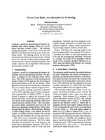
Fig. 1. Control of RIP1’s pro-life and pro-death activity by ubiquitination resolves the NF-jB paradox and the cooperation of TNFR signaling
in immunity. There are two cell-death checkpoints in the TNFR1 pathway. In the first checkpoint, ubiquitination of RIP1 by the E3 ligases
TRAF2, cIAP1 and cIAP2 hinders RIP1 from binding cell-death molecules early after TNF stimulation. This checkpoint does not require new
protein synthesis. In the second checkpoint, NF-jB activation by ubiquitinated RIP1 leads to the production of new proteins that can block
cell death, keeping cells alive in the long-term, which explains why cell survival is the default outcome of TNFR1 signaling. However, when
cell death is desirable, the early checkpoint can be rapidly altered by ligation of the other TNFR family members, which trigger degradation
of the E3 ligases. Nonubiquitinated RIP1 becomes a pro-death signaling molecule and quickly binds caspase 8 to initiate apoptosis. If cas-
pase activity is blocked, RIP1 binds RIP3 and triggers the back-up cell death pathway by programmed necrosis. Ubiquitination controls the
switch between RIP1’s pro-life and pro-death activity: this resolves the NF-jB paradox – ubiquitination of RIP1 prevents TNFR1 from trigger-
ing cell death until an effective pro-survival gene expression program has been implemented. NF-jB activated by TNFR1 drives expression
of immune and inflammatory genes during responses to pathogens. Some pathogens attempt to block NF-jB and dampen down inflamma-
tion, in order to prolong the survival of infected cells. The other TNFRs like TNFR2 can unmask the pro-death activity of RIP1 and trigger
pro-inflammatory necrotic death. This co-operation between the TNFRs allows TNFR1 to circumvent the immune evasion strategies used by
pathogens to block NF-jB activity or apoptosis.
Dual cell-death checkpoints during TNFR1 signaling M. A. O’Donnell and A. T. Ting
882 FEBS Journal 278 (2011) 877–887 ª 2011 The Authors Journal compilation ª 2011 FEBS
production of reactive oxygen species [54,61]. Fibro-
blasts from p65 ⁄ RelA knockout mice produce reactive
oxygen species when stimulated with TNF in the pres-
ence of caspase blockade and undergo cell death by
necrosis [55]. Soaking up the reactive oxygen species
with powerful pharmacological antioxidants can pre-
vent this necrotic cell death. NF-jB can drive expres-
sion of many proteins with antioxidant activity such as
ferritin, manganese superoxide dismutase and glutathi-
one S-transferase [62–64], which suggests that the
second cell-death checkpoint may block programmed
necrosis. The main cellular scavenger for free radicals
is catalase and degradation of this enzyme during
autophagy is associated with the necrotic cell death of
mouse fibrosarcoma cells [65]. In addition, it should be
remembered that the pro-survival function of NF-jB
was originally attributed to the inducible expression of
genes such as TRAF2, cIAP1 and cIAP2 [1], which
suggests that NF-jB activity might be important for
maintaining expression of the proteins that control the
early cell-death checkpoint. Whether or not cFLIP, the
main target responsible for NF-jB anti-apoptotic
activity [66], can prevent programmed necrosis has not
been fully addressed. Viral FLIPs such as MCF159
from molluscum contagiosum virus or K13 from
Kaposi’s sarcoma herpesvirus block necrotic cell death
[51] and knockdown of cFLIP can sensitize HeLa cells
to both TNF-induced apoptosis and necrosis [67].
Therefore, the contribution of NF-jB-dependent gene
transcription programs to protection from necrotic cell
death remains to be fully clarified.
It is clear from in vitro studies that the second cell-
death checkpoint, i.e. NF-jB activity, can prevent both
apoptosis and necrosis, which implies that there must
be a physiological situation in which loss of NF-jB
activity induces cell death as a favorable outcome.
NF-jB activation by TNF is required for expression
of immune and inflammatory genes that are important
for host responses to pathogens. Not surprisingly,
there are numerous examples of viral and bacterial
pathogens that trigger cell death when NF-jB is inhib-
ited, for example, by YopJ during Yersinia infection
[68] and herpes simplex virus triggered apoptosis
requires loss of NF-jB signaling [69]. However, many
viruses activate NF-jB in order to control their own
replication and promote cell survival. In such an event,
it may be advantageous to the host to possess the
capability to disrupt the first cell-death checkpoint in
order to trigger apoptosis or necrosis of the infected
cells. Induction of programmed necrosis in virally
infected cells by disruption of the first death check-
point may be particularly advantageous to the host
because this type of death may be more immunogenic.
Chan et al. have shown that TNFR2 is required for
necrotic cell death to occur during infection with
vaccinia virus, which encodes a potent inhibitor of
apoptotic and inflammatory caspases [51]. In the con-
text of vaccinia virus infection, the function of virus-
triggered programmed necrosis is clearly to enable
removal of infected cells and a pro-inflammatory
response to be initiated that circumvents the virus
immune evasion strategy [51,60]. The increasing num-
ber of pathogen components that have been reported
to block necrotic cell death [70] underscores the impor-
tance of programmed necrosis to the development of
protective immunity. Cell survival at both checkpoints
requires NEMO binding to ubiquitinated RIP1.
NEMO is a critical requirement for the activation of
several kinase complexes that mediate immune
responses to pathogens, from activation of NF-jBto
IRF3 [71,72] and thus required for cytokine and inter-
feron production. It is likely that pathogens may try to
downregulate NEMO protein levels in an attempt to
evade this response. However, pathogen-mediated loss
of NEMO should result in disruption of the first cell-
death checkpoint rendering infected cells sensitive to
programmed necrosis and the ensuing immunogenic
consequences of necrotic death may serve as a back-up
host defense mechanism. Support for this idea has
come from a recent report that an E3 ligase encoded
by Shigella induces degradation of NEMO and blocks
NF-jB-mediated immune gene expression programs
[73]. Other groups have shown that Shigella infection
of certain cell types leads to necrotic cell death [74,75].
Therefore, mammalian cells may have evolved the two
cell-death checkpoints in the TNFR1 pathway in order
to rapidly respond to interference with NEMO’s pro-
survival and immune functions (Fig. 1).
So what is the broader mechanism that enables
NEMO binding to ubiquitinated RIP1 to prevent both
apoptosis and programmed necrosis so effectively? The
fact that TNFR2 predisposes cells to undergo apopto-
sis or necrosis when stimulated with TNF, despite the
requirement for two very different sets of executioner
proteins, suggests that a common overall mechanism
underlies the inhibition of RIP1-dependent cell death
processes by NEMO. There are several possible effects
of NEMO binding to ubiquitinated RIP1 that we
envisage may restrain RIP1 from engaging different
cell death apparatus. The simplest explanation is that
ubiquitination of RIP1 at lysine 377 and the recruit-
ment of ubiquitin-binding proteins such as NEMO
may sterically hinder RIP1 from interacting with
downstream death mediators such as caspase 8 or
RIP3, which do not have ubiquitin-binding domains.
Ubiquitination of RIP1 may also prevent RIP1
M. A. O’Donnell and A. T. Ting Dual cell-death checkpoints during TNFR1 signaling
FEBS Journal 278 (2011) 877–887 ª 2011 The Authors Journal compilation ª 2011 FEBS 883
oligomerization, which has been suggested to play a
role in its ability to trigger cell death [76]. Oligomeriza-
tion of the death domain of RIP1 requires the presence
of an alpha-helix that is immediately adjacent to the
lysine 377 acceptor site of RIP1. This alpha-helix
region also contains the RIP homologous interaction
domain required for interaction with RIP3. Therefore,
it is conceivable that oligomerization of RIP1 might be
required for RIP1 to bind or activate either caspase 8
or RIP3 and that extensive modification of the inter-
mediate domain by ubiquitin chains would prevent
these structures from forming. Ubiquitination of
plasma membrane receptors is a well-known signal
that leads to receptor internalization. Endocytosis of
the interferon alpha receptor requires phosphorylation
and ubiquitination on a motif that is very similar to
the degradation motif of IjBa targeted by the IKK
complex [77]. As reported by Schutze et al., internali-
zation of TNFR1 is important for the activation of
downstream signaling pathways, particularly cell death
[78]. Therefore, ubiquitination of RIP1 when it is
recruited to TNFR1 might modulate the internaliza-
tion or subcellular trafficking of the TNFR1 complex,
which is another mechanism by which access of RIP1
to different cell death machinery might be regulated.
Alternatively, NEMO binding to RIP1 may regulate
the activity of kinase complexes such as IKKe or
IKKa ⁄ b that specifically phosphorylate and inhibit the
activity of cell death mediators, prior to the activation
of gene expression programs by these kinase com-
plexes. Reiley et al. [79] have shown that NEMO is
required for the IKK complex to phosphorylate and
inhibit the deubiquitinase CYLD, a critical component
of the necrotic machinery [80], as can the TNF-induc-
ible IKKe [81]. As alluded to earlier, activation of
these kinases may also influence internalization of the
TNFR1 complex itself.
In conclusion, there are two cell-death checkpoints
downstream of TNFR1 that determine whether cells
live or die. Pro-survival effects from the first checkpoint
that do not require gene expression can be as potent as
the pro-survival impact of new proteins synthesized
after the second checkpoint. A thorough examination
of the molecular mechanisms that regulate the pro-sur-
vival activity of NEMO binding to ubiquitinated RIP1
should reveal new strategies to instigate or prevent cell
death when appropriate in a clinical setting.
Acknowledgements
ATT is supported by NIH grant AI052417. MA O’D
is a recipient of a Research Fellowship Award from
the Crohn’s and Colitis Foundation of America.
References
1 Wang CY, Mayo MW, Korneluk RG, Goeddel DV &
Baldwin AS Jr (1998) NF-kappaB antiapoptosis: induc-
tion of TRAF1 and TRAF2 and c-IAP1 and c-IAP2 to
suppress caspase-8 activation. Science 281, 1680–1683.
2 Liu ZG, Hsu H, Goeddel DV & Karin M (1996)
Dissection of TNF receptor 1 effector functions: JNK
activation is not linked to apoptosis while NF-kappaB
activation prevents cell death. Cell 87 , 565–576.
3 Van Antwerp DJ, Martin SJ, Kafri T, Green DR &
Verma IM (1996) Suppression of TNF-alpha-induced
apoptosis by NF-kappaB. Science 274, 787–789.
4 Beg AA & Baltimore D (1996) An essential role for
NF-kappaB in preventing TNF-alpha-induced cell
death. Science 274, 782–784.
5 Natoli G, Costanzo A, Guido F, Moretti F &
Levrero M (1998) Apoptotic, non-apoptotic, and
anti-apoptotic pathways of tumor necrosis factor
signalling. Biochem Pharmacol 56, 915–920.
6 Stanger BZ, Leder P, Lee TH, Kim E & Seed B (1995)
RIP: a novel protein containing a death domain that
interacts with Fas ⁄ APO-1 (CD95) in yeast and causes
cell death. Cell 81, 513–523.
7 Hsu H, Huang J, Shu HB, Baichwal V & Goeddel DV
(1996) TNF-dependent recruitment of the protein kinase
RIP to the TNF receptor-1 signaling complex. Immunity
4, 387–396.
8 Ting AT, Pimentel-Muinos FX & Seed B (1996) RIP
mediates tumor necrosis factor receptor 1 activation of
NF-kappaB but not Fas ⁄ APO-1-initiated apoptosis.
EMBO J 15, 6189–6196.
9 Kelliher MA, Grimm S, Ishida Y, Kuo F, Stanger BZ
& Leder P (1998) The death domain kinase RIP medi-
ates the TNF-induced NF-kappaB signal. Immunity 8,
297–303.
10 Cusson N, Oikemus S, Kilpatrick ED, Cunningham L
& Kelliher M (2002) The death domain kinase RIP
protects thymocytes from tumor necrosis factor receptor
type 2-induced cell death. J Exp Med 196, 15–26.
11 Micheau O & Tschopp J (2003) Induction of TNF
receptor I-mediated apoptosis via two sequential
signaling complexes. Cell 114, 181–190.
12 Legler DF, Micheau O, Doucey MA, Tschopp J &
Bron C (2003) Recruitment of TNF receptor 1 to lipid
rafts is essential for TNFalpha-mediated NF-kappaB
activation. Immunity 18, 655–664.
13 Deng L, Wang C, Spencer E, Yang L, Braun A, You J,
Slaughter C, Pickart C & Chen ZJ (2000) Activation of
the IkappaB kinase complex by TRAF6 requires a
dimeric ubiquitin-conjugating enzyme complex and a
unique polyubiquitin chain. Cell 103, 351–361.
14 Wertz IE, O’Rourke KM, Zhou H, Eby M, Aravind L,
Seshagiri S, Wu P, Wiesmann C, Baker R, Boone DL,
et al. (2004) De-ubiquitination and ubiquitin ligase
Dual cell-death checkpoints during TNFR1 signaling M. A. O’Donnell and A. T. Ting
884 FEBS Journal 278 (2011) 877–887 ª 2011 The Authors Journal compilation ª 2011 FEBS
domains of A20 downregulate NF-kappaB signalling.
Nature 430, 694–699.
15 Ea CK, Deng L, Xia ZP, Pineda G & Chen ZJ (2006)
Activation of IKK by TNFalpha requires site-specific
ubiquitination of RIP1 and polyubiquitin binding by
NEMO. Mol Cell 22, 245–257.
16 Li H, Kobayashi M, Blonska M, You Y & Lin X
(2006) Ubiquitination of RIP is required for tumor
necrosis factor alpha-induced NF-kappaB activation.
J Biol Chem 281, 13636–13643.
17 Wu CJ, Conze DB, Li T, Srinivasula SM & Ashwell JD
(2006) Sensing of Lys 63-linked polyubiquitination by
NEMO is a key event in NF-kappaB activation
[corrected]. Nat Cell Biol 8, 398–406.
18 Tokunaga F, Sakata S, Saeki Y, Satomi Y, Kirisako T,
Kamei K, Nakagawa T, Kato M, Murata S, Yamaoka
S et al. (2009) Involvement of linear polyubiquitylation
of NEMO in NF-kappaB activation. Nat Cell Biol 11,
123–132.
19 Haas TL, Emmerich CH, Gerlach B, Schmukle AC,
Cordier SM, Rieser E, Feltham R, Vince J, Warnken
U, Wenger T et al. (2009) Recruitment of the linear
ubiquitin chain assembly complex stabilizes the
TNF-R1 signaling complex and is required for
TNF-mediated gene induction. Mol Cell 36,
831–844.
20 Rahighi S, Ikeda F, Kawasaki M, Akutsu M, Suzuki
N, Kato R, Kensche T, Uejima T, Bloor S, Komander
D et al. (2009) Specific recognition of linear ubiquitin
chains by NEMO is important for NF-kappaB activa-
tion. Cell 136, 1098–1109.
21 Laplantine E, Fontan E, Chiaravalli J, Lopez T,
Lakisic G, Veron M, Agou F & Israel A (2009) NEMO
specifically recognizes K63-linked poly-ubiquitin chains
through a new bipartite ubiquitin-binding domain.
EMBO J 28, 2885–2895.
22 Newton K, Matsumoto ML, Wertz IE, Kirkpatrick DS,
Lill JR, Tan J, Dugger D, Gordon N, Sidhu SS,
Fellouse FA et al. (2008) Ubiquitin chain editing
revealed by polyubiquitin linkage-specific antibodies.
Cell 134, 668–678.
23 Lee TH, Shank J, Cusson N & Kelliher MA (2004) The
kinase activity of Rip1 is not required for tumor
necrosis factor-alpha-induced IkappaB kinase or p38
MAP kinase activation or for the ubiquitination of
Rip1 by Traf2. J Biol Chem 279, 33185–33191.
24 Yeh WC, Shahinian A, Speiser D, Kraunus J, Billia F,
Wakeham A, de la Pompa JL, Ferrick D, Hum B,
Iscove N et al. (1997) Early lethality, functional
NF-kappaB activation, and increased sensitivity to
TNF-induced cell death in TRAF2-deficient mice.
Immunity 7, 715–725.
25 Bertrand MJ, Milutinovic S, Dickson KM, Ho WC,
Boudreault A, Durkin J, Gillard JW, Jaquith JB, Mor-
ris SJ & Barker PA (2008) cIAP1 and cIAP2 facilitate
cancer cell survival by functioning as E3 ligases that
promote RIP1 ubiquitination. Mol Cell 30, 689–700.
26 Varfolomeev E, Goncharov T, Fedorova AV,
Dynek JN, Zobel K, Deshayes K, Fairbrother WJ &
Vucic D (2008) c-IAP1 and c-IAP2 are critical
mediators of tumor necrosis factor alpha (TNFalpha)
-induced NF-kappaB activation. J Biol Chem 283,
24295–24299.
27 Alvarez SE, Harikumar KB, Hait NC, Allegood J,
Strub GM, Kim EY, Maceyka M, Jiang H, Luo C,
Kordula T et al. (2010) Sphingosine-1-phosphate is a
missing cofactor for the E3 ubiquitin ligase TRAF.
Nature 465, 1084–1088.
28 Zheng C, Kabaleeswaran V, Wang Y, Cheng G & Wu
H (2010) Crystal structures of the TRAF2:cIAP2 and
the TRAF1:TRAF2:cIAP2 complexes: affinity, specific-
ity, and regulation. Mol Cell 38, 101–113.
29 Csomos RA, Brady GF & Duckett CS (2009) Enhanced
cytoprotective effects of the inhibitor of apoptosis
protein cellular IAP1 through stabilization with
TRAF2. J Biol Chem 284, 20531–20539.
30 Natoli G, Costanzo A, Guido F, Moretti F,
Bernardo A, Burgio VL, Agresti C & Levrero M (1998)
Nuclear factor jB-independent cytoprotective pathways
originating at tumor necrosis factor receptor-associated
factor 2. J Biol Chem 273, 31262–31272.
31 Lee SY, Kaufman DR, Mora AL, Santana A,
Boothby M & Choi Y (1998) Stimulus-dependent
synergism of the antiapoptotic tumor necrosis factor
receptor-associated factor 2 (TRAF2) and nuclear
factor kappaB pathways. J Exp Med 188, 1381–1384.
32 O’Donnell MA, Legarda-Addison D, Skountzos P, Yeh
WC & Ting AT (2007) Ubiquitination of RIP1
regulates an NF-kappaB-independent cell-death switch
in TNF signaling. Curr Biol 17, 418–424.
33 Li X, Yang Y & Ashwell JD (2002) TNF-RII and
c-IAP1 mediate ubiquitination and degradation of
TRAF2. Nature 416, 345–347.
34 Chan FK & Lenardo MJ (2000) A crucial role for p80
TNF-R2 in amplifying p60 TNF-R1 apoptosis signals
in T lymphocytes. Eur J Immunol 30, 652–660.
35 Fotin-Mleczek M, Henkler F, Samel D, Reichwein M,
Hausser A, Parmryd I, Scheurich P, Schmid JA &
Wajant H. (2002) Apoptotic crosstalk of TNF recep-
tors: TNF-R2-induces depletion of TRAF2 and IAP
proteins and accelerates TNF-R1-dependent activation
of caspase-8. J Cell Sci 115, 2757–2770.
36 Vince JE, Chau D, Callus B, Wong WW, Hawkins CJ,
Schneider P, McKinlay M, Benetatos CA, Condon SM,
Chunduru SK et al. (2008) TWEAK-FN14 signaling
induces lysosomal degradation of a cIAP1–TRAF2
complex to sensitize tumor cells to TNFalpha. J Cell
Biol 182, 171–184.
37 Li L, Thomas RM, Suzuki H, De Brabander JK,
Wang X & Harran PG (2004) A small molecule Smac
M. A. O’Donnell and A. T. Ting Dual cell-death checkpoints during TNFR1 signaling
FEBS Journal 278 (2011) 877–887 ª 2011 The Authors Journal compilation ª 2011 FEBS 885
mimic potentiates TRAIL- and TNFalpha-mediated cell
death. Science 305, 1471–1474.
38 Varfolomeev E, Blankenship JW, Wayson SM, Fedor-
ova AV, Kayagaki N, Garg P, Zobel K, Dynek JN,
Elliott LO, Wallweber HJ et al. (2007) IAP antagonists
induce autoubiquitination of c-IAPs, NF-kappaB acti-
vation, and TNFalpha-dependent apoptosis. Cell 131,
669–681.
39 Wang L, Du F & Wang X (2008) TNF-alpha induces
two distinct caspase-8 activation pathways. Cell 133,
693–703.
40 Ermolaeva MA, Michallet MC, Papadopoulou N,
Utermohlen O, Kranidioti K, Kollias G, Tschopp J &
Pasparakis M (2008) Function of TRADD in tumor
necrosis factor receptor 1 signaling and in TRIF-depen-
dent inflammatory responses. Nat Immunol 9, 1037–
1046.
41 Pobezinskaya YL, Kim YS, Choksi S, Morgan MJ,
Li T, Liu C & Liu Z (2008) The function of TRADD
in signaling through tumor necrosis factor receptor 1
and TRIF-dependent Toll-like receptors. Nat Immunol
9, 1047–1054.
42 Zhang J, Cado D, Chen A, Kabra NH & Winoto A
(1998) Fas-mediated apoptosis and activation-induced
T-cell proliferation are defective in mice lacking
FADD ⁄ Mort1. Nature 392, 296–300.
43 Yeh WC, Pompa JL, McCurrach ME, Shu HB, Elia
AJ, Shahinian A, Ng M, Wakeham A, Khoo W,
Mitchell K et al. (1998) FADD: essential for embryo
development and signaling from some, but not all,
inducers of apoptosis. Science 279, 1954–1958.
44 Juo P, Kuo CJ, Yuan J & Blenis J (1998) Essential
requirement for caspase-8 ⁄ FLICE in the initiation of the
Fas-induced apoptotic cascade. Curr Biol 8, 1001–1008.
45 Varfolomeev EE, Schuchmann M, Luria V, Chiannilk-
ulchai N, Beckmann JS, Mett IL, Rebrikov D,
Brodianski VM, Kemper OC, Kollet O et al. (1998)
Targeted disruption of the mouse Caspase 8 gene
ablates cell death induction by the TNF receptors,
Fas ⁄ Apo1, and DR3 and is lethal prenatally. Immunity
9, 267–276.
46 Blankenship JW, Varfolomeev E, Goncharov T, Fedor-
ova AV, Kirkpatrick DS, Izrael-Tomasevic A, Phu L,
Arnott D, Aghajan M, Zobel K et al. (2009) Ubiquitin
binding modulates IAP antagonist-stimulated proteaso-
mal degradation of c-IAP1 and c-IAP2(1). Biochem J
417, 149–160.
47 Gyrd-Hansen M, Darding M, Miasari M, Santoro
MM, Zender L, Xue W, Tenev T, da Fonseca PC,
Zvelebil M, Bujnicki JM et al. (2008) IAPs contain an
evolutionarily conserved ubiquitin-binding domain that
regulates NF-kappaB as well as cell survival and
oncogenesis. Nat Cell Biol 10, 1309–1317.
48 Oshima S, Turer EE, Callahan JA, Chai S, Advincula
R, Barrera J, Shifrin N, Lee B, Benedict Yen TS,
Woo T et al. (2009) ABIN-1 is a ubiquitin sensor that
restricts cell death and sustains embryonic development.
Nature 457, 906–909.
49 Rudolph D, Yeh WC, Wakeham A, Rudolph B,
Nallainathan D, Potter J, Elia AJ & Mak TW (2000)
Severe liver degeneration and lack of NF-kappaB
activation in NEMO ⁄ IKKgamma-deficient mice. Genes
Dev 14
, 854–862.
50 Legarda-Addison D, Hase H, O’Donnell MA &
Ting AT (2009) NEMO ⁄ IKKgamma regulates an
early NF-kappaB-independent cell-death checkpoint
during TNF signaling. Cell Death Differ 16, 1279–1288.
51 Chan FK, Shisler J, Bixby JG, Felices M, Zheng L,
Appel M, Orenstein J, Moss B & Lenardo MJ (2003)
A role for tumor necrosis factor receptor-2 and recep-
tor-interacting protein in programmed necrosis and
antiviral responses. J Biol Chem 278, 51613–51621.
52 Holler N, Zaru R, Micheau O, Thome M, Attinger A,
Valitutt S, Bodmer JL, Schneider P, Seed B, Tschopp J
et al. (2000) Fas triggers an alternative, caspase-8-
independent cell death pathway using the kinase RIP as
effector molecule. Nat Immunol 1, 489–495.
53 Li M & Beg AA (2000) Induction of necrotic-like cell
death by tumor necrosis factor alpha and caspase
inhibitors: novel mechanism for killing virus-infected
cells. J Virol 74, 7470–7477.
54 Lin Y, Choksi S, Shen HM, Yang QF, Hur GM, Kim
YS, Tran JH, Nedospasov SA, Liu ZG et al. (2004)
Tumor necrosis factor-induced nonapoptotic cell death
requires receptor-interacting protein-mediated cellular
reactive oxygen species accumulation. J Biol Chem 279,
10822–10828.
55 Sakon S, Xue X, Takekawa M, Sasazuki T, Okazaki T,
Kojima Y, Piao JH, Yagita H, Okumura K, Doi T
et al. (2003) NF-kappaB inhibits TNF-induced accumu-
lation of ROS that mediate prolonged MAPK activa-
tion and necrotic cell death. EMBO J 22, 3898–3909.
56 Zheng L, Bidere N, Staudt D, Cubre A, Orenstein J,
Chan FK & Lenardo M (2006) Competitive control of
independent programs of tumor necrosis factor
receptor-induced cell death by TRADD and RIP1. Mol
Cell Biol 26, 3505–3513.
57 Vanden Berghe T, van Loo G, Saelens X, Van Gurp
M, Brouckaert G, Kalai M, Declercq W &
Vandenabeele P (2004) Differential signaling to apopto-
tic and necrotic cell death by Fas-associated death
domain protein FADD. J Biol Chem 279, 7925–7933.
58 He S, Wang L, Miao L, Wang T, Du F, Zhao L &
Wang X (2009) Receptor interacting protein kinase-3
determines cellular necrotic response to TNF-alpha.
Cell 137, 1100–1111.
59 Zhang DW, Shao J, Lin J, Zhang N, Lu BJ, Lin SC,
Dong MQ & Han J (2009) RIP3, an energy metabolism
regulator that switches TNF-induced cell death from
apoptosis to necrosis. Science 325, 332–336.
Dual cell-death checkpoints during TNFR1 signaling M. A. O’Donnell and A. T. Ting
886 FEBS Journal 278 (2011) 877–887 ª 2011 The Authors Journal compilation ª 2011 FEBS
60 Cho YS, Challa S, Moquin D, Genga R, Ray TD,
Guildford M & Chan FK (2009) Phosphorylation-dri-
ven assembly of the RIP1–RIP3 complex regulates
programmed necrosis and virus-induced inflammation.
Cell 137, 1112–1123.
61 Vercammen D, Beyaert R, Denecker G, Goossens V,
Van Loo G, Declercq W, Grooten J, Fiers W & Vande-
nabeele P (1998) Inhibition of caspases increases the
sensitivity of L929 cells to necrosis mediated by tumor
necrosis factor. J Exp Med 187, 1477–1485.
62 Sasazuki T, Okazaki T, Tada K, Sakon-Komazawa S,
Katano M, Tanaka M, Yagita H, Okumura K,
Tominaga N, Hayashizaki Y et al. (2004) Genome wide
analysis of TNF-inducible genes reveals that antioxidant
enzymes are induced by TNF and responsible for
elimination of ROS. Mol Immunol 41, 547–551.
63 Pham CG, Bubici C, Zazzeroni F, Papa S, Jones J,
Alvarez K, Jayawardena S, De Smaele E, Cong R,
Beaumont C et al. (2004) Ferritin heavy chain
upregulation by NF-kappaB inhibits TNFalpha-induced
apoptosis by suppressing reactive oxygen species. Cell
119, 529–542.
64 Wong GH & Goeddel DV (1988) Induction of manga-
nous superoxide dismutase by tumor necrosis factor:
possible protective mechanism. Science 242, 941–944.
65 Yu L, Wan F, Dutta S, Welsh S, Liu Z, Freundt E,
Baehrecke EH & Lenardo M (2006) Autophagic
programmed cell death by selective catalase degrada-
tion. Proc Natl Acad Sci USA 103, 4952–4957.
66 Yeh WC, Itie A, Elia AJ, Ng M, Shu HB, Wakeham A,
Mirtsos C, Suzuki N, Bonnard M, Goeddel DV et al.
(2000) Requirement for Casper (c-FLIP) in regulation
of death receptor-induced apoptosis and embryonic
development. Immunity 12, 633–642.
67 Nakajima A, Kojima Y, Nakayama M, Yagita H,
Okumura K & Nakano H (2008) Downregulation of
c-FLIP promotes caspase-dependent JNK activation
and reactive oxygen species accumulation in tumor cells.
Oncogene 27, 76–84.
68 Zhang Y, Ting AT, Marcu KB & Bliska JB (2005)
Inhibition of MAPK and NF-kappaB pathways is
necessary for rapid apoptosis in macrophages infected
with Yersinia. J Immunol 174, 7939–7949.
69 Goodkin ML, Ting AT & Blaho JA (2003) NF-kappaB
is required for apoptosis prevention during herpes
simplex virus type 1 infection. J Virol 77, 7261–7280.
70 Upton JW, Kaiser WJ & Mocarski ES (2010) Virus
inhibition of RIP3-dependent necrosis. Cell Host
Microbe 7, 302–313.
71 Hacker H & Karin M (2006) Regulation and function
of IKK and IKK-related kinases. Sci STKE 2006, re13.
72 Zhao T, Yang L, Sun Q, Arguello M, Ballard DW,
Hiscott J & Lin R (2007) The NEMO adaptor
bridges the nuclear factor-kappaB and interferon
regulatory factor signaling pathways. Nat Immunol 8,
592–600.
73 Ashida H, Kim M, Schmidt-Supprian M, Ma A,
Ogawa M & Sasakawa C (2010) A bacterial E3 ubiqu-
itin ligase IpaH9.8 targets NEMO ⁄ IKKgamma to dam-
pen the host NF-kappaB-mediated inflammatory
response. Nat Cell Biol 12, 66–73.
74 Galluzzi L & Kroemer G (2009) Shigella targets the
mitochondrial checkpoint of programmed necrosis. Cell
Host Microbe 5, 107–109.
75 Carneiro LA, Travassos LH, Soares F, Tattoli I,
Magalhaes JG, Bozza MT, Plotkowski MC, Sansonetti
PJ, Molkentin JD, Philpott DJ et al. (2009) Shigella
induces mitochondrial dysfunction and cell death in
nonmyleoid cells. Cell Host Microbe 5, 123–136.
76 Grimm S, Stanger BZ & Leder P (1996) RIP and
FADD: two ‘death domain’-containing proteins can
induce apoptosis by convergent, but dissociable,
pathways. Proc Natl Acad Sci USA
93, 10923–10927.
77 Kumar KG, Krolewski JJ & Fuchs SY (2004)
Phosphorylation and specific ubiquitin acceptor sites
are required for ubiquitination and degradation of the
IFNAR1 subunit of type I interferon receptor. J Biol
Chem 279, 46614–46620.
78 Schneider-Brachert W, Tchikov V, Neumeyer J, Jakob
M, Winoto-Morbach S, Held-Feindt J, Heinrich M,
Merkel O, Ehrenschwender M, Adam D et al. (2004)
Compartmentalization of TNF receptor 1 signaling:
internalized TNF receptosomes as death signaling vesi-
cles. Immunity 21, 415–428.
79 Reiley W, Zhang M, Wu X, Granger E & Sun SC
(2005) Regulation of the deubiquitinating enzyme
CYLD by IkappaB kinase gamma-dependent
phosphorylation. Mol Cell Biol 25, 3886–3895.
80 Hitomi J, Christofferson DE, Ng A, Yao J,
Degterev A, Xavier RJ & Yuan J (2008) Identification
of a molecular signaling network that regulates a
cellular necrotic cell death pathway. Cell 135,
1311–1323.
81 Hutti JE, Shen RR, Abbott DW, Zhou AY,
Sprott KM, Asara JM, Hahn WC & Cantley LC (2009)
Phosphorylation of the tumor suppressor CYLD
by the breast cancer oncogene IKKepsilon promotes
cell transformation. Mol Cell 34, 461–472.
M. A. O’Donnell and A. T. Ting Dual cell-death checkpoints during TNFR1 signaling
FEBS Journal 278 (2011) 877–887 ª 2011 The Authors Journal compilation ª 2011 FEBS 887