Báo cáo khoa học: Methanoferrodoxin represents a new class of superoxide reductase containing an iron–sulfur cluster docx
Bạn đang xem bản rút gọn của tài liệu. Xem và tải ngay bản đầy đủ của tài liệu tại đây (551.6 KB, 10 trang )
Methanoferrodoxin represents a new class of superoxide
reductase containing an iron–sulfur cluster
Christian Kra
¨
tzer
1
, Cornelia Welte
1
, Katerina Do
¨
rner
2
, Thorsten Friedrich
2
and Uwe Deppenmeier
1
1 Institut fu
¨
r Mikrobiologie und Biotechnologie, Universita
¨
t Bonn, Germany
2 Institut fu
¨
r Organische Chemie und Biochemie, Albert-Ludwigs-Universita
¨
t, Freiburg, Germany
Introduction
Oxidative stress is caused by reactive oxygen species
such as hydrogen peroxide (H
2
O
2
), the hydroxyl radi-
cal (OH
)
) and the superoxide anion radical (O
2
)
),
which are generated by the partial reduction of oxygen
[1]. Bacteria deal with oxidative stress with a set of
detoxifying enzymes. Superoxide dismutases (SODs)
were the first enzymes known to eliminate superoxide
by disproportionation to hydrogen peroxide and diox-
ygen [1]. Superoxide reductases (SORs) are a new fam-
ily of enzymes that were discovered in sulfate-reducing
bacteria of the Desulfovibrio genus [2,3], and catalyze
the reduction of superoxide to peroxide. SORs are pre-
dominantly found in anaerobic or microaerophilic
bacteria such as Desulfovibrio desulfuricans [2] and
Clostridium acetobutylicum [4], or anaerobic archaeons
such as Archaeoglobus fulgidus [5] and Pyrococcus
furiosus [6]. In the past decade, SORs from these
organisms and others have been studied in detail, and
a considerable amount of biochemical, crystallographic
and spectroscopic information has been reported [7].
Methanosarcina mazei is one of the methanogenic
archaeons, which are characterized by the ability to
generate methane as the major end product of energy
metabolism [8]. Many Methanosarcina strains are able
to utilize H
2
+CO
2
, methylated C
1
compounds or
acetate as energy and carbon sources, and are essential
for closing the cycle of organic matter on earth in
anaerobic environments. Methanogens are generally
considered to be sensitive towards aeration with
oxygen. However, it has been reported that some
methanogens are surprisingly oxygen-stable, and can
survive exposure to air for several hours, with the
Keywords
detoxification; iron–sulfur protein;
methanogenic archaea; oxygen radicals;
superoxide dismutase
Correspondence
U. Deppenmeier, Institut fu
¨
r Mikrobiologie
und Biotechnologie, Universita
¨
t Bonn,
Meckenheimer Allee 168, 53115 Bonn,
Germany
Fax: +49 228737576
Tel: +49 228735590
E-mail:
(Received 5 October 2010, revised
8 November 2010, accepted 17 November
2010)
doi:10.1111/j.1742-4658.2010.07964.x
Protein MM0632 from the methanogenic archaeon Methanosarcina mazei
showed strong superoxide reductase activity and rapidly decomposed
superoxide radicals to peroxides. The superoxide reductase activity of the
heterologously produced enzyme was determined by a cytochrome c assay
and in a test system with NADPH, ferredoxin:NADP
+
reductase, and
rubredoxin. Furthermore, EPR spectroscopy showed that MM0632 is the
first superoxide reductase that possesses an iron–sulfur cluster instead of a
second mononuclear iron center. We propose the name methanoferrodoxin
for this new class of superoxide reductase with an [Fe(NHis)
4
(SCys)] site as
the catalytic center and a [4Fe–4S] cluster as second prosthetic group that
is probably involved in electron transfer to the catalytic center.
Methanosarcina mazei grows only under anaerobic conditions, but is one of
the most aerotolerant methanogens. It is tempting to speculate that
methanoferrodoxin contributes to the protection of cells from oxygen
radicals formed by flavoproteins during periodic exposure to oxygen in nat-
ural environments.
Abbreviations
NROR, NADH-rubredoxin oxidoreductase; SOD, superoxide dismutase; SOR, superoxide reductase.
442 FEBS Journal 278 (2011) 442–451 ª 2010 The Authors Journal compilation ª 2010 FEBS
Methanosarcina species appearing to be the most aero-
tolerant methanogens [9]. The ability to cope with oxi-
dative stress is consistent with the finding that
methanogens are widespread in habitats that are peri-
odically exposed to oxygen, such as paddy soils.
In this article, we report that the protein MM0632
from M. mazei reductively decomposes superoxide to
peroxide. This SOR activity of the heterologously pro-
duced enzyme was determined with a cytochrome
c and an NADPH-dependent assay, respectively
[4,6,10,11]. EPR spectroscopy revealed that MM0632
possesses the typical catalytic nonheme [Fe(N-
His)
4
(SCys)] center and an iron–sulfur cluster instead
of a second mononuclear iron center. We propose the
name methanoferrodoxin for this SOR, and, together
with homologous proteins from other methanogenic
archaeons, this enzyme should be classified as a class
IV SOR.
Results
One mechanism protecting oxygen-sensitive bacteria
and archaeons from toxic oxygen reduction products
involves reduction of superoxide, rather than the clas-
sical disproportionation for its removal that occurs in
aerobic microorganisms [19,20]. A close inspection of
the genome of M. mazei [8] revealed the presence of an
ORF (MM0632) with significant similarities to genes
encoding SORs.
Figure 1 shows alignments of selected SORs from dif-
ferent prokaryotes in comparison with the amino acid
sequence of MM0632 from M. mazei. Different classes
of SORs can be distinguished. Desulfoferrodoxins or
class I SORs (also named 2Fe-SORs), represented by
enzymes from D. desulfuricans and A. fulgidus (Aful2),
contain a small N-terminal desulforedoxin-type domain
(domain I) with a rubredoxin-type [Fe(SCys)
4
] mono-
nuclear iron center, and a larger C-terminal domain
similar to neelaredoxin (domain II), with an [Fe(N-
His)
4
(SCys)] mononuclear iron center. Treponema palli-
dum contains a variant of desulfoferrodoxin (class III
SOR), composed of the C-terminal domain and a
N-terminal domain that does not contain an
[Fe(SCys)
4
] center. As evident from the alignment, the
critical Cys residues for binding iron center I (Fig. 1,
asterisks) are absent from MM0632. In contrast, the
four nitrogen ligands of His residues and the sulfur of
one Cys are conserved in MM0632 (Fig. 1, black
boxes), indicating that the neelaredoxin-type iron center
is present. This hypothesis is supported by the align-
ment of neelaredoxins (class II SORs or 1-Fe-SORs)
from different prokaryotes and MM0632 (Fig. 1).
Again the mononuclear iron-coordinating residues for
the [Fe(NHis)
4
(SCys)] center are present in all proteins.
Closer inspection of the alignment revealed that an inser-
tion in the middle of the neelaredoxin-like domain II of
MM0632 occurred that contains three Cys residues
[C(x)
7
CxxC motif]. Furthermore, there was an extension
of the C-terminal end of MM0632 containing one addi-
tional Cys (Fig. 1). These Cys residues are the only ones
present besides the Cys that coordinates the [Fe(N-
His)
4
(SCys)] center. Homologous insertions and exten-
sion were identified in proteins from the methanogenic
archaeons Methanococcoides burtonii, Methanohalophi-
lus mahii and Methanohalobium evestigatum (Fig. 1).
This interesting finding prompted the question of
whether a second metallocenter could be coordinated
by the four Cys residues next to the catalytic [Fe(N-
His
4
)(SCys)] center. A combined phi-psi-blast [21]
search indicated that the C(x)
7
CxxC motif is also pres-
ent in some ferredoxin-type proteins and iron–sulfur
binding domain proteins [e.g. YP_002890971 (NapF)
from Thauera sp. and YP_002990434 from Desulfovib-
rio salexigens]. It was also evident from the alignment
(Fig. 1) that the sequences containing the iron-binding
motifs were highly conserved, whereas the remaining
parts of the proteins showed only a low degree of simi-
larity. This was especially true for the C-terminal ends
of the proteins, with MM0632 being characterized by
an extension that contained a high percentage of
charged amino acids (Glu, Arg and Lys).
Protein properties
The gene encoding MM0632 from M. mazei was
cloned into pASK-IBA3 and heterologously overex-
pressed in Escherichia coli. The produced protein was
purified to apparent homogeneity in a single step with
a Strep-Tactin affinity matrix. A molecular mass of
20 kDa was found by SDS ⁄ PAGE, consistent with the
expected mass of 19.2 kDa of the protein monomer
(not shown). The native enzyme had a molecular mass
of 19 kDa when analyzed by gel filtration, indicating a
monomeric structure. Small amounts of dimers and tri-
mers were also observed, but contributed to < 10% of
the total protein (Fig. S1).
When MM0632 was overproduced aerobically in
E. coli, spectrometric analysis of the protein did not
show any prosthetic groups. However, after anaerobic
overproduction and purification, the protein prepara-
tion had a red color when hydrogen peroxide was added
to obtain the fully oxidized state (Fig. 2A). The spec-
trum showed a broad increase in absorption between
420 and 550 nm, with a peak at 470 nm. This absorption
vanished upon reduction with dithionate or ascorbate
(Fig. 2A and inset). After reconstitution of the protein
C. Kra
¨
tzer et al. Superoxide reductase from Methanosarcina mazei
FEBS Journal 278 (2011) 442–451 ª 2010 The Authors Journal compilation ª 2010 FEBS 443
with Fe
3+
, sulfide and dithiothreitol, the protein eluted
from the affinity column with a dark brown color. The
UV–visible spectrum of the reconstituted fully oxidized
protein revealed increased absorbance between 400 and
600 nm (Fig. 2B), with a broad peak at 420 nm and a
shoulder at 470 nm. This absorption disappeared by
reducing the preparation with sodium dithionite
(Fig. 2B and inset). Reduction with ascorbate led to
only a small decrease in absorbance between 400 and
600 nm (Fig. 2B). We also investigated whether the
reconstitution led to unspecific binding of iron ions. For
this purpose, the reconstitution was performed in the
absence of sulfide, but artificial incorporation of Fe
3+
was not detected (not shown).
The presence of iron–sulfur clusters was investigated
by quantification of nonheme iron and acid-labile sul-
fur after desalting and overnight dialysis against buf-
fer W with 5 mm EDTA and 1 mm dithioerythritol to
remove any possible contamination with nonenzyme
bound ions. The determination of iron and sulfur
yielded 5.7 ± 0.4 mol iron per mol enzyme and
4.5 ± 1.2 mol sulfur per mol enzyme, indicating the
presence of a mononuclear iron center and a [4Fe–4S]
cluster. In contrast, in the nonreconstituted protein,
1.3 ± 0.5 mol iron per mol enzyme was detected, and
acid-labile sulfide was not found.
EPR spectroscopy of MM0632
A representative EPR spectrum of reconstituted
MM0632 is shown in Fig. 3. The EPR spectrum of the
protein as isolated minus the spectrum of the dithio-
nite-reduced sample recorded at 6 K (Fig. 3A) showed
the typical EPR signal of a high-spin (S =5⁄ 2) ferric
site. This signal at g = 4.3 is attributed to the [Fe(N-
His)
4
(SCys)] center, and has also been detected in
the SORs from P. furiosus and Desulfovibrio vulgaris,
as well as in the D. desulfuricans desulfoferrodoxin
[22–24].
At higher fields, the spectrum of the dithionite-
reduced sample showed a distinct signal at 13 K
(Fig. 3B) that was barely detectable at 40 K and was
absent in the spectrum of the oxidized sample (data
not shown). The axial signal with g^ = 1.93 and
gk = 2.047 was assigned to a [4Fe–4S] cluster. These
findings, together with those of the UV–visible spec-
troscopy and iron–sulfur quantification, indicated that
the recombinant MM0632 from M. mazei was success-
fully produced in E. coli with a correctly incorporated
[Fe(NHis)
4
(SCys)] center and the [4Fe-4S] metallocen-
ter being reconstituted with Fe
3+
and sulfide. The
axial signal mentioned above was disturbed by the
contribution of another, as yet unknown, compound
Fig. 1. Alignment of SOR sequences. Alignment of amino acid sequences was performed with CLUSTALW [40]. The GenBank accession num-
bers of the proteins are as follows. Mma, M. mazei, methanoferrodoxin, MM0632: AAM30328. Mcc, Me. burtonii, methanoferrodoxin:
YP_565539. Mhal, Met. mahii, methanoferrodoxin: YP_003542283. Mhalo, Meth. evestigatum, methanoferrodoxin: YP_003727000. Mac,
M. acetivorans, neelaredoxin-like protein: NP_618610.1. Dgiga, Desulfovibrio gigas, neelaredoxin: O50258. Aful1, A. fulgidus, neelaredoxin-
like protein: O29903. Tocea, Thermosediminibacter oceani DSM 16646, neelaredoxin-like protein: YP_003826213. Ddesulf, D. desulfuricans,
desulfoferrodoxin: YP_002480584. Aful2, A. fulgidus, desulfoferrodoxin: NP_069667.1. Tpa1, T. pallidum, SOR type III: ADD72914.1. Amino
acids forming the [Fe(NHis)4(SCys)] are indicated by black boxes. Cys residues involved in the coordination of the [Fe(SCys)
4
] center are indi-
cated by asterisks. The insertion and extensions of the methanoferrodoxin-like proteins are boxed. Cys residues predicted to be involved in
the coordination of the [4Fe–4S] cluster are indicated by arrows.
Superoxide reductase from Methanosarcina mazei C. Kra
¨
tzer et al.
444 FEBS Journal 278 (2011) 442–451 ª 2010 The Authors Journal compilation ª 2010 FEBS
at g = 2.03. This signal most likely derives from a
low-spin S = ½Fe
3+
species, probably at the [Fe(N-
His
4
)(SCys)] site.
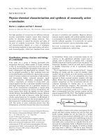
SOR activity of MM0632
The purified MM0632 product was tested for SOR
activity with the cytochrome c reduction assay. In this
test system, cytochrome c was reduced by superoxide,
which was provided continuously by xanthine, oxygen
and xanthine oxidase. The reduction of cytochrome c
was determined by the increase in absorbance at
550 nm. SOR functioned as a cytochrome c oxidase,
and withdrew electrons from cytochrome c to reduce
superoxide to peroxide (Fig. 4, inset). Cytochrome c
reduction decreased with increasing amounts of
MM0632. In contrast to the normal SODs, an excess
of the protein from M. mazei caused reoxidation of
reduced cytochrome c (Fig. 4). The enzymic activity of
MM0632 was calculated on the basis that 1 U of SOR
activity is defined by the amount of protein required
to inhibit the rate of cytochrome c reduction by 50%
[16]. In our test system, 72 ± 9 ng of protein led to
50% inhibition, representing an activity of 1 U. Hence,
the enzyme was highly active with 13 900 ±
1700 U mg
)1
protein, which is in the same order of
0
0.02
0.04
0.06
0.08
0.10
0.12
0.14
0.16
0.18
0
0.02
0.04
0.06
0.08
0.10
0.12
0.14
0.16
0.18
0.20
350 400 450 500 550 600 650 700
0
0.02
0.04
0.06
0.08
0.10
0.12
400 450 500 550 600 650
0
0.02
0.04
0.06
0.08
0.10
0.12
A
B
1
3
2
1
2
3
Absorbance Absorbance
Wavelength (nm)
350 400 450 500 550 600 650 700
Wavelength (nm)
400 450 500 550 600 650
Fig. 2. UV–visible spectra of M. mazei MM0632. (A) Nonreconsti-
tuted protein (0.2 mg mL
)1
): (1) H
2
O
2
-oxidized; (2) dithionite-
reduced; (3) ascorbate-reduced. Inset: oxidized–reduced spectrum.
(B) Reconstituted protein (0.1 mg mL
)1
): (1) H
2
O
2
-oxidized; (2)
ascorbate-reduced; (3) dithionite-reduced. Inset: oxidized–reduced
spectrum.
A
Magnetic field [mT]
4.3
110 120 130 140 150 160 170 180 190 200
280 300 320 340 360 380 400
B
Magnetic field [mT]
1.93
2.047
2.03
Fig. 3. X-band EPR spectra of MM0632. (A) difference spectrum
obtained by subtracting the spectrum of the dithionite-reduced
sample from that of the as-isolated enzyme. The spectra were
recorded at 6 K and 10 mW, and five scans were accumulated for
each spectrum. Other EPR conditions were as follows: microwave
frequency, 9.46 GHz; modulation amplitude, 1.0 mT; time constant,
0.164 s; scan rate, 17.9 mT min
)1
. (B) Spectrum of the dithionite-
reduced sample recorded at 13 K and 10 mW. Other EPR condi-
tions were as follows: microwave frequency, 9.46 GHz; modulation
amplitude, 0.6 mT; time constant, 0.164 s; scan rate, 17.9 mT
min
)1
. The g-values are indicated.
C. Kra
¨
tzer et al. Superoxide reductase from Methanosarcina mazei
FEBS Journal 278 (2011) 442–451 ª 2010 The Authors Journal compilation ª 2010 FEBS 445
magnitude as the specific activity of the SORs from
A. fulgidus [25]. In contrast, the SORs from C. acetobu-
tylicum and Desulfoarculus baarsii had lower activities
(160 and 53 U mg
)1
protein, respectively) [4,17]. The
reaction was dependent on the production of super-
oxide by xanthine and xanthine oxidase (not shown).
When cytochrome c was chemically reduced by sodium
dithionite and used in the assay in the absence of xan-
thine oxidase, no reaction was observed, even in the
presence of oxygen (not shown). These experiments
clearly indicated that oxygen cannot function as an
electron acceptor of MM0632. Thus, the M. mazei
protein functions as a cytochrome c–superoxide oxido-
reductase.
It has been shown that the activities of SORs signifi-
cantly decrease when acetylated cytochrome is used as
substrate [25]. Interestingly, the SOR from M. mazei
showed no inhibition when the acetylated form of
cytochrome c was used as electron donor in compari-
son with the nonacetylated form of cytochrome c (not
shown). The activity of nonreconstituted MM0632
missing the [4Fe–4S] cluster was also tested in the
cytochrome c assay. The protein containing only
[Fe(NHis)
4
(SCys)] showed an activity of 13 900 ±
2500 U mg
)1
protein (not shown), which is in the
same range as the activity of the reconstituted pro-
tein. Hence, it is obvious that the [4Fe–4S] cluster is
not necessary for cytochrome c-dependent superoxide
reduction.
It has been shown that SOD is able to inhibit the
reduction of cytochrome c by dismutation of superox-
ide to hydrogen peroxide [10]. Therefore, this enzyme
could compete with SOR and horse heart cyto-
chrome c for superoxide [16]. Indeed, the addition of
bovine SOD showed a clear effect on the catalytic effi-
ciency of SOR, because the former enzyme signifi-
cantly decreased the concentration of superoxide that
functions as an electron acceptor for MM0632
(Fig. 4). The standard photometric Nitro Blue tetrazo-
lium-dependent SOD assay indicated that MM0632
had a slow SOD activity of 25 U mg
)1
protein [18].
In C. acetobutylicum, an SOR acts as the terminal
component of a superoxide detoxification system that
transfers electrons from NADH to superoxide [4]. The
short electron transfer chain involves NADH-rubred-
oxin oxidoreductase, and a low molecular mass electron
transfer protein named rubredoxin. Rubredoxin has a
single [Fe(SCys)
4
] center as active site, and is known to
participate in electron transfer to SORs in various bac-
teria [6,26]. This electron pathway was reconstituted in
an in vitro assay with ferredoxin:NADP
+
reductase
(FNR) from spinach as a replacement for NADH-
rubredoxin oxidoreductase (Fig. 5). In our test system,
FNR, which uses NADPH as substrate, could donate
electrons to rubredoxin from C. acetobutylicum.
Reduced rubredoxin then functioned as an electron
donor for MM0632, which reduced superoxide gener-
ated by xanthine ⁄ xanthine oxidase.
As shown in Fig. 5, NADPH consumption was trig-
gered by addition of rubredoxin. The SOR activity of
MM0632 strongly depends on the presence of FNR, xan-
thine oxidase and rubredoxin. No activity was observed
when one of the proteins was omitted. FNR could not
0.46
0.48
0.5
0.52
0.54
0 50 100 150 200 250
1
2
3
4
5
6
Time (s)
Absorbance (550 nm)
Xanthine O
2
.
O
2
XO
SOR
+ O
2
O
2
.
O
2
2–
Cyt c red
Cyt c ox
Fig. 4. SOR activity of MM0632. Cytochrome c was reduced by
superoxide generated by xanthine and xanthine oxidase (inset). The
arrow indicates addition of MM0632 with subsequent oxidation of
cytochrome c: (1) 31 ng; (2) 62 ng; (3) 93 ng; (4) 124 ng; (5)
470 ng; (6) 470 ng plus SOD (40 U).
0.25
0.3
0.35
0.4
0.45
0.5
0.55
0.6
0 100 200 300 400 500 600 700 800
Time (s)
Absorbance (340 nm)
(2)
(3)
(1)
(4)
Fig. 5. Superoxide reduction as catalyzed by MM0632 with rubre-
doxin as electron donor. The reaction was monitored by measuring
the NADPH consumption spectrometrically at 340 nm. Addition of:
(1) NADPH, FNR, xanthine and xanthine oxidase; (2) 2 l
M
MM0632; (3) 6 lM rubredoxin; (4) 60 U of SOD.
Superoxide reductase from Methanosarcina mazei C. Kra
¨
tzer et al.
446 FEBS Journal 278 (2011) 442–451 ª 2010 The Authors Journal compilation ª 2010 FEBS
transfer electrons directly to MM0632, indicating that
MM0632 is a rubredoxin oxidase. The addition of SOD
to the assay resulted in decreased activity, owing to the
competitive consumption of superoxide (Fig. 5).
Discussion
SORs such as desulfoferrodoxin and neelaredoxin are
produced by anaerobic or microaerophilic prokaryotes,
and are widespread among the bacterial and archaeal
domains. Desulfoferrodoxin from sulfate reducers (e.g.
D. desulfuricans, Desulfoarculus baarsii and A. fuldigus)
is a protein containing a small N-terminal desulfore-
doxin-type domain with the mononuclear center
[Fe(SCys)
4
], and a larger C-terminal domain
containing an [Fe(NHis)
4
(SCys)] center. The [Fe(N-
His)
4
(SCys)] center is composed of a pentacoordinated
Fe
2+
with four equatorial His residues and one axial
Cys in a square pyramidal geometry [27,28]. The other
axial position is either coordinated to a Glu (oxidized
metal state), resulting in an octahedral geometry, or is
vacant when the metal is reduced, and it is probably
the superoxide-binding site [28].
The additional desulforedoxin-like iron center con-
sists of iron tetrahedrally coordinated to four Cys resi-
dues [24]. The [Fe(SCys)
4
] center is obviously not
involved in superoxide reduction [11]. In comparison,
neelaredoxins are much smaller, containing a single
iron site with (NHis)
4
(SCys) coordination, identical to
what occurs in the C-terminal domain of desulfoferro-
doxin [4,29–31].
We have shown that MM0632 from M. mazei func-
tions as an SOR and interacts with reduced rubredoxin
from C. acetobutylicum as an electron donor. Further-
more, EPR spectroscopy and sequence comparison
clearly revealed that the protein contains a
mononuclear iron center, which is most probably
coordinated by four His residues (His24, His51, His57
and His134), one Cys (Cys131) and, depending on the
oxidation state, one Glu (Glu21) [28]. Hence, the
center represents the typical neelaredoxin [Fe(N-
His)
4
(SCys)] center. Interestingly, the EPR data
showed the presence of a [4Fe–4S] cluster, indicated by
the axial signal with g^ = 1.93 and g
k
= 2.047. To
our knowledge, MM0632 is the first SOR containing
an [Fe(NHis)
4
(SCys)] center and an iron–sulfur cluster
described to date, and thus represents a new family of
SORs. By analogy with desulfoferrodoxin from sul-
fate-reducing bacteria, we propose to refer to MM0632
as methanoferrrodoxin, which is found in several
methanogenic archaeons (see below).
Sequence alignments indicated that methanoferro-
doxin is homologous with desulfoferrodoxins and
neelaredoxin, as well as with uncharacterized SORs from
hyperthermophilic archaeons and bacteria such as Ther-
motoga maritima, P. furiosus and A. fulgidus [5,28].
In addition, homologs were found in close relatives of
M. mazei, such as Methanosarcina acetivorans and Met-
hanococcus voltae. The residues that bind the [Fe(N-
His)
4
(SCys)] center are highly conserved among all
sequences analyzed. In contrast to all other SOR
sequences mentioned previously, we observed an inser-
tion in the amino acid sequence (position 104–126) of
MM0632 containing three Cys residues and an extended
highly charged C-terminus with an additional Cys
(Cys157). It is tempting to speculate that the additional
Cys residues in methanoferrodoxin are responsible for
the coordination of the [4Fe–4S] cluster. The same inser-
tion and extended C-terminal domain was found in the
predicted SORs from the methylotrophic methanogens
Me. burtonii (YP_565539), Met. mahii (YP_003542283)
and Meth. evestigatum (YP_003727000), indicating that
these organisms are also able to form SORs that belong
to the methanoferrodoxin protein family.
Possible physiological function of
methanoferrodoxin
In the cellular environment, superoxide can be gener-
ated from oxygen by electron leakage from b-type
cytochromes, which are present in the electron trans-
port chain of M. mazei, or by spontaneous oxidation
of reduced flavoproteins [32,33]. The flavin protein
AfpA from A. fulgidus was reported to generate super-
oxide as a byproduct of interaction with oxygen [34].
AfpA is similar to MM0635 from M. mazei, whose
coding region is located upstream of the mm0632 gene
[8]. Also, M. mazei contains flavoproteins, which are
essential for methanogenesis. Examples are the F
420
H
2
dehydrogenase, a key enzyme in membrane-bound
electron transport [35], and the F
420
-reducing hydroge-
nase, which is responsible for the H
2
-dependent reduc-
tion of the central electron carrier coenyzme F
420
[36].
These enzymes may be able to transfer electrons to
oxygen, forming superoxide, when oxygen enters the
natural environment of this organism. Under these
conditions, MM0632 could protect the cell from super-
oxide damage.
Rubredoxin is the only known electron donor for
SOR. It mediates electron transfer between an
oxidoreductase and the catalytic [Fe(NHis)
4
(SCys)]
center of the SOR. In this article, evidence is presented
that MM0632 also has rubredoxin oxidase activity,
which is probably also dependent on the [Fe(N-
His)
4
(SCys)] iron center. However, rubredoxin cannot
be the native electron donor in M. mazei, because the
C. Kra
¨
tzer et al. Superoxide reductase from Methanosarcina mazei
FEBS Journal 278 (2011) 442–451 ª 2010 The Authors Journal compilation ª 2010 FEBS 447
genome of M. mazei does not code for any rubredoxin
[8]. Thus, it is likely that an alternative electron donor
system is present in M. mazei. It is tempting to specu-
late that the [4Fe–4S] cluster of methanoferrodoxin
may be necessary for acceptance of electrons from the
undiscovered native electron donor in M. mazei.
Potential candidates that could transfer electrons to
methanoferrodoxin are ferredoxins and reduced
coenzyme F
420
[37].
In addition to the gene mm0632, which encodes
methanoferrodoxin, the M. mazei genome contains two
genes coding for a catalase (NP_634581, NP_633974)
and one gene coding for a SOD (NP_634447)[8]. Two
homologous proteins, a catalase (YP_304371) and a
SOD (CAB82579), from the close relative Methano-
sarcina barkeri are described in the literature [38,39].
Consequently, it is likely that they are also functional in
M. mazei. Therefore, the question arises of why
M. mazei possesses two systems to detoxify superoxide,
whereas M. barkeri is equipped only with a SOD. For
anaerobes in general, SODs have the disadvantage of
oxygen generation, which is circumvented by the
alternative reaction mechanism of SORs. Hence,
methanogens containing methanoferrodoxin, such as
M. mazei and Me. burtonii, may survive better under
oxygen stress conditions.
Experimental procedures
Reagents and proteins
Xanthine, xanthine oxidase (bovine milk), catalase and
SOD (bovine liver), FNR (spinach leaves), cytochrome c
and acetylated cytochrome c were purchased from Sigma-
Aldrich (Munich, Germany).
Cloning, expression and purification
The mm0632 gene was amplified by PCR, with chromosomal
DNA of M. mazei as template and the following primers:
mm0632for, 5¢-ATGGTAGGTCTCAAATGATAGGAA
ATGAAGAAAAAATAAATAAGC-3¢; and mm0632rev,
5¢-ATGGTAGGTCTCAGCGCTGGCTTTCCAGACGCA
TTTTTTGC-3¢.
The gene mm0632 was cloned via BsaI restriction sites in
plasmid pASK-IBA3 (IBA GmbH, Go
¨
ttingen, Germany),
resulting in a C-terminal strep-tag fusion. The coding
region of C. acetobutylicum rubredoxin (NP_349382) was
cloned in a pT vector, using the BamHI and the XmaI
restriction sites [12]. Overproduction of proteins was per-
formed in E. coli DH5a. Cells were grown on modified
maximal induction medium [MI; 3.2% (w ⁄ v) tryptone and
2% (w ⁄ v) yeast extract, with additions of M9 salts, 1 mm
CaCl
2
and 1 mm MgSO
4
]. Ampicillin (100 lgmL
)1
) was
added for plasmid maintenance.
For overproduction of rubredoxin, 40 lgmL
)1
FeNH
4
citrate was added to the cultures, which were grown aerobi-
cally at 30 °C for 16 h [4]. Cells were harvested by centrifu-
gation (8000 g, 10 min) and lysed by sonication. Protein
purification was performed aerobically according to the
manufacturer’s instructions (IBA GmbH, Go
¨
ttingen, Ger-
many). For the production of MM0632, the growth med-
ium was supplemented with l-cysteine (1 mm), FeNH
4
citrate (0.1 mg mL
)1
) and FeSO
4
.7H
2
O (0.1 mg mL
)1
) [13].
After induction of protein production, growth proceeded
under anaerobic conditions for 16 h at 28 °C. Cells were
harvested at 8000 g for 10 min under anaerobic conditions,
and all subsequent purification steps were performed in an
anaerobic chamber (Coy Laboratory Products, Grass Lake,
Michigan, USA) containing an atmosphere of 97% N
2
and
3% H
2
. Cells were lysed with B-Per (Pierce, Rockford, IL,
USA), and detergent and cell debris were removed by cen-
trifugation at 12 000 g for 20 min. MM0632 was reconsti-
tuted by addition of 1 mm FeCl
3
,1mm Na
2
S and 10 mm
dithiothreitol to the cleared lysate, and incubated for
30 min. Insoluble components were removed by
centrifugation at 12 000 g for 20 min. Protein purification
was performed anaerobically according to the manufac-
turer’s instructions (), except that
washing buffer W (50 mm Tris, 150 mm NaCl, pH 8) and
elution buffer E (50 mm Tris, 150 mm NaCl, 2.5 mm des-
thiobiotin, pH 8) were purged with N
2
. Aliquots of the
purified protein (50 lL) were diluted with 250 lL of buf-
fer W, and concentrated on Vivaspin ultrafiltration spin
columns (cut-off 3 kDa; Sartorius Stedim, Goettingen,
Germany) in an anaerobic chamber. This procedure was
repeated twice, and the final protein concentration was
adjusted to 1.5–2 mg mL
)1
. The protein was stored at
) 70 °C under an atmosphere of N
2
. Nonreconstituted pro-
tein was purified as described above, with the exception that
FeCl
3
,Na
2
S and 10 mm dithiothreitol were not added to the
cleared lysate.
Molecular sieve chromatography
A Superdex 75 chromatography column (Amersham Biosci-
enes, Piscataway, NJ, USA), with reference proteins cyto-
chrome c (12.4 kDa), myoglobin (17.8 kDa), chymotrypsin
(25 kDa) and albumin (43 kDa), was used to determine the
native mass of MM0632.
Quantification of iron and acid-labile sulfide
Purified protein was desalted on High-trap desalting col-
umns (GE Healthcare, Munich, Germany) or by dialysis
against buffer W containing 2.5 mm dithioerythritol and
5mm EDTA under anaerobic conditions Nonheme iron
was quantified as described by Landers & Sak [14].
Superoxide reductase from Methanosarcina mazei C. Kra
¨
tzer et al.
448 FEBS Journal 278 (2011) 442–451 ª 2010 The Authors Journal compilation ª 2010 FEBS
Acid-labile sulfide was quantified photometrically at
670 nm by measuring the formation of methylene blue after
the addition of N,N-dimethyl-p-phenylenediamine, with
Na
2
S as standard [15].
UV–visible spectroscopy
UV–visible spectra were recorded on a spectrophotometer
(TIDAS; J&M Analytik AG, Germany) from 275 to
700 nm, with a 0.5-cm quartz cuvette. The spectrum of a
preparation of MM0632 (1 mg mL
)1
) in buffer W was
recorded after titration with hydrogen peroxide, and
referred to as the oxidized state. A few grains of sodium
dithionite were added, and the spectrum of the reduced
protein was recorded. Protein concentrations were
determined with the BCA assay (Merck, Darmstadt,
Germany).
EPR spectroscopy
EPR measurements were conducted with a Bruker EMX 1 ⁄ 6
spectrometeroperatingatX-band. Thesampletemperaturewas
controlled with an Oxfordinstrument ESR-9 helium flow cryo-
stat.Themagneticfieldwascalibratedbyuseofastrongoraweak
pitch standard. The sample (300 lL; 10 mg protein mL
)1
) was
either measured as isolated or after reduction by a few grains of
sodiumdithionite.
Detection of SOR activity of MM0632 in a
cytochrome c assay
The SOR activity of MM0632 was measured with a stan-
dard cytochrome c reduction assay [4,16,17]. With this
assay, SOD and SOR activities were detected. SOD and
SOR compete with horse heart cytochrome c for the
superoxide anion, which is generated continuously by xan-
thine and xanthine oxidase under aerobic conditions. SOD
inhibits superoxide-dependent reduction of cytochrome c,
whereas SOR functions as a cytochrome c oxidase and
reduces superoxide with electrons derived from cyto-
chrome c. The assay was performed in 1.5-mL cuvettes
under aerobic conditions in 1 mL of buffer W. Catalase
(250 U mL
)1
) was added to prevent inhibition by perox-
ides. Addition of xanthine (0.2 mm) and xanthine oxidase
led to the generation of superoxide anions and reduction of
cytochrome c (40 lm), resulting in an increase in absor-
bance at 550 nm. The amount of xanthine oxidase was
adjusted to an increase in cytochrome c reduction of
0.025 ± 0.001 min
)1
at 550 nm [17]. Addition of MM0632
led to oxidation of cytochrome c, and a decrease in absor-
bance at 550 nm. Enzyme amounts between 0 and 100 ng
were used for the test, and the activities were linearly pro-
portional within this range of protein content. Higher
amounts of protein led to a decrease in specific activity,
indicating suboptimal concentrations of reduced cyto-
chrome c and ⁄ or superoxide. SOD, which competes for
superoxide consumption, was used as a control in this
assay. All enzymatic assays were performed on a Jasco
V-550 spectrometer.
SOD activity assay
SOD activity was quantified with the standard aerobic xan-
thine ⁄ xanthine oxidase assay in the presence of Nitro Blue
tetrazolium [18]. Superoxide generated by xanthine oxidase
reduces Nitro Blue tetrazolium to blue formazan, which
was detected at 560 nm. The assay (3 mL) was performed
in 50 mm KH
2
PO
4
(pH 7.6). One unit of activity was
defined as the amount of enzyme needed to inhibit 50% of
the reduction of Nitro Blue tetrazolium.
SOR–rubredoxin interaction assay
The aim of this assay was to test the interaction of
MM0632 with rubredoxin from C. acetobutylicum as elec-
tron donor. The activity was followed spectrometrically at
340 nm as an FNR-dependent decrease in the absorbance
of NADPH (e
340
= 6.22 mm
)1
cm
)1
). NADPH is oxidized
by FNR, and electrons are transferred to rubredoxin, which
serves as an electron donor for MM0632. Generation of
superoxide was performed in the same way as described for
the cytochrome c assay. The initial reaction mixture
included 100 lm NADPH, 500 U mL
)1
catalase, 0.06 lm
FNR and 0.2 mm xanthine in a buffer of 50 mm Mops and
0.1 mm EDTA at pH 7.5. Xanthine oxidase (5 lgmL
)1
),
rubredoxin (6 lm) from C. acetobutylicum and MM0632
(2 lm) were added to this premix. SOD was added as a
control to show that the activity depended on superoxide.
Acknowledgements
We thank E. Schwab for technical assistance and
P. Schweiger for critical reading of the manuscript. We
also thank O. Riebe for providing plasmid pTrd. This
work was supported by the Deutsche Forschungsgeme-
inschaft (grant De488 ⁄ 9-1).
References
1 Fridovich I (1995) Superoxide radical and superoxide
dismutases. Annu Rev Biochem 64, 97–112.
2 Moura I, Tavares P, Moura JJ, Ravi N, Huynh BH, Liu
MY & LeGall J (1990) Purification and characterization
of desulfoferrodoxin. A novel protein from Desulfovibrio
desulfuricans (ATCC 27774) and from Desulfovibrio
vulgaris (strain Hildenborough) that contains a distorted
rubredoxin center and a mononuclear ferrous center.
J Biol Chem 265, 21596–21602.
C. Kra
¨
tzer et al. Superoxide reductase from Methanosarcina mazei
FEBS Journal 278 (2011) 442–451 ª 2010 The Authors Journal compilation ª 2010 FEBS 449
3 Chen L, Sharma P, Le Gall J, Mariano AM, Teixeira
M & Xavier AV (1994) A blue non-heme iron
protein from Desulfovibrio gigas. Eur J Biochem 226,
613–618.
4 >Riebe O, Fischer RJ & Bahl H (2007) Desulfoferro-
doxin of Clostridium acetobutylicum functions as a
superoxide reductase. FEBS Lett 581, 5605–5610.
5 Abreu IA, Saraiva LM, Carita J, Huber H, Stetter KO,
Cabelli D & Teixeira M (2000) Oxygen detoxification in
the strict anaerobic archaeon Archaeoglobus fulgidus:
superoxide scavenging by neelaredoxin. Mol Microbiol
38, 322–334.
6 GrundenAM,JenneyFEJr,MaK,JiM,WeinbergMV&
AdamsMW(2005)In vitroreconstitutionofanNADPH-
dependentsuperoxidereductionpathwayfromPyrococcus
furiosus.ApplEnvironMicrobiol71,1522–1530.
7 Pinto AF, Rodrigues JV & Teixeira M (2010) Reduc-
tive elimination of superoxide: structure and mecha-
nism of superoxide reductases. Biochim Biophys Acta
1804, 285–297.
8 Deppenmeier U, Johann A, Hartsch T, Merkl R, Sch-
mitz RA, Martinez-Arias R, Henne A, Wiezer A, Bau-
mer S, Jacobi C et al. (2002) The genome of
Methanosarcina mazei: evidence for lateral gene transfer
between bacteria and archaea. J Mol Microbiol
Biotechnol 4, 453–461.
9 Tholen A, Pester M & Brune A (2007) Simultaneous
methanogenesis and oxygen reduction by
Methanobrevibacter cuticularis at low oxygen fluxes.
FEMS Microbiol Ecol 62, 303–312.
10 McCord JM & Fridovich I (1970) The utility of
superoxide dismutase in studying free radical reactions.
II. The mechanism of the mediation of cytochrome c
reduction by a variety of electron carriers. J Biol Chem
245, 1374–1377.
11 Emerson JP, Cabelli DE & Kurtz DM Jr (2003) An
engineered two-iron superoxide reductase lacking the
[Fe(SCys)(4)] site retains its catalytic properties in vitro
and in vivo. Proc Natl Acad Sci USA 100, 3802–3807.
12 Girbal L, Mortier-Barriere I, Raynaud F, Rouanet
C, Croux C & Soucaille P (2003) Development of a
sensitive gene expression reporter system and an
inducible promoter-repressor system for Clostridium
acetobutylicum. Appl Environ Microbiol 69, 4985–
4988.
13 Jaganaman S, Pinto A, Tarasev M & Ballou DP (2007)
High levels of expression of the iron–sulfur proteins
phthalate dioxygenase and phthalate dioxygenase
reductase in Escherichia coli. Protein Expr Purif 52,
273–279.
14 Landers JW & Zak B (1958) Determination of serum
copper and iron in single small sample. Tech Bull Regist
Med Technol 28, 98–100.
15 Beinert H (1983) Semi-micro methods for analysis of
labile sulfide and of labile sulfide plus sulfane sulfur in
unusually stable iron–sulfur proteins. Anal Biochem 131,
373–378.
16 McCord JM & Fridovich I (1968) The reduction of
cytochrome c
by milk xanthine oxidase. J Biol Chem
243, 5753–5760.
17 Lombard M, Fontecave M, Touati D & Niviere V (2000)
Reaction of the desulfoferrodoxin from Desulfoarculus
baarsii with superoxide anion. Evidence for a superoxide
reductase activity. J Biol Chem 275, 115–121.
18 Beauchamp C & Fridovich I (1971) Superoxide dismu-
tase: improved assays and an assay applicable to acryl-
amide gels. Anal Biochem 44, 276–287.
19 Abreu IA, Xavier AV, LeGall J, Cabelli DE & Teixeira
M (2002) Superoxide scavenging by neelaredoxin: dis-
mutation and reduction activities in anaerobes. J Biol
Inorg Chem 7 , 668–674.
20 Auchere F & Rusnak F (2002) What is the ultimate fate
of superoxide anion in vivo? J Biol Inorg Chem 7, 664–
667.
21 Altschul SF, Madden TL, Schaffer AA, Zhang J, Zhang
Z, Miller W & Lipman DJ (1997) Gapped BLAST and
PSI-BLAST: a new generation of protein database
search programs. Nucleic Acids Res 25, 3389–3402.
22 Clay MD, Jenney FE Jr, Hagedoorn PL, George GN,
Adams MW & Johnson MK (2002) Spectroscopic stud-
ies of Pyrococcus furiosus superoxide reductase: implica-
tions for active-site structures and the catalytic
mechanism. J Am Chem Soc 124, 788–805.
23 Clay MD, Emerson JP, Coulter ED, Kurtz DM Jr &
Johnson MK (2003) Spectroscopic characterization of
the [Fe(His)(4)(Cys)] site in 2Fe-superoxide reductase
from Desulfovibrio vulgaris. J Biol Inorg Chem 8, 671–
682.
24 Tavares P, Ravi N, Moura JJ, LeGall J, Huang YH,
Crouse BR, Johnson MK, Huynh BH & Moura I
(1994) Spectroscopic properties of desulfoferrodoxin
from Desulfovibrio desulfuricans (ATCC 27774). J Biol
Chem 269, 10504–10510.
25 Jenney FE Jr, Verhagen MF, Cui X & Adams MW
(1999) Anaerobic microbes: oxygen detoxification with-
out superoxide dismutase. Science 286, 306–309.
26 Riebe O, Fischer RJ, Wampler DA, Kurtz DM Jr & Bahl
H (2009) Pathway for H
2
O
2
and O
2
detoxification in
Clostridium acetobutylicum. Microbiology 155, 16–24.
27 Adam V, Royant A, Niviere V, Molina-Heredia FP &
Bourgeois D (2004) Structure of superoxide reductase
bound to ferrocyanide and active site expansion upon
X-ray-induced photo-reduction. Structure 12, 1729–1740.
28 Yeh AP, Hu Y, Jenney FE Jr, Adams MW & Rees DC
(2000) Structures of the superoxide reductase from Py-
rococcus furiosus in the oxidized and reduced states.
Biochemistry 39, 2499–2508.
29 Berthomieu C, Dupeyrat F, Fontecave M, Vermeglio A
& Niviere V (2002) Redox-dependent structural changes
in the superoxide reductase from Desulfoarculus baarsii
Superoxide reductase from Methanosarcina mazei C. Kra
¨
tzer et al.
450 FEBS Journal 278 (2011) 442–451 ª 2010 The Authors Journal compilation ª 2010 FEBS
and Treponema pallidum: a FTIR study. Biochemistry
41, 10360–10368.
30 Jovanovic T, Ascenso C, Hazlett KR, Sikkink R, Krebs
C, Litwiller R, Benson LM, Moura I, Moura JJ, Radolf
JD et al. (2000) Neelaredoxin, an iron-binding protein
from the syphilis spirochete, Treponema pallidum,isa
superoxide reductase. J Biol Chem 275, 28439–28448.
31 Silva G, Oliveira S, Gomes CM, Pacheco I, Liu MY,
Xavier AV, Teixeira M, Legall J & Rodrigues-pousada
C (1999) Desulfovibrio gigas neelaredoxin. A novel
superoxide dismutase integrated in a putative oxygen
sensory operon of an anaerobe. Eur J Biochem 259,
235–243.
32 Kamlage B & Blaut M (1992) Characterization of cyto-
chromes from Methanosarcina strain Gol and their
involvement in electron transport during growth on
methanol. J Bacteriol 174, 3921–3927.
33 Imlay JA (2002) How oxygen damages microbes: oxy-
gen tolerance and obligate anaerobiosis. Adv Microb
Physiol 46, 111–153.
34 Zhao T, Cruz F & Ferry JG (2001) Iron–sulfur flavo-
protein (Isf) from Methanosarcina thermophila is the
prototype of a widely distributed family. J Bacteriol
183, 6225–6233.
35 Baumer S, Ide T, Jacobi C, Johann A, Gottschalk G &
Deppenmeier U (2000) The F
420
H
2
dehydrogenase from
Methanosarcina mazei is a redox-driven proton pump
closely related to NADH dehydrogenases. J Biol Chem
275, 17968–17973.
36 Kulkarni G, Kridelbaugh DM, Guss AM & Metcalf
WW (2009) Hydrogen is a preferred intermediate in the
energy-conserving electron transport chain of Methano-
sarcina barkeri. Proc Natl Acad Sci USA 106, 15915–
15920.
37 Deppenmeier U (2002) The unique biochemistry of
methanogenesis. Prog Nucleic Acid Res Mol Biol 71,
223–283.
38 Shima S, Netrusov A, Sordel M, Wicke M, Hartmann
GC & Thauer RK (1999) Purification, characterization,
and primary structure of a monofunctional catalase from
Methanosarcina barkeri. Arch Microbiol 171, 317–323.
39 Brioukhanov A, Netrusov A, Sordel M, Thauer RK &
Shima S (2000) Protection of Methanosarcina barkeri
against oxidative stress: identification and characteriza-
tion of an iron superoxide dismutase. Arch Microbiol
174, 213–216.
40 Larkin MA, Blackshields G, Brown NP, Chenna R,
McGettigan PA, McWilliam H, Valentin F, Wallace
IM, Wilm A, Lopez R et al. (2007) Clustal W and Clus-
tal X version 2.0. Bioinformatics 23, 2947–2948.
Supporting information
The following supplementary material is available:
Fig. S1. SDS ⁄ PAGE analysis of the protein peaks
from gel filtration.
This supplementary material can be found in the
online version of this article.
Please note: As a service to our authors and readers,
this journal provides supporting information supplied
by the authors. Such materials are peer-reviewed and
may be re-organized for online delivery, but are not
copy-edited or typeset. Technical support issues arising
from supporting information (other than missing files)
should be addressed to the authors.
C. Kra
¨
tzer et al. Superoxide reductase from Methanosarcina mazei
FEBS Journal 278 (2011) 442–451 ª 2010 The Authors Journal compilation ª 2010 FEBS 451