Báo cáo khoa học: The mitochondrial protein frataxin is essential for heme biosynthesis in plants potx
Bạn đang xem bản rút gọn của tài liệu. Xem và tải ngay bản đầy đủ của tài liệu tại đây (395.01 KB, 12 trang )
The mitochondrial protein frataxin is essential for heme
biosynthesis in plants
Marı
´a
V. Maliandi
1
, Maria V. Busi
2
, Valeria R. Turowski
2
, Laura Leaden
2
, Alejandro Araya
3
and
Diego F. Gomez-Casati
2
1 Instituto de Investigaciones Biotecnolo
´
gicas-Instituto Tecnolo
´
gico de Chascomu
´
s (IIB-INTECH) CONICET ⁄ UNSAM, Argentina
2 Centro de Estudios Fotosinte
´
ticos y Bioquı
´
micos (CEFOBI-CONICET), Universidad Nacional de Rosario, Argentina
3 Microbiologie Cellulaire et Mole
´
culaire et Pathoge
´
nicite
´
, UMR 5234, Centre National de la Recherche Scientifique and Universite
´
Victor
Segalen-Bordeaux 2, France
Introduction
Frataxin, a mitochondrial protein encoded by the
nuclear genome, plays an essential role in mitochondria
biogenesis and is required for cellular iron homeostasis
regulation in different organisms [1–3]. Frataxin defi-
ciency in humans causes the cardio- and neurodegenera-
tive disease Friedreich’s ataxia, causing progressive
mitochondrial iron accumulation, severe disruption of
Fe–S cluster biosynthesis and increased oxidative stress
[4–8]. This protein is highly conserved from bacteria to
mammals and plants without major structural changes,
suggesting that frataxin could play an analogous role in
all these organisms. The frataxin (YFH1) null mutant of
Saccharomyces cerevisae displays a mitochondrial dys-
function phenotype characterized by a decrease in respi-
ration rate [4,9] and an increase in mitochondrial iron
content inducing hypersensitivity to oxidative stress [10].
In addition, it has also been reported that YFH1 binds
to the central iron sulfur cluster (ISC) assembly com-
plex, suggesting an important function in early steps of
Fe–S protein biogenesis [11]. Thus, it has been postu-
lated that this protein is involved in cellular respiration,
iron homeostasis and Fe–S cluster biogenesis [5,12–14].
Previously, we cloned and characterized the Arabid-
opsis thaliana frataxin homolog (AtFH) [15–18]. The
functionality of AtFH was assessed by complementa-
tion of a yeast frataxin null mutant, suggesting that
Keywords
Arabidopsis; catalase; frataxin;
hemeproteins; mitochondria
Correspondence
D. F. Gomez-Casati, Centro de Estudios
Fotosinte
´
ticos y Bioquı
´
micos (CEFOBI-
CONICET), Universidad Nacional de Rosario,
Suipacha 531, 2000, Rosario, Argentina
Fax: +54 341 437 0044
Tel: +54 341 437 1955
E-mail:
(Received 21 July 2010, revised 15 October
2010, accepted 18 November 2010)
doi:10.1111/j.1742-4658.2010.07968.x
Frataxin, a conserved mitochondrial protein implicated in cellular iron
homeostasis, has been involved as the iron chaperone that delivers iron for
the Fe–S cluster and heme biosynthesis. However, its role in iron metabo-
lism remains unclear, especially in photosynthetic organisms. In previous
work, we found that frataxin deficiency in Arabidopsis results in decreased
activity of the mitochondrial Fe–S proteins aconitase and succinate dehy-
drogenase, despite the increased expression of the respective genes, indicat-
ing an important role for Arabidopsis thaliana frataxin homolog (AtFH).
In this work, we explore the hypothesis that AtFH can participate in heme
formation in plants. For this purpose, we used two Arabidopsis lines, atfh-1
and as-AtFH, with deficiency in the expression of AtFH. Both lines present
alteration in several transcripts from the heme biosynthetic route with a
decrease in total heme content and a deficiency in catalase activity that was
rescued with the addition of exogenous hemin. Our data substantiate the
hypothesis that AtFH, apart from its role in protecting bioavailable iron
within mitochondria and the biogenesis of Fe–S groups, also plays a role
in the biosynthesis of heme groups in plants.
Abbreviations
ALA, 5-aminolevulinic acid; AtFH, Arabidopsis thaliana frataxin homolog; FC, ferrochelatase.
470 FEBS Journal 278 (2011) 470–481 ª 2010 The Authors Journal compilation ª 2010 FEBS
AtFH was involved in plant mitochondrial respiration
and stress responses [16]. Consistent with this hypothe-
sis, AtFH-deficient plants presented a retarded growth,
increased production of reactive oxygen species and the
induction of oxidative stress markers, characteristic of
an oxidative stress state. Interestingly, we also found
an induction of aconitase and succinate dehydrogenase
subunit (SDH2-1) transcripts, coding for two mito-
chondrial Fe–S-containing proteins. The fact that the
activities of both enzymes were reduced in cell extracts
indicates that AtFH also participates in Fe–S cluster
assembly or their insertion of Fe–S moiety into apopro-
teins [15]. Consistent with the critical role of AtFH in
cell physiology is the observation that homozygous null
mutants result in a lethal phenotype [15,19].
Studies in yeast lacking frataxin showed that mito-
chondrial iron is unavailable for heme synthesis, sug-
gesting that frataxin could have a role as a
mitochondrial iron donor involved in heme metabolism
[20–22]. Indeed, it has also been reported that human
frataxin interacts with ferrochelatase (FC), the enzyme
involved in iron assembly to protoporphyrin IX [21,23].
Moreover, Yoon & Cowan [24] demonstrated that fra-
taxin serves as a potential donor to FC for insertion of
iron into the protoporphyrin ring during heme synthe-
sis. Knocking down the expression of frataxin in
human cells revealed significant defects in the activity
of several Fe–S-containing proteins, a reduction of
heme a and concomitantly the cytochrome oxidase
activity, suggesting an important role of frataxin in the
biogenesis of heme-containing proteins [25].
Although the participation of frataxin in delivering
iron to heme synthesis is frequently mentioned in the lit-
erature, scarce direct evidence exists on the role of this
protein in the biogenesis of heme-containing proteins in
plants. To gain insight into this process, we decided to
study the role of frataxin using the enzyme catalase as a
model. Catalase (H
2
O
2
oxidoreductase, EC 1.11.1.6) is a
hemeprotein involved in the dismutation of H
2
O
2
to
water and oxygen. Together with superoxide dismutases
and hydroperoxidases, catalase is involved in a defense
system for the scavenging of superoxide radicals and
hydroperoxides [26]. In Arabidopsis, three genes named
CAT1, CAT2 and CAT3 encoding different catalase
subunits have been described [27]. Here we present
evidence that AtFH deficiency results in alteration of
mRNAs of heme pathway genes, and a deficiency in
heme content and catalase activity.
Results
It has been proposed that frataxin could be involved
in the regulation of iron availability within cells [5,28].
As this could have consequences on the biogenesis of
cellular Fe–S clusters and the heme groups, we decided
to investigate the effect of AtFH deficiency on heme
content and the activity of hemeproteins in Arabidopsis
plants.
Construction of the antisense as-AtFH line and
phenotypic characterization
The Arabidopsis knockdown mutant (atfh-1, SALK_
021263), deficient in frataxin expression [15], and a
frataxin-deficient transgenic antisense line (as-AtFH)
constructed by transformation with pCAMBIA1302
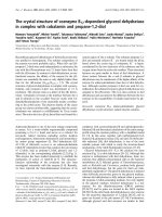
[29] (Fig. 1A) were used. Transcription analysis of
A
wt
atfh-1
as-AtFH
Fold change
B
wt
atfh-1 as-AtFH
kDa
17
AtFH
C
MCS
pCAMBIA 1302
EcoRIEcoRI
NPTII
CaMV35S
CaMV35S
as-AtFH
t-CAMV35S
*
*
0
1
2
3
*
*
wt
atfh-1
as-AtFH
LF
wt
atfh-1 as-AtFH
Fig. 1. (A) Scheme of the as-AtFH construct used to generate
transgenic plants expressing an AtFH fragment (564 bp) in anti-
sense orientation. as-AtFH is under the control of cauliflower
mosaic virus 35S (CaMV35S) promoter from pDH51 vector subcl-
oned at the EcoRI site from pCAMBIA 1302. MCS, multiple cloning
site; t-CAMV35S, 35S terminator; NPTII, kanamycin resistance
gene. (B) qRT-PCR analysis of AtFH expression in leaves (L) or
flowers (F) from wild-type (wt), atfh-1 and as-AtFH lines. The aster-
isk signals a statistically different result from the control value
(P < 0.05). Bars represent mean values (error ± standard deviation)
of three independent experiments. Relative AtFH expression levels
are shown as fold change values with respect to b-actin mRNA lev-
els. (C) Western-blot detection of AtFH protein in wild-type (wt),
atfh-1 and as-AtFH lines in leaves (left panel) or flowers (right
panel) using serum anti-recombinant AtFH.
M. V. Maliandi et al. Frataxin in heme synthesis in plants
FEBS Journal 278 (2011) 470–481 ª 2010 The Authors Journal compilation ª 2010 FEBS 471
mutants by qRT-PCR analysis showed that AtFH
mRNA levels were decreased in leaves and flowers of
both atfh-1 and as-AtFH lines (Fig. 1B). In addition,
AtFH protein levels determined by western blot using
specific antibodies showed a decrease of 50–70% in
atfh-1 and as-AtFH lines, respectively (Fig. 1C).
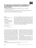
Using the growth conditions described in the experi-
mental section, the as-AtFH line showed retarded
growth (as also described for the atfh-1 line [15]) at
different developmental stages compared with wild-
type plants (Fig. 2). Moreover, as we reported previ-
ously for the atfh-1 line, we did not observe significant
differences in the morphology of as-AtFH roots, leaves
or flowers, but a decrease of 35% of fruit fresh
weight, alteration in silique length and a reduced num-
ber of viable seeds (28 ± 6 seeds per silique) compared
with 47 ± 5 seeds per silique found in the wild-type
(Fig. 2D).
Decrease in heme content in AtFH-deficient
plants
The heme content in rosette leaves was reduced to 34
and 41% in atfh-1 and as-AtFH plants, respectively,
whereas in flower tissues the levels fell to 25% in both
transgenic lines (Fig. 3). These results indicate that
AtFH-deficient plants have altered heme content,
agreeing with the proposed hypothesis. Thus, the fra-
taxin-deficient plants constitute a good model to study
the biogenesis of cellular hemeproteins.
Alteration of heme pathway transcripts in plants
with AtFH deficiency
To better understand the effect of AtFH deficiency on
heme biosynthesis, we evaluated the mRNA levels of
several transcripts coding for enzymes playing a role in
the heme metabolic pathway (see Fig. S1).
First, we investigated the expression levels of
HEMA1 (At1g58290) and HEMA2 (At1g09940), two
genes coding for glutamyl-tRNA reductase proteins
that catalyze the production of 5-aminolevulinic acid
(ALA). We found that HEMA1 is downregulated in
leaves without significant changes in flowers, whereas
HEMA2 is downregulated in both tissues (Fig. 4A).
The levels of GSA1 (At5g63570) and GSA2
(At3g48730), two glutamate-1-semialdehyde aminomu-
tase genes involved in the conversion of glutamate-
1-semialdehyde into 5-aminolevulinate were also
determined. GSA1 and GSA2 mRNA levels were
reduced 50% in leaves from AtFH-deficient lines,
compared with wild-type plants. By contrast, in flow-
ers, transcript levels of GSA1 and GSA2 presented an
augment of two- and three-fold compared with the
values found in wild-type plants (Fig. 4B).
We also evaluated the transcription levels of two
porphobilinogen synthase genes, HEMB1 (At1g69740)
and HEMB2 (At1g44318). A decrease in HEMB1 and
HEMB2 transcript levels was found in leaves, whereas
no change in HEMB1 transcript levels was found in
flowers (Fig. 4C). By contrast, a three- and eight-fold
induction in HEMB2 mRNA levels was found in flow-
ers of atfh-1 and as-AtFH lines, respectively (Fig. 4C).
Furthermore, coproporfirinogen oxidase (HEMF2,
At4g03205) mRNA levels in leaves showed a 50 and
70% decrease in atfh-1 and as-AtFH lines, compared
with wild-type, whereas no significant changes in their
amount were observed in flowers of these lines
(Fig. 4D).
Finally, we analyzed the expression of two FC
genes, AtFC-1 (At5g26030) and AtFC-2 (At2g30390).
AtFC-1 has been found to be expressed in all plant
A
B
C
D
wt
atfh-1
as-AtFH
wt wtatfh-1 as-AtFH atfh-1 as-AtFH
Fig. 2. Phenotype comparison of wild-type
(wt), atfh-1 and as-AtFH plants at different
stages of development: 14-day-old (A); 21-
day-old (B) and 40-day-old (C) growth plants.
(D) Morphology of siliques (8–10 days post
anthesis) from wild-type (wt), atfh-1 and
as-AtFH lines.
Frataxin in heme synthesis in plants M. V. Maliandi et al.
472 FEBS Journal 278 (2011) 470–481 ª 2010 The Authors Journal compilation ª 2010 FEBS
tissues and mainly in flowers and roots with an
enhanced expression under oxidative stress conditions
or tissue damage [30]. AtFC-2 is expressed in all plant
tissues, except in roots. An induction of 1.5–2-fold in
AtFC-1 levels was found in AtFH-deficient leaves by
QPCR analysis (Fig. 4E). By contrast, no significant
changes in AtFC-1 mRNA and a slight decrease in
AtFC-2 mRNA levels were detected in flowers (Fig. 4E).
In agreement with these results, AtFC activity in leaves
showed an increase of 15% in AtFH-deficient plants,
whereas no significant changes were observed in flowers
(not shown). These data suggest that AtFH deficiency
has a minor effect on AtFC activity.
AtFH deficiency affects catalase activity but not
their mRNA or protein levels
To assess the impact of AtFH deficiency on the activity
of heme-containing proteins, we decided to investigate
the catalase enzymes that catalyze the dismutation of
H
2
O
2
to H
2
O and O
2
. In plants, H
2
O
2
is removed essen-
tially by three enzymes: catalase, ascorbate peroxidase
and glutathione peroxidase [31]. Catalases do not con-
sume reducing power and have a very high reaction
rate, whereas ascorbate peroxidase and glutathione per-
oxidase require a source of reductant, ascorbate or glu-
tathione. Therefore, although plants contain different
H
2
O
2
metabolizing enzymes, catalases are highly active
enzymes in the absence of reductants as they primarily
catalyze a dismutase reaction [32]. H
2
O
2
consumption
was measured in the absence of other reductants and
using a protocol previously reported for the determina-
tion of catalase activity in plants (see Materials and
Methods section). Under this condition, the activity
detected can be attributed mainly to catalases.
Total catalase activity was determined in leaves and
flowers from AtFH-deficient lines. In both lines, a
decrease of 20% in catalase activity was found in
leaves (Fig. 5A), whereas a reduced activity of 15 and
40% was observed in flowers from atfh-1 and as-AtFH
lines, respectively.
In Arabidopsis, three genes coding for catalase,
CAT1 (At1g20630), CAT2 (At4g35090) and CAT3
(At1g20620), have been described. CAT2, located in
peroxisomes ⁄ glyoxisomes and cytosol, is the major
isoform in leaves, whereas CAT1 (located mainly in
cytosol and peroxisomes) and CAT3 (located in mito-
chondria) are less abundant [27]. Interestingly, the
mRNA levels of the genes encoding the three catalase
isoforms show no significant differences when com-
pared with wild-type plants (Fig. 5B). Western blot
analysis of leaf and flower extracts revealed with anti-
catalase IgG showed no significant differences between
AtFH-deficient and wild-type plants (Fig. 5C). These
results indicate that AtFH deficiency does not affect
catalase expression, but has an impact on the catalytic
activity in leaves and flowers.
Hemin rescues catalase activity in cell suspension
cultures and isolated mitochondria
To examine if the decrease in catalase activity results
from a heme deficiency, we determined the enzymatic
activity in atfh-1 and as-AtFH cell suspension cultures
using different concentrations of ALA, protoporphyrin
IX and hemin. It has been reported that hemin itself
has a catalase-like activity [33]. Therefore, we carried
out the assay of catalase activity in wild-type cells
without additions or in the presence of 1–10 lm hemin.
Under the conditions described above, the activity of
hemin does not have a significant contribution to the
total catalase activity (Fig. 6A). In agreement with the
data shown in Fig. 5A, we also observed a decrease in
catalase activity in Arabidopsis cells. On the other
hand, an almost complete restoration of catalase activ-
ity was observed in both AtFH-deficient lines after
incubation with 5 and 10 lm hemin (Fig. 6A), whereas
no changes were found in the presence of protopor-
phyrin IX or ALA (Fig. 6B, C). It should be noted
that no significant differences in AtFH mRNA levels
were detected after incubation with hemin, protopor-
phyrin IX or ALA (not shown).
The effect of hemin, protoporphyrin IX and ALA
treatment on catalase activity in isolated mitochondria
Heme (nmol·g
–1
FW)
*
*
wt
atfh-1
as-AtFH
wt
atfh-1
as-AtFH
L
F
*
*
0
5
10
15
Fig. 3. Noncovalently bound heme quantification in leaves (L, white
bars) or flowers (F, black bars) from wild-type (wt), atfh-1 and as-
AtFH lines. The asterisk signals a statistically different result from
the control value (P < 0.05). Values are the mean ± standard devia-
tion of four independent replicates.
M. V. Maliandi et al. Frataxin in heme synthesis in plants
FEBS Journal 278 (2011) 470–481 ª 2010 The Authors Journal compilation ª 2010 FEBS 473
from atfh-1 and as-AtFH plants was studied. A
decrease of 40 and 51% of catalase activity was found
in AtFH-deficient mitochondria. The activity was
almost completely restored after incubation of the
organelle suspension with 10 lm hemin (Fig. 6D). By
contrast, no significant changes in catalase activity
were observed in the presence of protoporphyrin IX or
ALA in atfh-1 and as-AtFH lines.
In addition, the catalase activity was not affected
when isolated mitochondria were incubated with pro-
toporphyrin IX in the presence of 1 or 5 lm Fe(II) in
citrate-buffered solutions (see Fig. S2). Moreover, the
levels of FC activity measured in isolated mitochondria
extracts were close to the background value (not
shown). These results agree with those previously
reported on the possibility that ferrous ions can be
inserted nonenzymatically into phorphyrin in the pres-
ence of reductants or fatty acids, but this reaction does
not occur in vivo [34].
Discussion
The understanding of the role of frataxin in iron
homeostasis in plants becomes highly relevant because
0
1
2
3
4
5
FL
GSA1 GSA2 GSA1 GSA2
0
1
2
3
4
5
HEMB1
FL
HEMB2
HEMB1
HEMB2
0.0
2.5
5.0
7.5
FL
AtFC1 AtFC2 AtFC1 AtFC2
0
1
2
3
4
HEMF2
FL
HEMF2
Fold change
Fold change
Fold change
BA
C
D
*
*
*
*
*
*
*
*
*
*
*
*
*
*
*
*
*
*
*
*
*
HEMA1
FL
HEMA2
HEMA1
HEMA2
*
*
*
*
*
*
0.0
0.5
1.0
1.5
E
Fig. 4. qRT-PCR analysis of genes involved
in the heme biosynthetic pathway: (A) glut-
amyl tRNA reductase (HEMA1, At1g58290
and HEMA2, At1g09940); (B) glutamate-
1-semialdehyde aminomutase (GSA1,
At5g63570 and GSA2, At3g48730);
(C) porphobilinogen synthase (HEMB1,
At1g69740 and HEMB2, At1g44318);
(D) coproporphyrinogen oxidase (HEMF2,
At4g03205); (E) FC (AtFC-1, At5g26030 and
AtFC-2, At2g30390). RNA was extracted
from rosette leaves (L) or flowers (F, stage
12) from wild-type (white bars), atfh-1 (grey
bars) and as-AtFH (black bars) plants. The
asterisk signals a statistically different result
from the control value (P < 0.05). Columns
represent mean values (error bars ± stan-
dard deviation) of three independent experi-
ments. Relative expression levels are
shown as fold change values with respect
to b-actin mRNA levels.
Frataxin in heme synthesis in plants M. V. Maliandi et al.
474 FEBS Journal 278 (2011) 470–481 ª 2010 The Authors Journal compilation ª 2010 FEBS
of its association with Fe–S clusters and heme groups,
the two main iron-containing prosthetic groups that
participate in the catalysis of numerous biochemical
reactions. However, the connection between both path-
ways, as well as the role of frataxin in iron metabo-
lism, remain unclear, especially in photosynthetic
organisms. Iron as a cofactor is involved in many cel-
lular processes: (a) biogenesis of Fe–S proteins accom-
plished by the Fe–S cluster machinery located in the
mitochondrial matrix [35] and (b) biogenesis of heme
groups and hemeproteins. The respiratory complexes
of the mitochondrial inner membrane involved in ener-
getic metabolism, aconitase and many other proteins
with different subcellular locations require Fe–S clus-
ters for activity [2,36]. On the other hand, cytochromes
and catalases require the presence of heme as a cofac-
tor for function [37,38].
Yeast cells lacking frataxin, YFH, are deficient in iron
use by FC and show low cytochrome content, suggest-
ing that the iron used in heme synthesis is under the
control of YFH [21]. Furthermore, yeast mutants with
deficiencies in the mitochondrial Fe–S cluster assembly
machinery display reduced levels of heme-containing
proteins such as cytochromes and cytochrome c oxidase,
suggesting a deficiency in the heme pathway [39].
In addition, Zhang et al. [40,41] reported that YFH and
two mitochondrial carrier proteins, MRS3 and MRS4
implicated in iron homeostasis, have a cooperative
CAT1 CAT2 CAT3
LFFLFL
wt wt
atfh-1 as-AtFH atfh-1 as-AtFH
L
FL
wt wt
atfh-1 as-AtFH atfh-1 as-AtFH
A
B
C
0
5
10
15
20
Activity (U·mg
–1
protein)
0.0
2.5
5.0
7.5
10.0
*
*
*
*
Fold change
F
Fig. 5. (A) Enzymatic activity of catalase from wild-type (wt), atfh-1
and as-AtFH lines analyzed in rosette leaves (L) or flower extracts
(F, stage 12). (B) qRT-PCR analysis of catalase genes in leaves (L)
and flowers (F) from wild-type, atfh-1 and as-AtFH lines (CAT1,
At1g20630; CAT2, At4g35090 and CAT3, At1g20620): wild-type
(white bars), atfh-1 (grey bars), as-AtFH (black bars). Columns rep-
resent mean values (error bars ± standard deviation) of three inde-
pendent experiments. Relative expression levels are shown as fold
change values with respect to b-actin mRNA levels. (C) Western
blot analysis of catalase protein from leaves (L) or flower (F)
extracts from wild-type (wt), atfh-1 and as-AtFH lines using specific
anti-catalase IgG.
B
*
*
wt
Activity (U·mg
–1
protein)Activity (U·mg
–1
protein)
A
*
*
*
*
0.00
0.25
0.50
0.75
wt
C
0.00
0.25
0.50
0.75
0
1
2
3
4
wt
0.00
0.25
0.50
0.75
wt
atfh-1 as-AtFH atfh-1 as-AtF
H
atfh-1 as-AtFHatfh-1 as-AtFH
D
Fig. 6. (A) Determination of total catalase activity in homogenates
obtained from cell culture extracts from wild-type (wt), atfh-1 and
as-AtFH lines in the absence (white bars) or in the presence of dif-
ferent concentrations of hemin: 0.5 l
M (light grey bars); 5 lM (dark
grey bars) or 10 l
M (black bars). (B, C) Determination of catalase
activity in homogenates obtained from cell culture extracts from
wild-type (wt), atfh-1 and as-AtFH lines in the absence (white bars)
or in the presence of different concentrations of protoporphyrin IX
(B) or ALA (C): 0.5 l
M (light grey bars); 5 lM (dark grey bars) or
10 l
M (black bars). (D) Total catalase activity determined in mito-
chondrial suspensions from wild-type (wt), atfh-1 and as-AtFH lines
without additions (white bars) or in the presence of 10 l
M protopor-
phyrin IX (light grey bars), ALA (dark grey bars) or 10 l
M hemin
(black bars). The asterisk indicates values statistically different from
the control (P < 0.05). Columns represent mean values (error
bars ± standard deviation) of three independent experiments.
M. V. Maliandi et al. Frataxin in heme synthesis in plants
FEBS Journal 278 (2011) 470–481 ª 2010 The Authors Journal compilation ª 2010 FEBS 475
function in providing iron for heme and Fe–S synthesis
in yeasts. Thus, it was proposed that frataxin could
have a role in the modulation of iron availability
within mitochondria for Fe–S and heme group synthe-
sis and frataxin deficiency might have an impact in
Fe–S and heme-containing protein biogenesis.
On the other hand, it has been reported that frataxin
interacts with FC and mediates iron delivery in the final
step of heme synthesis in human mitochondria [24].
However, there is no strong evidence for the presence
of FC in plant mitochondria. Cornah et al. [30] and
Masuda et al. [42] reported that most FC activity was
associated with plastids. Lister et al. [43] found that
either of the two FC isoforms from A. thaliana were
imported into chloroplasts in vitro. Masuda et al. [42]
found that GFP-fusion proteins with either of two iso-
forms of FC from cucumber were targeted to plastids,
but not to mitochondria. Indeed, the specific antibodies
against either of the two isoforms of FC detected sig-
nals only in plastids [42]. In Chlamydomonas rein-
hardtii, where a single gene encodes for FC, the protein
is targeted into the plastids, indicating that the FC
activity is not required to be present inside mitochon-
dria [44]. Thus, it has been suggested that in plants the
synthesis of heme takes place almost exclusively in
plastids and exported to cytosol and mitochondria [44–
46]. Consistent with these results, we found less than
0.3% FC total activity in isolated mitochondria, cor-
roborating the data reported by Cornah et al. [30].
It has been suggested that frataxin deficiency causes
defects late in the heme pathway. The transcriptome
analysis of human lymphoblasts derived from Frie-
dreich’s ataxia patients and frataxin-deficient mice
showed a decrease in the mRNA levels of copro-
porphyrinogen oxidase and delta-aminolevulinate syn-
thase 1, two enzymes involved in the heme biosynthetic
pathway, and also Isu1 and FC. These observations
support the idea that frataxin deficiency affects the
expression of many nuclear-encoded mitochondrial
genes [47]. This situation is associated with increased
levels of protoporphyrin IX, consistent with a defect
downstream of this metabolite in the heme pathway
[47]. In addition, reduced mitochondrial heme a and
heme c levels and a decreased activity of cytochrome
oxidase strongly suggest that frataxin is involved in late
stages of the heme biosynthetic pathway, i.e., the incor-
poration of iron into protoporphyrin IX to produce
heme [21,47,48]. It has been reported that the key con-
trol point of heme and chlorophyll synthesis in plants
is the formation of ALA from glutamate catalyzed by
glutamyl-tRNA reductase enzymes encoded by HEMA
genes [49]. HEMA1 has been associated with the provi-
sion of tetrapyrroles for chlorophyll and heme produc-
tion in photosynthetic tissues, whereas the role of
HEMA2 is to provide a background activity of glutam-
yl-tRNA reductase for heme production, mainly in
nonphotosynthetic tissues [50,51]. Thus, the downregu-
lation of both HEMA1 and HEMA2 transcripts is in
agreement with the observed heme deficiency in AtFH-
deficient plants.
Arabidopsis AtFH-deficient lines also showed a mod-
ification of the mRNA levels of other enzymes
involved in heme biosynthesis, such as GSA1 and 2,
HEMB1, and 2, HEMF2 and FC1 and FC2, indicat-
ing that an analogous situation occurs in plants. How-
ever, a different response was found when compared in
different organs. In flowers, GSA1 and GSA2 tran-
script levels were increased compared with leaves,
where the respective transcripts were downregulated or
remain unchanged. It should be noted that a differen-
tial response for some isoforms was observed in flow-
ers but not in leaves. The HEMB2 transcript level was
increased several fold, whereas HEMB1 mRNA levels
remained unchanged in flowers. Also, a decrease in
mRNA levels for AtFC2 contrast with the unmodified
expression pattern of AtFC1. The different expression
pattern of these genes in leaves and flowers could be
explained by a differential regulation, probably reflect-
ing the gene expression network specific to each organ.
These observations should be interpreted with caution,
as it is difficult to know whether the observed effect is
directly linked to AtFH deficiency or is the result of a
secondary event. Previously, we found that AtFH-defi-
cient plants present increased reactive oxygen species
formation [15,16]. The reactive oxygen species have
been implicated in complex gene expression responses,
particularly the induction of nuclear-encoded mito-
chondrial genes [52].
Catalase activity was reduced in AtFH-deficient
plants without significant reduction of catalase
mRNAs or protein levels. The fact that the decrease in
catalase activity correlates with the deficiency in heme
content, and the observation that the normal enzy-
matic activity is recovered after addition of hemin, but
not the iron-lacking tetrapyrrole protoporphyrin IX or
ALA, substantiate the hypothesis that AtFH would
have a major role in heme production required for the
formation of the active catalase holoenzyme. This
effect is particularly evident for the catalase activity
associated with the mitochondria fraction where CAT3
is the main isoform. These results are in accordance
with hemin rescue experiments performed in frataxin-
deficient neuronal cells, which showed increased activ-
ity of some Fe–S protein and cytochrome oxidase
restoring the normal phenotype [25], and with data
showing that recombinant erythropoietin, which
Frataxin in heme synthesis in plants M. V. Maliandi et al.
476 FEBS Journal 278 (2011) 470–481 ª 2010 The Authors Journal compilation ª 2010 FEBS
stimulates the synthesis of heme, can rescue the pheno-
type observed in frataxin-deficient cells [53].
In summary, AtFH-deficient plants present alter-
ation in several transcripts from the heme biosynthetic
route with a decrease in total heme content and a
deficiency of catalase activity that can be rescued by
exogenous hemin, indicating that AtFH, apart from its
role in protecting bioavailable iron within mitochon-
dria and the synthesis of Fe–S groups, also plays a role
in the production of heme groups and the activity of
hemeproteins in plants.
Materials and Methods
Plant material and growth conditions
Arabidopsis thaliana (var. Columbia Col-0) was used as the
wild-type reference plant. Two frataxin-deficient lines were
also used in these experiments: a T-DNA knockdown
mutant (atfh-1, SALK_021263) and an antisense line,
as-atfh. Mutant plants were selected in MS agar medium
containing 30 gÆmL
)1
kanamycin. Transgenic as-AtFH
plants were selected in MS medium containing 20 lgÆmL
)1
hygromycin. After 2 weeks, plants were transferred to soil
and grown in a greenhouse, at 25 °C under fluorescent
lamps (Grolux, Sylvania, Danvers, MA, USA and Cool
White, Philips, Amsterdam, The Netherlands) with an
intensity of 150 lmolÆm
)2
Æs
)1
using a 16 h light ⁄ 8 h dark
photoperiod. Arabidopsis cell suspension cultures were
grown in the dark (22 °C) in an orbital shaker (130 r.p.m.).
Isolation of RNA and qRT-PCR analysis
Total RNA was extracted from rosette leaves and flowers
(stage 12) using the RNA plant mini kit (Qiagen, Valencia,
CA, USA). Complementary DNA was synthesized using
random hexamers and the M-MLV reverse transcriptase
protocol (USB Corp., Cleveland, OH, USA). qRT-PCR
was carried out in a MiniOPTICON2 apparatus (BioRad,
Hercules, CA, USA), using the intercalation dye SYBR-
Green I (Invitrogen, Carlsbad, CA, USA) as a fluorescent
reporter and Go Taq polymerase (Promega, Madison, WI,
USA). Primers suitable for amplification of 150–250 bp
products for each gene under study were designed using the
primer3 software (see Table S1). Amplification of cDNA
was carried out under the following conditions: 2 min dena-
turation at 94 °C; 40–45 cycles at 94 °C for 15 s, 57 °C for
20 s, and 72 °C for 20 s, followed by 10 min extension at
72 °C. Three replicates were performed for each sample.
Melting curves for each PCR were determined by measur-
ing the decrease in fluorescence with increasing temperature
(from 65 to 98 °C). PCR products were run on a 2% (w ⁄ v)
agarose gel to confirm the size of the amplification products
and to verify the presence of a unique PCR product.
Relative transcript levels were calculated as a ratio of the
transcript abundance of the studied gene to the transcript
abundance of b-actin (At3g18780).
Production of as-atfh transgenic plants
To prepare the antisense construct of frataxin, a BamHI ⁄
SmaI fragment containing the AtFH coding sequence
(564 bp) was obtained by PCR (see primers used in
Table S1) and then cloned downstream from the cauliflower
mosaic virus 35S promoter into the pDH51 vector [54] previ-
ously digested with BamHI and SmaI. After verifying the
correct orientation of the insert, the resulting 35S:as-AtFH
expression cassette was excised as EcoRI restriction
fragments and subcloned into pCAMBIA 1320 [29]. The
recombinant plasmids were introduced into Agrobacte-
rium tumefaciens GV3101 strain by the freeze–thaw method
[55]. Arabidopsis was transformed using the floral dip method
[56]. The expression of the antisense version of AtFH was
verified by RT-PCR.
Determination of heme content
The content of noncovalently bound heme was determined
using 6 week rosette leaves or flowers (stage 12) from wild-
type, atfh-1 and as-AtFH, as previously described [57].
Extracted heme was spectrophotometrically quantified with
a Perkin–Elmer lambda 35 UV ⁄ Vis spectrometer by mea-
suring the absorbance at 398 nm (Perkin–Elmer, Boston,
MA, USA). Standard solutions of hemin (Sigma-Aldrich,
St Louis, MO, USA) were prepared by dissolving the solid
reagent in 50 mm sodium phosphate buffer, pH 7.4.
Enzyme assays
Homogenates from cell cultures were prepared as follows:
1–2 g of cells were centrifuged for 10 min at 3000g and the
pellet was ground to a powder with liquid nitrogen. The
powdered material was homogenized with extraction buffer
containing 450 mm sucrose, 15 mm Mops-KOH, 1.5 mm
EGTA and 6 gÆL
)1
polyvinylpyrrolidone, pH 7.4. The sus-
pension was incubated with 2 gÆL
)1
BSA, 0.2 mm phen-
ylmethanesulfonyl fluoride and 500 U cellulase (ICN
Biomedicals, Aurora, OH, USA) at 4 °C for 60 min. Cells
were disrupted using an ultrasonicator (VCX130, Sonics &
Materials, Newtown, CT, USA) and centrifuged at 10 000g
for 20 min at 4 °C and the supernatant collected. The
homogenate from Arabidopsis tissues (leaves and flowers)
was prepared as follows: 200 mg tissue was frozen under
liquid nitrogen and ground to a powder. The powdered
material was homogenized in extraction buffer (50 mm
KH
2
PO
4
pH 7.8, 0.5% v ⁄ v Triton X-100, 0.5 mm EDTA
and 1 mm phenylmethanesulfonyl fluoride). The homoge-
nate was centrifuged at 9500 g for 20 min at 4 °C and the
supernatant collected. Catalase activity was determined at
M. V. Maliandi et al. Frataxin in heme synthesis in plants
FEBS Journal 278 (2011) 470–481 ª 2010 The Authors Journal compilation ª 2010 FEBS 477
25 °C as described previously [58] with minor modifications
[59] by following the decrease in absorbance (A) at 240 nm
at 25 °C. The catalase assay medium contained 470 lLof
50 mm KH
2
PO
4
pH 7.0 and 10 mm H
2
O
2
as a substrate.
Homogenates used to determine FC activity were prepared
as previously described [60] and enzymatic activity was
measured according to previous methods [61].
Porphyrin and ALA treatments
Hemin, protoporphyrin IX or ALA (0–10 lm) were added
to 100 mL of Arabidopsis cell cultures and incubated at
24 °C for 18 h with orbital shaking. Catalase activity was
determined as described in the previous section. Mitochon-
dria suspensions (10 mgÆmL
)1
protein) were incubated in
a buffer containing 250 mm mannitol, 50 mm KCl, 2 mm
MgCl
2
,20mm Hepes pH 7.4, 1 mm K
2
HPO
4
,1mm dith-
iothreitol, 10 mm ATP, 20 lm ADP, 10 mm sodium succi-
nate and 10 lm hemin, protoporphyrin IX or ALA for 2 h
with constant shaking. After incubation, mitochondria were
recovered by centrifugation and resuspended in 10 mm
KH
2
PO
4
pH 7. After lysis using an ultrasonicator
(VCX130, Sonics & Materials) followed by centrifugation
at 12 000g for 10 min, the catalase activity was determined
in the supernatant using the assay described above.
Additional methods
Isolation of highly purified mitochondria from Arabidopis
leaves and flowers was carried out as described by Werhahn
et al. [62,63] with modifications. Under these conditions, the
mitochondrial fraction is essentially deprived of cytoplasmic
and plastid contamination. The mitochondrial pellet was
recovered with buffer containing 300 mm mannitol and
10 mm K
2
HPO
4
(pH 7.4) as previously described [15]. Pro-
teins were separated by electrophoresis on 12% SDS ⁄ PAGE
[64] and revealed by Coomassie Blue staining or electroblot-
ted on to nitrocellulose membranes (BioRad). Electroblotted
membranes were incubated with anti-recombinant AtFH or
anti-catalase (kindly provided by M. Nishimura, National
Institute for Basic Biology, Okazaki, Japan) polyclonal IgG.
The antigen–antibody complex was visualized with alkaline
phosphatase-linked anti-mouse IgG or anti-rabbit IgG, fol-
lowed by staining with 5-bromo-4-chloroindol-2-yl phos-
phate and Nitro Blue tetrazolium as described previously
[65]. Total protein was determined as described by Bradford
[66]. The relative protein levels in western blots were deter-
mined by densitometric analysis using the gel pro ana-
lyzer program (Media Cybernetics, Bethesda, MD, USA).
Statistical analyses
The significance of differences was determined using Stu-
dent’s t-test. Values statistically different from the control
(P < 0.05) are denoted with an asterisk in Figs 1, 3–6.
Acknowledgements
This work was supported by grants from PICS-CNRS
3641, the Universite
´
Victor Segalen Bordeaux 2, AN-
PCyT (PICT 00614 and 0729). MVM and VRT are
doctoral fellows from CONICET. LL is a doctoral fel-
low from ANPCyT. MVB and DGC are research
members from CONICET.
References
1 Bencze KZ, Kondapalli KC, Cook JD, McMahon S,
Millan-Pacheco C, Pastor N & Stemmler TL (2006) The
structure and function of frataxin. Crit Rev Biochem
Mol Biol 41, 269–291.
2 Lill R & Mu
¨
hlenhoff U (2008) Maturation of iron-sul-
fur proteins in eukaryotes: mechanisms, connected pro-
cesses, and diseases. Annu Rev Biochem 77, 669–700.
3 Lill R (2009) Function and biogenesis of iron-sulphur
proteins. Nature 460, 831–838.
4 Campuzano V, Montermini L, Molto MD, Pianese L,
Cossee M, Cavalcanti F, Monros E, Rodius F, Duclos
F, Monticelli A et al. (1996) Friedreich’s ataxia: auto-
somal recessive disease caused by an intronic GAA
triplet repeat expansion. Science 271, 1423–1427.
5 Babcock M, de Silva D, Oaks R, Davis-Kaplan S, Jiral-
erspong S, Montermini L, Pandolfo M & Kaplan J
(1997) Regulation of mitochondrial iron accumulation
by Yfh1p, a putative homolog of frataxin. Science 276,
1709–1712.
6 Pandolfo M (2002) Frataxin deficiency and mitochon-
drial dysfunction. Mitochondrion 2, 87–93.
7 Puccio H (2009) Multicellular models of Friedreich
ataxia. J Neurol 256(Suppl 1), 18–24.
8 Schmucker S & Puccio H (2010) Understanding the
molecular mechanisms of Friedreich’s ataxia to
develop therapeutic approaches. Hum Mol Genet 19,
R103–110.
9 Koutnikova H, Campuzano V, Foury F, Dolle P, Caz-
zalini O & Koenig M (1997) Studies of human, mouse
and yeast homologues indicate a mitochondrial function
for frataxin. Nat Genet 16, 345–351.
10 Rotig A, de Lonlay P, Chretien D, Foury F, Koenig
M, Sidi D, Munnich A & Rustin P (1997) Aconitase
and mitochondrial iron-sulphur protein deficiency in
Friedreich ataxia. Nat Genet 17, 215–217.
11 Gerber J, Muhlenhoff U & Lill R (2003) An interac-
tion between frataxin and Isu1 ⁄ Nfs1 that is crucial
for Fe ⁄ S cluster synthesis on Isu1. EMBO Rep 4,
906–911.
12 Chen OS, Hemenway S & Kaplan J (2002) Inhibition
of Fe-S cluster biosynthesis decreases mitochondrial
iron export: evidence that Yfh1p affects Fe-S
cluster synthesis. Proc Natl Acad Sci USA 99, 12321–
12326.
Frataxin in heme synthesis in plants M. V. Maliandi et al.
478 FEBS Journal 278 (2011) 470–481 ª 2010 The Authors Journal compilation ª 2010 FEBS
13 Huynen MA, Snel B, Bork P & Gibson TJ (2001) The
phylogenetic distribution of frataxin indicates a role in
iron-sulfur cluster protein assembly. Hum Mol Genet
10, 2463–2468.
14 Ristow M, Pfister MF, Yee AJ, Schubert M, Michael
L, Zhang CY, Ueki K, Michael MD II, Lowell BB &
Kahn CR (2000) Frataxin activates mitochondrial
energy conversion and oxidative phosphorylation. Proc
Natl Acad Sci USA 97, 12239–12243.
15 Busi MV, Maliandi MV, Valdez H, Clemente M,
Zabaleta EJ, Araya A & Gomez-Casati DF (2006)
Deficiency of Arabidopsis thaliana frataxin alters activity
of mitochondrial Fe-S proteins and induces oxidative
stress. Plant J 48, 873–882.
16 Busi MV, Zabaleta EJ, Araya A & Gomez-Casati DF
(2004) Functional and molecular characterization of the
frataxin homolog from Arabidopsis thaliana. FEBS Lett
576, 141–144.
17 Maliandi MV, Busi MV, Clemente M, Zabaleta EJ,
Araya A & Gomez-Casati DF (2007) Expression and
one-step purification of recombinant Arabidopsis thali-
ana frataxin homolog (AtFH). Protein Expr Purif 51,
157–161.
18 Martin M, Colman MJ, Gomez-Casati DF, Lamattina
L & Zabaleta EJ (2009) Nitric oxide accumulation is
required to protect against iron-mediated oxidative
stress in frataxin-deficient Arabidopsis plants. FEBS Lett
583, 542–548.
19 Vazzola V, Losa A, Soave C & Murgia I (2007) Knock-
out of frataxin gene causes embryo lethality in Arabid-
opsis. FEBS Lett 581, 667–672.
20 Becker EM, Greer JM, Ponka P & Richardson DR
(2002) Erythroid differentiation and protoporphyrin IX
down-regulate frataxin expression in Friend cells:
characterization of frataxin expression compared to
molecules involved in iron metabolism and
hemoglobinization. Blood 99, 3813–3822.
21 Lesuisse E, Santos R, Matzanke BF, Knight SAB,
Camadro JM & Dancis A (2003) Iron use for haeme
synthesis is under control of the yeast frataxin homo-
logue (Yfh1). Human Mol Genet 12, 879–889.
22 Park S, Gakh O, O’Neill HA, Mangravita A, Nichol H,
Ferreira GC & Isaya G (2003) Yeast frataxin sequen-
tially chaperones and stores iron by coupling protein
assembly with iron oxidation. J Biol Chem 278, 31340–
31351.
23 He Y, Alam SL, Proteasa SV, Zhang Y, Lesuisse E,
Dancis A & Stemmler TL (2004) Yeast frataxin solution
structure, iron binding, and ferrochelatase interaction.
Biochemistry 43, 16254–16262.
24 Yoon T & Cowan JA (2004) Frataxin-mediated iron
delivery to ferrochelatase in the final step of heme bio-
synthesis. J Biol Chem 279, 25943–25946.
25 Napoli E, Morin D, Bernhardt R, Buckpitt A & Corto-
passi G (2007) Hemin rescues adrenodoxin, heme a and
cytochrome oxidase activity in frataxin-deficient oligo-
dendroglioma cells. Biochim Biophys Acta 1772, 773–
780.
26 Beyer W, Imlay J & Fridovich I (1991) Superoxide
dismutases. Prog Nucleic Acid Res Mol Biol 40, 221–
253.
27 Frugoli JA, Zhong HH, Nuccio ML, McCourt P,
McPeek MA, Thomas TL & McClung CR (1996)
Catalase is encoded by a multigene family in
Arabidopsis thaliana (L.) Heynh. Plant Physiol 112,
327–336.
28 Radisky DC, Babcock MC & Kaplan J (1999) The
yeast frataxin homologue mediates mitochondrial iron
efflux. Evidence for a mitochondrial iron cycle. J Biol
Chem 274, 4497–4499.
29 Hajdukiewiez P, Svab Z & Maliga P (1994) The
small, versatile pPZP family of Agrobacterium binary
vectors for plant transformation. Plant Mol Biol 25,
989–994.
30 Cornah JE, Roper JM, Pal Singh D & Smith AG
(2002) Measurement of ferrochelatase activity using
a novel assay suggests that plastids are the major site
of haem biosynthesis in both photosynthetic and
non-photosynthetic cells of pea (Pisum sativum L.).
Biochem J 362, 423–432.
31 Rizhsky L, Hallak-Herr E, Van Breusegem F,
Rachmilevitch S, Barr JE, Rodermel S, Inze D &
Mittler R (2002) Double antisense plants lacking
ascorbate peroxidase and catalase are less sensitive to
oxidative stress than single antisense plants lacking
ascorbate peroxidase or catalase. Plant J 32, 329–342.
32 Willekens H, Chamnongpol S, Davey M, Schraudner
M, Langebartels C, Van Montagu M, Inze D & Van
Camp W (1997) Catalase is a sink for H2O2 and is
indispensable for stress defence in C3 plants. EMBO J
16, 4806–4816.
33 Grinberg LN, O’Brien PJ & Hrkal Z (1999) The effects
of heme-binding proteins on the peroxidative and cata-
latic activities of hemin. Free Radic Biol Med 27, 214–
219.
34 Taketani S & Tokunaga R (1984) Non-enzymatic heme
formation in the presence of fatty acids and thiol reduc-
tants. Biochim Biophys Acta 798, 226–230.
35 Lill R & Kispal G (2000) Maturation of cellular Fe-S
proteins: an essential function of mitochondria. Trends
Biochem Sci 25, 352–356.
36 Balk J & Lobreaux S (2005) Biogenesis of iron-sulfur
proteins in plants. Trends Plant Sci 10, 324–331.
37 Giege P, Grienenberger JM & Bonnard G (2008) Cyto-
chrome c biogenesis in mitochondria. Mitochondrion 8,
61–73.
38 Welinder KG (1992) Superfamily of plant, fungal and
bacterial peroxidases. Curr Opin Struct Biol 2, 388–393.
39 Lange H, Muhlenhoff U, Denzel M, Kispal G & Lill R
(2004) The heme synthesis defect of mutants impaired
M. V. Maliandi et al. Frataxin in heme synthesis in plants
FEBS Journal 278 (2011) 470–481 ª 2010 The Authors Journal compilation ª 2010 FEBS 479
in mitochondrial iron-sulfur protein biogenesis is caused
by reversible inhibition of ferrochelatase. J Biol Chem
279, 29101–29108.
40 Zhang Y, Lyver EK, Knight SAB, Lesuisse E & Dancis
A (2005) Frataxin and mitochondrial carrier proteins,
Mrs3p and Mrs4p, cooperate in providing iron for
heme synthesis. J Biol Chem 280, 19794–19807.
41 Zhang Y, Lyver ER, Knight SAB, Pain D, Lesuisse E
& Dancis A (2006) Mrs3p, Mrs4p, and frataxin provide
iron for Fe–S cluster synthesis in mitochondria. J Biol
Chem 281, 22493–22502.
42 Masuda T, Suzuki T, Shimada H, Ohta H & Takamiya
K (2003) Subcellular localization of two types of fer-
rochelatase in cucumber. Planta 217, 602–609.
43 Lister R, Chew O, Rudhe C, Lee MN & Whelan J
(2001) Arabidopsis thaliana ferrochelatase-I and -II are
not imported into Arabidopsis mitochondria. FEBS Lett
506, 291–295.
44 van Lis R, Atteia A, Nogaj LA & Beale SI (2005)
Subcellular localization and light-regulated expression
of protoporphyrinogen IX oxidase and ferrochelatase in
Chlamydomonas reinhardtii. Plant Physiol 139, 1946–
1958.
45 Mochizuki N, Tanaka R, Grimm B, Masuda T, Moulin
M, Smith AG, Tanaka A & Terry MJ (2008) The cell
biology of tetrapyrroles: a life and death struggle.
Trends Plant Sci 15, 488–498.
46 Tanaka R & Tanaka A (2007) Tetrapyrrole biosynthesis
in higher plants. Annu Rev Plant Biol 58, 321–346.
47 Schoenfeld RA, Napoli E, Wong A, Zhan S, Reut-
enauer L, Morin D, Buckpitt AR, Taroni F, Lonnerdal
B, Ristow M et al. (2005) Frataxin deficiency alters
heme pathway transcripts and decreases mitochondrial
heme metabolites in mammalian cells. Human Mol
Genet 14, 3787–3799.
48 Puccio H, Simon D, Cossee M, Criqui-Filipe P, Tiziano
F, Melki J, Hindelang C, Matyas R, Rustin P &
Koenig M (2001) Mouse models for Friedreich ataxia
exhibit cardiomyopathy, sensory nerve defect and Fe-S
enzyme deficiency followed by intramitochondrial iron
deposits. Nat Genet 27, 181–186.
49 Papenbrock J & Grimm B (2001) Regulatory network
of tetrapyrrole biosynthesis—studies of intracellular sig-
nalling involved in metabolic and developmental control
of plastids. Planta 213, 667–681.
50 Nagai S, Koide M, Takahashi S, Kikuta A, Aono M,
Sasaki-Sekimoto Y, Ohta H, Takamiya K & Masuda T
(2007) Induction of isoforms of tetrapyrrole biosyn-
thetic enzymes, AtHEMA2 and AtFC1, under stress
conditions and their physiological functions in
Arabidopsis. Plant Physiol 144, 1039–1051.
51 Ujwal ML, McCormac AC, Goulding A, Kumar AM,
Soll D & Terry MJ (2002) Divergent regulation of the
HEMA gene family encoding glutamyl-tRNA reductase
in Arabidopsis thaliana: expression of HEMA2 is
regulated by sugars, but is independent of light and
plastid signalling. Plant Mol Biol 50, 83–91.
52 Rhoads DM, Umbach AL, Subbaiah CC & Siedow JN
(2006) Mitochondrial reactive oxygen species. Contribu-
tion to oxidative stress and interorganellar signaling.
Plant Physiol 141, 357–366.
53 Sturm B, Stupphann D, Kaun C, Boesch S, Schranzho-
fer M, Wojta J, Goldenberg H & Scheiber-Mojdehkar
B (2005) Recombinant human erythropoietin: effects on
frataxin expression in vitro. Eur J Clin Invest 35 , 711–
717.
54 Pietrzak M, Shillito RD, Hohn T & Potrykus I (1986)
Expression in plants of two bacterial antibiotic resistance
genes after protoplast transformation with a new plant
expression vector. Nucleic Acids Res 14
, 5857–5868.
55 Weigel D & Glazebrook J (2002) Transformation of
Agrobacterium using the freeze–thaw method. In Arabi-
dopsis, A Laboratory manual, Cold Spring Harbor
Laboratory Press, New York, pp 125–126.
56 Clough SJ & Bent AF (1998) Floral dip‘ simplified
method for Agrobacterium-mediated transformation of
Arabidopsis thaliana. Plant J 16, 735–743.
57 Peter E & Grimm B (2009) GUN4 is required for post-
translational control of plant tetrapyrrole biosynthesis.
Mol Plant 2, 1198–1210.
58 Aebi H (1984) Catalase in vitro. Methods Enzymol 105,
121–126.
59 Saleh L & Plieth C (2009) Fingerprinting antioxidative
activities in plants. Plant Methods 5,2.
60 Rius SP, Casati P, Iglesias AA & Gomez-Casati DF
(2006) Characterization of an Arabidopsis thaliana
mutant lacking a cytosolic non-phosphorylating glycer-
aldehyde-3-phosphate dehydrogenase. Plant Mol Biol
61, 945–957.
61 Taketani S & Tokunaga R (1982) Purification and
substrate specificity of bovine liver-ferrochelatase. Eur J
Biochem 127, 443–447.
62 Kruft V, Eubel H, Jansch L, Werhahn W & Braun HP
(2001) Proteomic approach to identify novel mitochon-
drial proteins in Arabidopsis. Plant Physiol 127, 1694–
1710.
63 Werhahn W, Niemeyer A, Jansch L, Kruft V, Schmitz
UK & Braun H (2001) Purification and characterization
of the preprotein translocase of the outer mitochondrial
membrane from Arabidopsis. Identification of multiple
forms of TOM20. Plant Physiol 125, 943–954.
64 Laemmli UK (1970) Cleavage of structural proteins
during the assembly of the head of bacteriophage T4.
Nature 227, 680–685.
65 Bollag DM, Rozycki MD & Edelstein SJ (1996) Protein
methods, 2nd edn. Wiley-Liss, New York.
66 Bradford MM (1976) A rapid and sensitive method for
the quantitation of microgram quantities of protein
utilizing the principle of protein-dye binding. Anal
Biochem 72, 248–254.
Frataxin in heme synthesis in plants M. V. Maliandi et al.
480 FEBS Journal 278 (2011) 470–481 ª 2010 The Authors Journal compilation ª 2010 FEBS
Supporting information
The following supplementary material is available:
Fig. S1. AraCyc heme biosynthetic pathway.
Fig. S2. Determination of catalase activity in isolated
mitochondria in the presence of protoporphyrin and
Fe(II).
Table S1. Oligonucleotide primers used.
This supplementary material can be found in the
online version of this article.
Please note: As a service to our authors and readers,
this journal provides supporting information supplied
by the authors. Such materials are peer-reviewed and
may be re-organized for online delivery, but are not
copy-edited or typeset. Technical support issues arising
from supporting information (other than missing files)
should be addressed to the authors.
M. V. Maliandi et al. Frataxin in heme synthesis in plants
FEBS Journal 278 (2011) 470–481 ª 2010 The Authors Journal compilation ª 2010 FEBS 481