Báo cáo khoa học: Nuclear factor kappa B and tumor necrosis factor-alpha modulation of transcription of the mouse testis- and pre-implantation development-specific Rnf33⁄Trim60 gene pot
Bạn đang xem bản rút gọn của tài liệu. Xem và tải ngay bản đầy đủ của tài liệu tại đây (657 KB, 14 trang )
Nuclear factor kappa B and tumor necrosis factor-alpha
modulation of transcription of the mouse testis- and
pre-implantation development-specific Rnf33
⁄
Trim60 gene
Kong-Bung Choo
1,2,3
, Min-Chuan Hsu
1,2
, Yao-Hui Tsai
1,4
, Wan-Yi Lin
1
and Chiu-Jung Huang
4
1 Department of Medical Research and Education, Taipei Veterans General Hospital, Taipei, Taiwan
2 Department of Biotechnology and Laboratory Science in Medicine, Taipei Veterans General Hospital, Taipei, Taiwan
3 Institute of Clinical Medicine, National Yang Ming University, Taipei, Taiwan
4 Department of Animal Science and Graduate Institute of Biotechnology, School of Agriculture, Chinese Culture University, Yangmingshan,
Taipei, Taiwan
Introduction
We have previously reported two tripartite motif
(TRIM) ⁄ RING-Box-coiled coil (RBCC) protein
genes – Rnf33 ⁄ Trim60 and Rnf35 ⁄ Trim61 – that are
temporally expressed in the egg and in the pre-implan-
tation embryo of the mouse; both genes are silenced at
the blastocyst stage before placental implantation and
Keywords
nuclear factor-kappa B (NF-jB); p65 and p50
proteins; Rnf33; testis-specific gene
expression; tumor necrosis factor-alfa
(TNF-a)
Correspondence
Chiu-Jung Huang, PhD, Associate Professor,
Department of Animal Science and
Graduate Institute of Biotechnology, School
of Agriculture, Chinese Culture University,
55, Hwa-Kang Road, Yangmingshan,
Taipei 111, Taiwan
Fax: +886 2 28613100
Tel: +886 2 28610511 ext. 31231
E-mail:
(Received 29 July 2010, revised 15 November
2010, accepted 24 December 2010)
doi:10.1111/j.1742-4658.2010.08002.x
We have previously reported a mouse Rnf33 ⁄ Trim60 gene that is temporally
expressed in the pre-implantation embryo. The Rnf33 structural gene is com-
posed of a short noncoding exon 1 and an intronless coding exon 2. In the
present work, Rnf33 was shown to be expressed in the mouse testis and in the
testicular cell lines TM3 and TM4. To elucidate Rnf33 transcriptional modu-
lation, a 2.5-kb Rnf33 sequence, inclusive of the upstream regulatory region,
exon 1 and the associated intronic sequence, was dissected in transient trans-
fection and luciferase assays. An initiator and an atypical TATA-box were
shown to act as the core promoter elements of the gene. Deletion and muta-
genesis of the 2.5-kb sequence in luciferase constructs further demonstrated
that an intronic and palindromic kappa B (jB) sequence was an important
cis element targeted by the nuclear factor-jB (NF-jB) subunits p65 ⁄ RELA
and p50 ⁄ NFjB1, and also through modulation by tumor necrosis factor a.
Transcriptional up-regulation of Rnf33 by NF-jB and tumor necrosis factor-
a was directly demonstrated in TM3 and TM4 cells by real-time PCR quanti-
fication of the Rnf33 mRNA levels. Small interfering RNA knockdown of
p65 and p50 confirmed Rnf33 down-regulation by p65 ⁄ p50. Spermatogenesis
is regulated by a wide range of stimuli, including NF-jB, which, in turn, is
regulated by other signals. Hence, demonstration of NF-jB-regulated Rnf33
expression in testicular cells, particularly in Sertoli cells, implicates functional
involvement of the putative RNF33 protein in spermatogenesis through
association of the RNF33 protein with the microtubule via interaction with
kinesin motor proteins, as previously demonstrated [Huang et al.,submitted].
Abbreviations
AR, androgen receptor; aTATA, atypical TATA-box; ChIP, chromatin immunoprecipitation; EMSA, electrophoretic mobility shift assay;
EST, expressed sequence tag; GAPDH, glyceraldehyde-3-phosphate dehydrogenase; HIF-1a, hypoxia-inducible factor 1a; HRE, hypoxia-
response element; IKK, IjB kinase; Inr, initiator; KIF3A ⁄ KIF3B, kinesin-2 family members 3A and 3B; NF-jB, nuclear factor-kappa B; RBCC,
RING-Box-coiled coil; SF, serum-free; siRNA, small interfering RNA; SV40, simian virus 40; TFBSs, transcription factor-binding sites; TNF-a,
tumor necrosis factor a; TRIM, tripartitate motif; jB, kappa B.
FEBS Journal 278 (2011) 837–850 ª 2011 Chinese Culture University. Journal compilation ª 2011 FEBS 837
remain silenced throughout the remaining stages of
embryonic development [1,2]. The Rnf33 gene is
located 11.5 kb downstream of Rnf35; both Rnf33 and
Rnf35 are intronless in the coding sequences but each
gene is associated with a short noncoding exon 1 and
therefore with a single short intron of about 2.2 and
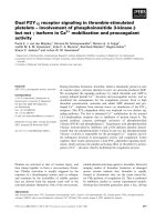
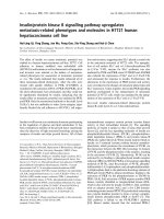
3.3 kb in size, respectively (Fig. 1) [1,2].
Transcriptional regulation of the upstream Rnf35
gene has been closely examined [3,4]. The Rnf35 pro-
moter is TATA-less but utilizes an initiator (Inr)
sequence as the core promoter element. Two transcrip-
tion factors have been identified that participate in
Rnf35 expression: the ubiquitous positive regulator
nuclear factor Y (NF-Y) that binds to Y-box motifs in
the upstream regulatory sequence, and the repressor
CCAAT-displacement protein (CDP) that targets a cis
sequence in exon 1 [3,4]. We have also shown, in a pre-
vious work, that the bulk of the Rnf33 transcripts in
the pre-implantation embryo are initiated from a
major promoter, designated P1 in Fig. 1, located
immediately upstream of the major transcription start
site [1]. Other weak Rnf33 transcription start sites have
also been identified in the early embryo, including
one that exploits the single major promoter of the
upstream Rnf35 gene, indicating occasional erratic co-
transcription of the Rnf35 and Rnf33 genes in early
development (Fig. 1) [1]. Rnf33 encodes a putative
TRIM protein composed of a typical RBCC and a
B30.2 domain; TRIM ⁄ RBCC proteins have been
implicated in development, cell growth, differentiation
and other biological functions [5]. In another study, we
have shown that RNF33 interacts with the kinesin-2
family members 3A (KIF3A) and 3B (KIF3B) motor
proteins in heterodimeric form, possibly contributing
to the KIF3A ⁄ KIF3B-dependent cargo-mobilization
process along the microtubule in the pre-implantation
embryo and in the testis [Huang, Huang, Chang, Hsu,
Lin & Choo, submitted]. Other TRIM proteins that
are associated with the microtubule have been shown
[6,7].
In this work, we aimed to further elucidate tran-
scriptional regulation of the Rnf33 retrogene. First of
all we reported expression of Rnf33 in the mouse
testis and testicular Sertoli and Leydig cell lines. We
identified a positive-acting kappa B (jB) element that
was located in the single intron of the Rnf33 gene
adjacent to exon 1; the intronic jB was targeted by
the p65 ⁄ RelA and p50 ⁄ NFjB1 nuclear factor-jB
(NF-jB) subunits, and the jB-dependent transcrip-
tion was also modulated by tumor necrosis factor
alpha (TNF-a). p65 ⁄ p50 transcriptional modulation
of Rnf33 in Sertoli cells was further confirmed by
small interfering RNA (siRNA) knockdown of
p65 ⁄ p50 expression, which resulted in Rnf33 down-
regulation. Our findings suggest possible functional
involvement of the putative RNF33 protein in sper-
matogenesis in Sertoli cells under the regulation of
NF-jB.
Results
Testicular expression of Rnf33
The Rnf33 gene has previously been shown to be
temporally expressed only in the mouse pre-implanta-
tion-stage embryo and not in the major tissues tested
[1]; this finding is supported by the approximate
expression profile based on the expressed sequence
tag (EST) database of GenBank (data not shown).
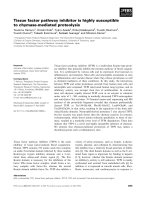
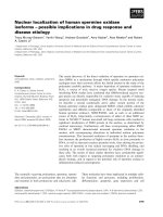
However, in this study we detected Rnf33 mRNA in
the testis and in two mouse testicular cell lines: TM3
and TM4 (Fig. 2). TM3 and TM4 are nontumorigenic
epithelial cell lines derived from mouse testicular
Leydig and Sertoli cells, respectively [8]. Leydig cells
are interstitial cells located adjacent to the seminifer-
ous tubules in the testicle; the cells synthesize and
secrete androgens in response to stimulation with the
pituitary luteinizing hormone. Sertoli cells form part
of the seminiferous tubule, and the main function of
the cells is to nurture the developing sperm cells
through spermatogenesis. The expression of Rnf33
mRNA in TM3 and TM4 cells suggests that Rnf33
transcription may occur in both the Leydig and Ser-
toli cells of the testis.
Rnf35 Rnf33
P1
CDP
NF-Y
Rnf35
Rnf33 TSS’s
Rnf33
Inr
Rnf35
promoter
1 kb
Fig. 1. Map of the Rnf35 and Rnf33 genes. The relative map positions of the intronless genes (boxes) are as established previously [1,2].
Thick horizontal bars denote untranslated regions; filled arrowheads with solid lines indicate major RNA start sites; arrowheads with dashed
lines denote minor Rnf33 RNA start sites used in pre-implantation embryos; and slanting dashed lines represent splicing events. P1 denotes
the major Rnf33 RNA start sites used in both the pre-implantation embryo and in the testis, as reported in this work. In the Rnf35 promoter,
an Inr element, two binding sites for nuclear factor Y (NF-Y) and one binding site for the CCAAT-displacement protein (CDP) are shown, as
reported previously [3,4]. TSS’s, transcriptional start sites.
NF-jB modulates testis-specific Rnf33 expression K B. Choo et al.
838 FEBS Journal 278 (2011) 837–850 ª 2011 Chinese Culture University. Journal compilation ª 2011 FEBS
Identification of the Rnf33 core promoter
elements and a cis-acting transcriptional
sequence in the intron
To elucidate cis-acting transcriptional elements of
Rnf33, a 2560-bp DNA sequence, designated as F1,
which included 1 kb of upstream regulatory sequence,
the 245-bp noncoding exon 1 and 1 kb of the flanking
intronic sequence (sequence )1287 to +1271, Fig. 3A),
was cloned into the promoter-less luciferase vector,
pGL3-Basic, for use in transient transfection and lucif-
erase assays in the permissive CHO-K1 cell line, as
previously described for the Rnf35 gene [3,4]. In the
F1 sequence, a putative atypical TATA-box (aTATA)
and a putative Inr element could be discerned
(Fig. 3A; sequence on top). For analysis of their roles
in transcription, aTATA was deleted in construct
F1DaT and Inr was mutated in construct F1mutI; both
the aTATA deletion and the Inr mutation were
included in the double mutant F1DaT ⁄ mutI (Fig. 3A,
left-hand panel). In transfection and luciferase assays,
a reduction of 35% or 25% in luciferase activity
was observed in cells transfected with F1DaT or
F1mutI, respectively, relative to the wild-type F1 luci-
ferase activity (Fig. 3A, right-hand panel). In cells
transfected with the double mutant, the luciferase
activity was further decreased to 50%, suggesting a
combined contribution of aTATA and Inr as the
Rnf33 core promoter elements. Interestingly, mutating
both aTATA and Inr failed to completely ablate tran-
scriptional activities, indicating the presence of other
important transcriptional cis sequences in the neigh-
borhood. This was supported by the detection of only
10% transcriptional suppression following transfec-
tion with the construct R1, in which the upstream
sequence is deleted but the two core promoter elements
and downstream sequences are retained. When both
aTATA and Inr were mutated in R1 in the R1DaT ⁄
mutI construct, a residual luciferase activity of 35%
was detected, indicating the presence of key transcrip-
tional regulatory elements in the retained exon 1 and
possibly also in the intron sequence. This hypothesis
gained support when progressive deletions of the in-
tronic sequence of F1 from the 3¢ end in constructs F2
and F3 led to a progressive reduction in transcriptional
activities. The first 3¢ segment deleted in the intron
sequence (designated R4) in the construct F2 resulted
in the abolishment of 80% of the relative luciferase
activity, despite the presence of exon 1 and the
upstream sequence (Fig. 3A). A further deletion in
construct F3 confirmed the importance of R4, albeit
with possible further contribution outside the R4
sequence. When one to three copies of the 305-bp R4
sequence were cloned in either the forward or reverse
orientations in front of the constitutive simian virus 40
(SV40) promoter, or 3¢ to the luciferase gene, in the
pGL3-promoter reporter plasmid, luciferase assays
showed that R4, in a single copy in either orientation,
up-regulated SV40 promoter activities when placed
upstream or downstream of the luciferase gene, indi-
cating enhancer-like functions (Fig. 3B, US-R4F and
DS-R4F). Furthermore, the enhanced transcriptional
activities were additive: up to three- or fivefold up-reg-
ulation in the SV40 promoter activities was achieved
when three copies of R4 were placed downstream of
the luciferase gene and in the forward or reverse orien-
tation, respectively (Fig. 3B, DS-3R4F and DS-3R4R).
Taken together, our data indicate that an aTATA and
an initiator act as the core promoter elements in Rnf33
transcription in the presence of crucial, positive cis-act-
ing transcriptional element(s) in the R4 sequence resid-
ing in the only intron of Rnf33.
Identification of a jB element as the crucial cis
regulatory sequence
To further dissect the transcriptional contribution, the
305-bp R4 was arbitrarily divided into three regions,
approximately equal in size, for luciferase assays. Puta-
tive transcription factor-binding sites (TFBSs) were
also identified by bioinformatics analysis (Fig. 4A).
The R4-1 section was found to contain a jB element
in the sequence 5¢-GGGAATTCCC-3¢, which is the
binding site for nuclear factor-j
B (NF-jB), a putative
hypoxia-response element (HRE) in the sequence
5¢-ACGTG-3¢ that is targeted by hypoxia-induced fac-
tor 1a (HIF-1 a) and a putative binding site for the
GATA transcription factor in the reverse orientation
[9,10]. No putative TFBSs were discernible in R4-2. In
R4-3, an N-box and two E-box motifs were predicted.
To delineate the possible contribution of these pre-
dicted TFBSs, the three R4 subsections were either
retained or deleted individually or in different combi-
nations from the F1 construct for luciferase assays
Te T3M TM4 Li
Rnf33
β
β
-actin
Fig. 2. Testicular expression of Rnf33. RNA samples were pre-
pared from the mouse testis (Te) and from the testicular cell lines
TM3 and TM4 for use in RT-PCR analysis with Rnf33-specific prim-
ers. Liver (Li) was included as a negative control, and b-actin was
used as a PCR control.
K B. Choo et al. NF-jB modulates testis-specific Rnf33 expression
FEBS Journal 278 (2011) 837–850 ª 2011 Chinese Culture University. Journal compilation ª 2011 FEBS 839
(Fig. 4A, left-hand panel). Deletion of R4-2 or R4-3
alone (constructs F1R4-1 ⁄ 3 and F1R4-1 ⁄ 2) did not
appreciably affect luciferase activities relative to that
of the parental F1 construct (Fig. 4A). However,
simultaneous deletion of both R4-2 and R4-3 (con-
struct F1R4-1) resulted in an increase, of 50%, in
luciferase activity, suggesting the possible presence of
negative regulator(s) in the deleted sequences. On the
other hand, when R4-1 alone was deleted in construct
F1R4-2 ⁄ 3, the relative luciferase activities were
almost abrogated. Furthermore, deletion of R4-1, in
combination with R4-2 or R4-3 deletion in constructs
A
B
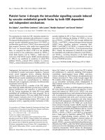
Fig. 3. Identification of the core promoter elements and an intronic cis-acting transcriptional regulatory sequence of Rnf33. (A) Identification
of the core promoter elements. In the experiments, mutation and deletion luciferase constructs were derived from the F1 fragment that con-
tained the upstream regulation region, exon 1 and 1 kb of the Rnf33 intron; the sequences were cloned in front of the promoter-less lucifer-
ase gene of pGL-Basic. In the sequence display at the top, the putative atypical TATA-box (denoted as aT) and the initiator (Inr) are boxed. In
the aT deletion mutant (F1DaT) constructs, six nucleotides (doubly underscored), including aT, were deleted (deletion denoted by D); in the
Inr mutant (F1mutI) constructs, a four-nucleotide mutation (indicated by downward-pointing arrows and the substituted nucleotides) was
introduced (the mutated sites are shown by crosses). In the R1 construct, the upstream regulatory sequence was deleted but aT and Inr
were preserved; and in R1DaT ⁄ mutI, the dT was deleted and Inr was mutated. F2 and F3 constructs carried three terminal serial deletions
of the intronic sequence of F1, as indicated. Transfection and luciferase assays were performed in CHO-K1 cells. The data shown are from
three independent experiments. Relative luciferase activities (RLU) were calculated by arbitrarily setting the luciferase activity of the wild-
type F1 construct as 10. The R4 sequence, identified as harboring cis-acting activities (see the text), is shown. (B) Confirmation of positive
cis transcriptional activities of R4. One or more copies of R4 (thick open arrows) were inserted upstream or downstream of the SV40
promoter (SV Pr, hatched boxes) of the pGL2-promoter vector in either the same (rightward-pointing) or reverse (leftward-pointing) orienta-
tion as the luciferase (Luc) gene, as displayed. The constructs were individually transfected into CHO-K1 cells for luciferase assays. The lucif-
erase activity of the parental pGL3 promoter was arbitrarily set as 1 for computation of the RLUs of other constructs. In both (A) and (B),
data were subjected to the Student’s t-test; *P < 0.05; **P < 0.01 relative to the controls.
NF-jB modulates testis-specific Rnf33 expression K B. Choo et al.
840 FEBS Journal 278 (2011) 837–850 ª 2011 Chinese Culture University. Journal compilation ª 2011 FEBS
F1R4-3 or F1R4-2, partially restored transcriptional
activity, consistent with the supposition of the presence
of negative cis regulator element(s) in R4-2 or R4-3, as
described above. Hence, deletion analysis further
mapped the presence of positive cis-acting transcrip-
tional element(s) to the 93-bp R4-1 sequence.
To investigate the contribution of the three dis-
cerned putative TFBSs in R4-1 to Rnf33 transcrip-
tional modulation, these sites were mutated
individually or in combination with one another, and
transfection and luciferase assays were carried out
(Fig. 4B). When the HRE was mutated in construct
F1MutH, a reduction in luciferase activity of 30%
was observed. Moreover, mutation of the jB element
in construct F1MutjB led to a reduction of 70% in
luciferase activity. When the double mutant
F1MutjB ⁄ H was similarly assayed, the reduction in
luciferase activity was not additive but remained at
70%, reflecting the dominant role of jB. However,
cross-talk between NF-jB and HIF-1 in Rnf33 tran-
scription cannot be ruled out because HIF-1a is also a
target gene of NF-jB [11,12]. On the other hand,
A
B
C
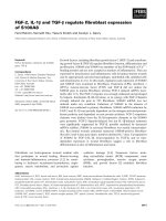
Fig. 4. Association of R4 transcriptional
activity with a jB element. (A) Further map-
ping of transcriptional activities to a subsec-
tion (R4-1) of R4. The R4 sequence was
arbitrarily divided into three sections – R4-1
to R4-3 – of approximately equal length. In
R4-1, the discerned putative TFBSs are jB,
HRE and GATA (denoted by vertical bars); in
R4-2, no TFBSs were identified; in R4-3, an
N-box (N) and two E-boxes (E) were
detected. The R4-1 to R4-3 subsections
were retained or deleted individually, or in
different combinations, from the parental F1
sequence (see Fig. 3A). In the panel of con-
structs displayed on the left, the R4 seg-
ment in F1 is magnified for clarity by
omitting the sequence between exon 1 and
R4 (denoted by the slanting double-break
symbols in the construct displays). (B)
Identification of jB as the major cis tran-
scriptional element in R4-1. The R4-1
sequence is shown at the top; the predicted
TFBSs are boxed; and mutations (denoted
by crosses) that were introduced into the
luciferase constructs are indicated by down-
ward-pointing arrows. (C) Confirmation of
positive transcriptional activity of the jB
element. In R4-1(jB)4, four jB copies were
cloned upstream of the SV40 promoter (SV
Pr, hatched boxes) of the pGL3 promoter.
Construct US-R4F that carried full-length R4
(see Fig. 3B) was included for comparison.
RLU, relative luciferase activities.
K B. Choo et al. NF-jB modulates testis-specific Rnf33 expression
FEBS Journal 278 (2011) 837–850 ª 2011 Chinese Culture University. Journal compilation ª 2011 FEBS 841
mutating the putative GATA-binding site (construct
F1MutG) had no discernible effects on transcriptional
activity, ruling out a role of the putative GATA-bind-
ing site in transcription. This conclusion is further
supported by the assay of the triple mutant
F1MutjB ⁄ H ⁄ G that yielded luciferase activities similar
to that obtained with the jB-only mutant construct.
To confirm the contribution of jB to transcription,
four copies of a 12-mer T
GGGAATTCCCC sequence,
which included the jB sequence (underlined), were
placed at the 5¢ end of the SV40 promoter of the
pGL3-promoter vector to generate construct R4-1(jB)4
for luciferase assays (Fig. 4C). While insertion of the
single-copy jB-containing R4 sequence resulted in a
1.5-fold increase in the SV40 promoter activity, the
presence of four copies of the 12-mer jB sequence
resulted in a significant eightfold increase in promoter
activity relative to the parental plasmid (Fig. 4C). Pro-
moter-activity analysis in luciferase assays firmly estab-
lished that the discerned jB element in R4-1 is the
primary cis-acting positive regulatory element, while
the putative HRE sequence may play a secondary role
in Rnf33 transcriptional regulation.
The Rnf33 jB element is targeted by the NF-jB
proteins p50 and p65
It has been well established that jB sequences are tar-
geted by the abundantly expressed NF-jB [13–15].
Involvement of the NF- jB in gene regulation in the
testis has also been described [16–20]. Among the five
NF-jB proteins, p50 ⁄ NFjB1 and p65 ⁄ RELA have
clearly been shown to be the major NF-jB proteins
expressed specifically in the testis [21–23]. Expression
of p50 and p65 was confirmed in the testis and estab-
lished in the TM3 and TM4 testicular cell lines by
RT-PCR and western blot analyses (Fig. 5). It is noted
in the western blots that while the p50 and p65 levels
were relatively constant in the testis, in the two testicu-
lar cell lines and in the control liver tissue, p65 levels
were found to be more than threefold higher in TM3
and TM4 cells than in the testis and liver tissues
(Fig. 5B).
To determine p50 and p65 targeting of the R4-1 jB
site, an electrophoretic mobility shift assay (EMSA)
was performed. In the presence of nuclear extracts pre-
pared from the TM3 and TM4 cells, a protein-induced
band shift was observed (Fig. 6A, lanes 2 and 7,
arrowhead). In the presence of increasing amounts of
the unlabeled wild-type probe sequence, the shifted
band was effectively competed out (Fig. 6A, lanes 3,
4, 8 and 9). A jB mutant oligonucleotide, however,
had little effect on the observed band shift (Fig. 6A,
lanes 5, 6, 10 and 11). The identity of the jB-bound
protein was further established in supershift assays
(Fig. 6B). On addition of an anti-p65 serum, the pro-
tein-induced bands in both TM3 and TM4 cells were
obliterated, indicating specific p65 targeting (Fig. 6B,
lanes 3 and 6); in the experiments, the supershifted
bands were not apparent, as previously reported in
similar assays in testicular cells [20]. However, addition
of an anti-p50 serum did not seem to appreciably
affect the protein-shifted band in both TM3 and TM4
cells (Fig. 6B, lanes 2 and 5). In in vivo p65-binding
assays carried out by chromatin immunoprecipitation
(ChIP), the anti-p65 and -p50 sera both yielded vari-
ous intensities of Rnf33-specific PCR bands from TM3
and TM4 cells and the testis, but not from the liver
(which does not express Rnf33) (Fig. 6C). In the mock
experiment in which the antibody treatment was omit-
ted, or in the case in which a pre-immune antiserum
was used, no specific PCR products were detected.
Taken together, EMSA and ChIP assays indicate that
the NF-jB subunit proteins p65, and possibly p50,
target the intronic jB site of Rnf33, resulting in tran-
scriptional activation of Rnf33 in the testis. The p65
protein seems to be preferred over p50 in targeting the
Rnf33 jB site, and the protein–target site interactions
also appear to be weak.
To further verify the specificity of NF-j
B transcrip-
tional modulation of Rnf33 expression, the expression
of p65 or p50 was knocked down by double-stranded
siRNA in TM4 cells (Fig. 7). When TM4 cells were
Te TM3 TM4 Li
p50
p65
p50
p65
β
-actin
Te TM3 TM4 Li
RL:
0.741 3.16 3.05
RL:
0.831 0.78 0.76
A
B
Fig. 5. Expression of the p65 and p50 NF-jB subunit proteins in
testicular cells. (A) RT-PCR detection of p50 and p65 transcripts in
the testis (Te) and in TM3 and TM4 cells. (B) Western blot analysis
of the p50 and p65 proteins. The relative protein levels (RL) were
computed by normalizing with the b-actin level and were calculated
relative to the level of the testis set as 1. Liver (Li) was included as
a control in the analyses.
NF-jB modulates testis-specific Rnf33 expression K B. Choo et al.
842 FEBS Journal 278 (2011) 837–850 ª 2011 Chinese Culture University. Journal compilation ª 2011 FEBS
transfected with the p65 siRNA, the relative mRNA
level of p65, as quantified by real-time RT-PCR, was
significantly reduced to 48% of that of nonspecific siR-
NA-transfected cells but the relative p50 mRNA level
was unaffected (Fig. 7A). Likewise, transfection with
p50 siRNA resulted in a significant reduction (by
50%) of the p50 transcript levels but not of the p65
transcript levels. Effective knockdown of p65 or p50
by the respective siRNA was supported by western
blot analysis, showing a reduction in the p65 and p50
protein levels of approximately 50% and 33%, respec-
tively, in the transfected cells (Fig. 7B). When p65 was
knocked down by siRNA, the mRNA level of Rnf33
was significantly reduced to 47.5% of that of the non-
specific control (Fig. 7A, Rnf33 panel). However, p50
knockdown did not have any significant effect on the
Rnf33 mRNA level. Likewise, when transfected with
the p65 siRNA, the RNF33 protein level was reduced
by approximately 33% relative to the cells transfected
with nonspecific siRNA, but transfection with p50 siR-
NA did not result in appreciable reduction of RNF33
protein (Fig. 7B, RNF33 panel). The results of the
siRNA experiments clearly support p65-modulated
Rnf33 expression and indicate that p50 seems to play a
lesser role than p65 in Rnf33 expression. Taken
together, our data demonstrate that Rnf33 transcrip-
tion is modulated by the NF-jB p65 protein, probably
in the form of the more ubiquitous p65–p50 hetero-
dimer, and possibly also in the p65–p65 homodimeric
form.
Rnf33 expression is up-regulated by TNF-a or
p50
⁄
p65 overexpression via the jB element
To further determine if the observed jB modulation of
Rnf33 transcription at the R4-1 jB site was TNF-a
dependent, the testicular TM3 and TM4 cells were
transfected with the jB-containing luciferase construct
(F1), with construct F1R4-2 ⁄ 3 (from which the jB-
containing R4-1 segment had been deleted) or with the
jB mutant construct F1MutkB (see Fig. 4 for con-
structs), and the transfected cells were treated with
TNF-a before luciferase assays were performed. The
results showed a 50%, significant, increase in luciferase
activity in the presence of TNF-a in F1-transfected
TM3 cells, and a fourfold, significantly higher lucifer-
ase activity in F1-tranfected TM4 cells relative to the
untreated cells (Fig. 8A). Consistent with previous
assays in CHO-K1 cells, luciferase activities were negli-
gible or were significantly lower when j B site deletion
or mutated constructs were assayed in both TM3 and
TM4 cells, and TNF-a did not elicit discernible effects
on the luciferase activity (Fig. 8A). Hence, Rnf33 pro-
moter activation in testicular cells is modulated by
TNF-a and the modulation is dependent on the pres-
ence of the jB site.
To test if the jB site is targeted by homodimeric or
heterodimeric p50 and p65 proteins, p50 and ⁄ or p65
overexpression plasmids were transiently co-transfected
with the jB-containing F1R4-1 construct or with
the jB mutant construct F1MutkB (see Fig. 4 for
Competitor:
+
+++ +–
– –
WT Mut
–
WT Mut
NE (g):
+
++ + +
TM4 TM3
123456 7891011
ns
ns
ns
p50:
p65:
TM4
TM3
123456
–+ – +––
––+–+–
Testis
TM4
TM3
Liver
Input
Mock
a
-p65
a
-p50
Pre
ABC
Fig. 6. Targeting of the R4-1 jB element by p65 and p50. (A) Electrophoretic mobility shift assay (EMSA) using a jB probe and nuclear
extracts (NE) prepared from the mouse testicular cell lines, TM3 and TM4. The open arrowhead indicates the position of the jB probe-
induced shifted band; other nonspecific (ns) bands are indicated by arrows. In the competition experiments (lanes 3–6 and 8–11), a 25- or a
250-fold molar excess of unlabeled wild-type (WT) or mutant (Mut) probe was used. (B) Supershift assays in TM3 and TM4 cells using an
anti-p50 (ap50) serum or an anti-p65 (ap65) serum. The arrowhead and arrows are as in (A) above. (C) ChIP assays of in vivo p65 and p50
binding to the jB site in nuclear extracts of testicular cells using antibodies against p65 (ap65) and p50 (ap50). A rabbit pre-immune serum
(Pre) was included as a control.
K B. Choo et al. NF-jB modulates testis-specific Rnf33 expression
FEBS Journal 278 (2011) 837–850 ª 2011 Chinese Culture University. Journal compilation ª 2011 FEBS 843
construct), and TM4 cells were used in the co-transfec-
tion experiments because TM4 cells had been shown
previously (Fig. 8A) to be more responsive to TNF-a
induction. No apparent effects of p50 overexpression
on luciferase activities were observed in the F1R4-1-
transfected cells (Fig. 8B). The luciferase activity was
twofold higher than that of untransfected cells when
p65 was overexpressed; more significantly, the lucifer-
ase activities were further increased to threefold those
of untransfected cells when p50 and p65 were co-over-
expressed (Fig. 8B). Consistent with the TNF-a
modulation demonstrated above (Fig. 8A), TNF-a
treatment elevated the luciferase activities in F1R4-1-
transfected cells to a level comparable to that when
p65 was overexpressed, but lower than that when p50
and p65 were co-overexpressed (Fig. 8B). On the other
hand, the luciferase activities were negligible and the
responsiveness to p50 ⁄ p65 NF-jB proteins and TNF-a
modulation was abolished when the F1MutkB jB
mutant was similarly assayed (Fig. 8B), unequivocally
demonstrating positive modulation by NF-jB and
TNF-a acting on the Rnf33 jB site.
As Rnf33 is expressed in testicular cells, the effects
of TNF-a and p50 ⁄ p65 overexpression on Rnf33 tran-
scription were directly assayed in these cells by real-
time quantitative RT-PCR in TM4 cells. Echoing the
luciferase assay data above (Fig. 8B), overexpression
of p50 resulted in an increase of only 40% in the
Rnf33 transcript level, but p65, p50 ⁄ p65 co-expression
or TNF-a significantly upregulated Rnf33 transcription
by 2.4- to 2.6-fold in TM4 cells (Fig. 8C), echoing the
findings in luciferase assays. However, the Rnf33
expression level did not change appreciably in the pres-
ence of TNF-a in TM3 cells (Fig. 8C), in agreement
with the luciferase assay data in Fig. 8A. Taken
together, data from luciferase assays and direct mea-
surements of Rnf33 mRNA levels in TM3 and TM4
cells clearly demonstrate that TNF-a, p65 (probably in
a homodimeric form) or the p50–p65 NF-jB hetero-
meric complex all serve to up-regulate Rnf33 expres-
sion in testicular cells via the intronic jB motif located
immediately downstream of exon 1 of Rnf33.
Possible NF-jB regulated expression of Rnf33 in
the pre-implantation embryo
In this study, Rnf33 transcriptional modulation was
investigated in the testis and in two testicular cell lines.
The question remains whether NF-jB is also involved
in Rnf33 transcription in the oocyte and in the pre-
implantation embryo where Rnf33 is expressed, as in
the testis. To investigate this possibility, the approxi-
mate temporal expression profiles of the p65 ⁄ Rela and
p50 ⁄ Nf-j
b1 genes were examined based on bioinfor-
matics analysis of the EST database in GenBank
(Table 1). Mouse EST sequences for p65 are found in
the oocyte, pre-implantation embryos and in the testis.
On the other hand, p50 ⁄ Nf- jb1 EST sequences are
found in the oocyte and in the testis but not in any of
the pre-implantation embryos. If NF-jB is experimen-
tally shown in subsequent studies to be involved in
transcriptional modulation of Rnf33 in oocyte and in
early development as in the testis, it is likely that
only the p65–p65 homodimer is involved, which is
highly consistent with data presented in this work in
the testis.
Discussion
In a previous work [1,2], we have shown that in the
fertilized egg and the zygote, Rnf33 transcription
recruits three minor promoters (one of which is located
upstream of the Rnf35 gene) and a major promoter,
0
2
4
6
8
10
12
14
16
RNF33
β
-actin
p65
p105/p50
siRNA:
NS p50p65
p65 p50 Rnf33
Relative mRNA level
**
**
**
NS
p65
p50
SiRNA
1.00 0.52 0.85
1.00 0.98 0.68
1.00 0.71
0.98
A
B
Fig. 7. Confirmation of NF-jB modulation of Rnf33 expression by
siRNA knockdown of p65 and p50. (A) p65 and p50 knockdown
and Rnf33 transcriptional down-regulation. TM4 cells were individu-
ally transfected with a nonspecific (NS), p65 or p50 siRNA for 48 h
before real-time RT-PCR quantification of the relative mRNA levels;
the mRNA levels of the NS-treated TM4 cells were arbitrarily set
as 10. Data presented are from three independent experiments;
**P < 0.01. (B) RNF33 protein reduction on p65 knockdown. siRNA
transfection was as described in (A). Representative western blots
of three independent experiments are shown. The precursor p105
protein was used to represent p50 levels in the western blot. Dis-
played below the blots are the computed relative levels of the
respective protein after normalization with the level of b-actin.
NF-jB modulates testis-specific Rnf33 expression K B. Choo et al.
844 FEBS Journal 278 (2011) 837–850 ª 2011 Chinese Culture University. Journal compilation ª 2011 FEBS
designated P1 in Fig. 1, which is dissected in this work
in the testis. At the four- and eight-cell embryonic
stages, multiple promoter usage is resolved into the use
of only the major promoter, and this is followed by
complete Rnf33 gene silencing at the blastocyst stage
and the remaining phases of embryonic development
[2]. Rnf33 is, however, reactivated specifically in the
testis in adult mice, as shown in this work. We have
further shown that Inr sequences act as the core
promoter element for both the Rnf33 and Rnf35 genes.
As Inr overlaps with the 5¢ end of exon 1, our studies
further attribute a critical role for noncoding untrans-
lated 5¢ exons and the acquired associated introns in
activating expression of intronless protein-encoding
genes, as for retrogenes [24–26]. Interestingly, mutating
both the Inr element and the aTATA of Rnf33 led to
the abolishment of only about 50% of promoter activi-
ties in luciferase assays, strongly suggesting that the
structure of the Rnf33 basal promoter is more complex
than the discerned Inr and aTATA. A 2-kb sequence
that encompasses the upstream regulatory region, exon
1 and the solo intron of both Rnf35 and Rnf33 is
found to be free from CpG islands (data not shown).
The combined characteristics of the core promoters
of Rnf35 and Rnf33 are highly consistent with the
general features of tightly regulated tissue-specific and
Table 1. Approximate expression profiles of p65 and p105 ⁄ p50 in the mouse pre-implantation embryos and the testis based on EST analy-
sis. Data shown are in transcripts per million.
Gene UniGene no. Oocyte Zygote Cleavage Morula Blastocyst Testis
p65 Mm.249966 51 140 36 0 57 42
p105 ⁄ p50 Mm.256765 51 0 0 0 0 25
012345
p50
p65
TNF-α
*
**
–––
+ – –
–+ –
+ + –
––+
RLU
F1R4-1
F1R4-1MutB
TM4
**
**
**
0 0.5 1 1.5 2
F1
F1R4-2/3
F1MutκB
+ TNF-α
– TNF-α
*
RLU
012345
01234
Relative Rnf33 mRNA level
p50
p65
TNF-α
–––
+ – –
–+ –
+ + –
––+
––+
TM4TM3
TM4
TM3
**
**
**
RLU
A
BC
Fig. 8. jB-dependent TNF-a and p50 ⁄ p65 modulation of Rnf33 promoter activity. (A) TNF-a up-regulation of Rnf33 promoter activity. TM3
and TM4 cells were transfected with the jB-containing construct F1, with construct F1R4-2 ⁄ 3 from which jB had been deleted (see Fig. 4A)
or with the jB mutant construct F1Mut j B (see Fig. 4B). TNF-a was added 24 h after transfection and the cells were cultured for a further
24-h period before being harvested for luciferase assays. Open and hatched bars represent relative luciferase activities (RLU) in the absence
or presence of TNF-a, respectively. (B) Transcriptional up-regulation by p50 and p65 overexpression. TM4 cells were transfected with either
construct F1R4-1 (see Fig. 4A) or with the jB mutant construct F1R4-1MutjB, or were co-transfected with either the p50 or p65 overex-
pression plasmid. TNF-a was also included in the assay for comparison. The cells were harvested for luciferase assays 48 h after transfec-
tion or 24 h after treatment with TNF-a. Open and gray bars represent luciferase activities of F1R4-1 and F1R4-1MutjB, respectively.
(C) TNF-a and p50 ⁄ p65 up-regulate Rnf33 transcription in testicular cells. TM4 cells were transfected with p50 and ⁄ or p65 overexpression
plasmids for 48 h before RNA was extracted and real-time quantitative RT-PCR assays for relative Rnf33 mRNA levels were carried out. For
analysis of the effect of TNF-a, both TM3 and TM4 cells were treated with TNF-a for 24 h before RNA was prepared and quantitative RT-
PCR assays were carried out. The Rnf33 mRNA level for the untreated TM4 cells was arbitrarily set as 1. Data presented are from three
independent experiments and were analyzed using the Student’s t-test;*P < 0.05; **P < 0.01.
K B. Choo et al. NF-jB modulates testis-specific Rnf33 expression
FEBS Journal 278 (2011) 837–850 ª 2011 Chinese Culture University. Journal compilation ª 2011 FEBS 845
temporal-specific promoters proposed based on gen-
ome-wide computation of the architecture of mamma-
lian promoters [27,28].
In this study, a jB element located in the only
intron of the Rnf33 gene was shown to be critical for
Rnf33 transcription; our data showed that the jB ele-
ment was targeted by the p65–p50 heterodimer and
possibly by the p65–p65 homodimeric complex, but
not by p50 alone, in the Sertoli cell-derived TM4 cells
(Fig. 7B,C). NF-jB is a transcription factor inducible
by multiple stimuli to regulate a wide range of genes.
Involvement of the NF-jB signaling pathway in the
regulation of genes involved in spermatogenesis and
other testicular functions in both Sertoli and Leydig
cells has been abundantly reported [16–20]. Among the
five known NF-jB proteins, p50 and p65 are the major
NF-jB proteins expressed in the testis [21–23]. NF-jB-
regulated expression of the testis-specific Rnf33 gene
echoes previous reports that expression of the cAMP-
response element-binding protein (CREB) and andro-
gen receptor (AR) genes in Sertoli cells is regulated by
the NF-jB p65–p50 heterodimer or by p65 alone, but
not by p50 alone [16,17,29]. Signaling pathways that
activate NF-jB have been well documented [13–15]. In
the canonical NF-jB activation pathway, degradation
of IjBa through phosphorylation by the activated IjB
kinase (IKK) complex leads to the release of cytoplas-
mic NF-jB and nuclear relocation of NF-jB. In an
IKK-independent pathway, external stimuli, including
hypoxia and genotoxic stresses, lead to NF-jB nuclear
localization. Involvement of NF-jBinRnf33 transcrip-
tional modulation is consistent with the fact that testis
is a highly dynamic site of active and continuous sper-
matogenesis and is therefore under constant molecular
and evolutionary stresses. Likewise, pre-implantation
development is also highly stressful. However, the
NF-jB signaling stimuli and potential co-activator(s)
involved in the demonstrated NF-jB modulation of
Rnf33 promoter activity in the testis and in early devel-
opment will need to be further identified.
The consensus jBsequenceis5¢-GGGRHTYYCC-3¢
(in which R is purine, Y is pyrimidine and H is A, C
or T). A ‘phosphorylation code’ has been proposed for
p65 that targets NF-jB activity to specific subsets of
genes via the recognition of distinct groups of the con-
sensus jB site [30]. In this code, the palindromic 5¢-
GGGAATTCCC-3¢ jB sequence of Rnf33 was shown
to tolerate a wider range of differential phosphoryla-
tion of the amino-terminal Rel homology domain in
p65, hence providing the jB palindrome with a wider
choice of utilization of differentially phosphorylated
versions of p65. We also showed, in the luciferase
assay, that in TM4 cells there was a basal level of
promoter activity, and TM4 cells treated with TNF-a
boasted the promoter activity (Fig. 7C) as a result of
NF-jB nuclear relocalization. On the other hand,
luciferase assays in both CHO-K1 and TM4 cells
showed that despite the mutation in the jB site, 30%
of the promoter activity remained (Figs 4B and 7B),
indicating participation of other cis-acting element(s)
and transcription factors in Rnf33 expression. One can-
didate cis element would be the adjacent HRE site tar-
geted by HIF-1a. Indeed, HIF-1a is transcriptionally
regulated by NF-jB, thus establishing cross-talk
between these two important transcription factors in
the testis [12].
Studies have established that TNF-a is a major cyto-
kine produced and released by germ cells and that
TNF-a receptors are found on Sertoli and Leydig cells
of the testis [31]. In the testis, TNF-a regulates sper-
matogenesis [32], modulates Leydig cell steroidogenesis
[33,34] and influences the expression of cell–cell adhe-
sion molecules in Sertoli cells [35,36]. There are abun-
dant examples of involvement of the TNF-a ⁄ NF-jB
network in transcriptional modulation. TNF-a induces
NF-jB binding to the promoter of the AR gene and
elevates AR promoter activities in Sertoli cells [17,29].
In Leydig cells, TNF-a-induced p50 and p65 specifi-
cally interact with the CCAAT ⁄ enhancer binding
protein beta (C ⁄ EBPb) to regulate the expression of
Nur77, a regulator of steroidogenic-enzyme genes
[18,20]. In investigating activation of the lipocalin-
2 gene, which is abundantly expressed in spermatogo-
nial cells but expressed at only very low levels in
Sertoli cells, Fujino et al. [19] demonstrated regulation
of Sertoli cells by spermatogonial cell-mediated lipoca-
lin-2 gene activation via an IKK-independent NF-jB
pathway. Expression of the Mullerian inhibiting sub-
stance (MIS), a key molecule in sex differentiation and
reproduction, is regulated by steroidogenic factor 1
(SF-1) also via the TNF-a ⁄ NF-jB pathway [37]. Most
importantly, NF-jB up-regulates Fas expression in
Sertoli cells, leading to apoptosis, a key event in the
delicate balance of pro-apoptotic and anti-apoptotic
signaling, to ensure optimal spermatogenesis [23,38].
The finding that Rnf33 is also under NF-jB regula-
tion in the testis is not surprising but functionally
rational. Spermatogenesis is tightly regulated by a
complex network of signals and stimuli and, as dis-
cussed above, one of the important identified stimuli is
NF-jB which, in turn, is also highly responsive to a
wide range of external signals. Furthermore, we have
shown that the putative RNF33 protein interacts
with the kinesin motor proteins KIF3A and KIF3B,
possibly contributing to cargo mobilization along
the microtubule [Huang, Huang, Chang, Hsu, Lin &
NF-jB modulates testis-specific Rnf33 expression K B. Choo et al.
846 FEBS Journal 278 (2011) 837–850 ª 2011 Chinese Culture University. Journal compilation ª 2011 FEBS
Choo, submitted]. The microtubule transportation sys-
tem, coupled with intercellular junctions, is essential in
the translocation and positioning of spermatids in the
spermatogenesis process [39,40]. Kinesin proteins have
indeed been shown to be present at junctions along
adjacent microtubules of spermatids and Sertoli cells,
thus contributing to spermatid translocation [41–43].
Hence, demonstration of Rnf33 expression in Sertoli
cells under the regulation of NF-jB may imply func-
tional involvement of the RNF33 protein in spermato-
genesis.
In summary, the present study demonstrates that
Rnf33 expression in the testis is regulated by an intron-
ic jB sequence modulated by the NF-jB subunits p65
and p50, and also by TNF-a. Although p65 is proba-
bly expressed in the oocyte and the pre-implantation
embryo, it remains to be shown if the TNF-a ⁄ NF-jB
signaling pathway also contributes to transcriptional
regulation of Rnf33 in pre-implantation development.
Experimental procedures
Cell lines and mice
The testicular TM3 and TM4 cell lines were obtained from
the Bioresource Collection and Research Center (BCRC),
Hsinchu, Taiwan; the cell lines were originally obtained by
BCRC from the American Type Culture Collection (ATCC,
Manassas, VA, USA) for maintenance and distribution in
Taiwan. The cells were cultured in a 1 : 1 mixture of Ham’s
F12 medium and Dulbecco’s modified Eagle’s medium con-
taining 1.2 gÆL
)1
of sodium bicarbonate, 4.5 gÆL
)1
of glu-
cose (TM3 only) and 15 mm Hepes, and 5% (v ⁄ v) horse
serum and 2.5% fetal bovine serum. C3H mice were used
in this work and were maintained at the Laboratory Ani-
mal Centre of the National Yang Ming University, Taipei.
This study was approved by the Institutional Animal Care
and Use Committee (IACUC) of the Taipei Veterans Gen-
eral Hospital, and the mice were killed according to
IACUC guidelines.
RT-PCR expression profiling and real-time
quantitative RT-PCR
To determine Rnf33 expression, RT-PCR was applied as pre-
viously described [1]. After the oligo(dT)-primed reverse
transcription reaction, PCR was carried out using primer
F1466 (5¢-GTGTGTGTCAAGCCCACTTTTCTG-3¢)of
the coding sequence and primer R1863 (5¢-GTGGG
TGGTGGATTTTGTTGTTTG-3¢) of the 3¢-UTR sequence
of Rnf33 to generate a 398-bp PCR product. PCR was per-
formed for 35 cycles using an annealing temperature of 65 °C
and an extension time of 30 s for each cycle. Mouse b-actin
primers were used as a control. Cellular Rnf33 mRNA levels
were quantified by real-time RT-PCR using the DyNAmoÔ
Flash SYBR
Ò
Green qPCR kit (Finnzymes, Espoo, Finland).
RNA samples extracted from treated cells were reverse tran-
scribed as described above. Real-time PCR was performed in
a LightCycler
Ò
480 (Roche, Mannheim, Germany) in 96-well
plates. The reaction mixture was 20 lL of cDNA, 1 · SYBR
Green PCR Master Mix (Finnzymes) and 0.4 lm each of for-
ward and reverse primers. PCR primers for Rnf33 were
F1466 and Rnf33-qPCR-R (5¢-GTTCTTAGAGGTCCA
TAGGTGACA-3¢). For normalization, the mRNA level of
the glyceraldehyde-3-phosphate dehydrogenase (Gapdh) gene
in each RNA preparation was determined using primers
GAPDH-F (5¢-GCCTCCTGCACCACCAACTG-3¢) and
GAPDH-R (5¢-CCAGTAGAGGCAGGGATGATGT-3¢).
The real-time PCR program was: pre-incubation at 50 °C for
2 min; initial denaturation at 95 °C for 7 min; and 45 cycles
at 95 °C for 10 s, 63 °C for 15 s and 72 °C for 30 s. The pro-
gram was terminated by a final extension at 60 °C for 1 min
and cooling at 40 °C for 5 min. The relative Rnf33 mRNA
levels were normalized to the mRNA level of the reference
Gadph gene. The melting curve of the amplification product
was always checked to ensure a single clean peak that repre-
sented good-quality real-time PCR data.
Construction of luciferase reporter plasmids and
site-specific mutagenesis
The 2560-bp F1 genomic fragment was PCR-amplified from
the mouse genomic BAC45 clone [2] using the DyNA-
zymeÔ II Hot Start DNA Polymerase (Finnzymes), and
the PCR product was cloned into the promoter-less pGL3-
Basic luciferase vector at the NheI and XhoI restriction
sites. Short deletions and site-specific mutations were car-
ried out using the commercial PhusionÔ Site-Directed
Mutagenesis kit (Finnzymes), following the manufacturer’s
instructions. The mutations and deletions in the constructs
were confirmed by sequencing.
Transient transfection, TNF-a treatment and
luciferase assays
Transient transfection was performed using the PLUSÔ
Reagent and LipofectamineÔ (Invitrogen, Carlsbad, CA,
USA), as previously described [3,4]. Luciferase assays were
performed 48 h post-transfection using the Dual-Lucifer-
ase
Ò
Reporter 1000 Assay kit (Promega, Madison, WI,
USA) in a 96-well microtiter plate, as instructed by the
manufacturer. The plate was read using a luminometer.
Transfection was typically carried out in duplicate, and for
each transfection sample, the luciferase assay was also car-
ried out in duplicate. Three or more independent experi-
ments of transfection and the associated luciferase assay
were performed for each construct. The p50 and p65
expression plasmids were a gift from Dr Neil Perkins; the
genes were under transcriptional regulation of the long
K B. Choo et al. NF-jB modulates testis-specific Rnf33 expression
FEBS Journal 278 (2011) 837–850 ª 2011 Chinese Culture University. Journal compilation ª 2011 FEBS 847
terminal repeat (LTR) of Rous sarcoma virus (RSV) [44].
In experiments in which TNF-a was used, TNF-a (Sigma-
Aldrich, St Louis, MO, USA) was typically added to the
untransfected cells or to the cells 24 h post-transfection, to
a final concentration of 20 ngÆ mL
)1
, and the cells were fur-
ther incubated for 24 h before being harvested for luciferase
assays or RT-PCR analysis. The Student’s t-test was used
for statistical analysis of the luciferase assay data; values of
P < 0.05 were considered significant.
Preparation of nuclear extracts and total protein
lysates
Nuclear extracts and protein lysates were prepared essen-
tially as previously described [3,4]. TM3 and TM4 cells cul-
tured in 100-mm dishes were harvested and the cell pellets
were washed three times with NaCl ⁄ P
i
before being gently
suspended in 300 lL of cold buffer A (10 mm Hepes, pH
7.9, 10 mm KCl, 1.5 mm MgCl
2
, 0.5 mm dithiothreitol,
0.5 mm phenylmethanesulfonyl fluoride). The cell suspen-
sion was kept on ice for 15 min followed by the addition of
CA630 (Sigma-Aldrich) to a final concentration of 0.5%
and then the cell suspension was briefly vortexed. The sam-
ples were spun down at 10 000 g for 30 s at 4 °C and the
cell pellets were resuspended in 80 lL (cultured cells) or
120 lL (for the testis) of ice-cold buffer C (20 mm Hepes,
pH 7.9, containing 0.42 m NaCl, 1.5 mm MgCl
2
, 0.2 mm
EDTA, 25% glycerol, 0.5 mm dithiothreitol and 0.5 mm
phenylmethanesulfonyl fluoride). The suspension was vigor-
ously rocked at 4 °C for 15 min on a shaking platform
before centrifugation at 10 000 g for 20 min at 4 ° C. Aliqu-
ots of the supernatant obtained were kept at )70 °C until
used. For total protein lysates, mouse testis or cultured
cells were resuspended in 120 or 80 lL, respectively, of cold
buffer C for 15 min on a shaking platform. Total
protein lysates were cleared by centrifugation at 10 000 g
for 20 min at 4 °C, aliquoted and kept at )70 °C until
used.
EMSA and supershift assays
The EMSA probes were prepared by end-labeling single-
stranded oligonucleotides with [
32
P]dATP[cP] using T4
DNA kinase (Promega) followed by annealing the comple-
mentary strands of the oligonucleotides at 20 pmolÆlL
)1
,as
previously described [45]. The binding reaction mixture con-
tained 100 mm Tris, 500 mm KCl, 10 mm dithiothreitol,
2.5% glycerol, 5 mm MgCl
2
,50ngÆlL
)1
of poly(dI Æ dC),
0.05% Nonidet P-40, 40 fmol of
32
P-labeled probe and 5 lg
of nuclear extracts, and the binding reaction was allowed to
proceed at room temperature for 30 min. In competition
assays, a 25- or a 250-fold molar excess of unlabeled dou-
ble-strand oligonucleotide was added to the binding reac-
tion and the reaction was also allowed to proceed at room
temperature for 30 min. Oligonucleotides containing the jB
sequence (underlined) that were used in the competition
experiments were: wild type, 5¢-AGGTCT
GGGAATTCCC
CCCGGA-3¢; and mutant 5¢-AGGTCT
GGGAATagggCCC
GGA-3¢ (mutated nucleotides shown in lowercase letters).
For supershift assays, 2.5 lg of a polyclonal anti-p65 serum
(sc-109x) (Santa Cruz Biotechnology, Santa Cruz, CA,
USA), or an anti-p50 serum (sc-7178x) (Santa Cruz), was
added to the reaction mixture and the reaction was incu-
bated at room temperature for 20 min. In all cases, binding
complexes were displayed on 6% polyacrylamide gels, fol-
lowed by blotting onto a positively charged nylon mem-
brane. Probe signals on the membrane were detected using
a Typhoon 8000 Molecular Dynamics PhosphoImager
(Amersham Pharmacia Biotech, Bucks., UK).
ChIP
ChIP was performed essentially as previously described [3].
After incubation with specific antibodies, the immunocom-
plexes were incubated at 68 °C in a water bath overnight to
reverse the cross-links in the samples, followed by digestion
with 10 lg of each of RNase A and proteinase K at 42 °C
for 1 h. After digestion, DNA samples were purified by
phenol ⁄ chloroform extraction followed by ethanol precipi-
tation. The DNA pellet was dissolved in 10 lLof
Tris–EDTA buffer. Three-microliter aliquots of each DNA
sample were used in PCR analysis in the presence of
40 pmol each of the Rnf33-specific RNF33-ChIP-F (5¢-AG
GGCATAAAGGAGGGCAGGGAAC-3¢) and RNF33-
ChIP-R (5¢-CATCAGCTTCCCTTATGAGAACAG-3¢)
primers in 10-lL PCR reaction volumes. The PCR was per-
formed for 33 cycles at an annealing temperature of
65.6 °C to generate a 300-bp amplification product that
was shown in a 1.5% agarose gel.
Transfection with siRNA
To knock down p50 or p65 expression, p50 (sc-29408),
p65 (sc-29411) or a nonspecific negative-control (sc-37007)
double-stranded RNA (Santa Cruz) was transfected indi-
vidually into 2 · 10
5
TM4 cells in a 3.5-cm petri dish
using Lipofectamine 2000 (Invitrogen), according to the
manufacturers’ instructions. Briefly, cells were first washed
twice with serum-free (SF) medium before transfection.
The siRNA oligonucleotides and 5 lL of Lipofectamine
2000 were prepared separately in 250 lL of SF medium.
The siRNA oligonucleotide was then added slowly into
Lipofectamine 2000 and the mixture was incubated for
20 min at room temperature before being added to
the cells in the presence of 500 lL of SF medium. A final
siRNA oligonucleotide concentration of 80 nm was rou-
tinely used. Six hours post-transfection, the SF medium
was replaced with complete medium. After 48 h of trans-
fection, cells were harvested for total RNA and protein
lysate preparations.
NF-jB modulates testis-specific Rnf33 expression K B. Choo et al.
848 FEBS Journal 278 (2011) 837–850 ª 2011 Chinese Culture University. Journal compilation ª 2011 FEBS
Acknowledgements
We thank Dr Neil D. Perkins, University of Bristol,
Bristol, UK, for the p50 and p65 expression plasmids.
This work was supported by the National Science
Council (Taiwan) grants NSC95-2311-B-075-001 and
NSC96-2311-B-075-001 to K.B.C. and C.J.H. as co-
principal investigators.
References
1 Choo KB, Chen HH, Liu TY & Chang CP (2002)
Different modes of regulation of transcription and
pre-mRNA processing of the structurally juxtaposed
homologs, Rnf33 and Rnf35, in eggs and in pre-
implantation embryos. Nucleic Acids Res 30, 4836–
4844.
2 Chen HH, Liu TY, Li H & Choo KB (2002) Use of a
common promoter by two juxtaposed and intronless
mouse early embryonic genes, Rnf33 and Rnf35:
implications in zygotic gene expression. Genomics 80,
140–143.
3 Huang CJ, Chang JG, Wu SC & Choo KB (2005)
Negative transcriptional modulation and silencing of
the bi-exonic Rnf35 gene in the preimplantation
embryo. Binding of the CCAAT-displacement
protein ⁄ Cux to the untranslated exon 1 sequence. J Biol
Chem 280, 30681–30688.
4 Huang CJ, Wu SC & Choo KB (2005) Transcriptional
modulation of the pre-implantation embryo-specific
Rnf35 gene by the Y-box protein NF-Y ⁄ CBF. Biochem
J 387, 367–375.
5 Meroni G & Diez-Roux G (2005) TRIM ⁄ RBCC, a
novel class of ‘single protein RING finger’ E3 ubiquitin
ligases. Bioessays 27, 1147–1157.
6 Maresca TJ, Niederstrasser H, Weis K & Heald R
(2005) Xnf7 contributes to spindle integrity through its
microtubule-bundling activity. Curr Biol 15, 1755–
1761.
7 Short KM & Cox TC (2006) Subclassification of the
RBCC ⁄ TRIM superfamily reveals a novel motif neces-
sary for microtubule binding. J Biol Chem 281 , 8970–
8980.
8 Mather JP (1980) Establishment and characterization
of two distinct mouse testicular epithelial cell lines.
Biol Reprod 23, 243–252.
9 Maxwell P & Salnikow K (2004) HIF-1: an oxygen and
metal responsive transcription factor. Cancer Biol Ther
3, 29–35.
10 Burch JB (2005) Regulation of GATA gene expression
during vertebrate development. Semin Cell Dev Biol 16,
71–81.
11 van Uden P, Kenneth NS & Rocha S (2008) Regulation
of hypoxia-inducible factor-1alpha by NF-kappaB.
Biochem J 412, 477–484.
12 Gorlach A & Bonello S (2008) The cross-talk between
NF-kappaB and HIF-1: further evidence for a signifi-
cant liaison. Biochem J 412, e17–e19.
13 Gilmore TD (2006) Introduction to NF-kappaB: play-
ers, pathways, perspectives. Oncogene 25, 6680–6684.
14 Perkins ND (2007) Integrating cell-signalling pathways
with NF-kappaB and IKK function. Nat Rev Mol Cell
Biol 8, 49–62.
15 Hayden MS & Ghosh S (2008) Shared principles in
NF-kappaB signaling. Cell 132, 344–362.
16 Delfino FJ & Walker WH (1999) NF-kappaB induces
cAMP-response element-binding protein gene transcrip-
tion in sertoli cells. J Biol Chem 274, 35607–35613.
17 Delfino FJ, Boustead JN, Fix C & Walker WH (2003)
NF-kappaB and TNF-alpha stimulate androgen recep-
tor expression in Sertoli cells. Mol Cell Endocrinol 201
,
1–12.
18 Hong CY, Park JH, Ahn RS, Im SY, Choi HS,
Soh J, Mellon SH & Lee K (2004) Molecular
mechanism of suppression of testicular steroidogenesis
by proinflammatory cytokine tumor necrosis factor
alpha. Mol Cell Biol 24, 2593–2604.
19 Fujino RS, Tanaka K, Morimatsu M, Tamura K,
Kogo H & Hara T (2006) Spermatogonial cell-mediated
activation of an IkappaBzeta-independent nuclear
factor-kappaB pathway in Sertoli cells induces
transcription of the lipocalin-2 gene. Mol Endocrinol
20, 904–915.
20 El-Asmar B, Giner XC & Tremblay JJ (2009) Tran-
scriptional cooperation between NF-kappaB p50 and
CCAAT ⁄ enhancer binding protein beta regulates Nur77
transcription in Leydig cells. J Mol Endocrinol 42,
131–138.
21 Delfino F & Walker WH (1998) Stage-specific nuclear
expression of NF-kappaB in mammalian testis. Mol
Endocrinol 12, 1696–1707.
22 Lilienbaum A, Sage J, Memet S, Rassoulzadegan M,
Cuzin F & Israel A (2000) NF-kappa B is developmen-
tally regulated during spermatogenesis in mice. Dev Dyn
219, 333–340.
23 Pentikainen V, Suomalainen L, Erkkila K, Martelin E,
Parvinen M, Pentikainen MO & Dunkel L (2002)
Nuclear factor-kappa B activation in human testicular
apoptosis. Am J Pathol 160, 205–218.
24 Huang CJ & Choo KB (2009) Retrogenes in preimplan-
tation embryo development: a unique mode of tran-
scriptional regulation. J Chin Med Assoc 72, 346–350.
25 Choo KB, Chuang TJ, Lin WY, Chang CM, Tsai YH
& Huang CJ (2010) Evolutionary expansion of SPOP
and associated TD ⁄ POZ gene family: impact of evolu-
tionary route on gene expression pattern. Gene 460,
39–47.
26 Fablet M, Bueno M, Potrzebowski L & Kaessmann H
(2009) Evolutionary origin and functions of retrogene
introns. Mol Biol Evol 26, 2147–2156.
K B. Choo et al. NF-jB modulates testis-specific Rnf33 expression
FEBS Journal 278 (2011) 837–850 ª 2011 Chinese Culture University. Journal compilation ª 2011 FEBS 849
27 Carninci P, Sandelin A, Lenhard B, Katayama S,
Shimokawa K, Ponjavic J, Semple CA, Taylor MS,
Engstro
¨
m PG, Frith MC et al. (2006) Genome-wide
analysis of mammalian promoter architecture and
evolution. Nat Genet 38, 626–635.
28 Sandelin A, Carninci P, Lenhard B, Ponjavic J, Hayash-
izaki Y & Hume DA (2007) Mammalian RNA poly-
merase II core promoters: insights from genome-wide
studies. Nat Rev Genet 8, 424–436.
29 Zhang L, Charron M, Wright WW, Chatterjee B,
Song CS, Roy AK & Brown TR (2004) Nuclear factor-
kappaB activates transcription of the androgen
receptor gene in Sertoli cells isolated from testes of
adult rats. Endocrinology 145, 781–789.
30 Anrather J, Racchumi G & Iadecola C (2005) cis-acting,
element-specific transcriptional activity of differentially
phosphorylated nuclear factor-kappa B. J Biol Chem
280, 244–252.
31 Lysiak JJ (2004) The role of tumor necrosis factor-
alpha and interleukin-1 in the mammalian testis and
their involvement in testicular torsion and autoimmune
orchitis. Reprod Biol Endocrinol 2,9.
32 De SK, Chen HL, Pace JL, Hunt JS, Terranova PF &
Enders GC (1993) Expression of tumor necrosis factor-
alpha in mouse spermatogenic cells. Endocrinology 133,
389–396.
33 Mauduit C, Gasnier F, Rey C, Chauvin MA,
Stocco DM, Louisot P & Benahmed M (1998) Tumor
necrosis factor-alpha inhibits leydig cell steroidogenesis
through a decrease in steroidogenic acute regulatory
protein expression. Endocrinology 139, 2863–2868.
34 Xiong Y & Hales DB (1993) The role of tumor necrosis
factor-alpha in the regulation of mouse Leydig cell ste-
roidogenesis. Endocrinology 132, 2438–2444.
35 Ziparo E, Riccioli A, Filippini A, De Cesaris P &
Barbacci E (1995) TNF-alpha induces surface
modifications in mouse Sertoli cells: physiopathological
implications. Ital J Anat Embryol 100(Suppl 1), 553–
562.
36 Riccioli A, Filippini A, De Cesaris P, Barbacci E,
Stefanini M, Starace G & Ziparo E (1995) Inflamma-
tory mediators increase surface expression of
integrin ligands, adhesion to lymphocytes, and secretion
of interleukin 6 in mouse Sertoli cells. Proc Natl Acad
Sci USA 92, 5808–5812.
37 Hong CY, Park JH, Seo KH, Kim JM, Im SY, Lee
JW, Choi HS & Lee K (2003) Expression of MIS in the
testis is downregulated by tumor necrosis factor alpha
through the negative regulation of SF-1 transactivation
by NF-kappa B. Mol Cell Biol 23, 6000–6012.
38 Starace D, Riccioli A, D’Alessio A, Giampietri C,
Petrungaro S, Galli R, Filippini A, Ziparo E & De
Cesaris P (2005) Characterization of signaling pathways
leading to Fas expression induced by TNF-alpha: piv-
otal role of NF-kappaB. FASEB J 19, 473–475.
39 Vogl AW, Vaid KS & Guttman JA (2008) The Sertoli
cell cytoskeleton. Adv Exp Med Biol 636, 186–211.
40 Lie PP, Mruk DD, Lee WM & Cheng CY (2010) Cyto-
skeletal dynamics and spermatogenesis. Philos Trans R
Soc Lond B Biol Sci 365, 1581–1592.
41 Miller MG, Mulholland DJ & Vogl AW (1999) Rat
testis motor proteins associated with spermatid
translocation (dynein) and spermatid flagella (kinesin-
II). Biol Reprod 60, 1047–1056.
42 Vaid KS, Guttman JA, Singaraja RR & Vogl AW
(2007) A kinesin is present at unique sertoli ⁄ spermatid
adherens junctions in rat and mouse testes. Biol Reprod
77, 1037–1048.
43 Wang W, Dang R, Zhu JQ & Yang WX (2010) Identifi-
cation and dynamic transcription of KIF3A homologue
gene in spermiogenesis of Octopus tankahkeei. Comp
Biochem Physiol A Mol Integr Physiol 157, 237–245.
44 Duckett CS, Perkins ND, Kowalik TF, Schmid RM,
Huang ES, Baldwin AS Jr & Nabel GJ (1993) Dimer-
ization of NF-KB2 with RelA(p65) regulates DNA
binding, transcriptional activation, and inhibition by an
I kappa B-alpha (MAD-3). Mol Cell Biol 13, 1315–
1322.
45 Supakar PC, Jung MH, Song CS, Chatterjee B &
Roy AK (1995) Nuclear factor kappa B functions as a
negative regulator for the rat androgen receptor gene
and NF-kappa B activity increases during the age-
dependent desensitization of the liver. J Biol Chem 270,
837–842.
NF-jB modulates testis-specific Rnf33 expression K B. Choo et al.
850 FEBS Journal 278 (2011) 837–850 ª 2011 Chinese Culture University. Journal compilation ª 2011 FEBS