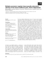
Báo cáo khoa học: Human papillomavirus 16 E7 protein inhibits interferon-c-mediated enhancement of keratinocyte antigen processing and T-cell lysis docx
Bạn đang xem bản rút gọn của tài liệu. Xem và tải ngay bản đầy đủ của tài liệu tại đây (734.85 KB, 9 trang )
Human papillomavirus 16 E7 protein inhibits
interferon-c-mediated enhancement of keratinocyte
antigen processing and T-cell lysis
Fang Zhou, Graham R. Leggatt and Ian H. Frazer
The University of Queensland Diamantina Institute, Princess Alexandra Hospital, Brisbane, Qld, Australia
Introduction
Persistent infection of the cervical epithelium with one
of a range of oncogenic human papillomaviruses
(HPVs) can initiate cervical cancer. The majority of
high-risk papillomavirus infections of immunocompe-
tent individuals are cleared, although this can take up
to 4 years, and a minority of apparently immunocom-
petent individuals will develop persisting infection [1].
These data suggest that HPV may have evolved mech-
anisms to enable infected epithelial cells to escape from
immune surveillance in vivo [2]. We established a skin
graft model to study the role in immune evasion of the
HPV 16 nonstructural protein E7, which is overexpres-
sed in premalignant lesions associated with HPV 16
infection. In this model, murine skin expressing
HPV 16 E7 as the product of a transgene in keratino-
cytes (KCs) from a keratin 14 promoter is grafted into
naı
¨
ve, otherwise syngeneic, mice. These mice fail to
reject such grafts, either spontaneously or after immu-
nization with E7 [3]. In contrast, skin grafts similarly
expressing ovalbumin (OVA) as a transgene product
are spontaneously rejected [4]. In vitro, HPV 16 E7-
specific cytotoxic T lymphocytes (CTLs) fail to kill
HPV 16 E7 transgenic KCs efficiently, but can kill
KCs pulsed with the dominant H-2D
b
restricted E7
peptide epitope [5] or with OVA [6]. A number of
studies have shown that HPV gene expression can
interfere with various components of antigen process-
ing by uncertain mechanisms [7–9]. Taken together,
these results suggest that the HPV 16 E7 may inhibit
cellular antigen processing and presentation to enable
Keywords
antigen processing; CD8 T cells; host–virus
interactions; papillomavirus
Correspondence
I. Frazer, The University of Queensland
Diamantina Institute, Princess Alexandra
Hospital, Ipswich Road, Woolloongabba,
Brisbane, Qld 4102, Australia
Fax: +61 7 3240 5946
Tel: +61 7 3240 5310
E-mail:
(Received 14 July 2010, revised 2
December 2010, accepted 10 January 2011)
doi:10.1111/j.1742-4658.2011.08011.x
Infection of epithelium with human papillomavirus (HPV) 16 is generally
prolonged, suggesting an ineffective virus-specific immune response, and
prolonged infection promotes anogenital cancer. To determine whether
poor antigen presentation by HPV-infected keratinocytes (KCs) contributes
to prolonged HPV infection, KCs and KCs expressing HPV 16 E7 protein
(E7-KCs) were compared for susceptibility to T-cell-mediated lysis directed
to ovalbumin (OVA) processed for presentation by the KCs. Interferon
(IFN)-c efficiently enhanced susceptibility to lysis of KCs presenting OVA,
but not of E7-KCs similarly presenting OVA. E7-KCs also exhibited
impaired IFN-c-induced upregulation of transcription of major histocom-
patibility complex class I antigen processing and presentation-associated
genes, and of membrane SIINFEKL–H-2K
b
complexes. Thus, expression
of HPV 16 E7 protein in KCs may inhibit enhancement by IFN-c of KC
sensitivity to T-cell lysis, by impairing antigen presentation.
Abbreviations
CTL, cytotoxic T lymphocyte; E7-KC, keratinocyte expressing HPV 16 E7; HPV, human papillomavirus; IFN, interferon; IRF, interferon
regulatory factor; KC, keratinocyte; MFI, mean fluorescence intensity; MHC, major histocompatibility complex; SD, standard deviation;
2-ME, 2-mercaptoethanol.
FEBS Journal 278 (2011) 955–963 ª 2011 The Authors Journal compilation ª 2011 FEBS 955
HPV to evade viral antigen-specific host immune
responses.
Several viruses have been demonstrated to interfere
with antigen processing and presentation by infected
cells, through inhibition of peptide recruitment to and
processing by the proteasome, or through blocking the
production and transport of major histocompatibility
complex (MHC) class I complexes [10–13]. Interferon
(IFN)-c plays an important role in facilitating CTL-
mediated immune effector function, through induction
of multiple genes associated with MHC class I antigen
presentation [14], and also contributes to the elimina-
tion of HPV infection [15].
HPV 16 E7 has been shown to interfere with the
transduction of IFN signaling [16–19]. HPV 16 E7
binds to the C-terminal transactivation domain of
interferon regulatory factor (IRF)-1 in vitro, and cul-
tured fibroblasts transduced with HPV 16 E7 demon-
strate reduced transcription of some IFN-c-inducible
gene products following overexpression of IRF-1 when
compared with untransduced cells [20]. In HPV 18
E6 ⁄ E7 transgenic mice, there is some evidence for
reduced transcription of the same gene products in the
cervix, as compared with nontransgenic animals [21].
We therefore investigated, using OVA as a model anti-
gen, whether HPV 16 E7, when expressed in KCs at
levels similar to those found in HPV infection, could
inhibit enhancement by IFN-c of antigen processing
and presentation, and CTL-mediated killing of KCs
expressing non-self-antigen.
Results
E7 does not affect the ability of IFN-
c
to
upregulate MHC class I expression and
exogenous CTL epitope presentation on KCs
We first wished to investigate whether expression of
HPV 16 E7 in KCs would alter the expression of mem-
brane-associated MHC, the presentation of processed
endogenous antigen as a peptide in association with
MHC, or CTL-mediated lysis directed at cell mem-
brane MHC–peptide complexes. As IFN-c can upregu-
late MHC class I expression on keratinocytes [22] and
enhances CTL epitope presentation and CTL-mediated
lysis of KCs [6], we also wished to investigate whether
E7 interfered with IFN-c-induced enhancement of anti-
gen processing and presentation. Therefore, KCs and
KCs from H-2
b
mice transgenic for HPV 16 E7
expressed from a keratin 14 promoter (E7-KCs) were
exposed in vitro to IFN-c. We first examined the effect
of E7 on IFN-c-mediated induction of MHC expres-
sion. Induction by IFN-c of the expression of MHC
class I on cultured E7-KCs was similar to induction on
nontransgenic KCs (Fig. 1A). We then exposed KCs
and E7-KCs to IFN-c and SIINFEKL, a peptide
A
CD
B
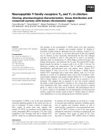
Fig. 1. Lysis of KCs and E7-KCs treated with IFN-c. Mouse KCs were exposed to SIINFEKL and IFN-c as shown. (A) Expression of MHC
class I on KCs and E7-KCs after exposure to IFN-c, as shown, was assessed by flow cytometry with an H-2K
b
-specific antibody. (B) Expres-
sion of SIINFEKL–H-2K
b
peptide complexes on KCs and E7-KCs after incubation with 10 lM SIINFEKL, and IFN-c, as shown, was assessed
with mouse antibody against SIINFEKL–H-2K
b
. (C) Expression of SIINFEKL–H-2K
b
complexes on KCs and E7-KCs after incubation with IFN-c
at 100 UÆmL
)1
and SIINFEKL, as shown, was assessed. (D) Susceptibility of SIINFEKL-exposed KCs to lysis by SIINFEKL-specific CTLs at an
effector ⁄ target ratio of 1 : 20 was compared for KCs and E7-KCs pulsed with three concentrations of SIINFEKL. The experiments in (A), (B)
and (C) were repeated twice, and those in (D) were repeated three times, with similar results. In (D), mean and SD from triplicate determina-
tions of percentage lysis for one experiment are shown.
HPV 16 E7 inhibits T-cell KC lysis F. Zhou et al.
956 FEBS Journal 278 (2011) 955–963 ª 2011 The Authors Journal compilation ª 2011 FEBS
derived from OVA that is able to associate with mem-
brane H-2K
b
MHC complexes without further process-
ing. KCs and E7-KCs showed similar dose-dependent
increases in the density of membrane SIINFEKL–
H-2K
b
complexes after IFN-c exposure (Fig. 1B).
Furthermore, KCs and E7-KCs exposed to a fixed
concentration of IFN-c and to increasing concentra-
tions of SIINFEKL displayed similar increased density
of SIINFEKL–H-2K
b
complexes (Fig. 1C). By select-
ing appropriate concentrations of peptide, we could
then compare the susceptibility of KCs and E7-KCs
expressing similar densities of SIINFEKL–H-2K
b
com-
plexes to lysis by a set number of E7-specific T cells,
to establish whether E7 expression might impact on
the sensitivity of KCs to T-cell lytic mechanisms. For
any given level of expression of MHC–peptide com-
plex, KCs and E7-KCs were equally susceptible to lysis
in vitro by SIINFEKL-specific CTLs (Fig. 1D). Thus,
endogenous expression of E7 has no effect on IFN-c-
induced enhancement of MHC expression by KCs, or
on the susceptibility of KCs pulsed with exogenous
SIINFEKL to CTL-mediated lysis.
E7 inhibits IFN-c-dependent upregulation of
presentation of endogenous antigen by KCs
As there was no observed effect of endogenous E7 on
MHC expression or on the susceptibility of KCs
expressing MHC–peptide complexes to T-cell-mediated
lysis, we next wished to test the hypothesis that endog-
enous E7 expression in KCs might inhibit the process-
ing and presentation of endogenously expressed
protein. We therefore compared KCs and E7-KCs,
each also expressing OVA endogenously as a transgene
product, for susceptibility to lysis by OVA-specific
T-cells, both with and without IFN-c pretreatment. E7
and OVA double-transgenic KCs and OVA single-
transgenic KCs, if not treated with IFN-c, were simi-
larly susceptible to lysis by OVA-specific T cells
(Fig. 2A). However, lysis of OVA transgenic KCs by
OVA-specific T cells was significantly increased follow-
ing IFN-c exposure, whereas lysis of E7 and OVA
double-transgenic KCs was not (Fig. 2A). These
results, together with those showing that E7 has no
effect on the presentation of exogenous peptide or on
T-cell-mediated lysis of cells sensitized by exogenous
peptide, with or without IFN-c exposure, allow the
conclusion that E7 inhibits the ability of IFN-c to
enhance the processing of endogenous antigen for pre-
sentation. To confirm these findings in an independent
system, we loaded KCs osmotically with OVA, using
previously established techniques. KCs and E7-KCs
loaded with OVA were treated with IFN-c, or left
untreated, and assessed for susceptibility to T-cell-med-
iated lysis. CTLs specific for SIINFEKL were equally
able to kill untreated KCs or E7-KCs when osmoti-
cally loaded with OVA, as expected. As predicted from
the findings with OVA transgenic KCs, lysis of KCs
osmotically loaded with OVA was similar whether the
cells were E7 transgenic or not, but after exposure to
IFN-c, substantially increased lysis of OVA-loaded
KCs was observed (Fig. 2B), whereas no such enhance-
ment was seen for osmotically loaded E7-KCs, con-
firming the findings with double-transgenic KCs that
endogenous E7 inhibits processing of endogenous anti-
gens by KCs for presentation to antigen-specific T cells
(Fig. 2B).
To further confirm this finding, we used an antibody
against SIINFEKL–H-2K
b
complexes to measure
MHC-associated presentation of SIINFEKL, derived
from osmotic loading of KCs with OVA. For KCs,
expression of SIINFEKL–H-2K
b
complexes was sub-
stantially upregulated in response to IFN-c, whereas
for E7-KCs, exposure to IFN-c failed to upregu-
late expression of SIINFEKL–H-2K
b
complexes
(Fig. 2C–E). Thus, endogenously expressed E7 inhibits
IFN-c-mediated enhancement of processing of endoge-
nous antigen by KCs, without inhibiting IFN-c-
mediated upregulation of MHC class I expression.
HPV 16 E7 blocks the ability of IFN-c to
upregulate the transcription of MHC class I
antigen processing and presentation-associated
genes in keratinocytes
As HPV 16 E7 expression in KCs impairs IFN-c-medi-
ated processing of endogenous OVA for presentation
of SIINFEKL (Fig. 2D), and HPV 16 E7 has been
suggested to inhibit IFN-c signal transduction, we
hypothesized that endogenously expressed E7 might
inhibit transcription of IFN-c-dependent genes that
are necessary for MHC class I antigen presentation.
To investigate whether HPV 16 E7 could reduce the
ability of IFN-c to upregulate transcription in KCs of
genes relevant to the processing and presentation
of endogenous antigen, we assessed the transcription
of three MHC class I antigen processing and presenta-
tion-associated genes (pa-28, tap-1 and irf-1) in KCs
and E7-KCs both before and after exposure of the
cells to IFN-c, using quantitative RT-PCR (Fig. 3).
Basal levels of expression of pa-28 and irf-1 were
somewhat higher in E7-KCs than in KCs (Fig. 3).
However, the increase in level of expression induced
by exposure to IFN-c was significantly blunted in E7
KCs, as compared with KCs, for pa28, tap-1 and irf-1
(Fig. 4). The maximal level of expression achieved
F. Zhou et al. HPV 16 E7 inhibits T-cell KC lysis
FEBS Journal 278 (2011) 955–963 ª 2011 The Authors Journal compilation ª 2011 FEBS 957
after IFN-c exposure was also significantly diminished
in E7-KCs, as compared with KCs, for tap-1 and irf-1
(Fig. 3).
Discussion
In this study, we show that expression of HPV 16 E7
as a transgene product in epithelial cells does not
directly impair, but rather slightly increases, MHC
class I expression. E7 expression is nevertheless associ-
ated with impairment of IFN-c-induced enhancement
of presentation of endogenous antigen to CTLs. For
E7-KCs, IFN-c treatment is less able to enhance the
transcription of genes regulating antigen presentation,
including tap-1, irf-1 and pa28. The reduction in gene
transcription is from 5- to 10-fold, which is sufficient
to reduce antigen presentation about five-fold and
impair T-cell-mediated killing in vitro, and may there-
fore be sufficient to explain the failure of E7-expressing
skin to be rejected in vitro.
Viruses use multiple strategies to make infected cells
of less interest to virus protein-specific immune effector
responses. Papillomavirus nonstructural viral proteins
have been shown to interact with several cellular pro-
cesses in a manner that could impair MHC class I
expression. HPV E5, when overexpressed as a trans-
gene product, can trap MHC class I molecules in the
Golgi [23]. HPV 16 E7, when overexpressed, can
repress the MHC class I heavy chain promoter, as well
as the promoters of tap1 and lmp2 [7], and can also
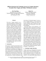
P < 0.05
AB
C
E
D
Fig. 2. HPV E7 impairs the enhancement by IFN-c of the presentation of endogenous antigen. (A) OVA transgenic KCs (KC-OVA) and KCs
expressing E7 and OVA (E7-KC-OVA) were compared for susceptibility to lysis by OVA-primed effector T cells, with or without IFN-c expo-
sure. (B) KCs and E7-KCs were loaded osmotically with OVA (OSM ⁄ OVA) or myoglobin (MYO), and, where indicated, exposed to IFN-c
(100 UÆmL
)1
for 48 h). OVA-loaded KCs with or without an E7 transgene were compared for susceptibility to lysis with and without IFN-c
pretreatment. Nontransgenic KCs treated with IFN-c (KC + IFN-c) are shown as a control. (C, D) Expression of SIINFEKL–MHC class I pep-
tide complexes on KCs (C) and E7-KCs (D) osmotically loaded with OVA and exposed to IFN-c (100 UÆmL
)1
) for 48 h was assessed with spe-
cific antibody by flow cytometry. (E) Processing of OVA for presentation by MHC as SIINFEKL, according to the protocols for (C) and (D).
OVA-loaded KCs exposed to IFN-c (KC + IFN + OVA) had significantly higher expression of SIINFEKL–MHC complexes than OVA-loaded E7
transgenic KCs exposed to IFN-c (E7-KC + IFN + OVA) (P = 0.02, t-test, n = 4). For the positive control (E7-KC + IFN + OVA + SIINFEKL),
MHC complexes were saturated with SIINFEKL added to the culture medium. For the negative controls, no IFN-c (KC + OVA; E7-KC + OVA)
or no OVA (E7-KC + IFN) was added. Means and SD values for MFI are shown.
HPV 16 E7 inhibits T-cell KC lysis F. Zhou et al.
958 FEBS Journal 278 (2011) 955–963 ª 2011 The Authors Journal compilation ª 2011 FEBS
bind to TAP1 and inhibit peptide transport [24], and
reduce the expression of MHC in cultured murine fi-
broblasts [25]. However, these reported effects of E7
do not seem, from our current study, to impact on the
ability of KCs expressing E7 at levels more typical of
those seen in HPV infection to present exogenous
peptide, or on the level of MHC class I expression.
Papillomavirus-associated cervical cancers express high
levels of E7, and demonstrate impaired membrane
expression of MHC class I complexes, which would be
expected to impair antigen presentation. However, in
cancer cells, reduced MHC class I display is associated
with low levels of TAP1 or TAP2 [26], as a result of
gene mutations associated with transformation, and
the contribution, if any, of overexpression of HPV 16
E7 to reduced MHC class I expression in these cells is
unclear.
We have recently shown that E7-specific CTLs that
are well able to kill E7-expressing transplantable
tumors fail to efficiently kill KCs expressing E7 as a
transgene product at levels commensurate with those in
infected cervical epithelium [5]. As impaired recognition
could be overcome by exposure to exogenous E7, it
probably reflects either low availability of E7 for pro-
cessing for presentation, or impaired antigen processing
in E7-expressing cells. To distinguish these possibilities,
we studied the processing and presentation of OVA
expressed as a transgene product in E7 transgenic and
control KCs, using antibody against SIINFEKL–H-
2K
b
. OVA presentation appeared to be normal in this
system, as cells with or without E7 were equally suscep-
tible to killing, although the fixed level of OVA expres-
sion did not exclude the possibility that high-level OVA
expression could overcome any restriction on process-
ing. We therefore tested cells loaded osmotically with
OVA, where lesser levels of OVA loading were still
AB
C
DE
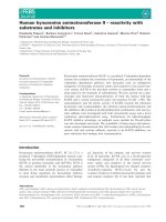
Fig. 3. Transcription of genes associated
with antigen processing and presentation in
KCs and E7-KCs after IFN-c treatment.
mRNA was extracted from KCs and E7-KCs.
KCs and E7-KCs were treated or not, as
shown, with IFN-c (IFN) at 100 UÆmL
)1
for
48 h. Expression levels of mRNA assessed
by RT-PCR with specific primers are shown
relative to a reference gene, rRNA adenine
dimethylase. Transcription after IFN-c expo-
sure was higher for KCs than for E7-KCs for
irf-1 (P = 0.02, n =7,t-test) and tap-1
(P < 0.01, n = 7). Differences for pa28 and
pias1 were nonsignificant by unpaired t-test
(n = 3). Error bars represent mean and SD
(n = 3).
Fig. 4. Upregulation of gene expression in KCs and E7-KCs follow-
ing IFN-c exposure. For each tested gene, the ratio of expression
level between cells exposed or not exposed to IFN-c is shown for
E7-KCs, and also for KCs. Significant differences in magnitude of
the IFN-c-induced change in expression between KCs and E7-KCs
were seen for irf-1 (P = 0.01, n =3,t-test), tap-1 (P = 0.05, n = 3),
and pa-28 (P = 0.05, n = 3). The change for pias1 was nonsignifi-
cant by unpaired t-test (n = 3). Error bars represent mean and SD.
F. Zhou et al. HPV 16 E7 inhibits T-cell KC lysis
FEBS Journal 278 (2011) 955–963 ª 2011 The Authors Journal compilation ª 2011 FEBS 959
equally able to sensitize cells to OVA, whether express-
ing E7 or not. However, in both the osmotic loading
model and the transgene model, induction by IFN-c of
increased expression of MHC–peptide complexes and
susceptibility to T-cell-mediated lysis was significantly
impaired if KCs expressed E7 as a transgene product.
Looking for a mechanism, we assessed levels of tran-
scription of genes whose protein products participate in
antigen expression. HPV 16 E7 attenuated the ability
of IFN-c to induce the transcription of several genes,
and also of IRF-1, a master regulator of IFN-inducible
genes. IFN-c is a potent inducer of antigen processing
and of MHC class I expression for many cell types [14].
Signal transduction occurs via the JAK–STAT path-
way, and upregulation of the expression of the down-
stream genes relevant to antigen processing and
presentation is mediated by members of the IRF family
[27], particularly IRF-1[28]. E7 blocked the ability of
IFN-c to efficiently induce irf-1 transcription to the
level observed in nontransgenic KCs. Thus, E7 may
block the ability of IFN-c to induce IRF-1 expression,
in turn inhibiting the expression of downstream genes
related to MHC class I antigen processing and
presentation. E7 could potentially also interfere with
IFN-c-mediated upregulation of IRF-1 expression by
inhibiting upstream transduction of IFN signaling
[16,17,29,30]. Furthermore, HPV 16 E7 can also inter-
fere with IRF-1 function without affecting IRF-1 tran-
scription and translation. This occurs through
alteration of the DNA-binding capacity and promoter
transactivation of IRF-1 without alteration of IRF-1
level [20,21,31], probably by direct binding to E7 [20].
Differences between the effects of E7 on induction by
IFN-c of IRF-1 mRNA in our study and on the
steady-state levels of IRF-1 measured by others may
reflect the different experimental systems, or effects of
E7 on post-transcriptional regulation of IRF-1 produc-
tion or destruction.
HPV 16 is a member of the mucosotropic a-papillo-
mavirus clade. Papillomaviruses from the genetically
and functionally distinct skin tropic b-clade use alter-
native means to impair antigen presentation. HPV 38
E6 inhibits STAT1 expression and phosphorylation
induced by IFN-b and IFN-c in human KCs, and
inhibits IRF-1, TAP1 and MHC class I expression in
host cells [29]. An impaired response of E7-KCs to
IFN-c has significant implications for immunotherapy
of HPV-associated skin lesions, which have proven
refractory to induced antigen-specific immunotherapy
[32,33]. IFN-c, secreted by CD8 T cells, by activated
NK and NKT cells and by dendritic cells [34,35], is
a key intermediate mediator of CD8 T-effector cell
function, enhancing antigen presentation as well as
polarizing the immune response to the Th1 type.
Impaired antigen presentation may thus be one of the
reasons why HPV infection is slow to clear in the face
of adequate cellular immunity, and why immunother-
apy has proven ineffective for persisting HPV infec-
tion. It may also explain why local administration of
supraphysiological concentrations of IFNs can contrib-
ute to the clearance of HPV-associated genital warts
[36]. Administration of proinflammatory mediators
that can enhance antigen presentation by an IFN-
independent pathway, perhaps through toll-like recep-
tor signaling [34], may therefore facilitate the immune
clearance of HPV-associated disease.
Experimental procedures
Immunogen, peptide and mice
An 8-mer peptide (SIINFEKL) corresponding to the major
CTL epitope of OVA was synthesized by AusPep (Park-
ville, Vic., Australia) to bind to MHC class I H-2K
b
(amino
acids 258–266 of OVA). C57BL ⁄ 6J mice, C57BL ⁄ 6J mice
expressing SIINFEKL [37] or HPV 16 E7 [38] from the
keratin 14 promoter, and C57BL ⁄ 6J mice expressing OVA
from the keratin 5 promoter [4], were bred under conven-
tional conditions in specific pathogen-free holding rooms in
the Princess Alexandra Hospital biological resources facility
(Brisbane, Qld, Australia). The protocols of these experi-
ments were approved by the institutional Animal Ethics
Committee.
Generation of effector cells
C57BL ⁄ 6J female mice (6–8 weeks of ages) were immunized
once with 100 lg of SIINFEKL ⁄ 30 lg of keyhole limpet
hemocyanin (Sigma Pharmaceuticals, Melbourne, Victoria,
Australia) and 30 lg of QuilA (Spikoside; ISCOTEC AB,
Lulea, Sweden). Lymph node cells were collected from
immunized mice 4 days after immunization. Lymphocytes
were cultured in filtered Click’s medium [50% Eagle’s ⁄
Ham’s amino acids (Sigma), 50% RPMI-1640 (Gibco;
Invitrogen, Carlsbad, CA, USA), 10% heat-inactivated
fetal bovine serum, containing 1 ngÆmL
)1
mouse inter-
leukin-2 (Pharmingen, San Diego, CA, USA) and 0.05 lm
SIINFEKL, 2 · 10
5
UÆmL
)1
penicillin ⁄ 2 · 10
5
UÆmL
)1
streptomycin, 200 mml-glutamine and 5 · 10
)5
m 2-mercap-
toethanol (2-ME)] for 4 days.
Generation of target cells
Isolation and culture of KCs from mouse skin has been
described previously [5]. In brief, KCs were cultured in
epidermal cell culture 3 : 1 medium [for 500 mL of 3 : 1
medium: 365 mL of DMEM (Gibco), 5 mL of
HPV 16 E7 inhibits T-cell KC lysis F. Zhou et al.
960 FEBS Journal 278 (2011) 955–963 ª 2011 The Authors Journal compilation ª 2011 FEBS
l-glutamine ⁄ penicillin ⁄ streptomycin, 200 mm ⁄ 2 · 10
5
U ⁄ 2
· 10
5
UÆmL
)1
, 125 mL of Ham’s F12, 50 mL of fetal
bovine serum, 500 lL of transferrin (Sigma), 5 mgÆmL
)1
in
0.1% BSA in NaCl ⁄ P
i
, 500 lL of insulin (Sigma),
5mgÆmL
)1
in 1 mm HCl, 500 lL of cholera toxin (Sigma),
8.4 mgÆmL
)1
in NaCl ⁄ P
i
, 100 lL of hydrocortisone
(Sigma), 1.2 mgÆmL
)1
in 90% ethanol in water, 1000 lLof
184 mm adenine (Sigma), 17 mgÆmL
)1
in 0.1% BSA in
NaCl ⁄ P
i
, 500 lL of gentamicin, 20 mgÆmL
)1
, 500 lLof
2-ME stock (35 lL of 2-ME in 10 mL of RPMI-1640 med-
ium for 2-ME stock)] for 48 h, and then transferred into
serum-free KC medium (Gibco) for 2 days. KCs were
seeded in 96-well plates at 2 · 10
4
cells per well for CTL
assays, or suspended at 5 · 10
5
cells per tube for FACS
experiments.
Osmotic loading of KCs
The techniques have been described previously [39]. In
brief, 5 · 10
6
KCs were suspended in 1 mL of RPMI-1640
osmotic loading buffer [25 mm Hepes, 0.5 m sucrose (w ⁄ v),
10% polyethylene glycol, pH 7.2] containing 10 mgÆmL
)1
OVA (Sigma) or myoglobin (Sigma), and incubated at
37 °C for 10 min. The cells were then diluted into 14 mL of
a mixture of 60% RPMI-1640 medium and 40% water,
and held at 37 °C for 2 min. The loaded KCs, pelleted at
300 g for 7 min, were resuspended in RPMI-1640 medium
and pelleted at 300 g for 5 min. Finally, the cells were
resuspended in culture medium and incubated with or with-
out IFN-c at 100 UÆ mL
)1
for 24 h [40].
CTL assay (
51
Cr release)
A standard 5-h
51
Cr release assay was conducted as
described previously [5]. CTL assay data were expressed as
percentage specific lysis according to the following formula:
The data were analyzed by t-test, and results were
regarded as significantly different when P < 0.05.
Flow cytometry
KCs (5 · 10
5
) were incubated with the first antibody (mouse
anti-SIINFEKL–H-2K
b
clone D25.1.1.16 [41], 50 lL per
sample, provided by D. Purcell, University of Melbourne),
mouse anti-H-2K
b
(clone AF6-88.5; Pharmingen), or mouse
anti-(rat IgM) (Pharmingen), an isotype control, for 1 h at
4 °C. Cells were then washed twice and incubated with the
secondary antibody [fluorescein isothiocyanate-conjugated
rabbit anti-mouse IgG (Dako Cytomation, Copenhagen,
Denmark)] for 1 h at 4 °C, washed, and fixed with 5%
formaldehyde. Data collected on a FACSCalibur (Becton
Dickinson, San Diego, CA, USA) were analyzed with
winmdi 2.8 (Joseph Trotter, Scripps Research Institute, La
Jolla, CA, USA). Viable KCs were selected preferentially by
excluding small particles. The change in mean fluorescence
intensity (MFI) was calculated as the difference in MFI
between test and isotype control samples.
Real-time PCR
mRNA was extracted from KCs and converted to cDNA
with the use of random primers and PowerScript RT (Gene-
Works, Hindmarsh, SA, Australia), according to the manu-
facturer’s protocol. cDNA samples dissolved in the PCR mix
buffer (FastStart SYBR Green Master; Roche Applied
Science, Mannheim, Germany) were used to conduct quanti-
tative PCR under the following conditions: 50 °C for 2 min;
95 °C for 10 min; and 40 cycles of 94 °C for 1 min, 55 °C for
1 min and 72 °C for 1 min. The following primers were
used: TAP1, forward, 5¢-ACC TGG CTA CGG TAC ACC
TG-3¢; TAP1, reverse, 5¢-CCT CTG AGC TCC CAC TTG
AC-3¢; IRF-1, forward, 5¢-CCT GGG TCA GGA CTT G-
GA TA-3¢; IRF-1, reverse, 5¢-TTC GGC TAT CTT CCC T-
TC CT-3¢; PA28, forward, 5¢-CCG CTC CTC CTT CTC
TTT CT-3¢; PA28, reverse, 5¢-AAG CCA AGG TGG
ATG TGT TC-3¢; JAK1, forward, 5¢-TCA ACC TTC CCA
AAG TGA CC-3¢; JAK1, reverse, 5¢-CAT GAC TCG CTG
CAT GAA CT-3¢; PIAS1, forward, 5¢-AAG TGC TCA -
CAG CCT TGG AT-3¢; PIAS1, reverse, 5¢-TCC CTA GGT
GCA TGT TCT CC-3¢; rRNA adenine dimethylase, for-
ward, 5¢-GGA GGG CCC ATC AGT TTA AT-3¢; rRNA
adenine dimethylase, reverse, 5¢-AAA CAA TTG CAT TGC
ATA GTGC-3¢. The data were analyzed with rotor-
gene 6000.
Statistical analysis
All experimental data, including DMFI of FACS data, were
analyzed with unpaired t-tests. Error bars represent mean
and standard deviation (SD). Results were regarded as
showing significant differences if P-values were < 0.05.
Acknowledgements
The authors are grateful to the staff of the biological
research facility at the Princess Alexandra Hospital for
their assistance. This work was funded by program
grant No. 352439 from the National Health and
% specific lysis ¼
mean sample release (c.p.m.) À mean spontaneous release
mean maximum release (c.p.m.) À mean spontaneous release (c.p.m.)
 100
F. Zhou et al. HPV 16 E7 inhibits T-cell KC lysis
FEBS Journal 278 (2011) 955–963 ª 2011 The Authors Journal compilation ª 2011 FEBS 961
Medical Research Council of Australia, and grants
from the Lions Medical Research Foundation, the
Australian Cancer Research Foundation, the Cancer
Council Queensland, and the Princess Alexandra hos-
pital Foundation. Scholarship funding to F. Zhou was
from the Cancer Collaborative Group, Princess Alex-
andra Hospital and from ANZ Trustees. I. Frazer was
recipient of a Queensland Government Premier’s fel-
lowship. The authors declare that they have no conflict
of interest or financial interests regarding the research
findings described in this article.
References
1 Koshiol JE, Schroeder JC, Jamieson DJ, Marshall SW,
Duerr A, Heilig CM, Shah KV, Klein RS, Cuuvin S
& Schuman P et al. (2006) Time to clearance of
human papillomavirus infection by type and human
immunodeficiency virus serostatus. Int J Cancer 119,
1623–1629.
2 Kanodia S, Fahey LM & Kast WM (2007) Mecha-
nisms used by human papillomaviruses to escape the
host immune response. Curr Cancer Drug Targets 7,
79–89.
3 Dunn LA, Evander M, Tindle RW, Bulloch AL, De
Kluyver RL, Fernando GJ, Lambert PF & Frazer IH
(1997) Presentation of the HPV16E7 protein by skin
grafts is insufficient to allow graft rejection in an E7-
primed animal. Virology 235, 94–103.
4 Azukizawa H, Kosaka H, Sano S, Heath WR, Takah-
ashi I, Gao XH, Sumikawa Y, Okabe M, Yoshikawa K
& Itami S (2003) Induction of T-cell-mediated skin dis-
ease specific for antigen transgenically expressed in
keratinocytes. Eur J Immunol 33, 1879–1888.
5 Leggatt GR, Dunn LA, De Kluyver R, Stewart T &
Frazer IH (2002) Interferon-gamma enhances cytotoxic
T lymphocyte recognition of endogenous peptide in
keratinocytes without lowering the requirement for sur-
face peptide. Immunol Cell Biol 80, 415–424.
6 Zhou F, Frazer IH & Leggatt GR (2009) Keratinocytes
efficiently process endogenous antigens for cytotoxic
T-cell mediated lysis. Exp Dermatol 18, 1053–1059.
7 Georgopoulos NT, Proffitt JL & Blair GE (2000) Tran-
scriptional regulation of the major histocompatibility
complex (MHC) class I heavy chain, TAP1 and LMP2
genes by the human papillomavirus (HPV) type 6b, 16
and 18 E7 oncoproteins. Oncogene 19, 4930–4935.
8 Evans M, Borysiewicz LK, Evans AS, Rowe M, Jones
M, Gileadi U, Cerundolo V & Man S (2001) Antigen
processing defects in cervical carcinomas limit the pre-
sentation of a CTL epitope from human papillomavi-
rus 16 E6. J Immunol 167, 5420–5428.
9 Thomas KJ, Smith KL, Youde SJ, Evans M, Fiander
AN, Borysiewicz LK & Man S (2008) HPV16 E6 29-38-
specific T cells kill cervical carcinoma cells despite
partial evasion of T-cell effector function. Int J Cancer
122, 2791–2799.
10 Hill A, Jugovic P, York I, Russ G, Bennink J, Yewdell
J, Ploegh H & Johnson D (1995) Herpes simplex virus
turns off the TAP to evade host immunity. Nature 375,
411–415.
11 Levitskaya J, Coram M, Levitsky V, Imreh S, Steiger-
wald-Mullen PM, Klein G, Kurilla MG & Masucci
MG (1995) Inhibition of antigen processing by the
internal repeat region of the Epstein–Barr virus nuclear
antigen-1. Nature 375, 685–688.
12 Ahn K, Meyer TH, Uebel S, Sempe
´
P, Djaballah H,
Yang Y, Peterson PA, Fruh K & Tamp R (1996)
Molecular mechanism and species specificity of TAP
inhibition by herpes simplex virus protein ICP47.
EMBO J 15, 3247–3255.
13 Ahn K, Gruhler A, Galocha B, Jones TR, Wiertz EJ,
Ploegh HL, Peterson PA, Yang Y & Fruh K (1997)
The ER-luminal domain of the HCMV glycoprotein
US6 inhibits peptide translocation by TAP. Immunity 6,
613–621.
14 Boehm U, Klamp T, Groot M & Howard JC (1997)
Cellular responses to interferon-gamma. Annu Rev
Immunol 15, 749–795.
15 Seresini S, Origoni M, Lillo F, Caputo L, Paganoni
AM, Vantini S, Longhi R, Taccagni G, Ferrari A,
Doglioni C, Secchi P & Protti MP (2007) IFN-gamma
produced by human papilloma virus-18 E6-specific
CD4 + T cells predicts the clinical outcome after
surgery in patients with high-grade cervical lesions.
J Immunol 179, 7176–7183.
16 Barnard P & McMillan NAJ (1999) The human papillo-
mavirus E7 oncoprotein abrogates signaling mediated
by interferon-a. Virology 259, 305–313.
17 Chang YE & Laimins LA (2000) Microarray analysis
identifies interferon-inducible genes and Stat-1 as major
transcriptional targets of human papillomavirus type 31.
J Virol 74
, 4174–4182.
18 Koromilas AE, Li S & Matlashewski G (2001)
Control of interferon signaling in human papillomavirus
infection. Cytokine Growth Factor Rev 12, 157–170.
19 Nees M, Geoghegan JM, Hyman T, Frank S, Miller L
& Woodworth CD (2001) Papillomavirus type 16 onco-
genes downregulate expression of interferon-responsive
genes and upregulate proliferation-associated and
NF-kappaB-responsive genes in cervical keratinocytes.
J Virol 75, 4283–4296.
20 Park JS, Kim EJ, Kwon HJ, Hwang ES, Namkoong SE
& Um SJ (2000) Inactivation of interferon regulatory
factor-1 tumor suppressor protein by HPV E7 oncopro-
tein – implication for the E7-mediated immune evasion
mechanism in cervical carcinogenesis. J Biol Chem 275 ,
6764–6769.
HPV 16 E7 inhibits T-cell KC lysis F. Zhou et al.
962 FEBS Journal 278 (2011) 955–963 ª 2011 The Authors Journal compilation ª 2011 FEBS
21 Um SJ, Rhyu JW, Kim EJ, Jeon KC, Hwang ES &
Park JS (2002) Abrogation of IRF-1 response by high-
risk HPV E7 protein in vivo. Cancer Lett 179, 205–212.
22 Niederwieser D, Aubock J, Troppmair J, Herold M,
Schuler G, Boeck G, Lotz J, Fritsch P & Huber C
(1988) IFN-mediated induction of MHC antigen expres-
sion on human keratinocytes and its influence on in vi-
tro alloimmune responses. J Immunol 140, 2556–2564.
23 Ashrafi GH, Haghshenas MR, Marchetti B, O’Brien
PM & Campo MS (2005) E5 protein of human papillo-
mavirus type 16 selectively downregulates surface HLA
class I. Int J Cancer 113, 276–283.
24 Vambutas A, DeVoti J, Pinn W, Steinberg BM & Bona-
gura VR (2001) Interaction of human papillomavirus
type 11 E7 protein with TAP-1 results in the reduction
of ATP-dependent peptide transport. Clin Immunol 101,
94–99.
25 Park JS, Boyer S, Mitchell K, Gilfor D, Birrer M,
Darlington G, El Deiry W, Firestone GL, Munger K,
Band V, Fisher PB & Dent P (2000) Expression of
human papilloma virus E7 protein causes apoptosis and
inhibits DNA synthesis in primary hepatocytes via
increased expression of p21Cip-1 ⁄ WAF1 ⁄ MDA6. J Biol
Chem 275, 18–28.
26 Ritz U, Momburg F, Pilch H, Huber C, Maeurer MJ
& Seliger B (2001) Deficient expression of components
of the MHC class I antigen processing machinery
in human cervical carcinoma. Int J Oncol 19,
1211–1220.
27 Tennant LM, Renard C, Chardon P & Powell PP
(2007) Regulation of porcine classical and nonclassical
MHC class I expression. Immunogenetics 59, 377–389.
28 Briscoe J, Guschin D, Rogers NC, Watling D, Muller
M, Horn F, Heinrich P, Stark GR & Kerr IM (1996)
JAKs, STATs and signal transduction in response to
the interferons and other cytokines. Phil Trans R Soc
Lond B Biol Sci 351, 167–171.
29 Cordano P, Gillan V, Bratlie S, Bouvard V, Banks L,
Tommasino M & Campo MS (2008) The E6E7 onco-
proteins of cutaneous human papillomavirus type 38
interfere with the interferon pathway. Virology 377,
408–418.
30 Li SY, Labrecque S, Gauzzi MC, Cuddihy AR,
Wong AHT, Pellegrini S, Matlashewski GJ &
Koromilas AE (1999) The human papilloma virus
(HPV)-18 E6 oncoprotein physically associates with
Tyk2 and impairs Jak-STAT activation by interferon-a.
Oncogene 18, 5727–5737.
31 Perea SE, Massimi P & Banks L (2000) Human papillo-
mavirus type 16 E7 impairs the activation of the inter-
feron regulatory factor-1. Int J Mol Med 5, 661–666.
32 Frazer IH (2004) Prevention of cervical cancer through
papillomavirus vaccination. Nat Rev Immunol 4, 46–55.
33 Frazer IH & Appleton S (2006) Vaccines to prevent and
treat human papillomavirus associated anogenital dis-
ease. In The Cervix, 2nd edn (Jordan JA & Singer A
eds), pp. 609–620. Blackwell Publishing, Oxford.
34 Wang RF, Miyahara Y & Wang HY (2008) Toll-like
receptors and immune regulation: implications for can-
cer therapy. Oncogene 27, 181–189.
35 Frucht DM, Fukao T, Bogdan C, Schindler H, O’Shea
JJ & Koyasu S (2001) IFN-gamma-production by anti-
gen-presenting cells: mechanisms emerge. Immunol
Today 22, 556–560.
36 Frazer IH & McMillan NA (1997) Papillomatosis and
condylomata acuminata. In Clinical Applications of the
Interferons (Stuart-Harris R & Penny RW eds), pp.
79–91. Chapman and Hall Medical, London.
37 Stefanski HE, Mayerova D, Jameson SC & Hogquist
KA (2001) A low affinity TCR ligand restores positive
selection of CD8 + T cells in vivo. J Immunol 166,
6602–6607.
38 Herber R, Liem A, Pitot H & Lambert PF (1996) Squa-
mous epithelial hyperplasia and carcinoma in mice
transgenic for the human papillomavirus type 16 E7
oncogene. J Virol 70, 1873–1881.
39 Moore MW, Carbone FR & Bevan MJ (1988) Intro-
duction of soluble protein into the class I pathway of
antigen processing and presentation. Cell 54, 777–785.
40 Carbone FR & Bevan MJ (1990) Class I-restricted pro-
cessing and presentation of exogenous cell-associated
antigen in vivo. J Exp Med 171, 377–387.
41 Porgador A, Yewdell JW, Deng Y, Bennink JR &
Germain RN (1997) Localization, quantitation, and
in situ detection of specific peptide–MHC class I
complexes using a monoclonal antibody. Immunity 6,
715–726.
F. Zhou et al. HPV 16 E7 inhibits T-cell KC lysis
FEBS Journal 278 (2011) 955–963 ª 2011 The Authors Journal compilation ª 2011 FEBS 963