Báo cáo khoa học: Cytoskeleton-modulating effectors of enteropathogenic and enterohemorrhagicEscherichia coli: role of EspL2 in adherence and an alternative pathway for modulating cytoskeleton through Annexin A2 function pot
Bạn đang xem bản rút gọn của tài liệu. Xem và tải ngay bản đầy đủ của tài liệu tại đây (389.64 KB, 6 trang )
MINIREVIEW
Cytoskeleton-modulating effectors of enteropathogenic
and enterohemorrhagic Escherichia coli: role of EspL2 in
adherence and an alternative pathway for modulating
cytoskeleton through Annexin A2 function
Toru Tobe
Department of Microbiology and Immunology, Graduate School of Medicine, Osaka University, Japan
Introduction
Adherence to the surface of host epithelia is a first step
in infection by enterohemorrhagic Escherichia coli
(EHEC) and enteropathogenic Escherichia coli (EPEC).
EHEC and EPEC are capable of intimate adherence to
the mucosal surface via a specific mechanism which is
mediated by the interaction between host-surface-
oriented translocated intimin receptor (Tir) and a bac-
terial outer membrane protein intimin [1]. Tir is one of
the effectors injected into host cells through a type III
secretion system (T3SS) and modulates cellular func-
tion [2]. Injected Tir is localized in the host-cell
membrane and forms a receptor for intimin. Binding of
intimin to the extracellular domain of Tir induces the
cytoplasmic domain of Tir to recruit the cellular factors
needed for actin polymerization [3]. By changing the F-
actin-based cytoskeleton, the morphology of bacteria-
adherent host cells is dramatically modified, leading
to disruption of the microvilli and the formation of a
pedestal-like structure beneath the adherent bacteria,
which is called an attaching ⁄ effacing (A⁄ E) lesion [4].
In addition, the host cell’s cytoskeleton is targeted
by other virulence factors, such as mitochondrial
Keywords
actin dynamics; A ⁄ E pathogen; colonization;
lipid raft; membrane morphology; prophage;
pseudopod-like structure; transcriptional
regulation; type III secretion; virulence factor
Correspondence
T. Tobe, Department of Microbiology and
Immunology, Graduate School of Medicine,
Osaka University, 2-2 Yamadaoka, Suita,
Osaka 565-0871, Japan
Fax: +81 6 6879 3309
Tel: +81 6 6879 3301
E-mail:
(Received 10 November 2009, revised 14
January 2010, accepted 3 February 2010)
doi:10.1111/j.1742-4658.2010.07654.x
Enterohemorrhagic Escherichia coli (EHEC) and enteropathogenic Escheri-
chia coli (EPEC) are attaching ⁄ effacing pathogens that possess a type III
secretion system and deliver a variety of effectors into host cells for suc-
cessful infection. EHEC produces at least 20 effector families with various
functions. Reorganization of the cellular cytoskeleton at the adherent site
is a hallmark of these pathogens. EspL2 of EHEC is a novel effector class
that can modulate the cellular cytoskeleton. By interacting with and activa-
ting Annexin A2, EspL2 contributes to the formation of a condensed
microcolony and may adhere to host cells in a translocated intimin recep-
tor-independent manner. The interaction of EspL2 with Annexin A2
increases F-actin bundling activity and strengthens the membrane–cytoskel-
eton linkage, resulting in the condensation of actin fibers and the induction
of a pseudopod-like structure. Membrane microdomains, namely the lipid
raft, which is rich in Annexin A2, may be a platform by which EHEC ⁄
EPEC infection modulates cellular signaling and the cytoskeleton.
Abbreviations
A ⁄ E, attaching ⁄ effacing; EHEC, enterohemorrhagic Escherichia coli; EPEC, enteropathogenic Escherichia coli; LEE, locus of enterocyte
effacement; Map, mitochondrial associated protein; SpLE3, Sakai prophage-like element 3; T3SS, type III secretion system; Tir, translocated
intimin receptor.
FEBS Journal 277 (2010) 2403–2408 ª 2010 The Author Journal compilation ª 2010 FEBS 2403
associated protein (Map), EspH and EspB [5]. EHEC
O157:H7 possesses > 20 types of effectors, the func-
tions of many of which are yet to be fully determined.
Among these, EspL2 is encoded by the espL2 gene on
Sakai prophage-like element 3 (SpLE3) which also car-
ries two other effector genes, nleB1 and nleE [6,7].
Interaction of EspL2 with Annexin A2
After the initial infection of epithelial cells, EHEC
delivers bacterial proteins, effector proteins, into cells
through the T3SS. Although EHEC O157:H7 Sakai
possesses > 60 genes that show similarity with the
amino acid sequences of known effectors, only 39
encode proteins that are secreted and translocated into
host cells through T3SS; the others are either pseudog-
enes or unexpressed genes [7]. EHEC O157:H7 Sakai
harbors two genes encoding EspL family proteins
(espL1 and espL2). Of these, EspL2 is secreted and
delivered into host cells in a T3SS-dependent manner,
whereas espL1 seems to be a dead gene because it
does not produce any detectable product or transcript
(T. Tobe, unpublished). Using FLAG-tagged EspL2
protein, T3SS-delivered EspL2 has been shown to
localize to the cytosolic side of the plasma membrane.
Adherence of EHEC induces an F-actin-rich structure
beneath the sites of bacterial adherence which extends
from the plasma membrane to the cytoplasm. EspL2
colocalizes with the F-actin structures although only at
the membrane proximal part. Isolation of host proteins
bound to EspL2 using affinity chromatography
revealed that Annexin A2 (also known as annexin II)
may be the target of EspL2 in host cells. Coprecipita-
tion of Annexin A2 with EspL2 from a purified mix-
ture of the two in vivo supported the interaction of
both proteins and, furthermore, indicated a direct
interaction between EspL2 and Annexin A2 [8]. Ann-
exin A2 binds to the cytosolic surface of the cellular
membrane and this has been linked to the formation
or stabilization of actin assembly sites [9]. Localization
of EspL2 at regions close to the membrane suggests
that EspL2 interacts directly with Annexin A2 on the
cellular membranes of infected cells.
Morphological changes in the
microcolony and surface of infected
host cells
Infection by an EHEC mutant strain lacking the espL2
gene showed little difference in terms of adherent capac-
ity and the morphology of host cells compared with a
wild-type strain. However, a microcolony of EHEC har-
boring multiple copies of the espL2 gene has a highly
condensed 3D structure and strongly induces extension
of the host-cell membrane, forming pseudopod-like
structures at the bacterial attachment sites [8]. Beneath
the bacterial adherence site, intimate attachment of
bacteria induces the formation of actin polymers and a
pedestal-like structure sometimes forms. Even bacteria
in the upper layer of the 3D colony are associated with
F-actin and the plasma membrane, indicating that
upward expansion of the bacterial colony is achieved by
the extension of actin polymers. By contrast, a microcol-
ony of EHEC espL2 mutant on epithelial cells is flat,
spreading two-dimensioally on the cell surface, and
bacterial density is lower than those formed by EHEC
harboring multiple copies of the espL2 gene with spaces
between bacteria (Fig. 1). Meanwhile, the wild-type
EHEC strain forms a microcolony of intermediate phe-
notype. In the microcolony of EHEC harboring multiple
copies of the of espL2 gene, the host-cell membrane is
extended to form filopodia or pseudopod-like structures
among the bacteria in the microcolony. However, adher-
ence of the EHEC espL2 mutant causes only a slight
EHEC
Tir
Annexin A2
F-actin
EspL2
EHEC
Δ
espL2
EHEC wild type
Nucleus
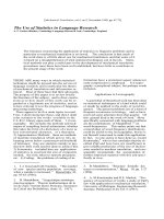
Fig. 1. Modulation of actin dynamism and membrane morphology
by the interaction of EspL2 and Annexin A2. Attachment of EHEC
induces actin reorganization beneath adherent sites via Tir-induced
actin polymerization. Formation of a membrane microdomain, the
lipid raft, is induced by EHEC microcolony formation, and lipid raft-
associated protein Annexin A2 also accumulates at the adherent
site. The interaction of EspL2 with Annexin A2 induces the conden-
sation of F-actin, leading to the formation of a condensed bacterial
colony and a pseudopod-like structure (right cell). By contrast, with-
out EspL2, attachment of EHEC induces the formation of F-actin
pedestals, but each pedestal is separated from others (left cell).
EspL2 effector modulates cytoskeleton T. Tobe
2404 FEBS Journal 277 (2010) 2403–2408 ª 2010 The Author Journal compilation ª 2010 FEBS
morphological change in the cell surface. The induction
of a pseudopod-like structure is observed even with
infection of an EHEC tir mutant harboring multiple
copies of the espL2 gene, which does not form intimate
attachment via Tir–intimin interaction and can not
induce actin polymerization at the site of adherence.
This clearly indicates that morphological changes in the
cell membrane, possibly with changes in the cytoskele-
ton, are induced by EspL2 without Tir-mediated actin
polymerization.
Other effectors have been shown to induce the forma-
tion of filopodia or a pseudopod-like structure, but the
induction is only transient before the formation of inti-
mate attachment via the Tir–intimin interaction [10,11].
Filopodia are formed at an early stage of infection and
later become dissipated when Tir is phosphorylated to
begin recruiting the cellular factors involved in actin
polymerization. Map is necessary for transient filopodia
formation and EspH is involved in supporting Tir-med-
iated actin polymerization [12,13]. Overexpression of
Map in EPEC not only increases the number of micro-
colonies with filopodia, but also prolongs the duration
of filopodia [12]. Deletion of the espH gene results in
enhanced filopodia formation [13]. These effectors seem
to act only at the early stages of adherence. Therefore,
it is unlikely that Map and ⁄ or EspH are necessary for
the formation of a condensed microcolony with F-actin
pedestals and induction of the morphological changes
in the cell surface caused by EspL2. Moreover, pheno-
types caused by EspL2, such as the formation of a pseu-
dopod-like structure into a microcolony, were similarly
observed with adherence of the tir mutant or the wild-
type strain [8], suggesting that EspL2-induced pseudo-
pod-like structure formation is distinct from
Map ⁄ EspH-regulated formation of filopodia.
Modification of activity of Annexin A2
by EspL2
Annexins are a family of proteins that bind to mem-
brane phospholipids in a Ca
2+
-dependent manner.
This property links Annexins to many cellular events
related to the membrane, such as membrane–cytoskele-
ton linkage, cell signaling, the assembly of certain
membrane domains, endocytosis and ion fluxes across
the membrane [14]. Annexin A2 binds directly and spe-
cifically to phophatidylinositol (4,5)-bisphosphate, and
this binding is responsible for recruiting the Annexin
A2–p11 complex to the submembraneous actin-assem-
bly site of EPEC-infected cells or Arf6-activated cells
[15]. In EPEC-infected cells, Annexin A2 accumulates
at sites of EPEC attachment where formation of actin-
rich pedestals is induced [16]. It has been suggested
that Annexin A2 at the cytoplasmic membrane surface
beneath the EPEC adherent site plays a role in reorga-
nization of the membrane ⁄ cytoskeleton following
EHEC infection [16]. Notably, recruitment of Annex-
in A2 to EPEC adherent sites is independent of
Tir-induced actin polymerization, suggesting that
Annexin A2 is not linked directly to actin pedestal
formation [16]. In addition, Annexin A2 has been
shown to interact directly with and bundle polymerized
actin [17,18]. Therefore, it is most likely that Annex-
in A2 at the EPEC adherent site links F-actin, most of
which is polyerimzed by Tir-activated cellular factors,
to the cytoplasmic membrane, affecting the morphology
of the membrane and cytoskeleton.
Morphological changes in the bacterial microcolony
and cellular membrane at the bacterial adherence site of
cells infected with EHEC harboring multiple copies
of the espL2 gene or espL2 mutant are may be the result
of membrane-associated cytoskeleton reorganization.
F-actin pedestals beneath the microcolony of an EHEC
strain with multiple copies of the espL2 gene also con-
densed along with adherent bacteria. In addition, pseu-
dopod-like structures are induced in infected cells. These
observations are explained by activation of Annexin A2:
strengthening the linkage between polymerized actins
and reorganization of the cytoskeleton–membrane inter-
action may result in the aggregation of actin pedestals
and the induction of membrane protrusions (Fig. 1). An
in vitro assay for the actin bundling activity of Annex-
in A2 clearly indicated enhancement of Annexin A2
activity in the presence of EspL2 protein [8]. Further-
more, depletion of Annexin A2 in EHEC-infected cells
reduced the EspL2-associated phenotypes and resulted
in the formation of a microcolony similar to that formed
by the espL2 mutant, even with EHEC harboring multi-
ple copies of the espL2 gene [8]. Consequently, it is
possible that injected EspL2 interacts with Annexin A2,
which is bound to the cytosolic plasma membrane, and
enhances the activity of bundling actin filament and
linking of the membrane to the cytoskeleton.
Role of the membrane microdomain,
the lipid raft, in EHEC
⁄
EPEC infection
Adherence of EPEC ⁄ EHEC to epithelial cells is closely
associated with the formation of a membrane microdo-
main, called a lipid raft. Lipid rafts are enriched in cho-
lesterol, sphingolipids and specific proteins that
mediate a variety of cellular functions, such as cell sig-
naling, cell adhesion, membrane trafficking, mem-
brane–actin interactions and membrane-domain
formation [19,20]. At EPEC-adherent loci, cholesterol
and glycosyl phosphatydilinositol-anchored proteins
T. Tobe EspL2 effector modulates cytoskeleton
FEBS Journal 277 (2010) 2403–2408 ª 2010 The Author Journal compilation ª 2010 FEBS 2405
accumulate in a Tir-independent manner [16]. Annex-
in A2 is a member of a group of proteins clustered at
the lipid raft, and is accumulated at EPEC- and
EHEC-adherent sites. Clustering of Annexin A2 at the
EPEC-adherent site is independent of actin pedestal
formation, indicating that actin reorganization induced
by the Tir–intimin interaction is not necessary for
Annexin A2 accumulation. Also, accumulation of
Annexin A2 at EHEC-adherent sites has been shown
to be independent of the espL2 gene [8]. Although
Annexin A2 binds directly to phophatidylinositol
(4,5)-bisphosphate, it is not clear whether Annexin A2
triggers the segregation of certain lipids or whether for-
mation of the lipid raft induces Annexin A2 clustering.
Although formation of a lipid raft is necessary to estab-
lish intimate adherence by bundle-forming pili-deficient
EPEC, a wild-type EPEC strain expressing bundle-
forming pili or EHEC can form intimate adherence
with actin pedestals even on cells treated with methyl-
b-cyclodextrin, an inhibitor of lipid raft formation [21].
Moreover, actin pedestal formation by EHEC is
observed in Annexin A2-depleted COS7 cells [8]. These
results suggest that formation of a membrane microdo-
main is enhanced by the adherence of EPEC ⁄ EHEC,
but EPEC ⁄ EHEC are not required for the intimate
adherence and reorganization of the actin cytoskeleton
by Tir. Formation of a lipid raft micordomain may
contribute to EPEC ⁄ EHEC adherence and assist in
microcolony formation by creating a microenvironment
that is preferable for reorganization of the cytoskeleton
at the adherent site or by modulating cell signaling.
Regulation and distribution of the espL
gene family in EHEC
⁄
EPEC
Orthologs of the EHEC O157:H7 Sakai espL2 gene
have been found in all EHEC and EPEC strains exam-
ined by hybridization or sequencing [22,23]. Further-
more, nucleotide sequences surrounding the espL2
genes of many EHEC and EPEC strains, including
EHEC O26:H-, EHEC O103:H2, rabbit EPEC O15:H-
and EPEC O127:H6, are highly conserved (99–100%
identity) and other two effector genes, nleB and nleE,
are found downstream of the espL2 gene in these
strains (NCBI database). EspL2 must be an essential
type III effector for the pathogenicity of EHEC and
EPEC. In addition, because the nucleotide sequences
of espL2 orthologs in sequenced strains of EHEC and
EPEC showed 99–100% similarity, it is likely that the
espL2 gene was recently acquired by EHEC ⁄ EPEC or
that the EspL2 protein is highly conserved among
EHEC ⁄ EPEC strains. Part of the EspL2 amino acid
sequence shows similarity with OspD3 ⁄ ShET ⁄ SenA of
Shigella spp. OspD3 has been shown to be an effector
protein secreted through the T3SS of Shigella flexneri
[24], but the role of OspD3 in Shigella infection
remains unknown. Although OspD3 ⁄ ShET ⁄ SenA was
reported to be an enterotoxin that was secreted by
E. coli K12 harboring the gene [25], EspL2 of EHEC
does not show any cytotoxic activity (A. Miyahara &
T. Tobe, unpublished). It is likely that EspL2 and
OspD3 have the same origin and have evolved as pro-
teins with different functions.
espL2
nleB1
nleE
nutrient starvation
butyrate
Environmental stimuli
espJ
tccP
LEE
Sp14 SpLE3
Pch
Ler
H-NS
tir
eae
(intimin)
map
espH
espB
ler
pchA
Sp4
etc
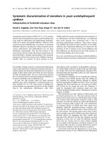
Fig. 2. Coordinated regulation of the espL2 gene on SpLE3, and LEE genes in EHEC O157:H7 Sakai. LEE genes are positively regulated by
Ler, which is encoded in LEE. Transcription of the LEE1 operon, including the ler gene, is positively regulated by Pch, which is encoded by
pchA on another prophage-like element Sp4. Effector genes located outside LEE are regulated by Pch (e.g. espL2, nleB1 and nleE on SpLE3)
or by Ler with Pch (e.g. espJ and tccP on Sp14). All the genes in the Pch–Ler regulon are repressed by H-NS, but once expression of pchA
and ler is stimulated by environmental signals, such as nutrient starvation or butyrate [27,28], transcription of the genes in the regulon are
activated coordinately.
EspL2 effector modulates cytoskeleton T. Tobe
2406 FEBS Journal 277 (2010) 2403–2408 ª 2010 The Author Journal compilation ª 2010 FEBS
The espL2 gene is on prophage-like element SpLE3
in EHEC O157:H7 Sakai, and two other effector
genes, nleB1 and nleE, are downstream of it on the
same element. Expression of these three genes is coor-
dinately regulated by locus of enterocyte effacement
(LEE) genes through the action of Pch [26] (Fig. 2).
LEE genes are composed of five operons and several
cistrons. Transcription of LEE operons and genes is
positively regulated by Ler, which is encoded by the
LEE1 operon, transcription of which is positively reg-
ulated by Pch regulators encoded on prophage-like
elements. The espL2 gene belongs to a subgroup of
the Pch–Ler regulon, expression of which is depen-
dent on Pch but less so on Ler [26]. Indeed, a chro-
matin immunoprecipitation assay showed that Pch,
but not Ler, binds to the chromosomal loci around
the espL2 gene [26]. Interestingly, the region contain-
ing the espL2–nleB1–nleE genes is often found next
to the LEE region. Consequently, espL2, together
with nleB1 and nleE, may be closely associated
with LEE genes in regulation and have roles in
pathogenicity.
Conclusions
Modulation of the cellular cytoskeleton seems to be
essential for establishing the tight adherence of EHEC
and EPEC. A main contributor must be the Tir-medi-
ated polymerization of actin beneath the adherent
bacteria. In addition, EHEC ⁄ EPEC translocate several
effectors that modulate organization of the actin fiber
and cytoskeleton. EspL2 is one effector that can mod-
ify membrane morphology and F-actin organization.
Although EspL2 by itself can not control F-actin orga-
nization, it modulates cytoskeletal and cell morphology
efficiently by enhancing the activity of Annexin A2.
EspL2 may also provide an alternative strategy for
EHEC ⁄ EPEC microcolony formation on enteric
epithelial cells.
Acknowledgments
I wish to thank Hilo Yen for critical reading. This
work was supported by Grants-in–Aid for Scientific
Research on Priority Areas (Applied Genomics) from
The Ministry of Education, Culture, Sports, Science
and Technology of Japan.
References
1 Campellone KG (2010) Cytoskeleton-modulating effec-
tors of enteropathogenic and enterohaemorrhagic
Escherichia coli: Tir, EspF
U
and actin pedestal
assembly. FEBS J 277, 2390–2402.
2 Caron E, Crepin VF, Simpson N, Knutton S, Gar-
mendia J & Frankel G (2006) Subversion of actin
dynamics by EPEC and EHEC. Curr Opin Microbiol
9, 40–45.
3 Hayward RD, Leong JM, Koronakis V & Campellone
KG (2006) Exploiting pathogenic Escherichia coli to
model transmembrane receptor signalling. Nat Rev
Microbiol 4, 358–370.
4 Kaper JB, Nataro JP & Mobley HL (2004) Pathogenic
Escherichia coli. Nat Rev Microbiol 2, 123–140.
5 Hamada D, Hamaguchi M, Suzuki KN, Sakata I &
Yanagihara I (2010) Cytoskeleton-modulating effectors
of enteropathogenic and enterohaemorrhagic Escheri-
chia coli: a case for EspB as an intrinsically less-ordered
effector. FEBS J 277, 2409–2415.
6 Hayashi T, Makino K, Ohnishi M, Kurokawa K, Ishii
K, Yokoyama K, Han CG, Ohtsubo E, Nakayama K,
Murata T et al. (2001) Complete genome sequence of
enterohemorrhagic Escherichia coli O157:H7 and geno-
mic comparison with a laboratory strain K-12. DNA
Res 8, 11–22.
7 Tobe T, Beatson SA, Taniguchi H, Abe H, Bailey CM,
Fivian A, Younis R, Matthews S, Marches O, Frankel
G et al. (2006) An extensive repertoire of type III secre-
tion effectors in Escherichia coli O157 and the role of
lambdoid phages in their dissemination. Proc Natl Acad
Sci USA 103, 14941–14946.
8 Miyahara A, Nakanishi N, Ooka T, Hayashi T,
Sugimoto N & Tobe T (2009) Enterohemorrhagic
Escherichia coli effector EspL2 induces actin micro-
filament aggregation through annexin 2 activation. Cell
Microbiol 11, 337–350.
9 Hayes MJ, Rescher U, Gerke V & Moss SE (2004)
Annexin–actin interactions. Traffic 5, 571–576.
10 Kenny B, Ellis S, Leard AD, Warawa J, Mellor H &
Jepson MA (2002) Co-ordinate regulation of distinct
host cell signalling pathways by multifunctional entero-
pathogenic Escherichia coli effector molecules. Mol
Microbiol 44, 1095–1107.
11 Alto NM, Shao F, Lazar CS, Brost RL, Chua G,
Mattoo S, McMahon SA, Ghosh P, Hughes TR, Boone
C et al. (2006) Identification of a bacterial type III
effector family with G protein mimicry functions. Cell
124, 133–145.
12 Berger CN, Crepin VF, Jepson MA, Arbeloa A &
Frankel G (2009) The mechanisms used by enteropatho-
genic Escherichia coli to control filopodia dynamics.
Cell Microbiol 11, 309–322.
13 Tu X, Nisan I, Yona C, Hanski E & Rosenshine I
(2003) EspH, a new cytoskeleton-modulating effector of
enterohaemorrhagic and enteropathogenic Escherichia
coli. Mol Microbiol
47, 595–606.
T. Tobe EspL2 effector modulates cytoskeleton
FEBS Journal 277 (2010) 2403–2408 ª 2010 The Author Journal compilation ª 2010 FEBS 2407
14 Gerke V, Creutz CE & Moss SE (2005) Annexins: link-
ing Ca
2+
signalling to membrane dynamics. Nat Rev
Mol Cell Biol 6, 449–461.
15 Rescher U, Ruhe D, Ludwig C, Zobiack N & Gerke
V (2004) Annexin 2 is a phosphatidylinositol (4,5)-
bisphosphate binding protein recruited to actin
assembly sites at cellular membranes. J Cell Sci 117,
3473–3480.
16 Zobiack N, Rescher U, Laarmann S, Michgehl S,
Schmidt MA & Gerke V (2002) Cell-surface attachment
of pedestal-forming enteropathogenic E. coli induces a
clustering of raft components and a recruitment of
annexin 2. J Cell Sci 115, 91–98.
17 Jones PG, Moore GJ & Waisman DM (1992)
A nonapeptide to the putative F-actin binding site of
annexin-II tetramer inhibits its calcium-dependent
activation of actin filament bundling. J Biol Chem
267, 13993–13997.
18 Falsey RR, Marron MT, Gunaherath GM, Shirahatti
N, Mahadevan D, Gunatilaka AA & Whitesell L
(2006) Actin microfilament aggregation induced by
withaferin A is mediated by annexin II. Nat Chem Biol
2, 33–38.
19 Brown DA & London E (2000) Structure and function
of sphingolipid- and cholesterol-rich membrane rafts.
J Biol Chem 275, 17221–17224.
20 Lajoie P, Goetz JG, Dennis JW & Nabi IR (2009)
Lattices, rafts, and scaffolds: domain regulation of
receptor signaling at the plasma membrane. J Cell Biol
185, 381–385.
21 Allen-Vercoe E, Waddell B, Livingstone S, Deans J &
DeVinney R (2006) Enteropathogenic Escherichia coli
Tir translocation and pedestal formation requires mem-
brane cholesterol in the absence of bundle-forming pili.
Cell Microbiol 8, 613–624.
22 Ogura Y, Kurokawa K, Ooka T, Tashiro K, Tobe T,
Ohnishi M, Nakayama K, Morimoto T, Terajima J,
Watanabe H et al. (2006) Complexity of the genomic
diversity in enterohemorrhagic Escherichia coli O157
revealed by the combinational use of the O157 Sakai
oligoDNA microarray and the whole genome PCR
scanning. DNA Res 13, 3–14.
23 Ogura Y, Ooka T, Asadulghani, Terajima J, Nougayre-
de JP, Kurokawa K, Tashiro K, Tobe T, Nakayama K,
Kuhara S et al. (2007) Extensive genomic diversity and
selective conservation of virulence-determinants in
enterohemorrhagic Escherichia coli strains of O157 and
non-O157 serotypes. Genome Biol 8, R138.
24 Buchrieser C, Glaser P, Rusniok C, Nedjari H,
d’Hauteville H, Kunst F, Sansonetti P & Parsot C (2000)
The virulence plasmid pWR100 and the repertoire of
proteins secreted by the type III secretion apparatus of
Shigella flexneri. Mol Microbiol 38, 760–771.
25 Nataro JP, Seriwatana J, Fasano A, Maneval DR, Guers
LD, Noriega F, Dubovsky F, Levine MM & Morris JG
Jr (1995) Identification and cloning of a novel plasmid-
encoded enterotoxin of enteroinvasive Escherichia coli
and Shigella strains. Infect Immun 63, 4721–4728.
26 Abe H, Miyahara A, Oshima T, Tashiro K, Ogura Y,
Kuhara S, Ogasawara N, Hayashi T & Tobe T (2008)
Global regulation by horizontally transferred regulators
establishes the pathogenicity of Escherichia coli. DNA
Res 15, 25–38.
27 Nakanishi N, Abe H, Ogura Y, Hayashi T, Tashiro K,
Kuhara S, Sugimoto N & Tobe T (2006) ppGpp with
DksA controls gene expression in the locus of
enterocyte effacement (LEE) pathogenicity island of
enterohaemorrhagic Escherichia coli through activation
of two virulence regulatory genes.
Mol Microbiol 61,
194–205.
28 Nakanishi N, Tashiro K, Kuhara S, Hayashi T,
Sugimoto N & Tobe T (2009) Regulation of virulence
by butyrate sensing in enterohaemorrhagic
Escherichia coli. Microbiology 155, 521–530.
EspL2 effector modulates cytoskeleton T. Tobe
2408 FEBS Journal 277 (2010) 2403–2408 ª 2010 The Author Journal compilation ª 2010 FEBS