Báo cáo khoa học: Glycan profiling of urine, amniotic fluid and ascitic fluid from galactosialidosis patients reveals novel oligosaccharides with reducing end hexose and aldohexonic acid residues ppt
Bạn đang xem bản rút gọn của tài liệu. Xem và tải ngay bản đầy đủ của tài liệu tại đây (727.18 KB, 17 trang )
Glycan profiling of urine, amniotic fluid and ascitic fluid
from galactosialidosis patients reveals novel
oligosaccharides with reducing end hexose and
aldohexonic acid residues
Cees Bruggink
1,2
, Ben J. H. M. Poorthuis
3
, Monique Piraud
4
, Roseline Froissart
4
, Andre
´
M. Deelder
1
and Manfred Wuhrer
1
1 Biomolecular Mass Spectrometry Unit, Department of Parasitology, Leiden University Medical Center, Leiden, The Netherlands
2 Dionex Benelux BV, Amsterdam, The Netherlands
3 Department of Medical Biochemistry, Academic Medical Center, Amsterdam, The Netherlands
4 Laboratoire des Maladies He
´
re
´
ditaires du Me
´
tabolisme et De
´
pistage Ne
´
onatal, Centre de Biologie Est, Hospices Civils de Lyon, Bron,
France
Introduction
Galactosialidosis is an autosomal recessive lysosomal
storage disease, caused by deficiency of both a-neurami-
nidase (EC 3.2.1.18) and b-galactosidase (EC 3.2.1.23)
activities [1], resulting from a defect in the protective
protein cathepsin A (EC 3.4.16.5). This lysosomal
protein protects a-neuraminidase and b-galactosidase
from proteolytic degradation [2] by formation of a
complex involving cathepsin A, b-galactosidase, a-neur-
Keywords
catabolism; clinical glycomics; HPAEC-PAD;
mass spectrometry; metabolic disorder
Correspondence
C. Bruggink, Biomolecular Mass
Spectrometry Unit, Department of
Parasitology, Leiden University Medical
Center, PO Box 9600, 2300 RC Leiden,
The Netherlands
Fax: +31 71 5266907
Tel: +31 71 5266079
E-mail:
Website: />81028091348221/811071049172556/
902270938532556/811120200332556/
(Received 4 March 2010, revised 16 April
2010, accepted 11 May 2010)
doi:10.1111/j.1742-4658.2010.07707.x
Urine, amniotic fluid and ascitic fluid samples of galactosialidosis patients
were analyzed and structurally characterized for free oligosaccharides using
capillary high-performance anion-exchange chromatography with pulsed
amperometric detection and online mass spectrometry. In addition to the
expected endo-b-N-acetylglucosaminidase-cleaved products of complex-type
sialylated N-glycans, O-sulfated oligosaccharide moieties were detected.
Moreover, novel carbohydrate moieties with reducing-end hexose residues
were detected. On the basis of structural features such as a hexose–N-ace-
tylhexosamine–hexose–hexose consensus sequence and di-sialic acid units,
these oligosaccharides are thought to represent, at least in part, glycan
moieties of glycosphingolipids. In addition, C
1
-oxidized, aldohexonic acid-
containing versions of most of these oligosaccharides were observed. These
observations suggest an alternative catabolism of glycosphingolipids in
galactosialidosis patients: oligosaccharide moieties from glycosphingolipids
would be released by a hitherto unknown ceramide glycanase activity. The
results show the potential and versatility of the analytical approach for
structural characterization of oligosaccharides in various body fluids.
Abbreviations
F, deoxyhexose; GluconA, gluconic acid; GD1b, Gal(b1-3)GalNAc(b1-4)(Neu5Ac(a2-8) Neu5Ac(a2-3))Gal(b1-4)Glc; GD3, Neu5Ac
(a2-8)Neu5Ac(a2-3)Gal(b1-4)Glc; GM1, Neu5Ac(a2-3)Gal(b1-3)GalNAc(b1-4)Gal(b1-4)Glc; GM2, GalNAc(b1-4)(Neu5Ac(a2-3))Gal(b1-4)Glc;
H or Hex, hexose; HexSO
3
, O-sulfated hexose; HPAEC, high-performance anion-exchange chromatography; MS, mass spectrometry;
N or HexNAc, N-acetylhexosamine; PAD, integrated pulsed amperometric detection; S or Neu5Ac, N-acetylneuraminic acid; SO
3
, sulfate;
X or HexonA, aldohexonic acid.
2970 FEBS Journal 277 (2010) 2970–2986 ª 2010 The Authors Journal compilation ª 2010 FEBS
aminidase and N-acetylgalactosamine-6-sulfate sulfatase
(EC 3.1.6.4) [3,4].
Galactosialidosis is characterized by excessive excre-
tion of sialyloligosaccharides in the urine, an increase in
the amount of bound sialic acid in various tissues, and
severe clinical symptoms [5,6]. Three clinical subtypes
can be distinguished, depending on the age of onset and
severity of the symptoms: the early infantile type with
fetal hydrops, ascites, visceromegaly, skeletal dysplasia
and early death, usually by 8–12 months of age; the late
infantile type with cardiac involvement, hepatospleno-
megaly, growth retardation and mild mental retarda-
tion; and the juvenile ⁄ adult type with progressive
neurological deterioration without visceromegaly.
Coarse faces, cherry red spots in the macula and verte-
bral changes are usually present [7,8]. Biochemical diag-
nosis is made by demonstration of increased excretion
of oligosaccharides by thin layer chromatography [9]
and by demonstrating a combined deficiency of a-neur-
aminidase and b-galactosidase in patient cells.
Several activity studies on the structural analysis of
sialyloligosaccharides from urine of galactosialidosis
patients [10,11] have been published. van Pelt et al.
[12] described 21 sialylated oligosaccharides. Twenty
of these were endo-b-N-acetylglucosaminidase-cleaved
products of complex-type sialylated N-glycans, and
one was a di-sialylated diantennary structure with an
intact N,N¢-diacetylchitobiose unit at the reducing end.
Here we report the analysis of oligosaccharides from
galactosialidosis patients using a previously described
capillary high-performance anion-exchange chromatog-
raphy (HPAEC) method with combined integrated
pulsed amperometric (PAD) and ion-trap mass spec-
trometric detection and analysis [13]. In addition to
urine samples, ascitic fluid and amniotic fluid obtained
from mothers pregnant with a galactosialidosis fetus
were analyzed. Amniotic fluid is of importance for pre-
natal diagnosis of many lysosomal storage disorders
such as galactosialidosis [14].
In addition to the expected endo-b-N-acetylglucosa-
minidase-cleaved products of complex-type sialylated
N-glycans, oligosaccharide structures that had not been
previously found were detected in the samples from
galactosialidosis patients. These newly found oligosac-
charide structures included O-sulfated oligosaccharide
moieties, carbohydrate moieties of glycosphingolipids,
and C
1
-oxidized (aldohexonic acid) carbohydrate moie-
ties of glycosphingolipids. On the basis of the presence
of carbohydrate moieties of glycosphingolipids, we
speculate about the potential involvement of a cera-
mide glycanase in the catabolism of glycosphingolipids
in humans.
Results
Glycans from seven urine samples from six galacto-
sialidosis patients, five amniotic fluid samples from
five mothers carrying a fetus suffering from galacto-
sialidosis, and two ascitic fluid samples were analyzed
by HPAEC-PAD-MS (Table 1). In addition, four
urine samples from healthy individuals were investi-
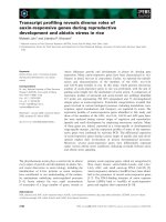
gated. Figure 1 shows a typical HPAEC-PAD chro-
matogram from a urine sample of a galactosialidosis
patient.
N-glycan-derived structures
The typical endo-b-N-acetylglucosaminidase cleavage
products of complex-type N-sialyloligosaccharides were
found in all urine samples, amniotic fluid samples and
ascitic fluid samples (see Fig. 2, n1–n6) [12]. A varying
number of isomers were detected for the various N-gly-
can compositions, and these were analyzed by MS ⁄ MS,
as summarized in Table 2. N-glycan-derived structure
Table 1. Information about the samples and patients. ND, not detected.
Sample code Details Creatinine (m
M)
U1 Urine from patient AB, 12 days old (Lyon, France) 1.0
U2 Urine from patient AV, 6 days old (Lyon, France) 2.3
U3, U4 Urine from patient MO (Lyon, France) ND
U5 Urine from patient BO, 127 days old (Lyon, France) 1.2
U6 Urine from patient B07 ⁄ 0175 (Amsterdam, The Netherlands) 1.6
U7 Urine from patient B07 ⁄ 0845.1, 8 weeks old (Leiden, The Netherlands) 0.5
Amfl1 Amniotic fluid from patient AB, 30 week fetus (Lyon, France) ND
Amfl2 Amniotic fluid from patient AS, 29 week fetus (Lyon, France) ND
Amfl3 Amniotic fluid from patient W, 23 weeks of amenorrhoea (Lyon, France) ND
Amfl4 Amniotic fluid from patient LA, 22 week fetus (Lyon, France) ND
Amfl5 Amniotic fluid from patient GG, protein 3.5 gÆL
)1
(Nijmegen, The Netherlands) 0.08
Asf1 Ascite fluid from patient AB (Lyon, France) ND
Asf2 Ascite fluid from patient AS (Lyon, France) ND
C. Bruggink et al. Novel oligosaccharides in galactosialidosis
FEBS Journal 277 (2010) 2970–2986 ª 2010 The Authors Journal compilation ª 2010 FEBS 2971
n1 had the composition HNS (H, hexose; N, N-acetyl-
hexosamine; S, N-acetylneuraminic acid), and two iso-
mers of n1 were detected. Tandem mass spectometry
indicated the structure Neu5Ac(a2–3 ⁄ 6)Gal(b1–4)
GlcNAc. On the basis of chromatographic retention [15]
in combination with the tandem mass spectrometric
data [16], we speculate that N-acetylneuraminic acid
(Neu5Ac) is (a2–6)-linked in the first n1 isomer and
(a2–3)-linked in the second isomer. Specifically, the rela-
tively low signal intensity of the fragment ion at m ⁄ z
655.2 from the second eluting isomer [16] suggests an
a2–3-linked Neu5Ac.
Moreover, larger complex sialyloligosaccharides
were found with the composition H
3–6
N
2–4
S
1–3
.In
accordance with literature data [12], we interpreted the
three isomers H
3
N
2
S as sialyl-mono antennary endo-b-
N-acetylglucosaminidase cleavage products of com-
plex-type N-glycan structures (Fig. 2, n2). Similarly,
the two isomers H
5
N
3
S were assigned to sialylated
diantennary structures (Fig. 2, n3), the two isomers
H
5
N
3
S
2
as di-sialylated diantennary structures (Fig. 2,
n4), the two isomers H
6
N
4
S
2
as di-sialylated trianten-
nary structures (Fig. 2, n5), and the three isomers
H
6
N
4
S
3
as tri-sialylated triantennary structures (Fig. 2,
n6). These assignments were corroborated by the
MS ⁄ MS data (Table 2).
In addition to the expected endo-b-N-acetylglucosa-
minidase-cleaved products of complex-type sialylated
N-glycans, some O-sulfated versions were also found in
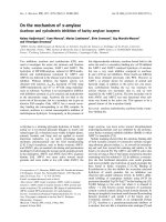
low amounts (see Table 3 and Fig. 2, s1–s4). The
detected carbohydrate HSO
3
NS eluted in the time win-
dow for double negatively charged carbohydrates
(Fig. 1). The MS ⁄ MS fragment ions Y
1
(m ⁄ z 219.9)
and Y
2
(m ⁄ z 462.0) indicated the sequence Neu5Ac–
HexSO
3
HexNAc (Fig. 3). The
0,2
A
3
ring fragment ion
at m ⁄ z 652.1 is typical of a 1–4 glycosidic link [16,17]
between HexSO
3
and HexNAc. The lack of significant
fragment ions between the fragment ions Y
1
and Y
2
is indicative of a 2–3 linkage between Neu5Ac and
HexSO
3
. These data are consistent with a Neu5Ac(a2–3)
Gal-6-SO
3
(b1–4)GlcNAc N-glycan antenna structure or
O-glycan structural motif [18]. Moreover, the presence
of complex O-sulfated sialylated oligosaccharides
with the composition H
3–5
SO
3
N
2–3
S
1–2
(see Table 2),
was indicated by MS. Based on observed retention
times, mass spectrometric data (Table 2) and literature
data, these glycans were assigned to sulfated variants of
the above-mentioned endo-b-N-acetylglucosaminidase
cleavage products of complex-type sialylated N-glycan
structures: the two isomers of composition H
3
SO
3
N
2
S
were assigned to O-sulfated sialylated monoantennary
glycans (Fig. 2, s2), the four isomers H
5
SO
3
N
3
Sas
Fig. 1. Capillary HPAEC-PAD chromatogram of oligosaccharides from a urine sample of a galactosialidosis patient. H, hexose; N, N-acetyl-
hexosamine; S, N-acetylneuraminic acid; X, aldohexonic acid. The numbers above the horizontal arrows represents the number of acidic
groups.
Novel oligosaccharides in galactosialidosis C. Bruggink et al.
2972 FEBS Journal 277 (2010) 2970–2986 ª 2010 The Authors Journal compilation ª 2010 FEBS
O-sulfated monosialylated diantennary glycans (Fig. 2,
s3), and the two isomers H
5
SO
3
N
3
S
2
as O-sulfated di-
sialylated diantennary glycans (Fig. 2, s4).
Glycans with reducing-end hexoses
In addition to the N-glycan-derived signals, the LC-MS ⁄
MS data provided evidence for the presence of a group
of oligosaccharides of composition H
0–3
N
0–1
S
0–2
(g1–g11, Table 2). Tandem mass spectrometry indicated
a sequence Hex–HexNAc–Hex–Hex or truncated
versions thereof for most of these oligosaccharides,
decorated with up to two Neu5Ac. Di-sialyl motifs
(Neu5Ac linked to Neu5Ac) were also observed. Struc-
tural characterization of these oligosaccharides is
described below.
Two isomers of the glycan H
2
were detected. The
retention time of the late-eluting H
2
isomer was identi-
cal to that of maltose (Glc(a1–4)Glc; Table 2). The
retention time of the early-eluting H
2
isomer was iden-
tical to that of lactose, and Fig. 4A shows the MS ⁄ MS
spectrum obtained. Fragment ion C
1
(m ⁄ z 178.9) indi-
cates the composition H
2
and the ring fragment ion
(m ⁄ z 220.8) corresponds to a loss of 120, which is
interpreted as a
2,4
A
2
ring fragment typical of a 1–4
linkage between the hexoses [16,17].
Four isomers were found with the composition H
2
S
(Table 2). The MS ⁄ MS spectrum of the first eluting
isomer with retention time of 10.5 min is shown in
Fig. 4B. The fragment ions B
1
,C
2
,Y
1
and Y
2
indicate
the sequence Neu5Ac–Hex–Hex. The ring fragments
0,2
A
3
and
0,2
A
3
-18 in combination with lack of the
0,3
A
3
ring fragment ion are typical of a 1–4-linkage
between the hexoses [16,17]. The lack of relevant ring
fragment ions between fragment ions B
1
and C
2
is
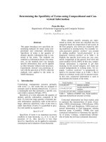
Fig. 2. Schematic overview of the proposed structures of free oligosaccharides in body liquids from galactosialidosis patients. The codes
n1–n6, s1–s4, g1–g11 and o1–o9 refer to Tables 2 and 3.
Fig. 3. Negative-ion fragmentation spectrum of the proposed
6¢-sulfated sialyl lactosamine.
C. Bruggink et al. Novel oligosaccharides in galactosialidosis
FEBS Journal 277 (2010) 2970–2986 ª 2010 The Authors Journal compilation ª 2010 FEBS 2973
Table 2. Structural data for detected oligosaccharide moieties.
Glycan
composition Species
Retention
time (min) Signal m ⁄ z MS ⁄ MS fragment ions References Proposed structure
HNS n1 22.3 673.6 [M–H]
)
655.2-H
2
O; 572.2
0,2
A
3
; 544.2
0,2
A
3
-H
2
O; 512.1
2,4
A
3
; 470.1 C
2
;
452.1 B
2
; 410.1
0,2
A
2
;392.2
0,2
A
2
-H
2
O; 380.2
0,3
A
2
; 350.1
0,4
A
2
;
332.1
0,4
A
2
-H
2
O; 308.0 C
1
; 290.0 B
1
Fig. 2, n1 Neu5Ac(a2–6)Gal(b1–4)Glc
NAc
26.9 673.6 [M–H]
)
655.2-H
2
O; 572.2
0,2
A
3
; 544.2
0,2
A
3
-H
2
O; 512.0
2,4
A
3
; 470.1 C
2
;
452.2 B
2
; 410.2
0,2
A
2
;392.2
0,2
A
2
-H
2
O; 380.0
0,3
A
2
; 308.0 C
1
;
290.0 B
1
Fig. 2, n1 Neu5Ac(a2–3)Gal(b1–4)
GlcNAc
H
3
N
2
S n2 22.3 1200.4 [M–H]
)
1182.6-H
2
O; 1122.5; 1099.4
0,2
A
6
; 1081.3
0,2
A
6
-H
2
O; 998.2;
979.3 B
5
; 943.3; 937.3
0,2
A
5
; 835.4 C
4
; 818.5; 747.9
2,4
A
5
Y
5
;
728.8 Z
4
; 686.6
2,4
X
4
; 655.0 B
3
; 536.2
Fig. 2, n2 Neu5Ac(a2–3 ⁄ 6)
Gal(b1–4)GlcNAc(b1–2)Man
(a1–6)Man(b1–4)GlcNAc
22.5 1200.4 [M–H]
)
;
1298.5 [M+HSO
4
]
)
1182.6-H
2
O; 1122.5; 1099.5
0,2
A
6
; 1081.5
0,2
A
6
-H
2
O; 1039.5
2,4
A
6
; 997.5 C
5
; 979.6 B
5
; 835.4 C
4
; 817.4 B
4
; 748.4
2,4
A
5
Y
5
;
673.4 C
3
; 655.4 B
3
; 572.3
0,2
A
3
; 526.1
3,5
A
3
; 470.2 C
2
; 452.2
B
2
; 424.2
1,5
A
2
; 410.1
0,2
A
3
Fig. 2, n2 Neu5Ac(a2–6)Gal(b1–4)
GlcNAc(b1–2)Man(a1–3)
Man(b1–4)GlcNAc
23.3 1200.4 [M–H]
)
;
1298.5 [M+HSO
4
]
)
1182.4-H
2
O; 1165.1; 1122.5; 1099.4
0,2
A
6
; 1081.3
0,2
A
6
-H
2
O;
1063.4; 1039.3
2,4
A
6
; 1021.4; 997.3 C
5
; 979.2 B
5
; 961.2;
910.4; 835.2 C
4
; 819.2
0,3
X
5
; 817.2 B
4
; 784.4; 779.4; 791.1;
775.4
0,2
A
4
; 773.4; 748.2
2,4
A
5
Y
5
; 744.3; 696.3; 674.0; 672.4;
655.3 B
3
; 619.1; 592.3; 586.1; 568.0; 554.2
0,2
A
3
-H
2
O; 536.1;
526.1
3,5
A
3
; 424.1
1,5
A
2
; 381.1
Fig. 2, n2 Neu5Ac(a2–3)Gal(b1–4)
GlcNAc(b1–2)Man(a1–3)
Man(b1–4)GlcNAc
H
5
N
3
S n3 23.7 1727.8 [M–H]
)
1709.8-H
2
O; 1668.8
2,4
X
5
; 1626.7
0,2
A
6
; 1608.7
0.2
A
6
-H
2
O;
1566.7
2,4
A
6
; 1524.7 C
5
; 1506.8 B
5
; 1316.6
0,2
X
5b
Y
5a
; 1275.5
2,4
A
6
Y
5a
; 1113.6
2,4
A
6
Y
4a
; 1053.7 Z
3a
; 979.3 C
5a
Z
2b
; 961.3
B
5a
Z
2b
; 835.3 C
4a
; 817.4 B
4a
Fig. 2, n3 Neu5Ac(a2–3 ⁄ 6)
Gal(b1–4)GlcNAc(b1–2)
Man (a1–6)[Gal(b1–4)
GlcNAc(b1–2)
Man(a1–3)]Man (b1–4)
GlcNAc
27.6 1727.8 [M–H]
)
Fig. 2, n3
H
5
N
3
S
2
n4 27.3 1009.0 [M–2H]
2)
1709.6 Z
5ab
; 1626.6
0,2
A
6
Y
5a
; 1548.7
2,4
A
6
Z
5a
;C
5
Y
5a
; 1050.5;
1000.3-H
2
O; 958.9
0,2
A
6
; 907.8
0,3
A
5b
; 835.6 C
4
; 817.7 B
4
;
655.6 B
3
; 290.2 B
1
Fig. 2, n4 Neu5Ac(a2–6)Gal(b1–4)
GlcNAc(b1–2)Man(a1–6)
[Neu5Ac(a2–6)
Gal(b1–4)GlcNAc
(b1–2)Man(a1–3)]
Man(b1–4)GlcNAc
28.0 1009.0 [M–2H]
2)
1797.6 B
5
; 1727.6 Y
5
; 1709.6 Z
5
; 1626.6
0,2
A
6
Y
5
; 1608.5
0,2
A
6
Z
5
; 1566.4
2,4
A
6
Y
5
; 1524.5 C
5
Y
5
; 1000.4-H
2
O; 958.8
0,2
A
6
;
907.4
0,3
A
5b
; 835.3 C
4
; 817.3 B
4
; 673.2 C
3
; 655.3 B
3
; 452.1 B
2
;
424.1
1,5
A
2
; 410.1
0,2
A
2
; 350.0
0,4
A
2
; 307.9 C
1
; 290.0 B
1
Fig. 2, n4 Neu5Ac(a2–3 ⁄ 6)Gal
(b1–4)GlcNAc(b1–2)Man
(a1–6)[Neu5Ac(a2–
3 ⁄ 6)Gal(b1–4)GlcNAc(b1–2)
Man(a1–3)]Man(b1–4)
GlcNAc
H
6
N
4
S
2
n5 27.4 1191.4 [M–2H]
2)
Fig. 2, n5
31.4 1191.4 [M–2H]
2)
Fig. 2, n5
Novel oligosaccharides in galactosialidosis C. Bruggink et al.
2974 FEBS Journal 277 (2010) 2970–2986 ª 2010 The Authors Journal compilation ª 2010 FEBS
Table 2. (Continued).
Glycan
composition Species
Retention
time (min) Signal m ⁄ z MS ⁄ MS fragment ions References Proposed structure
H
6
N
4
S
3
n6 31.3 891.3 [M–2H]
3)
;
1337.4 [M–2H]
2)
2075.8; 2074.9 Z
6
Y
6
or Y
6
Z
6
; 2016.0; 1995.1; 1992.9; 1974.6;
1973.8; 1931.8; 1890.6; 1889.8; 1871.8; 1608.4; 1474.4;
1473.6 B
4a
; 1328.7 –H
2
0; 1279.1
1,4
X
6abc
; 1203,4; 1202.3;
1201.3; 1200.5 C
4a
Y
5a
; 1192.1; 1191.4 Y
5
; 1184.7; 1183.9;
1182.4 Z
5
; 1152.8; 1142.7; 1141.0; 1133.6; 1132.8; 1111.5;
1111.1; 1092.6; 1090.0 C
5
Y
5a
; 1081.4; 1039.7; 1009.3; 1000.3;
981.0; 979.2 B
5
Y
2a
; 963.3; 962.2 B
5
Z
2a
; 944.4; 907.2 C
5
Y
3a
;
885.5 –H
2
O; 879.0; 860.0; 857.3
0,2
A
6
; 852.0
2,5
X
6
; 851.1
0,4
X
6
;
838.1; 837.4
2,4
A
6
; 835.5 C
4
; 823.3 C
5
; 817.2 B
4
; 798.3; 775.2
0,2
A
4
; 750.0; 745.3 C
4a
; 737.2; 736.1; 673.3 C
3
; 655.2 B
3
;
536.1; 470.1 C
2
; 424.1
1,5
A
2
; 306.2; 290.0 B
1
Fig. 2, n6 Neu5Ac(a2–3)Gal(b1–4)
GlcNAc(b1–2)Man(a1–6)
[Neu5Ac(a2–3)Gal
(b1–4)GlcNAc
(b1–2)][Neu5Ac
(a2–6)Gal(b1–4)GlcNAc
(b1–4))Man(a1–3)]Man
(b1–4)GlcNAc
32.4 891.3 [M–2H]
3)
Fig. 2, n6
34.6 891.3 [M–2H]
3)
Fig. 2, n6
HSO
3
NS s1 32.0 753.2 [M–H]
)
;
851.1 [M+HSO
4
]
)
709.1
1,3
X
3
; 652.1
0,2
A
3
; 638.2
1,4
X
3
; 469.9 C
2
; 462.0 Y
2
; 444.2
Z
2
; 370.0; 361.1
0,2
A
3
Y
2
; 352.0; 343.0
0,2
A
3
Z
2
; 331.9 B
2
0,4
X
3
-SO
3
; 301.0
2,4
A
3
Y
2
; 276.8 B
2
3,5
X
3
-SO
3
; 263.8
0,2
A
3
Z
2
-SO
3
;
258.9 C
2
Y
2
; 248.9 C
2
0,2
X
3
-SO
3
; 240.9 C
2
Z
2
; 219.9 Y
1
Fig. 2, s1 NeuAc(a2–3)Gal(6S)
(b1–4)GlcNAc
H
3
SO
3
N
2
S s2 36.5 639.7 [M–2H]
2)
990.2; 989.2 Y
5
; 987.0
0,3
A
5
; 971.1
3.5
A
5
; 951.2
1,5
A
5
-SO
3
;
915.3 C
4
; 890.2; 888.2
0,2
A
6
Y
5
; 886.1 C
5
0,3
X
6
; 871.5 C
4
1,3
X
6
;
870.1
0,2
A
6
Z
5
; 829.5; 828.2
2,4
A
6
Y
5
; 786.1 C
5
Y
5
; 768.0 C
5
Z
5
;
726.2 C
5
1,3
X
5
; 693.9 C
4
0,2
X
6
; 655.1 B
3
-SO
3
; 647.8
2,4
A
5
Z
5
;
630.6-H
2
O; 629.5 C
3
1,3
X
6
-SO
3
; 624.1 C
4
Y
5
; 622.2
0,3
A
3
; 620.3
B
3
1,4
X
6
; 611.3 B
3
1,3
X
6
-SO
3
; 600.5
3,5
A
6
Y
4
; 594.1; 589.1
0,2
A
6
;
588.1 B
4
Z
5
; 580.1; 571.9 C
5
1,5
X
5
; 559.0
2,4
A
6
; 541.1
2,5
A
6
-SO
3
;
538.6; 58.1 C
5
; 537.0
2,5
X
6
; 529.1 B
5
; 519.1
2,4
X
6
-SO
3
; 516.1
C
5
1,3
X
6
; 484.0 C
5
2,4
X
4
; 478.5
0,4
A
5
; 470.0 C
2
; 457.0 C
4
; 449.0
C
2
2,4
X
6
; 448.1 B
4
; 444.0 C
3
Z
5
; 427.5 C
5
0,2
X
6
; 418.8 B
5
0,2
X
6
;
398.0
1,5
A
3
Z
5
; 397.6 B
4
2,4
X
6
; 397.0
3,5
A
6
Y
3
; 380.0
1,4
A
2
; 375.9
C
3
; 357.1 B
2
3,5
X
6
; 302.1
3,5
A
3
; 292.7
2,4
A
6
Y
5
; 290.0 B
1
; 215.3
B
2
2,4
X
6
; 210.8 B
4
2,5
X
4
Fig. 2, s2 Neu5Ac(a2–3)Gal(6S)
(b1–4)GlcNAc(b1–2)Man
(a1–6)Man(b1–4)GlcNAc
38.9 639.7 [M–2H]
2)
Fig. 2, s2 Neu5Ac(a2–3 ⁄ 6)Gal(6S)
(b1–4)GlcNAc(b1–2)Man
(a1–3)Man(b1–4)GlcNAc
H
5
SO
3
N
3
S s3 34.6 903.3 [M–2H]
2)
1446.3
0,2
A
5
Z
4b
; 886.4; 859.2; 826
3,5
X
5b
; 774.1
2,5
A
6
Z
2a
; 757.6
E
3
-ion; 638.1 B
5
Z
5a
; 613.2
2,5
A
6
Z
3b
; 612.2 B
4a
2,5
X
6a
; 595.0;
594.2
3,5
A
6
Z
4a
; 483.1 Y
3a
2,5
X
5b
; 308.1 C
1
; 290.3 B
1
Fig. 2, s3 Gal(6S)(b1–4)GlcNAc
(b1–2)Man(a1–6)[Neu5Ac
(a2–3)Gal(b1–4)GlcNAc
(b1–2)Man(a1–3)]Man
(b1–4)GlcNAc
39.3 903.3 [M–2H]
2)
Fig. 2, s3
41.4 903.3 [M–2H]
2)
Fig. 2, s3
C. Bruggink et al. Novel oligosaccharides in galactosialidosis
FEBS Journal 277 (2010) 2970–2986 ª 2010 The Authors Journal compilation ª 2010 FEBS 2975
Table 2. (Continued).
Glycan
composition Species
Retention
time (min) Signal m ⁄ z MS ⁄ MS fragment ions References Proposed structure
45.4 903.3 [M–2H]
2)
Fig. 2, s3
H
5
SO
3
N
3
S
2
s4 39.6 1048.8 [M–2H]
2)
Fig. 2, s4 Neu5Ac(a2–3 ⁄ 6)Gal(6S)
(b1–4)GlcNAc(b1–2)Man
(a1–6)[Neu5Ac
(a2–3 ⁄ 6)Gal(b1–4)
GlcNAc(b1–2) Man
(a1–3)]Man(b1–4)GlcNAc
H
2
g1 7.6 341.2 [M–H]
)
323.0–H
2
O; 220.8
2,4
A
2
; 178.9 C
1
; 160.9 B
1
Fig. 2, g1;
Fig. 4A
Gal(b1–4)Glc
8.2 341.2 [M–H]
)
;
439.1 [M+HSO
4
]
)
281.0
0,2
A
2
; 235.0
3,5
A
2
; 220.8
2,4
A
2
; 178.9 C
1
; 160.9 B
1
Glc(a1–4)Glc
HS g2 24.4 470.2 [M–H]
)
410.0
0,2
A
2
; 379.9
0,3
A
2
; 370.0; 308.0 C
1
; 290.0 B
1
; 271.8;
220.0; 194.8; 169.9
NeuAc(a2–6)Gal
25.1 470.2 [M–H]
)
;
568.2 [M+HSO
4
]
)
357.8; 307.8 C
1
; 290.0 B
1
; 272.9; 269.7; 219.8; 201.7;
173.8; 169.9
Fig. 2, g2 NeuAc(a2–3)Gal
H
3
g3 7.6 503.2 [M–H]
)
;
601.2 [M+HSO
4
]
)
369.2
1,5
X
3
; 341.1 C
2
; 323.1 B
2
; 281.0
0,2
A
2
; 262.8
0,2
A
2
-H
2
O;
234.8
3,5
A
2
; 221.0
2,4
A
2
; 202.9
2,4
A
2
-H
2
O; 178.9 C
1
; 160.9 B
1
Hex(1–4)Hex(1–3)Hex
9.1 503.2 [M–H]
)
;
601.2 [M+HSO
4
]
)
341.1 C
2
; 323.1 B
1
; 250.9
0,3
A
2
; 220.9
0,4
A
2
; 178.9 C
1
; 160.9 B
1
Hex(1–6)Hex(1–3)Hex
20.4 503.2 [M–H]
)
;
601.2 [M+HSO
4
]
)
443.4
0,2
A
3
; 424.7
0,2
A
3
-H
2
O; 383.1
2,4
A
3
; 341.1 C
2
;322.8 B
2
;
295.4
1,5
A
2
; 280.9
0,2
A
2
; 237.0
2,5
X
2
; 234.7
3,5
A
2
; 220.8
2,4
A
2
;
178.9 C
1
; 160.8 B
1
Fig. 2, g3 Gal(a1–4)Gal(b1–4)Glc
S
2
g4 27.7 599.2 [M–H]
)
;
697.2 [M+HSO
4
]
)
581.1 –H
2
O; 511.1
0,2
A
2
; 495.0
2,5
A
2
; 410.0
0,4
A
2
; 380.0
1,5
X
2
;
308.0 C
1
; 290.0 B
1
Neu5Ac(a2–8)Neu5Ac
25.5 599.2 [M–H]
)
H
2
N g5 9.6 544.2 [M–H]
)
;
642.2 [M+HSO
4
]
)
526.1; 383.0; 290.0; 271.9; 169.9 Fig. 2, g5 GlcNAc(b1–4)Gal(b1–4)Glc
H
2
S g6 10.5 632.2 [M–H]
)
;
730.2 [M+HSO
4
]
)
614.0-H
2
O; 588.1
1,3
X
3
; 572.1
0,2
A
3
; 554.0
0,2
A
3
-H
2
O; 535.9;
470.0 C
2
; 411.0
0,2
X
3
; 408.0; 385.9; 341.1 Y
2
; 290.0 B
1
;
178.9 Y
1
Fig. 2, g6;
Fig. 4B
Neu5Ac(a2–3)Gal(b1–4)Glc
13.3 632.2 [M–H]
)
;
730.2 [M+HSO
4
]
)
614.0-H
2
O; 598.6; 588.1
1,3
X
3
; 534.3; 532.3; 472.1; 470.9;
469.9 C
2
; 456.2; 411.0
0,2
X
3
; 341.1 Y
2
; 307.9 C
1
; 290.0 B
1
;
178.9 Y
1
Neu5Ac(a2–3)Hex(1–3)Hex
or Neu5Ac(a2–3)Gal(1–3)Gal
14.6 632.2 [M–H]
)
;
730.2[M+HSO4]
)
614.0-H
2
O; 599.1; 588.1
1,3
X
3
; 535.0; 534.2; 524.1; 472.2;
470.1 C
2
; 411.1
0,2
X
3
; 434.0; 416.1; 411.0; 408.0 B
2
1,3
X
3
;
404.0; 386.0; 341.0 Y
2
; 337.0 B
2
1,4
X
3
; 305.9
0,4
A
2
-CO
2
; 292.0;
290.0 B
1
; 178.8 Y
1
Neu5Ac(a2–6)Hex(1–3)Hex
or Neu5Ac(a2–6)Gal
(b1–3)Glc
Novel oligosaccharides in galactosialidosis C. Bruggink et al.
2976 FEBS Journal 277 (2010) 2970–2986 ª 2010 The Authors Journal compilation ª 2010 FEBS
Table 2. (Continued).
Glycan
composition Species
Retention
time (min) Signal m ⁄ z MS ⁄ MS fragment ions References Proposed structure
20.9 632.2 [M–H]
)
;
730.2 [M+HSO
4
]
)
614.0-H
2
O; 572.1
0,2
A
3
; 571.2; 554.1
0,2
A
3
-H
2
O; 536.0; 512.1
2,4
A
3
;
494.0
2,4
A
3
-H
2
O; 472.0; 470.1 C
2
; 468.1; 452.0 B
2
; 441.0
0,3
X
3
;
411.0
0,2
X
3
; 410.0
0,2
A
2
; 408.1; 392.1
0,2
A
2
-H
2
O; 380.0
0,3
A
2
;
350.0
0,4
A
2
; 334.0; 332.0; 316.0; 308.0 C
1
; 306.0
0,4
A
2
-CO
2
;
290.0 B
1
Neu5Ac(a2–6)Hex(1–4)Glc or
Neu5Ac(a2–6)Gal(b1–4)Glc
H
3
N g7 9.5 706.2 [M–H]
)
;
804.3 [M+HSO
4
]
)
688.0-H
2
O or D-ion; 646.2
0,2
A
4
; 628.1
0,2
A
4
-H
2
O; 585.6
0,4
A
4
;
544.1 C
3
; 424.2
1,3
A
3
; 382.0 C
2
; 280.9
0,2
A
2
; 263.0
0,2
A
4
Z
2
Gal(b1–4)GlcNAc
(b1–2)Man(a1–6)Man
12.7 706.2 [M–H]
)
;
804.3 [M+HSO
4
]
)
;
902.3 [M+HSO
4
]
)
646.3
0,2
A
4
; 628.1
0,2
A
4
-H
2
O; 543.9 C
3
;381.9 C
2
;363.5 B
2
;202.1
C
2
Z
3
Fig. 2, g7;
Fig. 4D
Gal(b1–3)GalNAc
(b1–4)Gal(b1–4)Glc
H
2
NS g8 22.8 835.3 [M–H]
)
817.3-H
2
O; 715.2
2,4
A
3
; 673.0 C
3
; 655.3 B
3
; 572.0
0,2
A
3
Y
2b
;
494.2
2,4
A
3
Z
2b
; 290.1 B
1a
Fig. 2, g8 GalNAc(b1–4)[Neu5Ac
(a2–3)]Gal(b1–4)Glc
H
2
S
2
g9 29.1 923.3 [M–H]
)
632.2 Y
3
; 581.1 B
2
; 538.1 B
3
2,5
X
4
; 379.9 C
2
1,5
X
4
; 290.0 B
1
;
178.9 Y
1
Fig. 2, g9;
Fig. 4C
Neu5Ac(a2–8)Neu5Ac
(a2–3)Gal(b1–4)Glc
H
3
NS g10 22.0 997.3 [M–H]
)
;
1095.3 [M+HSO
4
]
)
979.2 –H
2
O; 937.2
0,2
A
5
; 920.1; 919.2
0,2
A
5
-H
2
O; 877.0
2,4
A
5
;
835.1 C
4
; 818.4; 776.5
0,2
A
4
; 717.1; 715.1
2,4
A
4
; 673.2 C
3
;
655.3 B
3
; 595.1; 586.1; 572.0
1,5
X
4
; 555.4; 554.3 B
3
2,4
X
5
;
526.2 Z
3
; 511.9
2,4
A
3
; 470.1 C
2
; 452.2 B
2
; 379.8; 383.0; 351.0
B
2
2,4
X
5
; 332.0 B
2
0,4
X
5
; 308.0 C
1
; 290.0 B
1
Fig. 2,
g10;
Fig. 4E
Neu5Ac(a2–3)Gal(b1–3)
GalNAc(b1–4)Gal(b1–4)
Glc
24.0 997.3 [M–H]
)
;
1095.3
[M+HSO
4
]
)
H
3
NS
2
g11 29.5 643.7 [M–2H]
2)
999.4; 998.2; 997.4 Y
3a
; 563.6 B
2a
-H
2
O; 562.6 C
3
; 471.1; 290.0
B
1a
; 271.9 B
1a
-H
2
O
Fig. 2, g11 Gal(b1–3)GalNAc
(b1–4)[Neu5Ac
(a2–8)Neu5Ac
(a2–3)]Gal(b1–4)Glc
X o1 13.3 195.1 [M–H]
)
Fig. 2, o1 GluconA
HX o2 19.2 357.2 [M–H]
)
;
455.1 [M+HSO
4
]
)
339.1-H
2
O; 321.0; 297.1
2,4
X
2
; 277.2; 258.7; 237.0
0,2
X
2
;
220.9
2,4
A
2
; 195.0 Y
1
; 178.9 C
1
; 176.9 Z
1
; 160.9 B
1
;
158.9 Z
1
-H
2
O
Fig. 2, o2;
Fig. 5A
Gal(b1–4)GluconA
SX o3 26.4 486.1 [M–H]
)
Fig. 2, o3
H
2
X o4 22.6 519.2 [M–H]
)
;
617.1 [M+HSO
4
]
)
382.7
2,4
A
3
; 356.9 Y
2
; 297.5
2,4
X
2
or
0,2
A
3
Y
2
; 221.0
2,4
A
2
; 177.1
Z
1
; 161.0 B
1
Fig. 2, o4 Gal(a1–4)Gal(b1–4)GluconA
HNX o5 24.3 560.2 [M–H]
)
;
658.2 [M+HSO
4
]
)
406.8; 399.0; 398.1; 396.0; 394.8; 323.0; 235.8; 179.0; 160.9 Fig. 2, o5
38.2 560.2 [M–H]
)
543.0; 540.9; 516.0; 480.0; 463.0; 462.2; 445.1; 349.2; 348.1;
345.0; 284.8
Fig. 2, o5
C. Bruggink et al. Novel oligosaccharides in galactosialidosis
FEBS Journal 277 (2010) 2970–2986 ª 2010 The Authors Journal compilation ª 2010 FEBS 2977
indicative of a 2–3 linkage between Neu5Ac and Hex.
These combined data are consistent with sialyllac-
tose (Neu5Ac(a2–3)Gal(b1–4)Glc) (g6, Table 2). The
MS ⁄ MS fragmentation spectra of the remaining three
isomers with the composition H
2
S are indicative of the
sequence Neu5Ac–Hex–Hex, for which the structure
has been partly elucidated (Table 2).
An oligosaccharide species with composition H
2
S
2
was detected at 29.1 min (g9, Table 2). The fragment
ion B
2
(m ⁄ z 581.2) consists of two N-acetylneuraminic
acids, indicating a sialic acid–sialic acid motif. Frag-
ment ion Y
3
(m ⁄ z 632.2) is in accordance with two
Hex decorated with Neu5Ac (Fig. 4C). These details
indicate the sequence Neu5Ac–Neu5Ac–Hex–Hex.
Two isomers were detected with the composition H
3
N
(m ⁄ z 706.2) (g7, Table 2). The MS ⁄ MS spectrum of
the isomer eluting at 12.7 min is shown in Fig. 4D.
The fragment ions B
2
(m ⁄ z 363.5) and C
2
(m ⁄ z 381.9)
corresponded to Hex linked to HexNAc. The fragments
C
3
(m ⁄ z 543.9) and C
2
(m ⁄ z 381.9) indicated two Hex at
the reducing end. Based on the ring fragment ions
0,2
A
4
and
0,2
A
4
-18 and the lack of
0,3
A
4
, a 1–4 linkage was
deduced for the two hexoses at the reducing terminus
[16,17], in accordance with a lactose core structure.
From the combined data, we postulate that this oligo-
saccharide has the glycan structure Hex–HexNAc–
Gal(b1–4)Glc.
Two isomers with the composition H
3
NS were
detected at m ⁄ z 997.3 (g10, Table 2). The MS ⁄ MS
spectrum of the isomer eluting at 22.0 min is shown in
Fig. 4E. The fragment ions B
1
,C
1
,B
2
,C
2
,B
3
,C
3
, and
C
4
are indicative of the sequence Neu5Ac–Hex–Hex-
NAc–Hex–Hex. The proposed linear sequence was
supported by the abundant signals B
3
and C
3
. The
lack of ring fragments between C
2
and C
1
is indicative
of a 2–3 linkage between Neu5Ac and the adjacent
hexose. No relevant ring fragments were observed
between C
2
and C
3
, which is consistent with a 1–3
linkage between Hex and HexNAc. The ring fragment
ions
0,2
A
4
and
2,4
A
4
, and the lack of
0,3
A
4
, are indica-
tive of a 1–4 linkage between HexNAc and the adja-
cent hexose. The ring fragment ions
0,2
A
5
,
0,2
A
5
-18
and
2,4
A
5
, and the lack of
0,3
A
5
, are indicative of a 1–
4 link between the reducing end Hex and the adjacent
Hex [16,17]. Based on these data, we propose the
structure Neu5Ac(a2–3)Hex(b1–3)HexNAc(b1–4)Gal
(b1–4)Glcb.
An oligosaccharide of composition H
3
N
1
S
2
was
detected (g11, Table 2). MS ⁄ MS analyses revealed an
intense signal at m ⁄ z 563.6 (B
2a
-H
2
O), which indicates
a di-sialic acid motif. This oligosaccharide was inter-
preted to be an extended version of g9, and the struc-
ture Hex–HexNAc–(Neu5Ac–Neu5Ac)–Hex–Hex is
Table 2. (Continued).
Glycan
composition Species
Retention
time (min) Signal m ⁄ z MS ⁄ MS fragment ions References Proposed structure
HSX o6 26.0 648.5 [M–H]
)
630.2-H
2
O; 604.3-CO
2
; 586.6-H
2
CO
3
; 544,2-C
3
H
4
O
4
; 510.2;
491.2; 428.0; 357.1 Y
2
; 339.0 Z
2
; 310.7
1,5
A
3
Y
2
; 307.9 C
1
Fig. 2, o6;
Fig. 5B
Neu5Ac(a2–3)Gal(b1–4)
GluconA
H
2
NX o7 21.4 722.4 [M–H]
)
;
820.3[M+HSO
4
]
)
704.2-H
2
O; 628.2; 602.3
0,2
X
4
; 586.2 [M-CH
2
OH(CHOH)
2
CO
2
H-H]
)
; 560.2 Y
3
; 543.9 C
3
; 421.8; 406.1 Z
3
-(CH
2
OH
(CHOH)
2
CO
2
H); 402.7; 357.0 Y
2
; 298.2; 267.9; 262.7; 234.0;
220.9 C
2
Y
3
Fig. 2, o7;
Fig. 5D
Gal(b1–3)GalNAc
(b1–4)Gal(b1–4)GluconA
HS
2
X o8 29.4 939.6 [M–H]
)
895.4-CO
2
; 841.5; 648.3 Y
3
; 604.2 Y
3
-CO
2
; 581.3 B
2
; 370.0;
357.1 Y
2
; 290.0 B
1
Fig. 2, o8;
Fig. 5C
Neu5Ac(a2–8)Neu5Ac
(a2–3)Gal(b1–4)GluconA
H
2
NSX o9 27.4 1013.4 [M–H]
)
;
1111.4 [M+HSO
4
]
)
995.5-H
2
O; 969.7-CO
2
; 951.7-H
2
CO
3
; 909.5-C
3
H
4
O
4
; 817.5 B
3
;
722.3 Y
2b
; 704.1 Z
2b
; 537,2; 406.2 Z
3a
Z
2b
-(CH
2
OH(CHOH)
2
CO
2
H; 380.1; 364.1 B
2a
; 357.1 Y
2
Y
2b
Fig. 2, o9;
Fig. 5E
Gal(b1–3)GalNAc(b1–4)
[Neu5Ac(a2–3)]Gal(b1–4)
GluconA
30.8 1013.4 [M–H]
)
H
2
F 8.5 487.2 [M–H]
)
;
585.3 [M+HSO
4
]
)
426.9
0,2
A
3
; 409.0
0,2
A
3
-H
2
O; 325.1 C
2
; 306.9 B
2
; 295.0
1,5
A
3
Y
2
;
246.0
2,5
A
3
Z
2
; 204.9
1,3
A
2
; 178.9 Y
1
; 162.9 C
1
; 160.9 Z
1
Fuc(a1–2)Gal(b1–4)Glc
H
2
NF 7.0 690.2 [M–H]
)
; 788.3
[M+HSO
4
]
)
592.1; 528.0 C
2a
; 526.7; 363.9 C
2a
Z
2b
; 348.1 C
2a
Z
2a
; 347.7;
244.0
2,4
A
2a
⁄ Z
2a
Y
2b
; 212.0
Hex(1–3)(Fuc4 ⁄ 3)HexNAc
(1–4)Hex
Novel oligosaccharides in galactosialidosis C. Bruggink et al.
2978 FEBS Journal 277 (2010) 2970–2986 ª 2010 The Authors Journal compilation ª 2010 FEBS
Table 3. Oligosaccharides observed in various body fluids of galactosialidosis patients. Mean retention time, mass to charge ratio and relative area are given for glycans detected in urine
(U), amniotic fluid (Amf) or ascitic fluid (Asf). H, hexose; N, N-acetylhexosamine; S, N-acetylneuraminic acid; X, aldohexonic acid; SO
3
, sulphate; +, trace amount; –, not detected.
Composition m ⁄ z Charge
Retention
time U1 U2 U3 U4 U5 U6 U7
Mean
for urine
samples Amf1 Amf2 Amf3 Amf4 Amf5 Asf1 Asf2
Mean for
amniotic
and
ascetic
samples
Mean
for all
samples
With
reducing-end
HexNAc
n1 HNS 673.4 [M–H]
)
22.3 4.1 14.5 18.3 15.9 15.1 9.6 15.0 13.2 22.5 19.9 22.7 25.4 20.5 19.2 19.1 21.3 17.3
n2 H
3
N
2
S 1200.4 [M–H]
)
23.0 2.8 10.6 3.4 7.5 0.9 4.0 0.5 4.2 6.0 7.3 5.0 4.7 4.7 3.7 5.8 5.3 4.8
n3 H
5
N
3
S 1727.8 [M–H]
)
23.9 0.3 0.6 0.2 0.6 0.2 0.4 0.1 0.3 + 0.5 0.2 0.8 0.3 – 0.1 0.4 0.4
n4 H
5
N
3
S
2
1009.0 [M–2H]
2)
27.3 5.9 17.6 3.6 9.6 2.2 5.5 1.2 6.5 18.7 22.3 16.5 17.5 22.0 9.1 14.3 17.2 11.9
n5 H
6
N
4
S
2
1191.4 [M–2H]
2)
27.4 0.5 1.3 0.5 1.1 0.4 0.4 0.1 0.6 1.2 1.4 0.9 1.4 1.5 0.4 – 1.1 0.8
n6 H
6
N
4
S
3
891.3 [M–3H]
3)
31.3 0.4 1.2 0.5 1.2 0.3 0.2 0.2 0.6 5.2 4.8 3.5 3.4 4.9 1.3 2.6 3.7 2.1
14.1 45.7 26.5 36.0 19.2 20.0 17.0 25.5 53.6 56.2 48.9 53.2 53.9 33.8 41.8 48.8 37.1
Sulfated
glycans
s1 HSO
3
NS 753.2 [M–H]
)
32.0 0.3 0.6 1.4 2.3 2.3 0.6 0.3 1.1 – – – 0.4 0.7 0.7 3.0 1.2 1.1
s2 H
3
SO
3
N
2
S 639.7 [M–2H]
2)
36.5 0.3 1.3 – 0.5 + 0.3 – 0.6 0.8 1.3 0.9 0.9 1.0 – – 1.0 0.8
s3 H
5
SO
3
N
3
S 903.3 [M–2H]
2)
34.6 – 0.3 – 0.2 + 0.2 – 0.2 0.3 – 0.4 – – 0.4 – 0.4 0.3
s4 H
5
SO
3
N
3
S
2
1048.8 [M–2H]
2)
39.6 + 0.2 0.1 0.2 – + – 0.1 ––––0.6––0.6 0.3
0.6 2.4 1.5 3.2 2.4 1.1 0.3 1.6 1.1 1.3 1.4 1.3 2.2 1.1 3.0 1.6 1.6
With
reducing-end
hexose or
disialyl motif
g1 H
2
341.2 [M–H]
)
7.6 1.2 1.9 5.3 3.6 4.2 1.2 39.9 8.2 0.9 0.4 – 0.7 1.5 2.3 2.3 1.3 5.0
g2 HS 470.2 [M–H]
–
24.4 3.4 4.4 9.4 8.6 6.0 2.6 5.1 5.6 19.1 14.4 21.9 14.7 13.4 36.1 23.1 20.4 13.0
g3 H
3
503.2 [M–H]
)
9.1 0.23.43.53.23.1–2.92.7 ––––0.6––0.6 2.4
g4 S
2
599.2 [M–H]
)
27.7 3.3 4.2 4.9 8.2 7.2 4.3 0.9 4.7 3.0 5.0 5.6 7.3 6.8 2.4 4.6 5.0 4.8
g5 H
2
N 544.2 [M–H]
)
9.6 0.9 1.8 3.4 4.3 3.3 0.9 1.6 2.3 1.2 0.8 0.9 1.3 1.1 0.9 1.1 1.1 1.7
g6 H
2
S 632.2 [M–H]
)
22.7 8.1 10.8 29.9 22.2 17.2 6.5 20.2 16.4 12.3 14.3 14.7 16.4 14.2 17.8 17.8 15.4 15.9
g7 H
3
N 706.2 [M–H]
)
12.7 1.8 0.8 2.4 0.8 0.4 0.5 0.3 1.0 0.2 – – 0.4 0.1 – – 0.2 0.8
g8 H
2
NS 835.3 [M–H]
)
22.8 1.5 4.1 2.0 4.2 0.9 1.9 0.6 2.2 4.1 3.5 4.0 2.1 3.4 1.0 3.5 3.1 2.6
g9 H
2
S
2
923.3 [M–H]
)
29.1 + 0.1 – – 0.1 + + 0.1 – 0.3 – 0.2 0.4 – 0.3 0.3 0.2
g10 H
3
NS 997.3 [M–H]
)
22.0 0.7 0.4 0.2 0.5 0.1 0.2 + 0.3 0.3 – – 0.3 0.3 – – 0.3 0.3
g11 H
3
NS
2
643.7 [M–2H]
2)
29.5 0.4 – 0.1 0.1 – – – 0.2 ––––0.2––0.2 0.2
21.6 31.8 61.0 55.7 42.3 18.1 71.6 43.1 41.1 38.6 47.1 43.6 41.9 60.6 52.8 46.5 44.8
With terminal
aldohexonic
acid
o1 X 195.1 [M–H]
)
13.3 62.3 1.6 1.0 2.0 3.1 18.6 8.9 13.9 0.9 – – – 1.1 2.2 + 1.4 10.2
o2 HX 357.2 [M–H]
)
19.2 0.8 17.3 3.0 2.2 27.9 37.0 1.5 12.8 1.3 1.0 – 1.3 0.2 0.9 1.8 1.1 7.4
o3 SX 486.1 [M–H]
)
26.4 0.2 0.1 – 0.2 0.2 0.1 – 0.2 0.4 0.5 – – 0.4 – – 0.4 0.3
o4 H
2
X 519.2 [M–H]
)
22.6 0.3 0.9 – – 1.5 0.5 – 0.8 1.5 1.6 2.3 – – 1.5 – 1.7 1.3
o5 HNX 560.2 [M–H]
)
24.3 0.2 0.1 6.1 0.3 0.9 0.2 0.5 1.2 0.1 0.7 0.4 0.6 0.3 – 0.6 0.4 0.8
o6 HSX 648.5 [M–H]
)
26.0 – – 0.8 0.5 2.7 3.8 – 1.9 ––––––– 1.9
o7 H
2
NX 722.4 [M–H]
)
21.4 –––––0.2–0.2 ––––––– 0.2
o8 HS
2
X 939.6 [M–H]
)
29.4 – + 0.1 – – 0.2 0.1 0.1 ––––––– 0.1
o9 H
2
NSX 1013.4 [M–H]
)
27.4 –––––0.3+0.3 ––––––– 0.3
63.7 20.1 11.1 5.2 36.1 60.8 11.1 29.7 4.2 3.9 2.7 1.9 2.0 4.6 2.4 3.1 16.4
C. Bruggink et al. Novel oligosaccharides in galactosialidosis
FEBS Journal 277 (2010) 2970–2986 ª 2010 The Authors Journal compilation ª 2010 FEBS 2979
proposed. Moreover, a Neu5Ac–Neu5Ac disaccharide
was detected (g4, Table 2), as well as oligosaccharides
of composition H
2
F
1
(where F stands for deoxyhexose)
and H
2
N
1
F
1
(Table 2).
Glycans with aldohexonic acid
In addition, evidence was obtained from the LC-
MS ⁄ MS data for the presence of C
1
-oxidized glycans
(Fig. 2, o1–o9). The innermost residue of these oligo-
saccharides was found to be an aldohexonic acid (X)
with a carboxyl group at C
1
. This monosaccharide
differs by +16 Da from hexose and by +2 Da from
hexuronic acid (oxidation of the alcohol group at C
6
).
The aldohexonic acid-containing oligosaccharides
(o1–o9) showed close structural similarities to the
above-mentioned glycans with reducing-end hexose
oligosaccharides (g1–g11). The structural interpretation
obtained for these glycans is presented below.
A component at m ⁄ z 357.2 was detected and inter-
preted as HX on the basis of the MS ⁄ MS spectrum
(Fig. 5A). Fragment ion B
1
(m ⁄ z 160.9) and C
1
(m ⁄ z
178.9) indicate terminal hexose, and Z
1
(m ⁄ z 176.9)
and Y
1
(m ⁄ z 195.0) result from aldohexonic acid. The
fragment ion with mass m ⁄ z 158.9 is interpreted as a
mass loss of 18 Da from the Z
1
ion. For the fragment
ion with mass m ⁄ z 220.9, carbon chain cleavages at
C
2
–C
3
and C
4
–C
5
of the aldohexonic acid were
assumed. A linkage of hexose to the C
4
of aldohexonic
acid is postulated. The proposed structure for HX is
Gal(b1–4)GluconA (gluconic acid), which may be
interpreted as the C
1
-oxidized form of lactose.
A glycan with the composition HSX (m ⁄ z 648.5)
was detected at retention time 26.0 min (Table 2). The
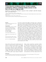
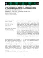
Fig. 4. Negative-ion fragmentation mass spectra of oligosaccharides with reducing-end hexose residues with the proposed structures: (A)
lactose, precursor ion m ⁄ z 341.2, g1; (B) sialyllactose, precursor ion m ⁄ z 632.2, g6; (C) lactose carrying a disialyl motif, precursor ion m ⁄ z
923.3, g9; (D) H
3
N tetrasaccharide, precursor ion m ⁄ z 706.2, g7; (E) H
3
NS pentasaccharide, precursor ion m ⁄ z 997.3, g10.
Novel oligosaccharides in galactosialidosis C. Bruggink et al.
2980 FEBS Journal 277 (2010) 2970–2986 ª 2010 The Authors Journal compilation ª 2010 FEBS
MS ⁄ MS spectrum is shown in Fig. 5B. The fragment
ions C
1
(m ⁄ z 307.9), Y
2
(m ⁄ z 357.1), Z
2
(m ⁄ z 339.0)
and [M–CH
2
OCH
2
OCOO–H]
)
(m ⁄ z 544.2) are indica-
tive of the sequence Neu5Ac–Hex–HexonA (aldohex-
onic acid). Fragment ions at m ⁄ z 604.3 [M–CO
2
–H]
)
and (m ⁄ z 586.3) [M–CO
2
–H
2
O–H]
)
are indicative of a
carboxylic acid. For the fragment ion with m ⁄ z 544.2,
cleavage between C
3
and C
4
in the aldohexonic acid is
proposed, indicating that the aldohexonic acid is
linked via C
4
to the adjacent hexose. Therefore, the
structure Neu5Ac(a2–3)Gal(b1–4)GluconA is pro-
posed, which represents the C
1
-oxidized version of
sialyllactose.
A glycan with the composition HS
2
X(m ⁄ z 939.6)
was observed at retention time 29.4 min (o8,
Table 2). The MS ⁄ MS spectrum (Fig. 5C) shows the
fragment ions B
1
(m ⁄ z 290.0), B
2
(m ⁄ z 581.3), Y
2
(m ⁄ z 357.1) and Y
3
(m ⁄ z 648.3), which is consistent
with the sequence Neu5Ac–Neu5Ac–Hex–HexonA.
The fragment ions Y
3
-CO
2
(m ⁄ z 604.2) and [M–
CO
2
–H]
)
(m ⁄ z 895.4) are indicative of a carboxylic
acid group.
A glycan with the composition H
2
NX (m ⁄ z 722.4)
was observed at retention time 21.4 min (o7,
Table 2). The MS ⁄ MS spectrum (Fig. 5D) shows the
fragment ions C
3
(m ⁄ z 543.9), Y
2
(m ⁄ z 357.0) and
Y
3
(m ⁄ z 560.2), which is consistent with the sequence
Hex–HexNAc–Hex–HexonA. For the fragment ions
with masses m ⁄ z 586.2 and m ⁄ z 406.1, carbon chain
cleavages at C
2
–C
3
and C
4
–C
5
of the aldohexonic
acid are assumed. The fragment ion with mass m ⁄ z
406.1 originated from fragment ion Z
3
. From
these details, the structure Hex–HexNAc–Gal(b1–
4)Glc (Fig. 2, o7) is proposed, which is interpreted
as the C
1
-oxidized version of oligosaccharide g7 (see
above).
A glycan with the composition H
2
NSX (m ⁄ z 1013.4)
was detected at retention time 27.4 min (o9, Table 2).
The fragment ions [M–CO
2
–H]
)
(m ⁄ z 969.7) and [M–
CO
2
–H
2
O–H]
)
(m ⁄ z 951.7) are indicative of a carbox-
ylic acid group (Fig. 5E). For fragment ion [M–CH
2
O-
CH
2
OCOO–H]
)
(m ⁄ z 909.5), a cleavage between C
3
and C
4
of the aldohexonic acid is proposed. Moreover,
the MS ⁄ MS spectrum shows the fragment ions B
2a
(m ⁄ z 364.1), Y
2
Y
2b
(m ⁄ z 357.1), Z
2b
(m ⁄ z 704.1), Y
2b
(m ⁄ z 722.3) and B
3
(m ⁄ z 817.5), which are consistent
with the sequence Hex–HexNAc–[Neu5Ac]–Hex–Hex-
onA.
Other C
1
-oxidized oligosaccharide moieties were
an aldohexonic acid carrying a sialic acid residue (o3),
oligosaccharide o4, which represents a C
1
-oxidized
version of g3, and o5, which is interpreted as
C
1
-oxidized version of g5 (for details, see Table 2).
Glycan profiling of body fluids
LC-MS data were obtained for four urine samples
from control individuals as well as seven urine
samples, five amniotic fluid samples and two ascitic
fluid samples from galactosialidosis patients. In the
four urine samples of healthy controls, lactose (m ⁄ z
341.2), sialylhexose (m ⁄ z 470.2) and sialyllactose (m ⁄ z
632.2) were detected (data not shown). For the body
fluid samples of galactosialidosis patients, the relative
abundances of the mass spectrometric signals are given
in Table 3. The two major classes of detected oligosac-
charides are the endo-b-N-acetylglucosaminidase-
cleaved products of complex-type sialylated N-glycans
derivatives (n1–n6) and oligosaccharides with reduc-
ing-end hexose residues or disialyl motifs (g1–g11),
with mean relative abundances of 37.1% and 44.8%,
respectively. Sulfated glycans (s1–s4), which are pre-
sumably derived from complex-type N-glycans,
accounted for a mean of 1.6% of all detected glycans.
The relative abundance of aldohexonic acid-based oli-
gosaccharides (o1–o9) differed considerably between
urine samples on the one hand (mean 29.7%) and
amniotic fluid and ascitic fluid samples on the other
(mean 3.1%).
In all samples, the same set of complex-type N-gly-
can-derived structures was found, with the exception
of H
5
N
3
S (n3) and H
6
N
4
S
2
(n5) in ascitic fluid samples
Asf1 and Asf2, respectively (Table 3). In all samples,
complex-type N-glycan derivatives with very high rela-
tive abundance were sialyl-N-acetyllactosamine (HNS;
n1), disialylated diantennary structures (H
5
N
3
S
2
; n4)
and sialylated monoantennary structures (H
3
N
2
S; n2).
In amniotic fluid and ascitic fluid, tri-sialylated trian-
tennary N-glycans (H
6
N
4
S
3
; n6) were clearly next in
order of relative abundance (Table 3).
Sulfated N-glycan derived structures were detected
in all samples (Table 3). In three urine samples, the
entire set of four sulfated N-glycans could be detected
(Table 3, U2, U4 and U6). In one urine sample (U2),
three isomers were detected for H
5
SO
3
N
3
S
2
(data not
shown).
Free oligosaccharides with reducing-end hexoses
were detected in all samples. In two samples (Table 3,
U1 and Amf5), the entire set of 11 oligosaccharides
(g1–g11) was detected. The most abundant species of
this glycan group in urine samples was sialyllactose
(relative mean abundance 16.4% for g6, H
2
S; Table 3),
while the proposed sialylgalactose was the most abun-
dant species in the amniotic and ascitic fluid samples
(mean 20.4% for g2, HS; Table 3). Sialyllactose was
observed with similar relative abundances in urine,
amniotic and ascitic fluid samples (g6, Table 3). In
C. Bruggink et al. Novel oligosaccharides in galactosialidosis
FEBS Journal 277 (2010) 2970–2986 ª 2010 The Authors Journal compilation ª 2010 FEBS 2981
urine sample U7, the relative amount of lactose was
high (39.9%), and was one or two orders of magnitude
lower for the other analyzed samples (g1, Table 3). The
disialyl glycan (g4) was detected in all samples and
had a mean relative abundance of 4.8%. Other glycans
containing a disialyl motif (g9 and g11) were detected
at low relative intensities (< 0.5%). Only in three of
the 14 samples analyzed were neither of these species
detected.
In the amniotic and ascitic fluid samples, aldohexonic
acid-containing oligosaccharides o1–o5 were detected.
In the urine samples, high levels of aldohexonic
acid-containing glycans were often observed, with the
exception of U4 (Table 3). In U1, U6 and U7, gluconic
acid (o1) has high abundance, and high levels of C
1
-oxi-
dized lactose (o2) were observed in urine samples U2,
U5 and U6.
Discussion
Using a prototype capillary HPAEC-PAD-MS system,
we observed N-glycan-derived oligosaccharide struc-
tures (Fig. 2, n1–6) in urine, amniotic fluid and ascitic
fluid samples from various galactosialidosis patients as
described previously [12]. The new set-up also allowed
detection of new oligosaccharides in the samples from
galactosialidosis patients: (a) O-sulfated oligosaccha-
ride moieties, (b) carbohydrate moieties with reducing-
end hexoses, and (c) oligosaccharides with C
1
-oxidized
hexose. The detection of relatively low amounts of
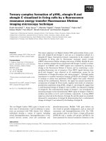
Fig. 5. Negative-ion fragmentation mass spectra of C
1
-oxidized oligosaccharides with the proposed structures: (A) C
1
-oxidized lactose, pre-
cursor ion m ⁄ z 357.2, o2; (B) C
1
-oxidized sialyllactose, precursor ion m ⁄ z 648.5, o6; (C) C
1
-oxidized lactose carrying a disialyl motif, precursor
ion m ⁄ z 939.6, o8; (D) C
1
-oxidized version of H
3
N tetrasaccharide, precursor ion m ⁄ z 722.4; o7; (E) C
1
-oxidized version of the H
3
NS tetrasac-
charide, precursor ion m ⁄ z 1013.4; o9.
Novel oligosaccharides in galactosialidosis C. Bruggink et al.
2982 FEBS Journal 277 (2010) 2970–2986 ª 2010 The Authors Journal compilation ª 2010 FEBS
O-sulfated oligosaccharide moieties and C
1
-oxidized
carbohydrate moieties, especially in the amniotic and
ascitic fluid samples, is made possible by the sensitivity
gain achieved by coupling of a capillary HPAEC-PAD
to the MS system compared to use of a normal-bore
HPAEC-PAD [13,19]. Importantly, the analytical set-
up allows analysis of glycans with reducing ends,
reduced termini and C
1
oxidation, which makes it
more broadly applicable than methods that depend on
reducing ends for reductive amination reactions [20].
An important aspect of HPAEC is its ability to sepa-
rate structural isomers, as documented previously
[13,15]. Hence, HPAEC-PAD-MS represents a valu-
able addition to the repertoire of LC-MS methods for
oligosaccharide analysis.
Almost all carbohydrate structures described here
are terminated with galactose and ⁄ or sialic acid resi-
dues, which can be explained by the defect of cathep-
sin A in galactosialidosis patients, resulting in
insufficient protection of b-galactosidase and a-neur-
aminidase against excessive intra-lysosomal degrada-
tion [2]. Cathepsin A is one of four enzymes in a
lysosomal multi-enzyme complex comprising N-acetyl-
galactosamine-6-sulfate sulfatase, b-galactosidase,
cathepsin A and a-neuraminidase [3,4].
The enzyme N-acetylgalactosamine-6-sulfatase or
galactose-6-sulfatase has been shown to be specific for
6-sulfated galactose and N-acetylgalactosamine [21,22].
The structures s1–s4 (Fig. 2) are interpreted as being
derived from complex-type N-linked carbohydrates.
6¢-sulfated sialyllactosamine (s1) has also been found
on O-linked glycan moieties [18], which may therefore
represent an alternative source of this glycan.
Tandem mass spectrometry provided evidence that
at least some of the oligosaccharide chains with hexose
at the reducing end have a Gal(b1–4)Glc (lactose) core
structure. This group of glycans (g1–g11) shares struc-
tural features with milk oligosaccharides, plasma
oligosaccharides and previously described urinary
oligosaccharides from healthy individuals [23–25]. The
structures g1 and g6 in Fig. 2 can be interpreted as
lactose (g1) and sialyllactose (g6), which are known to
be present in various body fluids [24,26–28]. Moreover,
the tetrasaccharide g7 may be interpreted as lacto-
N-tetraose, and g10 may represent a sialylated version
thereof. As these structures are in part identical with
milk sugars, they may be of limited diagnostic value.
Several glycosyltransferases have been identified in
urine and amniotic fluid [29–32], but no-one, to the
best of our knowledge, has demonstrated that glycosyl-
transferases are active in these fluids.
Notably, the detected structures g4 (S
2
), g8 (H
2
NS),
g9 (H
2
S
2
), g10 (H
3
NS) and g11 (H
3
NS
2
) all exhibited
structural motifs that are typically found on glycos-
phingolipids. g4 is interpreted as a predominantly
glycosphingolipid-derived disialyl motif, and the oligo-
saccharides g8, g9, g10 and g11 are postulated to repre-
sent, at least in part, reducing-end glycan moieties of the
gangliosides GM2, GD3, GM1 and GD1b, respectively
(Fig. 2, g8–g11). In addition, the structures g5 and g7
may also be interpreted as partly glycosphingolipid-
derived (ganglio-, lacto- or lactoneo- series).
To our knowledge, such intact oligosaccharide moie-
ties have hitherto not been described as glycosphingo-
lipid degradation products. According to the literature,
catabolism of glycosphingolipids starts from the non-
reducing end while the glycan is still bound to the
ceramide, and is performed by a variety of exoglyco-
sidases, which are often also involved in the degrada-
tion of N-glycans and O-glycans [33,34]. This process
leads to the release of monosaccharides and results in
glucosylceramide and galactosylceramide, which may
be degraded further by glycosidic bond cleavage. Addi-
tional proteins such as saposins (sphingolipid activator
proteins) are required for the catabolism of glycosphin-
golipids [35]. A blockage of glycosphingolipid degrada-
tion, as occurs in Fabry’s disease as a result of a lack
of a-galactosidase activity, leads to accumulation of
the glycosphingolipid substrate, which in Fabry’s
disease is globotriaosylceramide [34]. Consequently, in
galactosialidosis, only intact glycosphingolipids would
be expected to be secreted, not the glycan moieties as
described here. Our finding of free oligosaccharide
moieties presumably derived from glycosphingolipids
implies the existence of an endoglycosylceramidase
involved in an alternative glycosphingolipid catabolic
pathway. While such an enzyme has not been
described for vertebrates, endoglycoceramidases (EC
3.2.1.123) have been found and characterized for inver-
tebrates [36–39]. The enzymatic activity of the postu-
lated endoglycoceramidase may depend on saposins
[35], and may represent a side activity of glucosylce-
ramidase (EC 3.2.1.45) facilitated by specific saposins.
With regard to the disaccharide of two sialic acid
residues (Fig. 2, g4), it is unclear which enzyme would
catalyze the release of this disaccharide unit from
gangliosides.
The last group of newly found oligosaccharides is
characterized by C
1
-oxidized hexose residues (o1–o9).
This group of glycans appears to be strongly related to
the above-described glycans with reducing-end hexoses
(g1–g11), suggesting C
1
oxidation of these oligosaccha-
ride moieties. The glycans o8 and o9 may be inter-
preted as C
1
-oxidized versions of ganglioside-derived
glycan moieties (Fig. 2). The C
1
-oxidized oligosaccha-
rides were found in urine samples at relatively high
C. Bruggink et al. Novel oligosaccharides in galactosialidosis
FEBS Journal 277 (2010) 2970–2986 ª 2010 The Authors Journal compilation ª 2010 FEBS 2983
amounts (mean 30%; Table 3). C
1
-oxidized carbohy-
drate moieties were also found in amniotic fluid sam-
ples, albeit at lower relative amounts (mean 3%;
Table 3). The cause of C
1
oxidation of the reducing
end is unknown. We can exclude the possibility that
these species were observed due to oxidation of reduc-
ing sugars during the chromatographic process and the
subsequent MS detection, as we observed chromato-
graphic separation of the reducing glycans from the
C
1
-oxidized species, clearly indicating that these species
were already present in the samples prior to HPAEC-
PAD-MS analysis. With regard to the origin of the
C
1
-oxidized glycans, it is possible to speculate about a
non-enzymatic oxidation reaction that may have
occurred before the urine and amniotic samples were
collected, or during sample storage. Alternatively, an
enzymatic oxidation may be postulated. The possibility
that an enzyme of microbial origin is responsible, as
described for Escherichia coli [40–44], appears not to
be likely, as the oxidation products were not only
observed in urine samples, but also in amniotic fluid,
which is considered to be sterile. Alternatively, it could
be speculated that a human enzymatic activity might
be present in the liver or kidney, for example, that
causes C
1
oxidation of glycosphingolipid glycan moie-
ties. This enzyme may act in conjunction with the
postulated endoglycoceramidase.
Together with our previous study on G
M1
gangliosi-
dosis [13], this study shows the potential value of capil-
lary HPAEC-PAD-MS for analyzing oligosaccharides
from clinical samples. This prototype analytical system
features femtomolar sensitivity for both pulsed ampero-
metric detection and mass spectrometric detection [13].
Moreover, it allows the analysis of oligosaccharides in
both positive-ion mode [13] and negative-ion mode, as
shown here. Based on the excellent MS ⁄ MS features of
the ion trap mass spectrometer, informative fragment
spectra of sodium adducts [13] and deprotonated spe-
cies (this study) can be obtained with minute amounts
of material, thus allowing insights into defects of glyco-
conjugate degradation and lysosomal storage diseases.
Experimental procedures
Materials
Analytical reagent-grade sodium hydroxide (50% w ⁄ w),
sodium acetate, sulfuric acid and sodium chloride were
obtained from J.T. Baker (Deventer, The Netherlands).
Acetonitrile was obtained from Biosolve (Valkenswaard,
The Netherlands). All solutions were prepared using water
from a Milli-Q synthesis system from Millipore BV
(Amsterdam, The Netherlands). Details of the urine, amni-
otic fluid and ascitic fluid samples are given in Table 1.
Capillary HPAEC
The capillary chromatographic system consists of a modi-
fied BioLC system from Dionex (Sunnyvale, CA), compris-
ing a microbore GP40 gradient pump, a Famos micro
autosampler with a full polyaryletherketone (PAEK) injec-
tor equipped with a 1 lL loop, and an ED40 electrochemi-
cal detector. BioLC control, data acquisition from the
ED40 detector and signal integration are supported by
chromeleon software (Dionex). This modified system has
been described in detail previously [13]. A prototype capil-
lary column 250 mm long with internal diameter 0.4 mm,
packed with CarboPac PA200 resin, was manufactured by
Dionex. The GP40 flow rate was 0.53 mLÆmin
)1
, and the
eluent flow was split using a custom-made polyether ether
ketone (PEEK) splitter to 10 lLÆmin
)1
. The pump was pro-
vided with the following eluents: eluent A, water; eluent B,
500 mm NaOH; eluent C, 500 mm NaOAc. All separations
were performed at room temperature. The following ter-
nary gradient was used for the separation: 76% A + 24%
B()20 to )14 min), isocratic sodium hydroxide wash; 88%
A + 12% B ()14 to 0 min), isocratic equilibration of the
column; 42.6% A + 12% B + 45.4% C (0–40 min), linear
sodium acetate gradient was used for the separation. The
ED40 detector applies the following waveform to the elec-
trochemical cell: E
1
= 0.1 V (t
d
= 0.00–0.20 s, t
1
= 0.20–
0.40 s), E
2
= )2.0 V (t
2
= 0.41–0.42 s), E
3
= 0.6 V
(t
3
= 0.43 s), E
4
= )0.1 V (t
4
= 0.44–0.50 s) versus an
Ag ⁄ AgCl reference electrode [45]. A gold work electrode
and a 25 lm gasket were installed.
Mass spectrometry
Coupled to the chromatographic system was an
Esquire 3000 ion-trap mass spectrometer from Bruker Dal-
tonics (Bremen, Germany), equipped with an electrospray
ionization source. To convert the HPAEC eluate into an
ESI-compatible solution, an in-line prototype desalter (Dio-
nex) was used, continuously regenerated with diluted sulfu-
ric acid [13]. A modified microbore AGP-1 from Dionex
was used as an auxiliary pump: to obtain efficient ioniza-
tion of the eluted carbohydrates, 50% acetonitrile was
pumped into the eluent flow via a MicroTEE (P-775, Up-
church Scientific, Oak Harbor, WA, USA) at a flow rate of
4.6 lLÆmin
)1
. The mixture was directed to the electrospray
ionization interface of the Esquire 3000. The carbohydrates
were detected using the MS in the negative-ion mode. The
MS was operated under the following conditions: dry tem-
perature 325 °C, nebulizer 103 kPa, dry gas 7 LÆmin
)1
, tar-
get mass m ⁄ z 850, scan speed 13 000 m ⁄ z per s in MS and
MS ⁄ MS mode. For tandem MS, automatic selection of
three precursors was applied.
Novel oligosaccharides in galactosialidosis C. Bruggink et al.
2984 FEBS Journal 277 (2010) 2970–2986 ª 2010 The Authors Journal compilation ª 2010 FEBS
Sample preparation
Oligosaccharides of the samples were isolated by graphi-
tized carbon solid-phase extraction, as described previously
[46]. A 200 lL sample was diluted with 1800 l L demineral-
ized water and loaded on a Carbograph SPE (210142) from
Alltech Associates Inc. (Deerfield, IL, USA). The cartridge
was washed with 6 mL of demineralized water. The oligo-
saccharides were eluted from the column using 3 mL of
25% acetonitrile containing 0.05% trifluoroacetic acid. The
eluate was evaporated under a nitrogen stream at room
temperature until the volume had decreased by 50%. The
remaining solution was lyophilized and reconstituted with
200 lL demineralized water.
Acknowledgements
We would like to thank Professor Ron Wevers
(Radboud University Nijmegen Medical Center, The
Netherlands) and Dr Pim Onkenhout (Leiden Univer-
sity Medical Center, The Netherlands) for kindly pro-
viding samples, Dr Cornelis H. Hokke for fruitful
discussions, Rob Bruggink for providing essential input
for producing the capillary desalter, and Chris Pohl,
Yan Liu, Victor Barretto and Franck van Veen from
Dionex for essential support of this research.
References
1 Wenger DA, Tarby TJ & Wharton C (1978)
Macular cherry-red spots and myoclonus with demen-
tia: coexistent neuraminidase and beta-galactosidase
deficiencies. Biochem Biophys Res Commun 82, 589–
595.
2 D’Azzo A, Hoogeveen A, Reuser AJ, Robinson D &
Galjaard H (1982) Molecular defect in combined
b-galactosidase and neuraminidase deficiency in man.
Proc Natl Acad Sci USA 79, 4535–4539.
3 Pshezhetsky AV & Potier M (1996) Association of
N-acetylgalactosamine-6-sulfate sulfatase with the
multienzyme lysosomal complex of b-galactosidase,
cathepsin A, and neuraminidase. Possible implication
for intralysosomal catabolism of keratan sulfate. J Biol
Chem 271, 28359–28365.
4 Pshezhetsky AV & Ashmarina M (2001) Lysosomal
multienzyme complex: biochemistry, genetics, and
molecular pathophysiology. Prog Nucleic Acid Res Mol
Biol 69, 81–114.
5 Lowden JA & O’Brien JS (1979) Sialidosis: a review of
human neuraminidase deficiency. Am J Hum Genet 31,
1–18.
6 Takahashi Y, Nakamura Y, Yamaguchi S & Orii T
(1991) Urinary oligosaccharide excretion and severity of
galactosialidosis and sialidosis. Clin Chim Acta 203,
199–210.
7 Groener J, Maaswinkel-Mooy P, Smit V, van der
Hoeven M, Bakker J, Campos Y & D’Azzo A (2003)
New mutations in two Dutch patients with early infan-
tile galactosialidosis. Mol Genet Metab 78, 222–228.
8 Okamura-Oho Y, Zhang S & Callahan JW (1994) The
biochemistry and clinical features of galactosialidosis.
Biochim Biophys Acta 1225, 244–254.
9 Sewell AC (1981) Simple laboratory determination of
excess oligosacchariduria. Clin Chem 27, 243–245.
10 van Pelt J, Kamerling JP, Vliegenthart JF, Hoogeveen
AT & Galjaard H (1988) A comparative study of the
accumulated sialic acid-containing oligosaccharides
from cultured human galactosialidosis and sialidosis
fibroblasts. Clin Chim Acta 174, 325–335.
11 van Pelt J, Kamerling JP, Vliegenthart JF, Verheijen
FW & Galjaard H (1988) Isolation and structural char-
acterization of sialic acid-containing storage material
from mucolipidosis I (sialidosis) fibroblasts. Biochim
Biophys Acta 965, 36–45.
12 van Pelt J, Hard K, Kamerling JP, Vliegenthart JF,
Reuser AJ & Galjaard H (1989) Isolation and structural
characterization of twenty-one sialyloligosaccharides
from galactosialidosis urine. An intact N,N’-diacetylchi-
tobiose unit at the reducing end of a diantennary
structure. Biol Chem Hoppe Seyler 370, 191–203.
13 Bruggink C, Wuhrer M, Koeleman CA, Barreto V, Liu
Y, Pohl C, Ingendoh A, Hokke CH & Deelder AM
(2005) Oligosaccharide analysis by capillary-scale high-
pH anion-exchange chromatography with on-line ion-
trap mass spectrometry. J Chromatogr B 829, 136–143.
14 Ramsay SL, Maire I, Bindloss C, Fuller M, Whitfield
PD, Piraud M, Hopwood JJ & Meikle PJ (2004)
Determination of oligosaccharides and glycolipids in
amniotic fluid by electrospray ionisation tandem mass
spectrometry: in utero indicators of lysosomal storage
diseases. Mol Genet Metab 83, 231–238.
15 Townsend RR, Hardy MR, Hindsgaul O & Lee YC
(1988) High-performance anion-exchange chromatogra-
phy of oligosaccharides using pellicular resins and pulsed
amperometric detection. Anal Biochem 174, 459–470.
16 Harvey DJ (2005) Fragmentation of negative ions from
carbohydrates: part 3. Fragmentation of hybrid and
complex N-linked glycans. J Am Soc Mass Spectrom
16, 647–659.
17 Pfenninger A, Karas M, Finke B & Stahl B (2002)
Structural analysis of underivatized neutral human milk
oligosaccharides in the negative ion mode by nano-elec-
trospray MS
n
(part 1: methodology). J Am Soc Mass
Spectrom 13, 1331–1340.
18 Hemmerich S, Leffler H & Rosen SD (1995) Structure
of the O-glycans in GlyCAM-1, an endothelial-derived
ligand for L-selectin. J Biol Chem 270 , 12035–12047.
19 van der Hoeven RAM, Hofte AJP, Tjaden UR, van der
Greef J, Torto N, Gorton L, Marko-Varga G &
Bruggink C (1998) Sensitivity improvement in the
C. Bruggink et al. Novel oligosaccharides in galactosialidosis
FEBS Journal 277 (2010) 2970–2986 ª 2010 The Authors Journal compilation ª 2010 FEBS 2985
analysis of oligosaccharides by online high-performance
anion-exchange chromatography ⁄ ionspray mass
spectrometry. Rapid Commun Mass Spectrom 12, 69–74.
20 Anumula KR (2006) Advances in fluorescence
derivatization methods for high-performance liquid
chromatographic analysis of glycoprotein
carbohydrates. Anal Biochem 350, 1–23.
21 Glossl J, Truppe W & Kresse H (1979) Purification and
properties of N-acetylgalactosamine 6-sulphate
sulphatase from human placenta. Biochem J 181, 37–
46.
22 Glossl J & Kresse H (1982) Impaired degradation of
keratan sulphate by Morquio A fibroblasts. Biochem J
203, 335–338.
23 Koseki M & Tsurumi K (1977) A convenient method
for the isolation of 3¢-sialyllactose from normal human
urine. J Biochem 82, 1785–1788.
24 Arthur PG, Kent JC & Hartmann PE (1989)
Microanalysis of the metabolic intermediates of lactose
synthesis in human milk and plasma using biolumines-
cent methods. Anal Biochem 176, 449–456.
25 Ninonuevo MR, Park Y, Yin H, Zhang J, Ward RE,
Clowers BH, German JB, Freeman SL, Killeen K,
Grimm R et al. (2006) A strategy for annotating the
human milk glycome. J Agric Food Chem 54, 7471–
7480.
26 Ramsay SL, Meikle PJ, Hopwood JJ & Clements PR
(2005) Profiling oligosaccharidurias by electrospray
tandem mass spectrometry: quantifying reducing
oligosaccharides. Anal Biochem 345, 30–46.
27 An Y, Young SP, Hillman SL, Van Hove JL, Chen YT &
Millington DS (2000) Liquid chromatographic assay for
a glucose tetrasaccharide, a putative biomarker for the
diagnosis of Pompe disease. Anal Biochem 287, 136–143.
28 Maury CP & Wegelius O (1981) Urinary sialyloligosac-
charide excretion as an indicator of disease activity in
rheumatoid arthritis. Rheumatol Int 1, 7–10.
29 Chester MA (1974) The occurrence of a beta-galactosyl-
transferase in normal human urine. FEBS Lett 46,
59–62.
30 Serafini-Cessi F, Malagolini N & Dall’Olio F (1988)
Characterization and partial purification of b-N-acety-
lgalactosaminyltransferase from urine of Sd(a+)
individuals. Arch Biochem Biophys 266, 573–582.
31 Nelson JD, Jato-Rodriguez JJ & Mookerjea S (1973)
Occurrence of soluble glycosyltransferases in human
amniotic fluid. Biochem Biophys Res Commun 55, 530–
537.
32 Nelson JD, Jato-Rodriguez JJ & Mookerjea S (1974)
The occurrence and properties of soluble UDP-galac-
tose:glycoprotein galactosyltransferase in human
amniotic fluid. Can J Biochem 52, 42–50.
33 Sandhoff K & Kolter T (2003) Biosynthesis and
degradation of mammalian glycosphingolipids. Philos
Trans R Soc Lond B Biol Sci
358, 847–861.
34 Kolter T & Sandhoff K (2006) Sphingolipid metabolism
diseases. Biochim Biophys Acta 1758, 2057–2079.
35 Kolter T & Sandhoff K (2005) Principles of lysosomal
membrane digestion: stimulation of sphingolipid
degradation by sphingolipid activator proteins and
anionic lysosomal lipids. Annu Rev Cell Dev Biol 21,
81–103.
36 Ito M & Yamagata T (1986) A novel glycosphingolipid-
degrading enzyme cleaves the linkage between the
oligosaccharide and ceramide of neutral and acidic
glycosphingolipids. J Biol Chem 261, 14278–14282.
37 Ito M & Yamagata T (1989) Purification and character-
ization of glycosphingolipid-specific endoglycosidases
(endoglycoceramidases) from a mutant strain of
Rhodococcus sp. Evidence for three molecular species of
endoglycoceramidase with different specificities. J Biol
Chem 264, 9510–9519.
38 Li YT & Li SC (1989) Ceramide glycanase from leech,
Hirudo medicinalis, and earthworm, Lumbricus terres-
tris. Methods Enzymol 179, 479–487.
39 Horibata Y, Sakaguchi K, Okino N, Iida H, Inagaki
M, Fujisawa T, Hama Y & Ito M (2004) Unique
catabolic pathway of glycosphingolipids in a hydrozoan,
Hydra magnipapillata , involving endoglycoceramidase.
J Biol Chem 279, 33379–33389.
40 Hugh R & Leifson E (1953) The taxonomic significance
of fermentative versus oxidative metabolism of carbohy-
drates by various gram negative bacteria. J Bacteriol 66,
24–26.
41 Taylor WH & Juni E (1961) Pathways for biosynthesis of
a bacterial capsular polysaccharide. I. Carbohydrate
metabolism and terminal oxidation mechanisms of a
capsule-producing coccus. J Bacteriol 81, 694–703.
42 Hommes RW, Postma PW, Tempest DW, Neijssel OM,
Dokter P & Dione JA (1984) Evidence of a quinoprotein
glucose dehydrogenase apoenzyme in several strains of
Escherichia coli. FEMS Microbiol Lett 24, 329–333.
43 Liu ST, Lee LY, Tai CY, Hung CH, Chang YS,
Wolfram JH, Rogers R & Goldstein AH (1992) Cloning
of an Erwinia herbicola gene necessary for gluconic acid
production and enhanced mineral phosphate solubiliza-
tion in Escherichia coli HB101: nucleotide sequence and
probable involvement in biosynthesis of the coenzyme
pyrroloquinoline quinone. J Bacteriol 174, 5814–5819.
44 Yan Z, Caldwell GW & McDonell PA (1999)
Identification of a gluconic acid derivative attached to
the N-terminus of histidine-tagged proteins expressed in
bacteria. Biochem Biophys Res Commun 262, 793–800.
45 Rocklin RD, Clarke AP & Weitzhandler M (1998)
Improved long-term reproducibility for pulsed
amperometric detection of carbohydrates via a new qua-
druple-potential waveform. Anal Chem 70, 1496–1501.
46 Packer NH, Lawson MA, Jardine DR & Redmond JW
(1998) A general approach to desalting oligosaccharides
released from glycoproteins. Glycoconj J 15, 737–747.
Novel oligosaccharides in galactosialidosis C. Bruggink et al.
2986 FEBS Journal 277 (2010) 2970–2986 ª 2010 The Authors Journal compilation ª 2010 FEBS