Differential Diagnosis in Internal Medicine From Symptom to Diagnosis_2 ppt
Bạn đang xem bản rút gọn của tài liệu. Xem và tải ngay bản đầy đủ của tài liệu tại đây (35.27 MB, 589 trang )
Siegenthaler, Differential Diagnosis in Internal Medicine, © 2007 Thieme
All rights reserved. Usage subject to terms and conditions of license.
518
18
Pulmonary Opacities
K. E. Bloch and E. W. Russi
18
18
Siegenthaler, Differential Diagnosis in Internal Medicine, © 2007 Thieme
All rights reserved. Usage subject to terms and conditions of license.
519
18.1 Infectious Pulmonary Infiltrates
(Pneumonias)
521
Bacterial Pneumonia 523
Classification 523
Clinical Presentation of Bacterial
Pneumonias 524
Pneumonias Due to Gram-Positive
Microorganisms 524
Pneumonias Due to Gram-Negative
Bacteria and Microorganisms not
Identifiable under Light Microscopy 526
Pneumonia Due to Multiple Gram-Positive
and Gram-Negative Organisms (“Mixed
Flora”) 528
Pulmonary Tuberculosis 530
Primary Tuberculosis 531
Postprimary Pulmonary Tuberculosis 531
Exsudative Pulmonary Tuberculosis 531
Tuberculous Cavity 531
Miliary Tuberculosis 533
Fibroproliferative Tuberculosis 534
Tuberculoma 534
Disease Due to Mycobacteria Other
Than Tuberculosis (MOTT) 535
Viral Pneumonia 536
Influenza Pneumonia 536
Adenovirus Pneumonia 536
Severe Acute Respiratory Syndrome (SARS) 536
Hantavirus Pneumonia 536
Pneumonia Due to Nonpneumotropic
Viruses 536
Fungal Pneumonia 537
Fungus Infection in Immunocompromised
Patients 537
Pneumonia Due to Yeasts and Molds 537
Pneumocystis carinii Pneumonia 537
Endemic Fungal Infection 539
Allergic Bronchopulmonary Aspergillosis
and Mycetoma 539
Pulmonary Parasitosis 540
18.2 Noninfectious Pulmonary
Infiltrates
540
Physical or Chemical Pneumonitis 540
Radiation Pneumonitis 541
Lipoid Pneumonia 541
Infiltrates Due to Chronic Congestive Heart
Failure 541
Pulmonary Infarction − Infarction
Pneumonia 543
Pneumonia Associated with Bronchiectasis 545
Pneumonia Due to Bacterial Superinfection 545
Chronic Pneumonia 545
Other Noninfectious Pulmonary Infiltrates 545
18.3 Eosinophilic Pulmonary Infiltrates
546
Transient Eosinophilic Pulmonary Infiltrates
(Löffler) 546
Pulmonary Eosinophilia with Parasitosis
and Tropical Pulmonary Eosinophilia 546
Allergic Bronchopulmonary Aspergillosis
(ABPA) 546
Drug-Induced Pulmonary Eosinophilia 547
Acute Eosinophilic Pneumonia 547
Chronic Eosinophilic Pneumonia 547
Eosinophilic Infiltrates with Asthma 547
Allergic Granulomatosis and Angiitis
(Churg−Strauss Syndrome) 547
Hypereosinophilic Syndrome 547
Diagnostic Criteria: 547
18.4 Diffuse Parenchymal Lung Disease
(DPLD)/Pulmonary Fibrosis
548
Idiopathic Interstitial Pneumonia 549
Idiopathic Pulmonary Fibrosis (IPF) 550
Nonspecific Interstitial Pneumonia (NSIP) 551
Cryptogenic Organizing Pneumonia
(Idiopathic Bronchiolitis Obliterans
Organizing Pneumonia [BOOP]) 551
Pulmonary Opacities
520
Acute Interstitial Pneumonia (AIP,
Hamman−Rich Syndrome) 553
Respiratory Bronchiolitis-Associated
Interstitial Lung Disease (RB-ILD) 554
Desquamative Interstitial Pneumonia (DIP) 554
Lymphoid Interstitial Pneumonia (LIP) 554
Interstitial Pneumonia in Association
with Collagen Vascular Disease 554
Toxic and Drug-Induced Interstitial
Pneumonia 556
Extrinsic Allergic Alveolitis
(Hypersensitivity Pneumonitis) 556
Pneumoconiosis 557
Silicosis 557
Silicatosis and Other Pneumoconioses 559
Diffuse Granulomatous Pulmonary
Diseases 561
Other Diffuse Parenchymal Lung
Diseases and Orphan Lung Diseases 561
Alveolar Cell Carcinoma, Bronchoalveolar
Cell Carcinoma, and Pulmonary
Adenomatosis 561
Lymphangiosis Carcinomatosa 561
Kaposi Sarcoma 561
Pulmonary Hemosiderosis 561
Goodpasture Syndrome 561
Antiphospholipid Syndrome 564
Pulmonary Alveolar Proteinosis (PAP) 564
Microlithiasis Alveolaris 564
Langerhans Cell Histiocytosis 564
Lymphangioleiomyomatosis (LAM) 564
Formation of Cysts and Honeycombing 565
18.5 Pulmonary Nodules
566
Solitary Pulmonary Nodules 567
Malignant Neoplasms 567
Benign Tumors 569
Inflammatory Pulmonary Nodules 569
Tuberculoma 569
Echinococcosis 569
Pulmonary Nodules of Various Etiology 570
Multiple Pulmonary Nodules 570
Metastasis 570
Wegener Granulomatosis 570
Arteriovenous Aneurysms 572
18.6 Cavernous and Cystic Lung
Diseases
573
Tuberculous Cavitary Lesion 573
Pulmonary Abscess 573
Pulmonary Abscess Due to Aspiration 573
Pulmonary Abscess Formation as a
Complication of Bacterial Pneumonia 574
Metastatic Lung Abscess 574
Lung Cysts 574
Cavernous and Cystic Lesions
of Various Etiologies 574
18.7 Atelectasis
57 4
18.8 Middle Lobe Syndrome
576
18.9 Opacities in the Cardiophrenic
Angle
578
Cysts and Hernias 578
Lung Sequestration 578
18
Pulmonary Opacities
Siegenthaler, Differential Diagnosis in Internal Medicine, © 2007 Thieme
All rights reserved. Usage subject to terms and conditions of license.
521
Radiologic Morphology of Pulmonary Opacities
By definition, the diagnosis of pulmonary opacities re-
quires a radiographic examination. Although the pres-
ence of opacities may be suspected on clinical grounds,
chest percussion and auscultation are often normal or
equivocal. Opacities of the lung parenchyma may be re-
lated to extravascular fluid accumulation (exudate, tran-
sudate) and to infiltration of inflammatory, fibrotic, or
neoplastic cells. The following discussion refers to con-
ventional chest radiography. A larger amount of anatomi-
cal information, and a significantly higher spatial resolu-
tion, can be obtained by a computed tomography (CT)
scan of the chest including spiral acquisition in thin slices
and visualization in high-resolution techniques, with and
without application of a contrast agent. However, a CT
scan is generally not performed as the initial radiologic
examination due to its higher costs and radiation expo-
sure. For the description of CT images a specialized no-
menclature, different from that used to describe conven-
tional chest radiography, is applied.
Pulmonary opacities are characterized by their size, dis-
tribution, and pattern.
Size. Based on their size and extension, localized opaci-
ties (such as those found in lobar pneumonia or tuber-
culoma) are differentiated from diffuse opacities (such as
those found in fibrosing alveolitis, or pneumoconiosis).
Pattern of Infiltrates. Inflammatory or neoplastic infil-
trates are typically associated with opacities that pre-
serve the lung structure. Infiltrates may occur with an aci-
nar or interstitial pattern:
ț Acinar Infiltrates. Diseases that affect the pulmonary
acini (e. g., pneumoconioses) have the following
characteristics:
− homogeneous density
− tendency for confluence
− air bronchograms
− absence of lung volume loss.
ț Interstitial Infiltrates. Diseases that predominantly af-
fect the pulmonary interstitium (e. g., fibrosing alve-
olitis) have the following characteristics:
− ground-glass opacity
− inhomogeneous density
− linear and reticular densities (Kerley A, B, and C
lines)
− noduli
− “honeycombing”
− loss of lung volume.
Consolidation. This term refers to a dense opacity that
conceals the structure of the affected lung parenchyma.
Consolidation is seen in pneumonia, lung cancer, or
metastasis.
Mass Lesions. A typical characteristic of a pulmonary
mass is its tendency to encroach upon adjacent lung
parenchyma and other structures. This is not only seen in
neoplasia but also in inflammatory diseases (pulmonary
abscess).
Nodules. A nodule is defined as a rounded, clearly de-
lineated opacity of less than 3 cm in diameter. Solitary or
multiple pulmonary nodules may be related to cancer, in-
fection, or organic and inorganic dust exposure (such as
found in silicosis).
Opacities with Central Hypodensity. These opacities
occur due to necrotic destruction such as found in an ab-
scess (bacterial abscess, cavity due to infection with My-
cobacterium tuberculosis), pulmonary infarct, neoplasia,
or vasculitis (Wegener granulomatosis).
Mixed Patterns of Pulmonary Opacities. The simul-
taneous occurrence of various types of opacities is com-
mon.
Additional Diagnostic Criteria. Certain lung diseases
are associated with a distinct distribution and pattern of
radiologic opacities. The high-resolution CT scan may be
diagnostic in some of these diseases (e. g., Langerhans
cell histiocytosis, lymphangioleiomyomatosis, or asper-
gilloma). In contrast, other diseases occur with variable,
nonspecific findings that preclude a definitive diagnosis
by chest radiography or even by CT scan (e. g., sarcoido-
sis, or amiodarone pneumopathy).
18.1 Infectious Pulmonar y Infiltrates (Pneumonias)
Infectious pulmonary infiltrates result from an inflam-
matory response to the infection caused by microor-
ganisms. The clinical manifestation is determined by the
type of the infectious agent (bacteria, viruses, fungi, or
parasites) and the immunologic response of the host.
The following discussion is arranged according to the in-
fectious agent (Tab. 18.1).
Infectious Pulmonary Infiltrates (Pneumonias)
Siegenthaler, Differential Diagnosis in Internal Medicine, © 2007 Thieme
All rights reserved. Usage subject to terms and conditions of license.
522
Table 18.1 Classification of inflammatory infiltrates
Infectious pneumonia
Bacterial pneumonia
− due to Gram-positive organisms
− Streptococcus pneumoniae (pneumococci type 1−90)
− Streptococcus
− Staphylococcus
− Actinomyces
− Nocardia
− due to Gram-negative organisms and organism not identifiable by light-microscopy
− Haemophilus influenzae
− Klebsiella
− Branhamella (Moraxella) catarrhalis
− Escherichia coli
− Proteus
− Pseudomonas
− Serratia
− Mycoplasma pneumoniae
− Chlamydia pneumoniae
− Chlamydia psittaci (ornithosis)
− Brucella (Bang pneumonia)
− Legionella pneumophila
− Rickettsia (Q-fever)
− Bacillus anthracis
− due to Gram-positive and Gram-negative anerobic organisms (Bacteroides, Fusobacterium)
− due to Mycobacterium tuberculosis complex
− M. tuberculosis
− M. bovis
− M. africanum
− due to atypical mycobacteria
− M. avium-intracelluare complex
− M. kansasii
− M. fortuitum, M. abscessus, and M. chelonei
Viral pneumonia
− Influenza virus
− Adenovirus
− Coronavirus (SARS)
− Hantavirus
− pneumonia due to virus not primarily affecting the lungs (measles, Epstein−Barr virus)
Fungal pneumonia
− fungal infection in the immunocompromised host
− candidiasis (moniliasis)
− aspergillosis
− Pneumocystis carinii
− Mucor mycosis (geotrichosis)
− cryptococcosis (torulosis)
− endemic fungal infections
− blastomycosis
− histoplasmosis
− coccidioidomycosis
Pneumonia due to parasites
− Toxoplasma gondii
Noninfectious pneumonia
Physical−chemical pneumonia
Pneumonia with eosinophilia (see Eosinophilic Pulmonary Infiltrates p. 546)
Inflammatory pulmonary infiltrates in collagen vascular disease (see Diffuse Parenchymal Lung Disease/Pulmonary
Fibrosis p. 548)
Due to circulatory failure
− cardiogenic pulmonary edema
− infarction pneumonia
18
Pulmonary Opacities
Siegenthaler, Differential Diagnosis in Internal Medicine, © 2007 Thieme
All rights reserved. Usage subject to terms and conditions of license.
523
Prognostic Factors in Community-Acquired Pneumonia
Infectious Pulmonary Infiltrates (Pneumonias)
In a patient with community-acquired pneumonia, the
severity of the illness and the need for hospital care have
to be assessed. The following factors may aid in this
assessment:
−age 50 years
− co-morbidity such as a neoplasia, congestive heart
failure, chronic obstructive lung disease, or renal
and hepatic diseases
− altered consciousness
− tachycardia 125 beats/min.
− respiratory rate 30/min.
− systolic blood pressure 90 mmHg
− body temperature 35 °C or 40 °C.
If none of the above-mentioned risk factors is present,
the clinical course is generally favorable and treatment of
the pneumonia may be performed at home.
The following findings represent additional risk factors:
− acidosis: arterial pH 7.35
− serum urea ͧ 30 mg/dL (11 mmol/L)
− serum sodium 130 mmol/L
− serum glucose ͧ 250 mg/dL (14 mmol/L)
− hematocrit 30%
− leukocyte count 4000 × 10
6
/L or 20 000 10
6
/L
− arterial oxygen partial pressure: Po
2
60 mmHg
− multilobar pulmonary infiltrates
− pleural effusion.
Bacterial Pneumonia
Bacterial pneumonias are still among the leading causes
of death due to infectious diseases despite the wide-
spread use of antibiotics. The organisms responsible
vary according to where the infection is acquired (at
home, in the hospital or another institution, in the in-
tensive care unit), host factors such as the age of the
patient, co-morbidities, and the individual immune re-
sponse. Streptococcus pneumoniae (pneumococcus),
Haemophilus influenzae, Gram-negative enteric bacilli,
Staphylococcus aureus, and Legionella pneumophila are
the most common causes of bacterial pneumonia. To-
gether with Mycoplasma pneumoniae, Chlamydia pneu-
moniae, and the respiratory viruses, they account for the
majority of community-acquired pneumonias. In con-
trast, hospital-acquired pneumonias are more com-
monly caused by Gram-negative organisms.
Classification
According to Where the Infection Was Acquired and the
Host Defense. One of the major determinants of the type
of microorganism and course of the pneumonia is
whether it has been acquired in the community (“com-
munity-acquired pneumonia”) or in a special setting
such as in a hospital (“hospital-acquired pneumonia”),
nursing home, prison, or in other institutions where
many people live closely together.
Another major determinant of the susceptibility to
certain infections, and of particular clinical presenta-
tions, is the ability of a patient to create an effective im-
mune response. In nonimmunocompromised patients,
bacterial pneumonias generally occur with an acute
course (with high fever, cough, and production of putrid
sputum), whereas bacterial pneumonias may initially
have a less dramatic course in the immunocompromised
host. Nevertheless, neither the clinical presentation,
laboratory tests, nor the radiographic appearance allows
a reliable diagnosis of the infectious agent of pneumonia.
Thus, the term atypical pneumonia is inappropriate and,
at best, of historical interest. Originally, atypical pneu-
monias were thought to be caused by microorganisms
other than Streptococcus pneumoniae. The first recog-
nized “atypical agents” were Mycoplasma pneumoniae;
subsequently Chlamydia pneumoniae and Legionella
pneumophila were identified. Since pneumonia caused
by Streptococcus pneumoniae and other bacteria may
have a clinical and radiologic presentation similar to in-
fections by the “atypical agents,” the distinction between
typical and atypical pneumonia is clinically useless.
According to the Infectious Agent. Another classification
of pneumonia is based on the microorganism. However,
the identification of the agents responsible is often not
feasible or successful. Even in patients hospitalized for
treatment of pneumonia, the microorganism is iden-
tified in less than half of cases.
Other Classifications. These are based on the mode of
transmission of the infection and the radiologic appear-
ance. These classifications may help to identify the
potential spectrum of microorganisms responsible: for
example, community-acquired aspiration pneumonia is
generally caused by a mixed flora of anaerobic and aero-
bic bacteria. In contrast, in hospital-acquired aspiration
pneumonia, Gram-negative bacteria are often involved.
Hematogeneous pneumonias are often caused by
Staphylococcus spp. Special conditions predispose the
patient to certain infections. Examples are HIV infection
(Pneumocystis carinii, Mycobacterium tuberculosis, and
mycobacteria other than tuberculosis), chemotherapy-
induced agranulocytosis (bacterial pneumonias, inva-
sive fungal infections), prolonged corticosteroid therapy
(Pneumocystis carinii), and immunoglobulin def iciency
(infections by capsulated microorganisms).
Siegenthaler, Differential Diagnosis in Internal Medicine, © 2007 Thieme
All rights reserved. Usage subject to terms and conditions of license.
524
a b
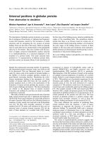
Fig. 18.1 Pneumococcal pneumonia. Homogeneous confluent
infiltrate of the right upper lobe (lobar pneumonia) in a 32-
year-old man.
a Posterio-anterior view.
b Lateral view.
18
Pulmonary Opacities
Although often used, the radiologic classification
based on the extension and pattern of infiltrates is clini-
cally of minor relevance. The following patterns may be
identified: localized or diffuse forms, unilateral or bi-
lateral, and lobar or segmental pneumonic infiltrates. If
the opacities are confluent and show an air broncho-
gram the pattern is called acinar. If the infiltrates are ill-
defined of linear, reticular, or nodular shape, and if there
is no air bronchogram, the pattern is called interstitial. A
solitary abscess is a typical complication of aspiration
pneumonia. Multiple pulmonary abscesses suggest a
hematogenic spread, such as occurs in staphylococcal
infection. Pulmonary infarction may occur as a con-
sequence of an invasive Aspergillus infection or may
occur in Pseudomonas aeruginosa pneumonia.
Clinical Presentation of Bacterial Pneumonias
Pneumonias Due to Gram-Positive
Microorganisms
As already observed by Hans Gram in 1884, the majority
of bacterial pneumonias are caused by Gram-positive
bacteria, namely Streptococcus pneumoniae, staphylo-
cocci, and streptococci. Peptococci and peptostreptococci,
Actinomyces israelii, Nocardia asteroides, Bacillus an-
thracis, and Mycobacterium tuberculosis are also positive
for staining.
Pneumococcus Pneumonia. Currently, 20−60 % of com-
munity-acquired bacterial pneumonias are caused by
Streptococcus pneumoniae, and 5% of patients with this
infection still die as a consequence. Patients with a com-
promised immune defense system, immunoglobulin
deficiency, hemoglobinopathies, or following splen-
ectomy are at particular risk.
The clinical presentation of Pneumococcal pneumonia
classically starts with sudden onset, high fever, chills,
and pleuritic chest pain. Untreated, the fever remains at
Siegenthaler, Differential Diagnosis in Internal Medicine, © 2007 Thieme
All rights reserved. Usage subject to terms and conditions of license.
525
Fig. 18.2 Staphylococcus pneumonia with focal
and confluent infiltrates in the middle and lower
lobes in a patient with Hodgkin lymphoma (lobu-
lar pneumonia or bronchopneumonia) in a 33-
year-old man.
Infectious Pulmonary Infiltrates (Pneumonias)
temperatures up to 39−40 °C. The pulse rate is also ele-
vated.
The following findings can occur:
➤ Chest percussion: dullness, increased fremitus.
➤ Auscultation: bronchial breath sounds, end-inspira-
tory rales, often perioral herpes infection, leukocyto-
sis up to 30 000/μL (30 × 10
9
/L) with pronounced left
shift, toxic granulations, and lymphopenia.
➤ Radiologic: in the chest radiograph the infiltrates are
dense, homogeneous, and clearly delineated with air
bronchograms. They may extend to entire lobes
(lobar pneumonia) (Fig. 18.1) or consist in one, or
more rarely, in multiple ill-defined infiltrates.
➤ Sputum: microscopic examination of the frothy
sputum reveals Gram-positive diplococci. Abundant
amounts of Gram-positive diplococci identified in
putrid sputum are diagnostic for Streptococcus pneu-
moniae. Since pneumococci are normal saprophytes
of the oropharyngeal mucosa (carrier rate 5−70%),
only moderate or minor amounts of Gram-positive
diplococci do not allow a definitive identification of
Streptococcus pneumoniae.
➤ Blood cultures: in hospitalized patients blood
cultures are positive in one-fourth to one-third of
cases.
The resorption of infiltrates takes place within four to
eight weeks. Delayed resorption of infiltrates may indi-
cate another diagnosis (tuberculosis, neoplasia) or a
complication, and may occur in patients in a reduced
general condition, such as from alcohol abuse, diabetes,
or chronic obstructive airway disease.
Complications of Pneumococcal pneumonia include
atelectasis, pleural empyema, parapneumonic effusion,
delaye d resorption, pulmonary abscess formation (in
approximately 2% of cases), and pericarditis. Minor ef-
fusions are common (60%), major effusions are rare
(5%). Metastatic spread of the infection leading to septic
arthritis, endocarditis, meningitis, or peritonitis is
mainly seen in immunocompromised patients or after
splenectomy.
Streptococcal and Staphylococcal Pneumonia. While
streptococcal pneumonias are relatively rare in adults,
staphylococcal infection accounts for 3−5% of bacterial
pneumoniasinoutpatients,andfor 6−24 %inhospitalized
patients. Streptococcal and staphylococcal pneumonias
mayoccurasacomplicationofinfluenzaviralinfection.In
addition, streptococcal pneumonia may result from a
spread of an upper airway infection to the lungs. Strepto-
coccal and staphylococcal pneumonias generally have an
acute course with severe febrile illness. The chest radio-
graph shows multiple patchy infiltrates spreading over
one or several lobes (bronchopneumonia; Fig. 18.2). Ab-
scess formation is a common complication that typically
occurs in staphylococcal pneumonia. The diagnosis is
confirmed by blood cultures, which are positive in
approximately 20% of staphylococcal pneumonias.
Pulmonary Actinomycosis and Nocardiosis. Pulmonary
actinomycosis has a prolonged course with fever,
phlegm, pleural pain, and leukocytosis. It is commonly
associated with manifestation of the infection at other
sites, in particular in the oral cavity and the jaw. The
Siegenthaler, Differential Diagnosis in Internal Medicine, © 2007 Thieme
All rights reserved. Usage subject to terms and conditions of license.
526
Fig. 18.3 Combined Pneumocystis
carinii and Nocardia asteroides pneu-
monia in a in a 50-year-old male
patient receiving chemotherapy.
18
Pulmonary Opacities
typical radiologic manifestation includes pleural-based
infiltrates that may extend beyond the visceral pleura to
the chest wall or to adjacent lobes. The microscopical
identification of Actinomyces in the sputum or bronchial
washings suggests the diagnosis (yellow sulfur granula
corresponding to Actinomyces israeli colonies). The
definitive cultural diagnosis may require several weeks.
Pulmonary infection by the mandatory aerobic and
weakly acid-fast staining Nocardia spp. (Nocardia
asteroides, Nocardia brasiliensis) predominantly occurs
in the immunocompromised host or in patients with
preexisting chronic lung diseases, e. g., pulmonary alve-
olar proteinosis (Fig. 18.3).
Pneumonias Due to Gram-Negative
Bacteria and Microorganisms not
Identifiable under Light Microscopy
ThisincludespneumoniasduetoHaemophilusinfluenzae,
Gram-negative Enterobacteriaceae (Klebsiella pneu-
moniae, Escherichia coli, Proteus, Enterobacter, and Serra-
tia), Pseudomonas aeruginosa, and Branhamella (Morax-
ella) catarrhalis. Legionnaires disease, Q-fever, and Bang
disease are also caused by Gram-negative bacteria (i. e.,
Legionella pneumophila, Rickettsia, and Brucella, respec-
tively). Mycoplasma pneumoniae and Chlamydia pneu-
moniae are both common causes of community-acquired
pneumonias.Thesemicroorganismscannot bevisualized
by light microscopy due to their small size. It has been
estimated that 9−20% of community-acquired pneu-
monias and more than 40 % of hospital-acquired pneu-
monias are due to Gram-negative bacteria.
Haemophilus influenzae Pneumonia. Haemophilus influ-
enzae in its capsulated or uncapsulated form is thought
to cause 3−10% of community-acquired pneumonias. It
commonly occurs in patients with chronic lung disease,
such as chronic obstructive pulmonary disease (COPD)
or bronchiectasis, who are also susceptible to Klebsiella
infection.
Pneumonias Due to Gram-Negative Enterobacteria. Kleb-
siella spp. along with Haemophilus influenzae are the
most common Gram-negative microorganisms causing
community-acquired pneumonia. Alcoholics, diabetics,
and patients with chronic airway disease are most sus-
ceptible. The clinical presentation is similar to that of
other bacterial pneumonias such as caused by Strepto-
coccus pneumoniae. The diagnosis is made by identifica-
tion of Klebsiella pneumoniae (Gram-negative, capsu-
lated diplobacillus) in the sputum or blood culture (50−
70% of blood cultures are positive).
Siegenthaler, Differential Diagnosis in Internal Medicine, © 2007 Thieme
All rights reserved. Usage subject to terms and conditions of license.
527
Infectious Pulmonary Infiltrates (Pneumonias)
Pseudomonas aeruginosa Pneumonia. This predomi-
nantly occurs in severely ill, hospitalized patients, and
in patients with bronchiectasis. More than 40% of hospi-
tal-acquired pneumonias are due to Pseudomonas and
other Gram-negative bacteria. These nosocomial pneu-
monias may be related to long-term antibiotic therapy,
immunosuppression, cytotoxic chemotherapy, or me-
chanical ventilation. Bacteriemic Pseudomonas aerugi-
nosa pneumonia is associated with a high mortality rate
(i. e., 60−70 %) despite appropriate antibiotic treatment.
Mycoplasma Pneumonia. Mycoplasma pneumoniae are
estimated to be the causative agent in 3−35% of commu-
nity-acquired pneumonias. The microorganisms are
spread to close contacts by aerosol formation during
coughing. Mycoplasma pneumonias are therefore com-
monly observed in preschools and schools, and among
military personnel. The incubation period is approxi-
mately three weeks. Following the infection, lifelong
immunity develops.
Mycoplasma pneumoniae infections may pass
without apparent symptoms, but more severe infec-
tions, with fever, bronchitis, and pneumonia, may also
occur. The disease usually starts with headaches,
malaise, and fever. The cough is usually nonproductive.
Less than 10% of patients present clinically with a typi-
cal pneumonia. Pulmonary auscultation may be normal.
Associated symptoms may include pharyngitis, rhi-
nosinusitis, and otitis (sometimes with protracted
hemorrhagic, bullous myringitis).
The chest radiograph in Mycoplasma pneumonia
shows nonspecific alterations, usually consisting in
bronchopneumonic infiltrates. Mycoplasma infections
may be associated with extrapulmonary manifestations
such as hemolysis (cold agglutinins), a rash, and arthritis.
This small microorganism cannot be seen under light
microscopy. A cultural identification may take several
weeks and is therefore clinically not useful. Instead, a
serologic examination (complement fixation) may sug-
gest a Mycoplasma infection by an increase in IgM and,
subsequently, in IgG antibodies. A four-fold increase in
antibody titers over the course of the disease or an in-
dividual titer of 1:32 are suggestive of the diagnosis.
An increase in antibodies may be expecte d within seven
to nine days after infection with a maximal response
after three to four weeks.
Chlamydia Pneumonia. Chlamydia are mandatory intra-
cellular microorganisms. Worldwide, their main mani-
festation consists of genitourinary and ophthalmic in-
fections (Chlamydia trachomatis). Pulmonary infections
are due to Chlamydia pneumoniae and, less frequently,
Chlamydia psittaci.
The clinical presentation of Chlamydia pneumoniae is
nonspecific. Symptoms may start acutely with pharyn-
gitis and a hoarse voice. Pulmonary infiltrates may
occur as ground-glass opacities and be unilatral or bi-
lateral.
A serologic diagnosis is not feasible, since immuniza-
tion without previous clinical manifestation is common.
Legionella Pneumonia (Legionnaires disease). Legionella
are ubiquitous organisms that are commonly found in
water from sprinklers, air conditioners, and humidifica-
tion systems. The infection occurs by inhalation of aero-
sol of water containing Legionella. It is not always as-
sociated with clinical disease; 1.5−20% of healthy sub-
jects have circulating antibodies.
Legionella pneumonia may be acquired in the com-
munity and in the hospital and its prevalence varies
largely, i. e., 1−22.5%. Men are more commonly affected
than women and there is a seasonal predilection in the
period from June through November.
Symptoms at the beginning of the disease include
fatigue, malaise, joint pain, and headache. One to two
days after infection, the disease manifests fever, nonpro-
ductive cough, pleuritic chest pain, nausea, vomiting, di-
arrhea, abdominal pain, and abnormal neurologic signs.
A laboratory examination may reveal a moderate
leukocytosis, proteinuria hematuria, and hypophos-
phatemia.
Radiologically, diffuse, patchy, and homogeneous
confluent infiltrates are found (Fig. 18.4). Pleural ef fu-
sion is present in 50% of patients, but abscess formation
is rare. The diagnosis of Legionella infection is based on
culture, serologic, and immunologic testing. In clinical
practice, identification of Legionella antigen in the urine
is most useful, as it provides a rapid diagnosis within
hours, whereas the cell culture diagnosis takes several
days.
Rickettsia Pneumonia. This is caused by Coxiella burnetii,
a Gram-negative coccus, which presents clinically as
Q-Fever. Although Q-fever was first described in
Queensland the letter Q stands for query, alluding to the
unknown etiology of the disease at that time. As Coxiella
is found in animal milk and secretions, farmers, animal
dealers, and veterinarians are exposed to the infection.
It can present as a flulike disease with fever, cough, my-
algia, and headaches. During physical examination
splenomegaly, enlarged and tender cervical lymph
nodes, and bradycardia may be found. Rickettsia pneu-
monia cannot be distinguished clinically, radiologically,
or histologically from pneumonia due to Mycoplasma
pneumoniae. The chest radiograph may show segmental
consolidation of lower lobes, patchy infiltrates, and
ground-glass opacities.
The differential diagnosis includes infectious mono-
nucleosis. Additional information on Q-fever is provided
in Chapter 4.
Brucella Pneumonia. Pulmonary infiltrates in Bang dis-
ease are rare and have no typical appearance. Infiltrates
may appear in the perihilar area. Diagnosis is made
serologically. Clinical presentation is described in Chap-
ter 4.
Branhamella catarrhalis Pneumonia. Branhamella or
Moraxella catarrhalis, a Gram-negative diplococcus,
may cause bronchopneumonia in patients with COPD or
with immunosuppression.
Siegenthaler, Differential Diagnosis in Internal Medicine, © 2007 Thieme
All rights reserved. Usage subject to terms and conditions of license.
528
ba
Fig. 18.4 Legionella pneumonia in a 51-year-old woman.
a In the postero-anterior chest radiograph the paracardial in-
filtrate is clearly delineated from the border of the heart
(negative silhouette phenomenon). It therefore has to be
posterior to the heart.
b This is confirmed in the lateral view.
18
Pulmonary Opacities
Psittacosis−Ornithosis. This disease is caused by Chlamy-
dia psittaci and occurs in veterinarians, zoo personnel,
and bird fanciers. As Chlamydia psittaci is not only found
in parakeets and budgerigars, but in other birds such as
chickens and pigeons, the disease has been named or-
nithosis.
If transmitted by parakeets, the clinical course may
be particularly severe. After an incubation period of 10−
14 days, severe headaches, epistaxis (in 25% of patients),
and fever with a temperature of up to 39 °C occur. The
chest radiograph shows dense, irregular infiltrates. After
an initial moderate leukocytosis with pronounced left
shift, leukopenia develops.
The diagnosis relies on a history of exposure to birds
and a positive serologic test (complement fixation titer
1:16 or higher). However, the test becomes positive only
10−14 days after onset of the disease. Asymptomatic in-
fection or a mild form of the disease (with cough, flulike
illness, and with a positive antibody reaction) are com-
mon, in particular in persons with repetitive or chronic
exposure.
Anthrax Pneumonia. Bacillus anthracis is transmitted by
contact with sheep, goat, and cattle, or with their hair or
wool, or by contaminated meat. The most common form
is cutaneous infection. Gastrointestinal manifestations
include abdominal pain, diarrhea, and life-threatening
gastrointestinal hemorrhage. Recently, anthrax has
gained renewed interest since it has been used in bio-
logical warfare and bioterrorism. Inhalation of even
small amounts of anthrax spores may cause a severe,
lethal pneumonia that is unresponsive to treatment.
Pneumonia Due to Multiple Gram-
Positive and Gram-Negative Organisms
(“Mixed Flora”)
Anaerobic and Aspiration Pneumonia. Pneumonia with
anaerobic organisms such as Fusobacteriae and
Bacteroides spp. is most commonly due to oropharyn-
geal aspiration. Rarely, it may result from hemato-
geneous spread from an intra-abdominal abscess.
Elderly persons and patients with impaired upper air-
way reflexes (due to neurologic disease or after upper
airway surgery [such as tonsillectomy]), alcoholics, and
Siegenthaler, Differential Diagnosis in Internal Medicine, © 2007 Thieme
All rights reserved. Usage subject to terms and conditions of license.
529
Fig. 18.5 Pulmonary abscess in the right
upper lung field in a 25-year-old man.
Infectious Pulmonary Infiltrates (Pneumonias)
those with reduced general health are at particular risk
of aspiration pneumonia.
Depending on body position, aspiration occurs into
the posterior segments of upper and lower lobes (in
supine patients) or into the basal lower lobe segments,
in particular on the right side (in sitting patients).
Accordingly, infiltrates are seen at these locations in
the chest radiograph. There is a tendency for abscess
formation and for empyema (Fig. 18.5). The differential
diagnosis of aspiration pneumonia related to infection
with anaerobic organisms includes massive aspiration
of acidic gastric content, which may cause an acute res-
piratory distress syndrome (ARDS) by chemical irrita-
tion. The radiologic findings include varying diffuse,
patchy, or confluent infiltrates and consolidation
(Fig. 18.6).
Siegenthaler, Differential Diagnosis in Internal Medicine, © 2007 Thieme
All rights reserved. Usage subject to terms and conditions of license.
530
a
b
Fig. 18.6 Acute respiratory distress syndrome
(ARDS) due to staphylococcal sepsis.
a Bilateral infiltrates with an air bronchogram
(arrow), endotracheal tube.
b CT scan with consolidations in the dorsal, de-
pendent lung zones.
18
Pulmonary Opacities
Pulmonary Tuberculosis
Diagnosis of Tuberculosis. The proof of a tuberculous
origin of pulmonary infiltrates is the identification of
Mycobacterium tuberculosis in the sputum, bronchial
secretions, or bronchial lavage (smear and culture).
Sputum examination has to be performed at least three
times and should be followed, if appropriate, by bron-
choscopy with bronchial washings or lavage.
The diagnosis of tuberculosis requires cell culture
identification of M. tuberculosis complex (in particular
M. tuberculosis and M. bovis). The distinction between
M. tuberculosis and mycobacteria other than tuberculo-
sis (MOTT) is generally not feasible by microscopic
examination alone, but requires cultural and molecular
biologic testing (e. g., by PCR = polymerase chain reac-
tion or MTD = gene probe amplified Mycobacterium
tuberculosis direct test). A negative PCR from a smear
positive for acid-fast bacilli makes the diagnosis of
tuberculosis highly unlikely. A positive PCR alone,
however, does not differentiate between active and in-
active tuberculosis.
There is no criterion for the definitive diagnosis of
tuberculosis infection other than a positive culture.
Siegenthaler, Differential Diagnosis in Internal Medicine, © 2007 Thieme
All rights reserved. Usage subject to terms and conditions of license.
531
Infectious Pulmonary Infiltrates (Pneumonias)
The general clinical manifestations of pulmonary tuber-
culosis are nonspecific. They include night sweats, low-
grade fever, weight loss, and chronic cough. Similar
symptoms are also caused by other diseases. A positive
skin reaction to tuberculin (i. e., a positive Mantoux
test) suggests infection with tuberculosis (or previous
BCG vaccination!) but does not allow the identification
of patients with active disease. In addition, immuno-
suppressed patients may have a negative skin reaction
despite M. tuberculosis infection. Detecting Mycobac-
terium tuberculosis-specific gamma interferon-produc-
ing T cells in blood samples from patients with sus-
pected tuberculosis has promise as a novel diagnostic
test, but its role in various settings has yet to be deter-
mined.
Classification. A distinction is made between primary
and postprimary tuberculosis, which can be associated
with acute and chronic pulmonary infiltrates.
The American Thoracic Society recommends the fol-
lowing classification of persons exposed to contacts
with tuberculosis or who have tuberculosis disease:
➤ stage 0: not exposed, not infected
➤ stage 1: exposed, infected (tuberculin skin test nega-
tive)
➤ stage 2: exposed, infected (tuberculin skin test posi-
tive)
➤ stage 3: tuberculosis disease.
Primary Tuberculosis
Clinical Features. Primary tuberculosis may occur at any
age in areas of low prevalence, whereas it affects mainly
children in areas of high prevalence. The presentation is
nonspecific with low-grade fever (usually no more than
38 °C). Therefore, diagnosis is often missed.
Laboratory Tests. The differential blood count reveals
moderate left shift and monocytosis in nearly 50% of the
cases, but there is generally no lymphocytosis or lym-
phopenia. The erythrocyte sedimentation rate is mod-
erately elevated as is the C-reactive protein (CRP).
Tuberculin Reaction. The tuberculin skin test is generally
strongly positive. Conversion occurs four to six weeks
after the infection.
Chest Radiograph. There is a peripheral infiltrate with
hilar adenopathy (Ghon primary complex).
Course. Primary tuberculosis extends over several
weeks to months but usually passes unnoticed. A small
calcified nodule (Ghon lesion) may be evident later. The
following complications may occur: necrotic cavity for-
mation in the area of the pulmonary infiltrate (primary
phthysis), bronchial compression and obstruction, or
perforation of a lymph node into the bronchial lumen
with bronchogenic spread, resulting in bronchial tuber-
culosis and caseous pneumonia. A hematogenic spread
may lead to miliary tuberculosis. Exsudative pleuritis is
also a manifestation of primary tuberculosis. In general,
primary tuberculosis heals without leaving residua or
results in the formation of calcified scars or tuberculo-
mata.
Postprimary Pulmonary Tuberculosis
Definition. The term postprimary tuberculosis (reactiva-
tion) refers to the manifestation of disease in a host who
has been previously infected with tuberculosis and has
acquired cellular immunity.
Pathogenesis. Postprimary tuberculosis represents an
endogenous reactivation of (radiologically often not ap-
parent) preexisting foci. The direct progression from
primary to postprimary tub erculosis is rare. However,
postprimary tuberculosis may also result from ex-
ogenous reinfection.
Clinical Features. Typical manifestation includes a
chronic cough, subfebrile temperature, poor appetite,
and weight loss for several weeks. In addition to pulmo-
nary tuberculosis, the infection may be reactivated in
other organs (tuberculous lymphadenitis, urogenital
tuberculosis, vertebral spine meningitis). A distinction
is made according to the following forms of the disease:
➤ localized manifestations: (exsudative pulmonary
tuberculosis, tuberculous cavity)
➤ generalized manifestation: (miliary tuberculosis)
➤ chronic: fibroproliferative lesions.
Exudative Pulmonary Tuberculosis
This term reflects extensive infection of the pulmonary
parenchyma with tubercle bacilli. The foci of infection
contain exudative inflammation and liquefied necrotic
areas with cavities (Figs. 18.7,18.8). The caseous necro-
sis and the subsequent cavitary destruction are thought
to reflect a pronounced hypersensitivity reaction to the
tuberculous antigen. Typically, the apical and posterior
segments of the upper lobes and the apical regions of
the lower lobes are involved. In the immunosuppressed
patient (e. g., with HIV infection), the chest radiograph
may not show the typical location of infiltrates and they
may involve lower lobes and may not cavitate.
Tuberculous Cavity
The cavity represents a typical manifestation of postpri-
mary tuberculosis (Fig. 18.8). It is a thick-walled cavity,
mostly in the apical and posterior segments of the upper
lobes and in the apical segments of the lower lobes.
Siegenthaler, Differential Diagnosis in Internal Medicine, © 2007 Thieme
All rights reserved. Usage subject to terms and conditions of license.
Siegenthaler, Differential Diagnosis in Internal Medicine, © 2007 Thieme
All rights reserved. Usage subject to terms and conditions of license.
532
a b
Fig. 18.7 Exudative pulmonary tuberculosis with patchy and
partly confluent infiltrates in a 39-year-old woman.
Before (a) and after (b) chemotherapy.
Fig. 18.8 Tuberculous cavity in the left upper
lobe in a 43-year-old man.
18
Pulmonary Opacities
Siegenthaler, Differential Diagnosis in Internal Medicine, © 2007 Thieme
All rights reserved. Usage subject to terms and conditions of license.
533
Fig. 18.9 Miliary tuberculosis. The
patient has miliary noduli that are
evenly distributed over their entire
lungs.
Infectious Pulmonary Infiltrates (Pneumonias)
Chest Radiograph. The differential diagnosis of a tuber-
culous cavity includes an abscess (with an air−liquid
level), aspergilloma (i. e., a fungus ball in a preformed
cavity), and squamous cell carcinoma with central
necrosis. If a cavity is not visible in the conventional
chest radiograph, it may show up in a CT scan of the
chest.
Microbiologic Diagnosis. If a cavity exists, the sputum is
usually positive for acid-fast bacilli. A repeated negative
sputum examination makes the diagnosis of a tuber-
culous cavity unlikely. If the sputum is negative in the
direct microscopic inspection, cultural identification of
the tubercle bacilli is still possible. Alternatively,
bronchial secretions or postbronchoscopic sputum may
reveal the organisms.
Miliary Tuberculosis
See also Chapter 4.
Pathogenesis. Miliary tuberculosis represents a hemato-
geneous spread of tubercle bacilli. In children, miliary
tuberculosis may occur as a primary infection. In adults
it is more likely to b e a reactivation triggered by a re-
duced cellular immune defense (poor nutrition, alco-
holism, diabetes, HIV infection, or advanced age). Mili-
ary tuberculosis may have an acute or chronic course
with only mild symptoms. This may occur in elderly
persons in whom the diagnosis may only be made post-
mortem.
Chest Radiograph. Radiographically, miliary tuberculosis
has typical features. They consist of multiple nodules,
1−3 mm in diameter, evenly distributed over the entire
lung parenchyma (Fig. 18.9). With initiation of anti-
tuberculous therapy, the miliary foci generally disap-
pear after several weeks to months without remaining
residua. Exceptionally, a patient may succumb to the in-
fection even without the miliary nodules seen in the
chest radiograph if the nodules are very small ( 1mm
in diameter).
Differential Diagnosis. The following diseases have to be
considered in the differential diagnosis of miliary tuber-
culosis:
➤ sarcoidosis
➤ silicosis
➤ exogen allergic alveolitis
➤ histiocytosis X (Langerhans cell histiocytosis)
➤ hematogeneous spread of a neoplasm
➤ microlithiasis alveolaris.
Siegenthaler, Differential Diagnosis in Internal Medicine, © 2007 Thieme
All rights reserved. Usage subject to terms and conditions of license.
534
Fig. 18.10 Fibroproductive pulmonary tuber-
culosis with multiple foci in both apical and
middle lung fields. The hilum is apically displaced
in a 31-year-old man.
Fig. 18.11 Calcified tuberculoma in a 74-year-old woman.
18
Pulmonary Opacities
Fibroproliferative Tuberculosis
In contrast to acute forms of tuberculosis, fibroprolifera-
tive tuberculosis does not show patchy infiltrates but
linear, partly calcified, densities with signs of traction
and distortion of the lung structure, mostly in the upper
lobes (Fig. 18.10). The identification of tubercle bacilli
may be difficult.
Tuberculoma
A special manifestation of tuberculosis is tuberculoma.
It is identified in the chest radiograph as a rounded,
moderately dense opacity of up to 5 cm in diameter
(Fig. 18.11), sometimes with lobulation or calcification.
Siegenthaler, Differential Diagnosis in Internal Medicine, © 2007 Thieme
All rights reserved. Usage subject to terms and conditions of license.
535
a
b
Fig. 18.12 Mycobacterium avium-intracellurare infection.
a CT scan with a cavitary lesion in the right upper lobe.
b Several nodular and patchy infiltrates in the middle lobe, the
lingula, and in the left lower lobe (arrows).
Infectious Pulmonary Infiltrates (Pneumonias)
Disease Due to Mycobacteria Other Than Tuberculosis (MOTT)
Mycobacteria other than tuberculosis (MOTT), also
called atypical mycobacteria, may present clinically in a
similar way as M. tuberculosis complex.
Diagnosis. Radiologically, multiple noduli and infiltrates
are seen. If there are cavities, they tend to be rather thin-
walled with less extension of the inflammation to the
adjacent parenchyma. Suspicion of infection with atypi-
cal mycobacteria arises if pulmonary disease does not
respond to the standard antituberculous therapy
patients with a history of previous tuberculosis, chronic
lung disease (bronchiectasis), or HIV.
The diagnosis of disease due to MOTT requires the re-
peated, cell culture identification of the organism (at
least three times) in sputum specimens, other secre-
tions, or biopsy material.
Causitive Agent Characteristics. Atypical mycobacteria
differ from the mandatory human pathogen M. tuber-
culosis in that they are ubiquitous saprophytes that sur-
vive in soil and water and rarely cause infection and dis-
ease. Some MOTT grow more rapidly in culture (M. for-
tuitum and M. chelonei) than the slow-growing tubercle
bacilli. M. avium-intracellulare, M. kansasii, M. scrofu-
laceum, M. xenopi, and M. szulgai are also slow-growing
MOTT, some of them forming pigments (M. kansasii and
M. scrofulaceum).
Manifestations. M. kansasii and M. avium-intracellulare
may cause pulmonary disease. Rarely, M. scrofulaceum,
M. szulgai, M. simiae, and M. fortuitum-chelonei, as well
as M. abscessus, may represent the cause of pulmonary
abscess formation.
A rare, but fairly characteristic, disease is observed
with M. avium-intracellulare (MAI) pulmonary infection
in patients (mostly women) suf fering from bronchiecta-
sis. The main symptom is chronic cough, usually non-
productive. The high-resolution chest CT (HRCT) scan
shows bronchiectasis with peribronchial infiltrates and
so-called “tree in bud” alterations (Fig. 18.12), corre-
sponding to peribronchial infection and inflammation.
Additional manifestations of infection with MOTT
may include lymphadenitis (M. scrofulaceum), soft
tissue abscesses, and wound infection (M. fortuitum-
chelonei).
536
18
Pulmonary Opacities
Viral Pneumonia
Influenza Pneumonia
Influenza pneumonia is fairly common and has been de-
scribed as an acutely progressive and fatal disease
during influenza epidemics. Although influenza pneu-
monias without other pathogens have been observed,
secondary infection with bacteria such as pneumococci,
streptococci, and in particular staphylococci, are also
common.
Pneumonia due to paramyxoviruses (parainfluenza,
mumps, measles, and respiratory syncitial viruses) are
common in children, but rarely occur in adults. Re-
oviruses also rarely cause pneumonia in adults and usu-
ally occur as “common cold infections.”
Adenovirus Pneumonia
Adenovirus infection is associated with pulmonary in-
filtrates in 10−20% of infections. The general symptoms
of adenovirus infection may give a hint that this
organism is causing pneumonia. In military recruits ad-
enovirus is common, as approximately 50% of the mili-
tary personnel in training camps acquire adenovirus
infection.
Acute fever with a temperature up to 39 °C, cough,
headaches, nausea, vomiting, meningitis, pharyngitis,
conjunctivitis, and lymphadenopathy precede the pneu-
monia. Leukocytosis of approximately 10000/μl(10×
10
9
/L) may be found. The duration of fever is two to
three days. The diagnosis relies on complement fixation
with a rise in antibody titers. The virus may also be iden-
tified in sputum and stool samples. Pulmonary infil-
trates are hazy and not very dense (Fig. 18.13). Pulmo-
nary auscultation is unremarkable.
Severe Acute Respiratory Syndrome
(SARS)
On March 15 2003, patients with an acutely progressive
respiratory disease were found in the Chinese province
of Guangdong, and in Hong Kong, Vietnam, Singapore,
and Canada and this finding gained worldwide interest.
The World Health Organization (WHO) coined the term
“severe acute respiratory syndrome” (SARS), and has
coordinated the efforts of identification of cases and
modes of transmission. Thanks to major scientific ef-
forts, the cause of the disease, a hitherto unknown
Coronavirus, was identified and techniques for diagnosis
and treatment guidelines were established.
Clinical Features. The disease comprises two phases
beginning with fever ( 38 °C), malaise, myalgia, and
headaches. Three to seven days later cough, dyspnea,
and subsequently respiratory failure develop. The chest
radiograph shows bilateral infiltrates and, in severe
cases, white lungs with a clinical picture of ARDS occur
(see Fig. 18.6).
Diagnosis. Evidently, SARS can not be clinically differen-
tiated from other potentially severe respiratory infec-
tions such as influenza pneumonia. The diagnosis is
made in the appropriate clinical setting (prior residence
in an endemic or epidemic SARS area). Identification of
the SARS pathogen requires special laboratory tests. The
treatment is supportive as causative therapy is currently
not available. Mortality rate depends on age and is from
10% in young patients to 40% in patients age d over 60
years.
Hantavirus Pneumonia
In 1993, patients with an acute febrile illness were ob-
served in the Southwest USA. They suffered from pro-
gressive respiratory failure and fatal shock within a few
days. Mortality rate was 50−70 %. Subsequent investiga-
tions revealed that the syndrome was due to an RNA
virus, belonging to the Bunyaviridae family, the so-
called Hantavirus. The reservoirs of this virus are mice
and rats. The infection is acquired by inhalation of aero-
solized animal excretions. Human-to-human transmis-
sion has not been observed.
The diagnosis relies on the serologic identification of
IgM antibodies or a four-fold increase in IgG antibody
titers. The differential diagnosis includes mainly other
virus diseases such as influenza.
Pneumonia Due to Nonpneumotropic
Viruses
Viruses that are not primarily pneumotropic may still
cause pneumonia. This is the case in measles pneumonia
(see Fig. 18.13). The typical exanthema is the clue to the
diagnosis. More difficult is the diagnosis of pneumonia
related to Epstein−Barr virus infection (mononucleosis
infectiosa), erythema exsudativum multiforme, hepatitis
epidemica, and choriomeningitis. In immunosuppressed
patients the varicella-zoster virus, and cytomegaloviruses
may also cause pneumonia. In varicella-zoster pneu-
monia, multiple miliary nodules with calcifications
typically occur.
Siegenthaler, Differential Diagnosis in Internal Medicine, © 2007 Thieme
All rights reserved. Usage subject to terms and conditions of license.
537
Fig. 18.13 Viral pneumonia; bilateral ground-
glass opacities of middle and lower lung fields.
Multiple fine reticulonodular opacities. Histologi-
cally, a giant cell interstitial pneumonia typical
for measles pneumonia was found in this patient.
Infectious Pulmonary Infiltrates (Pneumonias)
Fungal Pneumonia
It is crucial to distinguish pulmonary fungal infections
affecting predominantly or exclusively immunocom-
promised patients from those also observed in im-
munocompetent patients. Conditions associated with
impaired immune response that predispose to fungal
infections include agranulocytosis, neutropenia, corti-
costeroid treatment, and AIDS. In contrast, endemic fun-
gal infections occur in immunocompetent residents of
certain areas. Bronchopulmonary disease may also be
related to an immunologic reaction to fungal antigens
without invasive or only semi-invasive growth in the
bronchial tree (allergic bronchopulmonary aspergillo-
sis).
Fungus Infection in Immuno-
compromised Patients
Pneumonia Due to Yeasts and Molds
The most important agents are the yeasts Candida and
Cryptococcus, and the molds Aspergillus and Mucor. All
are found worldwide.
Diagnosis. These fungal infections are associated with
variable chest radiographic manifestations (Fig. 18.14),
sometimes mimicking bronchopneumonia, chronic
tuberculous infection, interstitial pneumonitis, or lung
cancer. The fungi are ubiquitous, and therefore their
identification in the sputum does not necessarily signify
a pulmonary infection. Invasive growth of the fungus
has to be suspected from the clinical presentation in the
particular setting of a patient. The def initive diagnosis
requires a histological proof of invasive growth along
with the cultural identification of the fungus.
Candidiasis and Aspergillosis. Candidiasis (moniliasis)
and aspergillosis are seen most commonly. As men-
tioned above, infections with the causative fungi occur
nearly exclusively in patients with immune deficiency.
Empirical treatment is generally initiated without delay
to prevent life-threatening progression of the infection
in severely ill patients. CT images in invasive aspergillo-
sis typically reveal multiple opacities, at times with cen-
tral necrosis. Pulmonary infiltrates in a patient with
positive blood cultures for Candida albicans are sugges-
tive of a generalized fungal infection with pulmonary
involvement.
Pneumocystis carinii Pneumonia
Pneumocystis carinii pneumonia is mainly observed in
patients with cancer who are receiving chemotherapy,
and as a complication of AIDS (see Fig. 18.3). Rapidly
progressive dyspnea associated with ground-glass
opacities, micronodular or homogeneous confluent in-
filtrates in the chest radiograph are consistent with
Pneumocystis pneumonia in the immunocompromised
host. The direct identification of the fungus in alveolar
macrophages may be made in spontaneous sputum or
Siegenthaler, Differential Diagnosis in Internal Medicine, © 2007 Thieme
All rights reserved. Usage subject to terms and conditions of license.
Siegenthaler, Differential Diagnosis in Internal Medicine, © 2007 Thieme
All rights reserved. Usage subject to terms and conditions of license.
538
a
b
Fig. 18.14 Invasive aspergillosis of the lung in a
patient with ill-defined pneumonic infiltrates in
both lower lobes.
a Right lower lobe.
b Left lower lobe.
18
Pulmonary Opacities
Siegenthaler, Differential Diagnosis in Internal Medicine, © 2007 Thieme
All rights reserved. Usage subject to terms and conditions of license.
539
a b
Fig. 18.15 Inactive disseminated histoplasmosis. Typically,
there are multiple calcified nodules in both lungs and calcified
hilar lymph nodes.
a Postero-anterior view.
b Close view of the right hilum.
Infectious Pulmonary Infiltrates (Pneumonias)
after sputum provocation by inhalation of 2−5 % sodium
chloride solution. The most sensitive diagnostic means
are bronchial lavage and transbronchial lung biopsies.
Endemic Fungal Infection
These infections are caused by dimorphic fungi (Histo-
plasma, Coccidioides, Paracoccidioides, Blastomyces),
which are mainly found in North and South America. In
the USA histoplasmosis and coccidioidomycosis are
common. Histoplasma spp. infection may pass unno-
ticed or manifest itself as an acute disease resulting in
multiple, partly calcified pulmonary nodules
(Fig. 18.15). The differential diagnosis includes tuber-
culous granuloma and nodules after varicella pneu-
monia. The diagnosis of histoplasmosis is supported by
the typical clinical presentation and serologic tests.
Allergic Bronchopulmonary Aspergillosis
and Mycetoma
Endobronchial growth of Aspergillus fumigatus may in-
duce immunologic type I and III reactions. Clinically, this
corresponds to bronchial asthma and allergic bron-
chopulmonary aspergillosis, respectively (see below).
The endobronchial fungi are usually noninvasive, but
semi-invasive growth may occur. Bronchopulmonary
aspergillosis is found in combination with atopy in
patients with bronchial asthma or cystic fibrosis. The di-
agnostic criteria include: bronchial asthma, bronchiec-
tasis, variable pulmonary infiltrates, increased total IgE,
specific anti-Aspergillus IgE, precipitins, a positive skin
reaction to Aspergillus, identification of Aspergillus in
the sputum, and blood eosinophilia. Radiologically
characteristic are digit-like opacities and central
bronchiectasis.
Aspergillomas (or mycetomas, fungus balls) develop
when Aspergillus grows as a clump in a preformed pul-
monary cavity, in particular in a inactive tuberculous
cavity. The radiologic documentation of a cavity with a
rounded central opacity in the dependent part is typical
(Fig. 18.16). The differential diagnosis includes pulmo-
nary abscess formation, a partly necrotic bronchial
cancer, and an Echinococcus cyst.
540
Fig. 18.16 Aspergilloma of the lung. A rim of air surrounds the
fungus ball within a tuberculous cavity seen here in a 28-year-
old man.
The following diseases should be considered in patients
with HIV infection and pulmonary infiltrates:
➤ tuberculosis
➤ atypical mycobacteriosis
➤ opportunistic infections (Pneumocystis carinii, Cyto-
megalovirus, Epstein−Barr virus, toxoplasmosis, fungi)
➤ Kaposi sarcoma (p. 561).
➤ noninfectious lymphocytic interstitial pneumonitis
➤ lymphocytic granulomatosis.
18.2 Noninfectious Pulmonar y Infiltrates
See Tab. 18.1.
Physical or Chemical Pneumonitis
If an infection cannot be identified as the cause of a
pneumonia; noninfectious physical and chemical injur-
ies of the lungs have to be considered. For example, ion-
izing radiation (Fig. 18.17), metal fumes (manganese,
cadmium, mercury, iron, aluminum), and gases (nitric
oxide [silo fillers disease, p. 557], sulfur oxide, ozone,
ammonia, phosgene, or chlorine) may damage bronchi-
oli and alveoli. Depending on the extent and the type of
the exposure bronchiolitis, pulmonary edema, pneumoni-
tis, and finally fibrosis may occur.
18
Pulmonary Opacities
Pulmonary Parasitosis
Various parasites may cause pulmonary disease. Hel-
minths (Ascaris, Ancylostoma, Strongyloides, and Filaria)
are associated with transient or chronic eosinophilic
lung infiltrates, echinococci cause pulmonary cysts, and
protozoans, such as Toxoplasma gondii, cause interstitial
pneumonia in immunocompromise d patients.
Differential Diagnosis of Pulmonary Infiltrates in HIV Infection
Siegenthaler, Differential Diagnosis in Internal Medicine, © 2007 Thieme
All rights reserved. Usage subject to terms and conditions of license.
541
Fig. 18.17 Radiation pneumonitis in the right
middle and lower lung field in a 54-year-old
woman who had received radiation therapy for
breast cancer.
Radiation Pneumonitis
Pulmonary radiation injury often goes unnoticed. It oc-
curs after radiation therapy of cancers of the breast,
lungs, esophagus, and mediastinum (thymoma, Hodg-
kin and non-Hodgkin lymphoma). The incidence of
radiation pneumonitis varies depending on the extent
and protocol of application, and it has significantly
decreased with technical improvements. Radiation
therapy of breast or lung cancer leads to alterations in
the chest radiograph in about one-third of patients, and
in 10 % of patients it causes clinical symptoms.
Clinical Features. Radiation pneumonitis is observed be-
tween one and six months after the end of treatment. It
is associated with a slowly progressive cough, fever, and
dyspnea, but it often does not cause any symptoms.
After a duration of up to one month, spontaneous resti-
tution usually occurs.
Rarely, radiation pneumonitis results in fibrosis,
sometimes even without clinically apparent acute
manifestation.
Diagnosis. Pulmonar y function tests reveal a restrictive
ventilatory defect and a reduced diffusing capacity. Ac-
cordingly, the chest radiograph shows a reduced lung
volume and focal or confluent, patchy or reticular infil-
trates (see Fig. 18.17). Radiation therapy of breast
cancer may cause bronchiolitis obliterans organizing
pneumonia (BOOP), a syndrome associated with dys-
pnea, fatigue, and varying unilateral and bilateral infil-
trates.
Lipoid Pneumonia
Chronic repeated inhalation of oily substances such as
nose drops into the bronchi may cause pulmonary infil-
trates, predominantly in the lower lobes. The diagnosis
relies on the typical history and fat droplets in the
sputum, which may be found even weeks after discon-
tinuation of the application of nose drops.
Infiltrates Due to Chronic Congestive
Heart Failure
Chest Radiography. Chronic congestive heart failure may
lead to localized unilateral or bilateral reticular infil-
trates due to pulmonary interstitial edema (Fig. 18.18).
Associated radiologic signs are cardiomegaly, redis-
tribution of pulmonary circulation from predominantly
lower zones to lower and upper zones, increased caliber
of pulmonary veins, Kerley A and B lines, and pleural ef-
fusions. Exclusively left-sided infiltrates are rarely due
to congestive heart failure. A localized interlobar effu-
sion may mimic a pulmonary mass (tumor) (Fig. 18.19),
which disappears after treatment of heart failure
(“vanishing tumor”).
Noninfectious Pulmonary Infiltrates
Siegenthaler, Differential Diagnosis in Internal Medicine, © 2007 Thieme
All rights reserved. Usage subject to terms and conditions of license.
542
a b
Fig. 18.18 Interstitial pulmonary infiltrates in a
patient with congestive heart failure.
Fig. 18.19 Right-sided interlobar effusion (vanishing tumor).
in a 46-year-old man.
Before (a) and after (b) treatment.
18
Pulmonary Opacities
Siegenthaler, Differential Diagnosis in Internal Medicine, © 2007 Thieme
All rights reserved. Usage subject to terms and conditions of license.