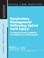
Respiratory Management Following Spinal Cord Injury: A Clinical Practice Guideline for Health-Care Professionals ppt
Bạn đang xem bản rút gọn của tài liệu. Xem và tải ngay bản đầy đủ của tài liệu tại đây (299.67 KB, 62 trang )
Respiratory
Management
Following Spinal
Cord Injury:
A Clinical Practice Guideline
for Health-Care Professionals
Administrative and financial support provided by
Paralyzed Veterans of America
SPINAL CORD MEDICINE
CLINICAL PRACTICE GUIDELINE: RESPIRATORY
MANAGEMENT
Consortium for Spinal Cord Medicine
Member Organizations
American Academy of Orthopedic Surgeons
American Academy of Physical Medicine and Rehabilitation
American Association of Neurological Surgeons
American Association of Spinal Cord Injury Nurses
American Association of Spinal Cord Injury Psychologists and Social Workers
American College of Emergency Physicians
American Congress of Rehabilitation Medicine
American Occupational Therapy Association
American Paraplegia Society
American Physical Therapy Association
American Psychological Association
American Spinal Injury Association
Association of Academic Physiatrists
Association of Rehabilitation Nurses
Christopher Reeve Paralysis Foundation
Congress of Neurological Surgeons
Insurance Rehabilitation Study Group
International Spinal Cord Society
Paralyzed Veterans of America
U.S. Department of Veterans Affairs
United Spinal Association
CLINICAL PRACTICE GUIDELINE
Spinal Cord Medicine
Respiratory Management
Following Spinal Cord Injury:
A Clinical Practice Guideline
for Health-Care Professionals
Consortium for Spinal Cord Medicine
Administrative and financial support provided by Paralyzed Veterans of America
© Copyright 2005, Paralyzed Veterans of America
No copyright ownership claim is made to any portion of these materials contributed by departments or employees
of the United States Government.
This guideline has been prepared based on the scientific and professional information available in
2004. Users of this guideline should periodically review this material to ensure that the advice
herein is consistent with current reasonable clinical practice.
January 2005
ISBN: 0-929819-16-0
CLINICAL PRACTICE GUIDELINE
iii
v Preface
vi Acknowledgments
vii Panel Members
viii Contributors
1 Summary of Recommendations
5 The Consortium for Spinal Cord Medicine
5 GUIDELINE DEVELOPMENT PROCESS
6 METHODOLOGY
6 THE LITERATURE SEARCH
6 GRADING OF ARTICLES
7 GRADING THE GUIDELINE RECOMMENDATIONS
7 GRADING OF PANEL CONSENSUS
8 Recommendations
8 INITIAL ASSESSMENT OF ACUTE SCI
9 PREVENTION AND TREATMENT OF ATELECTASIS AND PNEUMONIA
12 MEDICATIONS
13 MECHANICAL VENTILATION
14 INDICATIONS FOR MECHANICAL VENTILATION
14 RESPIRATORY FAILURE
14 INTRACTABLE ATELECTASIS
15 LARGE VERSUS SMALL TIDAL VOLUMES
16 SURFACTANT, POSITIVE-END EXPIRATORY PRESSURE (PEEP), AND ATELECTASIS
16 COMPLICATIONS OF SHORT-TERM AND LONG-TERM VENTILATION
16 ATELECTASIS
16 PNEUMONIA
17 PULMONARY EMBOLISM AND PLEURAL EFFUSION
17 LONG-TERM VENTILATION
18 CUFF DEFLATIONS
18 WEANING FROM THE VENTILATOR
18 PROGRESSIVE VENTILATOR-FREE BREATHING VERSUS SYNCHRONIZED
INTERMITTENT MANDATORY VENTILATION
19 PARTIAL WEANING
19 ELECTROPHRENIC RESPIRATION
20 SLEEP-DISORDERED BREATHING
21 DYSPHAGIA AND ASPIRATION
23 PSYCHOSOCIAL ASSESSMENT AND TREATMENT
23 ADJUSTMENT TO VENTILATOR-DEPENDENT TETRAPLEGIA
23 ENHANCEMENT OF COPING SKILLS AND WELLNESS
23 AFFECTIVE STATUS
23 SUBSTANCE ABUSE
24 PAIN
24 SECONDARY MILD BRAIN INJURY
24 DECISION-MAKING CAPACITY
25 ADVANCE DIRECTIVES
25 FAMILY CAREGIVING
25 INTIMACY AND SEXUALITY
25 ESTABLISHMENT OF AN EFFECTIVE COMMUNICATION SYSTEM
26 EDUCATION PROGRAM DEVELOPMENT
27 DISCHARGE PLANNING
27 HOME MODIFICATIONS
Table of Contents
27 CAREGIVERS
28 DURABLE MEDICAL EQUIPMENT
28 TRANSPORTATION
28 FINANCES
28 LEISURE
29 VOCATIONAL PURSUITS
29 TRANSITION RESOURCES
30 Recommendations for Future Research
31 Appendix A: Respiratory Care Protocol
34 Appendix B: Protocol for Ventilator-Dependent Quadriplegic
Patients
36 Appendix C: Wean Protocol for Ventilator-Dependent
Quadriplegic Patients
37 Appendix D: Wean Discontinuation Protocol
38 Appendix E: Cuff Deflation Protocol for Ventilator-Dependent
Quadriplegic Patients
40 Appendix F: Cuff Deflation Discontinuation Protocol
41 Appendix G: High Cuff Pressures Protocol
42 Appendix H: Post-Tracheoplasty/Post-Extubation Protocol
43 Appendix I: Criteria for Decannulation of Trach Patients
44 Appendix J: Evaluation of High Peak Pressure on Mechanically
Ventilated Patients
45 References
49 Index
iv
RESPIRATORY MANAGEMENT FOLLOWING SPINAL CORD INJURY
CLINICAL PRACTICE GUIDELINE
v
Preface
O
ur panel attempted to develop guidelines that would meet the needs of a per-
son with recent onset spinal cord injury who is in respiratory distress. This
document represents the best recommendations that we could provide given
the availability of scientific evidence. As chairman of the panel writing these
guidelines, my goal was to gather and disseminate the best available knowledge
and information about managing the respiratory needs of patients with ventila-
tion problems. I know from many years of personal experience that the acute
respiratory management of persons with spinal cord injuries is highly variable,
and there is a great need for development of scientifically based standards of
care. Unfortunately, our review of the available literature demonstrated that
there are not widely accepted guidelines for some aspects of respiratory man-
agement because appropriate research studies have not been published for
some of the topics that needed coverage. Because the scientific basis of many
of our recommendations is not clearly established, wherever necessary, we
developed consensus-based recommendations.
Many questions still need to be answered. What is the appropriate
way to ventilate a person who has partial or complete paralysis of the
muscles of respiration? What are the criteria for weaning from the venti-
lator? How much work does ventilation require? How can patients who
have impaired ventilation put forth the additional effort required for other
activities without becoming exhausted? Again, from my personal experi-
ence, many patients are suffering because of lack of answers that would
allow widespread agreement on these management issues.
From the earliest days of the Consortium for Spinal Cord Medicine,
we have known that scientific evidence was not always available to defini-
tively settle all the issues that could be raised on a topic. So we include
an analysis of needs for future research studies. The members of our
excellent panel hope that future studies will clarify the problems and
define solutions. Despite shortcomings pointed out during the review
process, this document may help medical providers become more atten-
tive to the needs of such patients.
I extend heart-felt gratitude to my colleagues on the panel for their
faithful work and to the reviewers for their valuable input! I also want to
extend my great appreciation to the Paralyzed Veterans of America for
making this effort possible. It was my great pleasure to work with all of
you!
Kenneth C. Parsons, MD
Chair, Steering Committee
Consortium for Spinal Cord Medicine
Acknowledgments
The chairman and members of the respiratory management guideline
development panel wish to express special appreciation to the individuals and
professional organizations who are members of the Consortium for Spinal Cord
Medicine and to the expert clinicians and health-care providers who reviewed the
draft document. Special thanks go to the consumers, advocacy organizations, and
the staff of the numerous medical facilities and spinal cord injury rehabilitation
centers who contributed their time and expertise to the development of this
guideline.
Douglas McCrory, MD, and colleagues at Duke Evidence-based Practice Center
(EPC), Center for Clinical Health Policy Research in Durham, North Carolina, served
as consultant methodologists. They masterfully conducted the initial and secondary-
level literature searches, evaluated the quality and strength of the scientific evidence,
constructed evidence tables, and graded the quality of research for all identified
literature citations. This included an update and expansion to the original scope of
work in the EPC Evidence Report, Treatment of Pulmonary Disease Following
Cervical Spinal Cord Injury, developed under contract 290-97-0014 with the Agency
for Healthcare Research and Quality (AHRQ).
Members of the consortium steering committee, representing 19 professional,
payer, and consumer organizations, were joined in the guideline development
process by 30 expert reviewers. Through their critical analysis and thoughtful
comments, the recommendations were refined and additional supporting evidence
from the scientific literature was identified. The quality of the technical assistance
by these dedicated reviewers contributed significantly to the professional consensus
building that is hopefully achieved through the guideline development process.
William H. Archambault, Esq., conducted a comprehensive analysis of the legal and
health policy issues associated with this complex, multifaceted topic. In addition,
the consortium and development panel are most appreciative for the excellent
consultation and editing of the education section provided by Theresa Chase, RN,
director of patient education at Craig Hospital, Englewood, Colorado.
The guideline development panel is grateful for the many technical support
services provided by various departments of the Paralyzed Veterans of America
(PVA). In particular, the panel recognizes J. Paul Thomas and Kim S. Nalle in the
Consortium Coordinating Office for their help in organizing and managing the
process; James A. Angelo, Kelly Saxton, and Karen Long in the Communications
Department for their guidance in writing, formatting, and creating art; and medical
editor Joellen Talbot for her excellent technical review and editing of the clinical
practice guideline (CPG). Appreciation is expressed for the steadfast commitment
and enthusiastic advocacy of the entire PVA Board of Directors and of PVA’s senior
officers, including National President Randy L. Pleva, Sr.; Immediate Past President
Joseph L. Fox, Sr.; Executive Director Delatorro L. McNeal; Deputy Executive
Director John C. Bollinger; and Director of Research, Education, and Practice
Guidelines Thomas E. Stripling. PVA’s generous financial support has made the CPG
consortium and its guideline development process a successful venture.
vi
RESPIRATORY MANAGEMENT FOLLOWING SPINAL CORD INJURY
Panel Members
Kenneth C. Parsons, MD
Panel Chair
(Physical Medicine and Rehabilitation)
Institute for Rehabilitation Research
Houston, TX
Richard Buhrer, MN, RN, CRRN-A
(SCI Nursing)
VA Puget Sound Health Care System
Seattle, WA
Stephen P. Burns, MD
(Physical Medicine and Rehabilitation)
VA Puget Sound Health Care System
Seattle, WA
Lester Butt, PhD, ABPP
(Psychology)
Craig Hospital
Englewood, CO
Fina Jimenez, RN, MEd
(SCI Nursing)
Vancouver Hospital and Health Sciences Center
Vancouver, BC, Canada
Steven Kirshblum, MD
(Physical Medicine and Rehabilitation)
Kessler Institute for Rehabilitation
West Orange, NJ
Douglas McCrory, MD
(Evidence-based Methodology)
Duke Evidence-based Practice Center
Duke University Medical Center
Durham, NC
W. Peter Peterson, MD (Ret.)
(Pulmonary Disease and Internal Medicine)
Denver, CO
Louis R. Saporito, BA, RRT
(Respiratory Therapy)
Wayne, NJ
Patricia Tracy, LCSW
(Social Work)
Craig Hospital
Englewood, CO
CLINICAL PRACTICE GUIDELINE
vii
Consortium Member Organizations
and Steering Committee
Representatives
American Academy of Orthopedic Surgeons
E. Byron Marsolais, MD
American Academy of Physical Medicine and Rehabilitation
Michael L. Boninger, MD
American Association of Neurological Surgeons
Paul C. McCormick, MD
American Association of Spinal Cord Injury Nurses
Linda Love, RN, MS
American Association of Spinal Cord Injury Psychologists and
Social Workers
Romel W. Mackelprang, DSW
American College of Emergency Physicians
William C. Dalsey, MD, FACEP
American Congress of Rehabilitation Medicine
Marilyn Pires, MS, RN, CRRN-A, FAAN
American Occupational Therapy Association
Theresa Gregorio-Torres, MA, OTR
American Paraplegia Society
Lawrence C. Vogel, MD
American Physical Therapy Association
Montez Howard, PT, MEd
American Psychological Association
Donald G. Kewman, PhD, ABPP
American Spinal Injury Association
Michael Priebe, MD
Association of Academic Physiatrists
William O. McKinley, MD
Association of Rehabilitation Nurses
Audrey Nelson, PhD, RN
Christopher Reeve Paralysis Foundation
Samuel Maddox
Congress of Neurological Surgeons
Paul C. McCormick, MD
Insurance Rehabilitation Study Group
Louis A. Papastrat, MBA, CDMS, CCM
International Spinal Cord Society
John F. Ditunno, Jr., MD
Paralyzed Veterans of America
James Dudley, BSME
U.S. Department of Veterans Affairs
Margaret C. Hammond, MD
United Spinal Association
Vivian Beyda, DrPH
Expert Reviewers
American Academy of Physical Medicine and Rehabilitation
David Chen, MD
Rehabilitation Institute of Chicago
Michael Y. Lee, MD
University of North Carolina at Chapel Hill
Steven A. Stiens, MD, MS
University of Washington
American Association of Spinal Cord Injury Nurses
Cathy Farnan, RN, MS, CRRN, ONC
Thomas Jefferson University Hospital
Jeanne Mervine, MS, RN, CRNN
Schwab Rehabilitation Hospital
Mary Ann Reilly, BSN, MS, CRRN
Santa Clara Valley Medical Center
American Association of Spinal Cord Injury Psychologists and
Social Workers
Charles H. Bombardier, PhD
Rehabilitation Medicine, University of Washington School
of Medicine
Terrie Price, PhD
Rehabilitation Institute of Kansas City
American Congress of Rehabilitation Medicine
Marilyn Pires, RN, MS, CRRN-A, FAAN
Rancho Los Amigos National Rehabilitation Center
Karen Wunch, MS, RN, CRRN, CNAA, FACRM
Rancho Los Amigos National Rehabilitation Center
American Occupational Therapy Association
Franki Cassaday, OTR
Craig Hospital
Gabriella G. Stiefbold, OTR, ATP
Kessler Institute for Rehabilitation
American Paraplegia Society
David W. Hess, PhD, ABPP (RP)
Virginia Commonwealth University
Michael Y. Lee, MD
University of North Carolina at Chapel Hill
Steven A. Stiens, MD, MS
University of Washington
viii
RESPIRATORY MANAGEMENT FOLLOWING SPINAL CORD INJURY
Contributors
American Physical Therapy Association
Elizabeth Alvarez, PT
University of Maryland Medical Center
R. Adams Cowley Shock Trauma Center
Kendra L. Betz, MS, PT
VA Puget Sound Health Care System
American Spinal Injury Association
John Bach, MD
The University Hospital
University of Medicine and Dentistry of New Jersey
David Chen, MD
Rehabilitation Institute of Chicago
Association of Rehabilitation Nurses
E. Catherine Cash, RN, MSN
James A. Haley VA Medical Center
Iliene Page, BSN, MSN, ARNP-C
James A. Haley VA Medical Center
Insurance Rehabilitation Study Group
Louis A. Papastrat, MBA, CCM, CDMS,
Vice President
Medical Management AmReHealthCare
Adam L. Seidner, MD, MPH
Travelers Property Casualty Company
James Urso, BA
Travelers Property Casualty Company
U.S. Department of Veterans Affairs
Jeffrey Harrow, MD, PhD
Spinal Cord Injury Service
James A. Haley VA Medical Center
Steve H. Linder, MD
VA Palo Alto Health Care System
United Spinal Association
Harry G. Goshgarian, PhD
Wayne State University, School of Medicine
Consulting Reviewer
Robert Levine, MD
University of Texas School of Medicine, Houston
CLINICAL PRACTICE GUIDELINE
ix
Initial Assessment
of Acute SCI
1. Guide the initial management of people presenting
with suspected or possible spinal cord injury in the
field and in the emergency department using the
American Heart Association and the American Col-
lege of Surgeons’ principles of basic life support,
advanced cardiac life support, and advanced trau-
ma life support.
2. Perform an initial history and physical exam to
include the following:
Relevant past medical history.
Prior history of lung disease.
Current medications.
Substance abuse.
Neurologic impairment.
Coexisting injuries.
3. The initial laboratory assessment should include:
Arterial blood gases.
Routine laboratory studies (complete blood
count, chemistry panel, coagulation profile,
cardiac enzyme profile, urinalysis, toxicology
screen).
Chest x-ray.
EKG.
Conduct periodic assessments of respiratory func-
tion to include:
Respiratory complaints.
Physical examination of the respiratory
system.
Chest imaging as indicated.
Continuous pulse oximetry.
Performance of the respiratory muscles:
vital capacity (VC) and maximal negative
inspiratory pressure.
Forced expiratory volume in 1 second
(FEV
1
) or peak cough flow.
Neurological level and extent of impairment.
4. Monitor oxygen saturation and end tidal CO
2
to
measure the quality of gas exchange during the
first several days after injury in correlation with
patient expression of respiratory distress.
Prevention and Treatment
of Atelectasis and Pneumonia
5. Monitor indicators for development of atelectasis
or infection, including:
Rising temperature.
Change in respiratory rate.
Shortness of breath.
Increasing pulse rate.
Increasing anxiety.
Increased volume of secretions, frequency of
suctioning, and tenacity of secretions.
Declining vital capacity.
Declining peak expiratory flow rate,
especially during cough.
6. Intubate the patient for the following reasons:
Intractable respiratory failure, especially if
continuous positive airway pressure (CPAP)
and bi-level positive airway pressure (BiPAP)
or noninvasive ventilation has failed.
Demonstrable aspiration or high risk for
aspiration plus respiratory compromise.
7. If the vital capacity shows a measurable decline,
investigate pulmonary mechanics and ventilation
with more specific tests.
8. Implement the following steps to clear the airway
of secretions:
Assisted coughing.
Use of an in-exsufflator/exsufflator.
Intermittent Positive Pressure Breathing
(IPPB) “stretch.”
Glossopharyngeal breathing.
Deep breathing and coughing.
Incentive spirometry.
Chest physiotherapy.
CLINICAL PRACTICE GUIDELINE
1
Summary of Recommendations
Intrapulmonary percussive ventilation.
Continuous positive airway pressure (CPAP)
and bi-level positive airway pressure
(BiPAP).
Bronchoscopy.
Positioning (Trendelenburg or supine).
9. Determine the status of the movement of the
diaphragm (right and left side) by performing a
diaphragm fluoroscopy.
10. Successful treatment of atelectasis or pneumonia
requires reexpansion of the affected lung tissue.
Various methods include:
Deep breathing and voluntary coughing.
Assisted coughing techniques.
Insufflation—exsufflation treatment.
IPPB “stretch.”
Glossopharyngeal breathing.
Incentive spirometry.
Chest physiotherapy.
Intrapulmonary percussive ventilation (IPV).
Continuous positive airway pressure (CPAP)
and bi-level positive airway pressure
(BiPAP).
Bronchoscopy with bronchial lavage.
Positioning the patient in the supine or
Trendelenburg position.
Abdominal binder.
Medications.
Mechanical Ventilation
Indications for Mechanical
Ventilation
Respiratory Failure
Intractable Atelectasis
11. If the patient needs mechanical ventilation, use a
protocol that includes increasing ventilator tidal
volumes to resolve or prevent atelectasis.
12. Set the ventilator so that the patient does not over-
ride the ventilator settings.
Surfactant, Positive-End
Expiratory Pressure (PEEP),
and Atelectasis
13. Recognize the role of surfactant in atelectasis,
especially when the patient is on the ventilator.
Complications of Short-Term
and Long-Term Ventilation
Atelectasis
14. Use a protocol for ventilation that guards against
high ventilator peak inspiratory pressures. Con-
sider the possibility of a “trapped” or deformed
lung in individuals who have trouble weaning and
have had a chest tube or chest surgery.
Pneumonia
15. Employ active efforts to prevent pneumonia,
atelectasis, and aspiration.
Pulmonary Embolism and Pleural
Effusion
16. Monitor ventilated patients closely for pulmonary
embolism and pleural effusion.
Long-Term Ventilation
17. Evaluate the need for long-term ventilation.
Order equipment as soon as possible.
If a ventilator is needed, recommend that
patients also have a backup ventilator.
Weaning from the Ventilator
18. Consider using progressive ventilator-free breath-
ing (PVFB) over synchronized intermittent manda-
tory ventilation (SIMV).
PVFB Versus SIMV
Partial Weaning
Electrophrenic Respiration
19. For apneic patients, consider evaluation for elec-
trophrenic respiration.
20. Consider the advantages of acute and long-term
use of noninvasive ventilation over initial intuba-
tion and long-term tracheostomy if the treatment
staff has the expertise and experience in the use of
such devices.
2
RESPIRATORY MANAGEMENT FOLLOWING SPINAL CORD INJURY
Sleep-Disordered Breathing
21. Perform a polysomnographic evaluation for those
patients with excessive daytime sleepiness or other
symptoms of sleep-disordered breathing.
22. Prescribe positive airway pressure therapy if sleep-
disordered breathing is diagnosed.
Dysphagia and Aspiration
23. Evaluate the patient for the following risk factors:
Supine position.
Spinal shock.
Slowing of gastrointestinal tract.
Gastric reflux.
Inability to turn the head to spit out
regurgitated material.
Medications that slow gastrointestinal activity
or cause nausea and vomiting.
Recent anterior cervical spine surgery.
Presence of a tracheostomy.
Advanced age.
24. Prevent aspiration by involving all caregivers,
including respiratory therapists, speech therapists,
physical therapists, pharmacists, nurses, and
physicians, in the care of the patient.
Institute an alert system for patients with a
high risk for aspiration.
Position the patient properly.
Ensure easy access to a nurse call light and
alarm system.
Have the patient sit when eating, if possible.
Screen patients without a tracheostomy who
have risk factors or signs and symptoms of
dysphagia.
If the patient is found to be aspirating and is
on large ventilator tidal volumes, monitor the
peak inspiratory pressure closely.
25. Consider a tracheostomy for patients who are aspi-
rating. If the patient has a tracheostomy and is
aspirating, the tracheostomy cuff should only be
deflated when the speech therapist—and possibly
a nurse or respiratory therapist as well—is pre-
sent. (All involved personnel should be expert in
suctioning.) Monitor SPO
2
as an early indicator of
an aspiration impact.
Psychosocial Assessment
and Treatment
Adjustment to Ventilator-Dependent
Tetraplegia
26. Consider the manner in which the individual is
accommodating to the spinal cord injury, including
the individual’s post-injury psychological state.
Enhancement of Coping Skills and
Wellness
27. Assist the patient and family in the development,
enhancement, and use of coping skills and health
promotion behaviors.
Affective Status
28. Monitor the patient’s post-injury feeling states,
specifically for the emergence of depression and
anxiety.
Substance Abuse
29. Assess the patient for the presence of comorbid
substance abuse beginning in the acute rehabilita-
tion setting.
Pain
30. Assess the patient’s level of pain, if any, and estab-
lish the type of pain to determine the most appro-
priate physical and psychological treatment
modalities.
Secondary Mild Brain Injury
31. Assess for possible comorbid brain trauma as indi-
cated by the clinical situation.
Decision-Making Capacity
32. Determine the individual’s capacity to make
decisions and give informed consent on medical-
related issues by examining the following:
Organicity.
Medications.
Psychological reactions.
Pre-morbid substance abuse.
Pain.
CLINICAL PRACTICE GUIDELINE
3
Advance Directives
33. Discuss advance directives, specifically the living
will and durable power for medical health care,
with the competent patient or the patient’s proxy
to determine the validity of the documents post
trauma.
Family Caregiving
34. As appropriate, assess and support family
functioning.
Intimacy and Sexuality
35. Explore issues of intimacy and sexuality with the
patient and other appropriate parties.
Establishment of an Effective
Communication System
36. Assess the patient’s ability to communicate, and
ensure that all staff can effectively interact with
the patient to determine his or her needs and
concerns.
Education Program
Development
37. Plan, design, implement, and evaluate an educa-
tional program to help individuals with SCI and
their families and caregivers gain the knowledge
and skills that will enable the individual to main-
tain respiratory health, prevent pulmonary compli-
cations, return home, and resume life in the
community as fully as possible.
Discharge Planning
38. Working with the multidisciplinary rehabilitation
team, the patient, and his or her family, develop a
discharge plan to assist the individual with
ventilator-dependent spinal cord injury in
transitioning from the health-care facility to a
less restrictive environment, preferably a home
setting.
Home Modifications
39. Evaluate and then modify the home environment
to accommodate the demands of wheelchair
access and respiratory equipment.
Caregivers
40. Home health-care workers, family members, pri-
vately hired assistants, and others trained in per-
sonal care and respiratory management of the
individual with spinal cord injury should provide
care or be available to assist the patient 24 hours a
day. Efficient care of the patient depends on care-
ful charting by home caregivers and proper man-
agement of the home medical supply inventory.
Durable Medical Equipment
41. Prescribe the appropriate durable medical equip-
ment for home use based on the evaluations of
therapy staff and the patient. Consider emergency
provisions (e.g., backup generator and alarms)
and assistive technology as part of a safe and
effective environment.
Transportation
42. Use a van equipped with a lift and tie downs or
accessible public transportation to transport the
person with ventilator-dependent spinal cord
injury. The patient should be accompanied by an
attendant trained in personal and respiratory care.
Finances
43. Evaluate thoroughly the patient’s personal and
financial resources and provide expert guidance in
applying for benefits and coordinating assets to
maximize all available resources.
Leisure
44. Explore and provide information on diversionary
pursuits, leisure interests, local community
resources, and adaptive recreational equipment.
Vocational Pursuits
45. Arrange a vocational evaluation to determine spe-
cial aptitudes, interests, and physical abilities; fac-
tor in the need for transportation and attendant
services.
Transition Resources
46. Identify medical and other transition resources in
the home community, including:
Local specialists.
Respiratory services.
Home supply and durable medical equipment
vendors.
Pharmacies.
Home health-care services.
Advocacy groups.
4
RESPIRATORY MANAGEMENT FOLLOWING SPINAL CORD INJURY
CLINICAL PRACTICE GUIDELINE
5
S
eventeen organizations, including PVA, joined in a
consortium in June 1995 to develop clinical prac-
tice guidelines in spinal cord medicine. A steering
committee governs consortium operation, leading
the guideline development process, identifying top-
ics, and selecting panels of experts for each topic.
The steering committee is composed of one repre-
sentative with clinical practice guideline experience
from each consortium member organization. PVA
provides financial resources, administrative sup-
port, and programmatic coordination of consortium
activities.
After studying the processes used to develop
other guidelines, the consortium steering commit-
tee unanimously agreed on a new, modified,
clinical/epidemiologic evidence-based model
derived from the Agency for Healthcare Research
and Quality (AHRQ). The model is:
Interdisciplinary, to reflect the numerous
informational needs of the spinal cord
medicine practice community.
Responsive, with a time line of 12 months
for completion of each set of guidelines.
Reality-based, to make the best use of the
time and energy of the busy clinicians who
serve as panel members and field expert
reviewers.
The consortium’s approach to the develop-
ment of evidence-based guidelines is both innova-
tive and cost-efficient. The process recognizes the
specialized needs of the national spinal cord medi-
cine community, encourages the participation of
both payer representatives and consumers with
spinal cord injury, and emphasizes the use of grad-
ed evidence available in the international scientific
literature.
The Consortium for Spinal Cord Medicine is
unique to the clinical practice guidelines field in that
it employs highly effective management strategies
based on the availability of resources in the health-
care community; it is coordinated by a recognized
national consumer organization with a reputation
for providing effective service and advocacy for
people with spinal cord injury and disease; and it
includes third-party and reinsurance payer organiza-
tions at every level of the development and dissemi-
nation processes. The consortium expects to
initiate work on two or more topics per year, with
evaluation and revision of previously completed
guidelines as new research demands.
Guideline Development
Process
The guideline development process adopted
by the Consortium for Spinal Cord Medicine con-
sists of twelve steps, leading to panel consensus
and organizational endorsement. After the steer-
ing committee chooses a topic, a panel of experts
is selected. Panel members must have demon-
strated leadership in the topic area through inde-
pendent scientific investigation and publication.
Following a detailed explication and specification
of the topic by select steering committee and
panel members, consultant methodologists review
the international literature; prepare evidence tables
that grade and rank the quality of the research,
and conduct statistical meta-analyses and other
specialized studies as needed. The panel chair
then assigns specific sections of the topic to the
panel members based on their area of expertise.
Writing begins on each component using the refer-
ences and other materials furnished by the
methodology support group.
After the panel members complete their sec-
tions, a draft document is generated during the
first full meeting of the panel. The panel incorpo-
rates new literature citations and other evidence-
based information not previously available. At this
point, charts, graphs, algorithms, and other visual
aids, as well as a complete bibliography, are
added, and the full document is sent to legal coun-
sel for review.
After legal analysis to consider antitrust,
restraint-of-trade, and health policy matters, the
draft document is reviewed by clinical experts from
each of the consortium organizations plus other
select clinical experts and consumers. The review
comments are assembled, analyzed, and entered
into a database, and the document is revised to
reflect the reviewers’ comments. Following a sec-
ond legal review, the draft document is distributed
to all consortium organization governing boards.
Final technical details are negotiated among the
panel chair, members of the organizations’ boards,
and expert panelists. If substantive changes are
required, the draft receives a final legal review.
The document is then ready for editing, formatting,
and preparation for publication.
The benefits of clinical practice guidelines for
the spinal cord medicine practice community are
The Consortium for
Spinal Cord Medicine
numerous. Among the more significant applica-
tions and results are the following:
Clinical practice options and care standards.
Medical and health professional education
and training.
Building blocks for pathways and algorithms.
Evaluation studies of guideline use and
outcomes.
Research gap identification.
Cost and policy studies for improved
quantification.
Primary source for consumer information
and public education.
Knowledge base for improved professional
consensus building.
Methodology
Literature Search
For this guideline on respiratory management,
a literature search was designed to identify empiri-
cal evidence on patients with acute traumatic cer-
vical SCI, regardless of the degree of completeness
of injury. We focused on the period of days to
months following acute injury as well as on the
long-term followup over years. Excluded from
consideration were nonpulmonary complications
of SCI and venous thromboembolism/pulmonary
embolus. The evidence does not cover patients
with SCI occurring below the cervical level or res-
piratory muscle weakness caused by neuromuscu-
lar or other spinal cord diseases, such as
Guillain-Barré syndrome and polio. The databases
searched for literature were MEDLINE (1966–Dec
2000), HealthSTAR (1975–Dec 2000), Cumulative
Index to Nursing & Allied Health Literature
(CINAHL) (1983–Jan 2001), and EMBASE
(1980–Feb 2000). The search strategies com-
bined an SCI concept (implemented using MeSH
terms spinal cord injuries, paraplegia, and quadri-
plegia [exploded] and text words for tetraplegia,
quadriplegia, and paraplegia) with a pulmonary
disease concept. The search was limited to arti-
cles pertaining to humans and published in the
English language.
Empirical studies or review articles were
included after screening by the following criteria:
1. The study population includes traumatic
cervical SCI.
2. The study question relates to the research
questions described above.
3. The study includes data on health outcomes,
health services utilization, economic
outcomes, or physiological measures related
to respiratory status.
4. The study design is controlled trial,
prospective trial with historical controls,
prospective or retrospective cohort study, or
case series with 10 or more subjects.
Articles were excluded when the study popula-
tion was children (all subjects or mean age < 18
years) or when the study design a case series with
fewer than 10 subjects or a case report. Each arti-
cle was independently reviewed by at least two
investigators.
Grading of Articles
For grading internal validity, the investigators
employed the hierarchy outlined in Table 1.
TABLE 1
Hierarchy of the Levels of Scientific Evidence
Level Description
I Large randomized trials with clear-cut results
(and low risk of error)
II Small randomized trials with uncertain results
(and moderate to high risk of error)
III Nonrandomized trials with concurrent or con-
temporaneous controls
IV Nonrandomized trials with historical controls
V Case series with no controls
Each study was also evaluated for factors
affecting external validity using the following
criteria:
Were the criteria for selection of patients
described?
Were patients included in the study
adequately characterized with regard to level
and completeness of SCI?
Were criteria for outcomes clearly defined
(e.g., timing, measurement, reliability)?
Was the clinical care of patients adequately
described to be able to be reproduced?
Were the results reported according to level
of injury (minimum high cervical [C4 or
above] versus low cervical [below C4]) or
ventilation status (independently breathing
versus ventilator dependent)?
6
RESPIRATORY MANAGEMENT FOLLOWING SPINAL CORD INJURY
These items were not aggregated into an over-
all quality score, but were considered individually.
Studies meeting the above criteria were summa-
rized in the AHRQ evidence report or in update
reports, which included additional topics searched
expressly for this guideline, prepared for the
expert guideline panel. Additional studies that do
not meet the above criteria are cited in some sec-
tions of the report when sufficient high-quality evi-
dence on the target population was not available.
These studies are not graded according to the
quality criteria.
Grading the Guideline
Recommendations
After panel members had drafted their sec-
tions of the guideline, each recommendation was
graded according to the level of scientific evidence
supporting it. The framework used by the
methodology team is outlined in Table 2. It should
be emphasized that these ratings, like the evidence
table ratings, represent the strength of the sup-
porting evidence, not the strength of the recom-
mendation itself. The strength of the
recommendation is indicated by the language
describing the rationale.
TABLE 2
Categories of the Strength of Evidence
Associated with the Recommendations
Category Description
A The guideline recommendation is supported by
one or more level I studies.
B The guideline recommendation is supported by
one or more level II studies.
C The guideline recommendation is supported
only by one or more level III, IV, or V studies.
Sources: Sackett, D.L., Rules of evidence and clinical recommen-
dation on the use of antithrombotic agents, Chest 95 (2 Suppl)
(1989), 2S-4S; and the U.S. Preventive Health Services Task Force,
Guide to Clinical Preventive Services, 2nd ed. (Baltimore:
Williams and Wilkins, 1996).
Category A requires that the recommendation
be supported by scientific evidence from at least
one properly designed and implemented random-
ized, controlled trial, providing statistical results
that consistently support the guideline statement.
Category B requires that the recommendation be
supported by scientific evidence from at least one
small randomized trial with uncertain results; this
category also may include small randomized trials
with certain results where statistical power is low.
Category C recommendations are supported by
either nonrandomized, controlled trials or by trials
for which no controls are used.
If the literature supporting a recommendation
comes from two or more levels, the number and
level of the studies are reported (e.g., in the case
of a recommendation that is supported by two
studies, one a level III, the other a level V, the “Sci-
entific evidence” is indicated as “III/V”). In situa-
tions in which no published literature exists,
consensus of the panel members and outside
expert reviewers was used to develop the recom-
mendation and is indicated as “Expert consensus.”
Grading of Panel Consensus
The level of agreement with the recommenda-
tion among panel members was assessed as either
low, moderate, or strong. Each panel member
was asked to indicate his or her level of agree-
ment on a 5-point scale, with 1 corresponding to
neutrality and 5 representing maximum agree-
ment. Scores were aggregated across the panel
members and an arithmetic mean was calculated.
This mean score was then translated into low,
moderate, or strong, as shown in Table 3. A
panel member could abstain from the voting
process for a variety of reasons, including, but not
limited to, lack of expertise associated with the
particular recommendation.
TABLE 3
Levels of Panel Agreement with the
Recommendations
Level Mean Agreement Score
Low 1.0 to less than 2.33
Moderate 2.33 to less than 3.67
Strong 3.67 to 5.0
CLINICAL PRACTICE GUIDELINE
7
Initial Assessment
of Acute SCI
1. Guide the initial management of people pre-
senting with suspected or possible spinal cord
injury in the field and in the emergency
department using American Heart Association
and American College of Surgeons principles
of basic life support, advanced cardiac life
support, and advanced trauma life support.
(Scientific evidence–V; Grade of recommendation–C;
Strength of panel opinion–Strong)
Guidelines from the American Heart Associa-
tion and the American College of Surgeons suggest
the professional standard for emergency care of
respiratory and cardiovascular emergencies. The
guidelines are evidence based and are regularly
reviewed and changed as warranted. They apply
to the needs of spinal cord injured individuals dur-
ing the emergency and urgent phases of care.
2. Perform an initial history and physical exam
to include the following:
Relevant past medical history.
Prior history of lung disease.
Current medications.
Substance abuse.
Neurologic impairment.
Coexisting injuries.
(Scientific evidence–NA; Grade of recommendation–NA;
Strength of panel opinion–Strong)
Note: See Recommendation 3 Rationale.
3. The initial laboratory assessment should
include:
Arterial blood gases.
Routine laboratory studies (complete blood
count, chemistry panel, coagulation profile,
cardiac enzyme profile, urinalysis, toxicology
screen).
Chest x-ray.
EKG.
Conduct periodic assessments of respiratory
function to include:
Respiratory complaints.
Physical examination of the respiratory
system.
Chest imaging as indicated.
Continuous pulse oximetry.
Performance of the respiratory muscles:
vital capacity (VC) and maximal negative
inspiratory pressure.
Forced expiratory volume in 1 second
(FEV1) or peak cough flow.
Neurological level and extent of impairment.
(Scientific evidence–NA; Grade of recommendation–NA;
Strength of panel opinion–Strong)
Pulmonary problems are a common comorbid-
ity of spinal cord injury, especially among cervical
and higher thoracic injuries. Clinical assessment,
including respiratory rate and pattern, patient
complaints, chest auscultation, and percussion,
significantly contributes to the initial and ongoing
management of people with higher spinal cord
injuries. Objective, reproducible measures of pul-
monary mechanics should document all sequential
trends in respiratory function. By following these
parameters closely, new deficits in function can be
identified and treated in a controlled fashion
before they become clinically urgent.
In the hours and days after injury, the neuro-
logical level of injury can ascend, causing changes
in respiratory function requiring urgent attention.
In addition, people with high-level cervical injuries
(C3–5) may fatigue over the course of the first few
days after their injury, especially since they are
unable to cough up their secretions. Common
methods of evaluation, such as chest roent-
genograms (x-rays), have been shown to miss sig-
nificant respiratory pathologies and cannot be
relied on as the sole evidence of normal function.
Nevertheless, chest x-rays provide useful diagnostic
information when pathology is identified.
4. Monitor oxygen saturation and end tidal CO
2
to measure the quality of gas exchange during
the first several days after injury in correlation
with patient expression of respiratory distress.
(Scientific evidence–NA; Grade of recommendation–NA;
Strength of panel opinion–Strong)
8
RESPIRATORY MANAGEMENT FOLLOWING SPINAL CORD INJURY
Recommendations
Monitoring oxygen saturation is a noninvasive
way of following the quality of gas exchange. This
can be a means of identifying changes in function
and developing pathologies early before they
become clinically urgent. Consider arterial blood
gas depending on patient complaints and deterio-
ration in oxygen saturation. Decline in oxygen sat-
uration and increased requirement for O
2
supplementation may be associated with CO
2
retention and herald the need for initiation of
mechanical ventilation.
Prevention and
Treatment of
Atelectasis and
Pneumonia
Pneumonia, atelectasis, and other respiratory
complications, reported to occur in 40–70% of
patients with tetraplegia, are the leading cause of
mortality (Bellamy et al., 1973; Carter, 1987;
Kiwerski, 1992; Reines and Harris, 1987). In one
study, 60% of C3 and C4 patients on a ventilator
who were transferred to a tertiary care facility had
atelectasis (Peterson et al., 1999).
5. Monitor indicators for development of atelec-
tasis or infection, including:
Rising temperature.
Change in respiratory rate.
Shortness of breath.
Increasing pulse rate.
Increasing anxiety.
Increased volume of secretions, frequency of
suctioning, and tenacity of secretions.
Declining vital capacity.
Declining peak expiratory flow rate,
especially during cough.
Note: If atelectasis or pneumonia is present on the chest
x-ray, institute additional treatment and follow serial chest
radiographs. If temperature, respiratory rate, vital capaci-
ty, or peak expiratory flow rate is trending in an adverse
direction, obtain a chest radiograph.
(Scientific evidence–V; Grade of recommendation–C;
Strength of panel opinion–Strong)
Because the incidence of atelectasis and
pneumonia is so high in the tetraplegic patient,
special attention needs to be given to monitoring
the patient for these complications. The most
common location for atelectasis is the left lower
lobe. The physician should attempt to roll the
patient to the side or sit him or her up to fully
evaluate the left lower lobe, often missed when
auscultating over the anterior chest wall (Sugar-
man, 1985).
Other methods of evaluating the patient
should be used, including the serial determination
of the vital capacity, the peak expiratory flow
rate, the negative inspiratory force (NIF), and
oximetry. These should be followed on an indi-
vidual flow sheet designed for this purpose or on
a graph. If any of these measures are deteriorat-
ing, a chest radiograph should be performed. A
chest radiograph should also be performed if the
vital signs are deteriorating, if subjective dyspnea
increases, or if the quantity of sputum changes.
The higher the level of spinal cord injury, the
greater the risk of pulmonary complications.
Wang et al. (1997) documented a reduction in
peak expiratory flow rate in tetraplegic patients.
Because peak expiratory flow rate is important in
cough, it would be expected that the higher the
level of SCI, the greater the likelihood of reten-
tion of secretions and atelectasis.
6. Intubate the patient for the following rea-
sons:
Intractable respiratory failure, especially if
continuous positive airway pressure (CPAP)
and bi-level positive airway pressure
(BiPAP) or noninvasive ventilation has
failed.
Demonstrable aspiration or high risk for
aspiration plus respiratory compromise.
(Scientific evidence–III; Grade of recommendation–C;
Strength of panel opinion–Strong)
The decision to intubate the SCI patient is
often difficult. There is evidence that patients
have fewer respiratory complications on noninva-
sive ventilation than with invasive ventilation
(Bach et al., 1998). However, unless the physi-
cians and other staff caring for the patient have
adequate experience in caring for tetraplegic
patients who are not on a ventilator, it may be
safer for the patient to be intubated and ventilat-
ed using the protocol outlined in Appendix A. In
these situations, it is also desirable to transfer the
patient to a specialized center with expertise in
caring for tetraplegic patients (Applebaum, 1979;
Bellamy et al., 1973).
CLINICAL PRACTICE GUIDELINE
9
7. If the vital capacity shows a measurable
decline, investigate pulmonary mechanics and
ventilation with more specific tests.
(Scientific evidence–NA; Grade of recommendation–NA;
Strength of panel opinion–Strong)
The quickest and simplest way to follow the
patient is to perform the vital capacity serially at
the bedside. If the patient’s vital signs deteriorate,
especially the heart rate and respiratory rate, or if
the vital capacity declines, confirmatory measure-
ment of peak expiratory flow rate, FEV1, and NIF
may suggest that the patient is developing atelecta-
sis or pneumonia and that a chest radiograph is
indicated. A change in the chest radiograph may
indicate that a change in the medical management
of the respiratory problems is warranted.
Deterioration of the patient’s vital capacity,
peak expiratory flow rate, FEV1, or NIF may also
indicate an ascending level of injury. Therefore,
deterioration in respiratory status needs to be cor-
related with any ongoing changes in level of injury
as well as with changes in the patient’s lung status.
Whatever the reason, if the ventilatory status dete-
riorates significantly, the patient may need
mechanical ventilation. (See Mechanical Venti-
lation on page 13.) Abdominal complications,
such as distended bowel, can put pressure on the
diaphragm and thus add to the problem of basal
atelectasis. Therefore, abdominal complications
need to be diagnosed and treated expeditiously.
8. Implement the following steps to clear the
airway of secretions:
Assisted coughing.
Use of an in-exsufflator/exsufflator.
IPPB “stretch.”
Glossopharyngeal breathing.
Deep breathing and coughing.
Incentive spirometry.
Chest physiotherapy.
Intrapulmonary percussive ventilation.
Continuous positive airway pressure (CPAP)
and bi-level positive airway pressure
(BiPAP).
Bronchoscopy.
Positioning (Trendelenburg or supine).
(Scientific evidence–NA; Grade of recommendation–NA;
Strength of panel opinion–Strong)
The ability of the patient to clear secretions
can be assessed in the physical examination. The
patient can be asked to cough, and the forceful-
ness of the cough can be estimated. The move-
ment of the chest and of the abdomen with deep
breaths can also be observed. These signs can
be used singly or in combination, and also
together with medications. (See Medications on
page 12.)
9. Determine the status of the movement of the
diaphragm (right and left side) by performing
a diaphragm fluoroscopy.
(Scientific evidence–NA; Grade of recommendation–NA;
Strength of panel opinion–Strong)
Patients with unilateral diaphragm paralysis
may be more likely to develop atelectasis on the
side of the paralysis of the diaphragm. Whether
diaphragmatic paralysis is present can usually be
inferred from the level of injury noted on radiolog-
ical examinations of the cervical spine and from
the neurological examination, which defines the
level of sensation and paralysis of the extremities,
neck, and chest muscles. However, there are
some patients with unexpected bilateral or unilat-
eral diaphragmatic paralysis.
Respiratory complications may be treated
whether the patient is on or off the ventilator. If a
patient has intractable unilateral atelectasis, this is
a good indication for performing fluoroscopy of
the diaphragms. Also, if a patient is unable to
wean from the ventilator, diaphragm fluoroscopy
may indicate whether there is paralysis of one or
both diaphragms. Basal atelectasis, if it is adja-
cent to the diaphragm, can obliterate the
diaphragm radiographically, and movement of the
diaphragms sometimes may not be detectable fluo-
roscopically. In this situation, the atelectasis may
have to be radiographically cleared before ade-
quate fluoroscopic evaluation can be performed.
10. Successful treatment of atelectasis or pneu-
monia requires reexpansion of the affected
lung tissue. Various methods include:
Deep breathing and voluntary coughing.
Assisted coughing techniques.
Insufflation—exsufflation treatment.
IPPB “stretch.”
Glossopharyngeal breathing.
Incentive spirometry.
Chest physiotherapy.
10
RESPIRATORY MANAGEMENT FOLLOWING SPINAL CORD INJURY
Intrapulmonary percussive ventilation (IPV).
Continuous positive airway pressure (CPAP)
and bi-level positive airway pressure
(BiPAP).
Bronchoscopy with bronchial lavage.
Positioning the patient in the supine or
Trendelenburg position.
Abdominal binder.
Medications.
(Scientific evidence–III/IV; Grade of recommendation–C;
Strength of panel opinion–Strong)
Deep breathing and voluntary coughing is a
standard treatment for any patient in the postoper-
ative state and for those with pneumonia, atelecta-
sis, or bronchitis. There are no studies
documenting effectiveness in people with tetraple-
gia. The vital capacity often improves with time
after injury, which should help with lung inflation.
Assisted coughing is used extensively. Its use is
often associated with use of IPPB or insufflator
treatments, but it can also be helpful with postural
drainage or simply to clear secretions from the
throat. Manually assisted coughing has been
shown to result in a statistically significant
increase in expiratory peak airflow (Jaeger et al.,
1993; Kirby et al., 1966). No study shows that
assisted coughing by itself results in a lower inci-
dence of atelectasis or pneumonia.
Insufflation—exsufflation treatment with a
“coughalator” or an “in-exsufflator” machine has
been used extensively. This machine delivers a
deep breath and assists with exhalation by “suck-
ing” the air out. It is often accompanied by
“assisted coughing.” The object is to improve the
rate of airflow on exhalation, thereby improving
the clearance of mucus. The effectiveness of
increasing the rate of airflow has been document-
ed. The pressure for inspiration and the negative
pressure on expiration can be set on the machine.
Normally, pressures are set at a low level, perhaps
10cm H
2
O to start, and then increased to as high
as 40cm as the individual becomes used to the
sensation of the deep breath and the suction on
exhalation (Bach, 1991; Bach and Alba, 1990a;
Bach et al., 1998).
IPPB “stretch” is similar to the in-exsufflation
treatment described above. IPPB is administered,
usually with a bronchodilator, starting at a level of
pressure of 10–15cm and increasing the pressure
as the treatment progresses to as high as the
machine will go, but not exceeding 40cm of pres-
sure (see Appendix A on page 31).
Glossopharyngeal breathing can be used to help
the patient obtain a deeper breath. Glossopharyn-
geal breathing is accomplished by “gulping” a rapid
series of mouthfuls of air and forcing the air into
the lungs, and then exhaling the accumulated air.
It can be used to help with coughing, often along
with assisted coughing. Montero et al. (1967)
showed improvement from 35% predicted to 65%
of predicted vital capacity after training in glos-
sopharyngeal breathing and also improvements in
maximum expiratory flow rate, maximum breath-
ing capacity, and breath-holding time. Loudness of
the voice also improved (Montero et al., 1967).
Incentive spirometry is a technique that uses a
simple bedside device allowing the patient to see
how deep a breath is being taken. It is widely
used with other patients as well, such as the able-
bodied patient who is post-op. It is something
that the tetraplegic patient’s family members can
help with, thereby involving them in the daily
care of their loved one. The concept is a good
one, although there are no documented studies
indicating efficacy in tetraplegic patients.
Chest physiotherapy, along with positioning of
the patient, is a logical form of therapy to prevent
and treat respiratory complications. However,
some patients may not be able to assume the head
down position to facilitate drainage of the lower
lobes because of the effect of gravity pulling their
abdominal contents against their diaphragm, there-
by further compromising their already limited abil-
ity to take a deep breath. Also, positioning of the
patient with the head down may increase gastro-
esophageal reflux or emesis. Positioning is some-
times difficult for patients with halo-vest
immobilization. There are no studies indicating the
efficacy of chest physiotherapy and positioning in
tetraplegic patients.
Intrapulmonary percussive ventilation (IPV)
can be done with the ventilator, and a similar con-
cept can be used in the form of a “flutter valve”
during nebulizer treatments. Patients report that
secretions are loosened with these techniques;
however, there are no reports that objectively doc-
ument the efficacy of these procedures.
CPAP and BiPAP can be used to rest the nonin-
tubated patient and also to give the patient a deep
breath to help with managing secretions. A face-
mask or a mouthpiece can be used. These tech-
niques are used extensively in some institutions.
CPAP and BiPAP may be useful in the short term
CLINICAL PRACTICE GUIDELINE
11
to get the patient over the acute phase after injury
and may keep some patients from needing intuba-
tion or a tracheostomy.
Bronchoscopy can be useful in clearing the lungs
of mucus that the patient cannot raise, even with
the help of the above listed modalities. The bron-
choscopy can be performed whether the patient is
on or off the ventilator. It should be kept in mind
that the bronchoscopy is used to clear the airway
of secretions, not to inflate the lung (unless it is
done with a method for inflating the lung through
the bronchoscopy). Just clearing the lungs of the
mucus will not be adequate treatment by itself.
Other treatments must be instituted to inflate the
lungs and prevent reaccumulation of secretions.
Positioning the patient in the supine or
Trendelenburg position improves ventilation.
Forner et al. (1977) studied 20 patients with C4–8
tetraplegia and found that the mean value of the
forced vital capacity was 300ml higher in the
supine or Trendelenburg positions than in the sit-
ting position. Linn et al. (2000) studied the vital
capacities of patients when supine and when sit-
ting. They found that most tetraplegic patients
had increases in vital capacity and FEV1 when
supine, compared to the erect position.
Abdominal binders offer no pulmonary advan-
tage for the typical patient with cervical spinal
cord injury when positioned supine in bed. How-
ever, the observed 16–28% increment of vital
capacity of tetraplegic patients when supine, com-
pared to sitting, can be eliminated by wearing an
abdominal binder (Estenne and DeTroyer, 1987;
Fugl-Meyer, 1971). An abdominal binder acts to
keep the abdominal contents from falling forward
and exerts a traction effect on the diaphragm.
Therefore, especially in the early phases of injury,
it is helpful for the patient to wear a binder when
sitting up in a chair. Some patients will regain
some muscle tone in the abdomen and/or adapt to
the problem in time after the injury; these patients
can sometimes stop using the abdominal binder.
Medications
Consider the following in a comprehensive
medical management program.
Bronchodilators. Long-acting and short-acting
Beta agonists should be used concomitantly to
reduce respiratory complications in tetraplegics
and those with lower level lesions that are prone
to respiratory complications. In addition to the
direct benefits of bronchodilation, these agents
promote the production of surfactant and help
diminish atelectasis. Studies have not assessed the
long-term benefits of bronchodilator therapy in
this population but do suggest that use may mimic
the reduction in respiratory symptoms seen with
airway hyperactivity in able-bodied patients.
Spungen et al. (1993) and Almenoff et al.
(1995) demonstrated that greater than 40% of
nonacute dyspneic tetraplegics administered
metaproterenol or ipratropium responded with an
improvement in FEV1 of at least 12%. Although
the use of ipratropium is recommended initially, it
should be discontinued after stabilization since the
anticholinergic effects may thicken secretions and
diminish optimal respiratory capacity. There is
also evidence in the literature that atropine blocks
the release of surfactant from the type II alveolar
cells. Because ipratropium is an atropine ana-
logue, some experts believe that ipratropium
should not be used in spinal cord injured patients,
since the production of surfactant is essential for
prevention and treatment of atelectasis.
Cromolyn sodium. Cromolyn sodium is an
inhaled anti-inflammatory agent that is used in
asthma. Theoretically, since tetraplegic patients
have bronchospasm and inflammation, it would be
helpful in tetraplegia; however, there are no stud-
ies of cromolyn sodium in tetraplegia.
Steroids. Other than in the setting of acute spinal
cord injury and those with an asthmatic compo-
nent of reactive airway disease, these agents
should be reserved for short-term use in acute res-
piratory distress. Aged patients administered
intravenous high-dose methylprednisolone in the
acute setting post injury were noted to be more
prone to develop atelectasis and pneumonia
(Matsumoto et al., 2001).
Antibiotics. Although pneumonia commonly
occurs in the post-injury period and has a high
mortality rate among pulmonary complications in
SCI patients (DeVivo et al., 1989; Lanig and Peter-
son, 2000), in the absence of signs and symptoms
of infection, the use of antibiotics for treatment of
bacterial colonization will only foster the develop-
ment of resistant organisms and is not recom-
mended. When treatment is warranted and culture
results are not yet available for optimal antibiotic
selection, empiric therapy should be directed to
cover nosocomial bacteria (Montgomerie, 1997).
Anticoagulation. Current guidelines established
by the Consortium for Spinal Cord Medicine call
for prophylaxis with low molecular weight heparin
or adjusted dose unfractionated heparin and
12
RESPIRATORY MANAGEMENT FOLLOWING SPINAL CORD INJURY
should begin within 72 hours of injury. Treatment
should continue for 8 weeks in patients with
uncomplicated complete motor lesions and for 12
weeks or until discharge from rehabilitation for
those with complete motor lesions and additional
risk factors. These recommendations also apply to
patients with inferior vena cava filters (see the
Consortium for Spinal Cord Medicine Clinical
Practice Guideline: Prevention of Thromboem-
bolism in Spinal Cord Injury, 2nd ed. (1999)).
Vaccinations. Although studies indicating a
decreased incidence of influenza or pneumococcal
pneumonia after vaccination are lacking in this
population, vaccinations are recommended. There
are no studies evaluating the efficacy of influenza
vaccines, but Darouiche et al. (1993) found no dif-
ferences in the immune responses to five pneumo-
coccal polysaccharides in 40 SCI and 40
able-bodied subjects after receiving the pneumo-
coccal vaccine. Adverse reactions occurred in
approximately one-third of each group. Waites et
al. (1998) evaluated the immune response in 87
SCI patients and found that at 2 months post
injection 95% of patients that received the vaccine
and 35% of the placebo group developed an
immune response to at least one of the five
serotypes tested. Approximately 93% of the vacci-
nated patients maintained a two-fold increase in
antibody concentration to at least one serotype at
12 months post injection. This study indicated
adequate pneumococcal vaccine response in SCI
patients irrespective of the time of administration.
Methylxanthines. Methylxanthines may be of
benefit in improving diaphragmatic contractility
and respiratory function in this population. Stud-
ies of methylxanthines in the SCI population are
lacking, and studies in other populations have pro-
duced mixed results. In a small study by Aubier et
al. (1981) the efficacy of aminophylline was
demonstrated via improved contractility in eight
able-bodied subjects after diaphragmatic fatigue
was induced via resistive breathing. A theo-
phylline study in chronic obstructive pulmonary
disease (COPD) patients by Murciano et al. (1984)
produced similar results. However, another study
in COPD patients by Foxworth et al. (1988) found
no improvement in diaphragmatic contractility or
respiratory response to theophylline.
Anabolic steroids. Correction of malnutrition is
recommended for optimal effect on strength and
endurance of the diaphragm and accessory mus-
cles, which may assist with ventilator weaning.
Short-term treatment with anabolic steroids has
demonstrated promising results in this area.
Spungen et al. (1999) investigated the use of
oxandrolone for strengthening of the respiratory
musculature in a small uncontrolled case series.
Ten complete tetraplegics were titrated to a dose
of 20mg/day and treated for 30 days. Spirometry
was measured at baseline and at the end of the
trial. Forced Vital Cadrity (FVC) increased from
2.8L to 3.0L by the end of the trial and maximal
inspiratory and expiratory pressures improved by
approximately 10%. Subjective symptoms of rest-
ing dyspnea also improved.
Mucolytics. The solubilizing effect of this therapy
may make tenacious secretions easier to eliminate
and may be of benefit when secretion management
via other modalities has not provided adequate
results. Nebulized sodium bicarbonate is frequently
used for this purpose. Nebulized acetylcysteine is
also effective for loosening secretions, although it
may be irritating and trigger reflex bronchospasm.
Hydrating agents. Isotonic sterile saline given by
inhalation is useful in mobilizing secretions thick-
ened due to dehydration.
Mechanical Ventilation
The assessment of the need for mechanical
ventilation can be done using the following:
Patient’s symptoms.
Physical examination.
Vital capacity.
FEV1 and NIF.
Peak expiratory flow rate.
Chest radiographs.
Arterial blood gases.
The patient may complain of feeling short of
breath or may experience decreased alertness or
increased anxiety. Physical examination may
reveal increased respiratory effort along with
increased respiratory rate. The strength of the
cough effort may be observed to be deteriorating,
and the movement of the chest and abdomen may
diminish. If the vital capacity deteriorates to the
point that it is less than 10–15cc/kg of ideal body
weight (approximately 1000cc for an average
80kg person) and is on a downward trend, serious
consideration should be given to mechanical venti-
lation of the patient.
Gardner et al. (1986) emphasized that ventila-
tion should be started before the patient reaches
the point of cardiac or respiratory arrest, since
CLINICAL PRACTICE GUIDELINE
13
arrest can cause further damage to the spinal cord
secondary to hypoxemia or hypotension. The
authors suggested hourly clinical and respiratory
volume and negative inspiratory pressure assess-
ments, if indicated. They advocated early transfer
to experienced spinal cord injury centers.
Indications for Mechanical
Ventilation
Respiratory failure, atelectasis, and recurrent
pneumonia are common problems in the
tetraplegic patient (Bellamy et al., 1973; Carter,
1987; Kiwerski, 1992; Reines and Harris, 1987).
In tetraplegic patients, the forces favoring airway
closure are greater than the forces favoring open-
ing of airways. The factors favoring airway clo-
sure are:
Weakness of inspiratory musculature.
Loss of surfactant.
Water in the alveoli (which can occur
because of aggressive fluid resuscitation in
the initial phase of the injury, when the
patient may have been hypotensive).
Pressure of subdiaphragmatic organs on the
lung.
The major factor favoring opening of the air-
ways is the negative force generated during inhala-
tion. This force is greatly reduced in the
tetraplegic patient due to paralysis. Mucus can
also block the inflow of air, and the paralyzed
patient has trouble keeping the airways free of
mucus because of the weakness of the cough.
When the airways close, lung compliance reduces
because of the loss of surfactant production.
Atelectatic lung produces no surfactant, but hyper-
inflation enhances surfactant production. If the
compliance of the lung is reduced because of air-
way closure or plugging by mucus, it becomes
more difficult for the patient to generate a breath.
If it is more difficult to breathe, the patient
fatigues and develops respiratory failure. If the
airways can be kept open or can be reexpanded
with treatment, it becomes easier for the patient to
breathe. Therefore, it is very important to keep
the lungs expanded, and efforts need to be maxi-
mized to effect deep breaths and clear the airways
of mucus.
Respiratory Failure
Respiratory failure is an indication for ventila-
tion. This is defined as pO
2
less than 50, or pCO
2
over 50, by arterial blood gas testing, while the
patient is on room air.
Intractable Atelectasis
The patient’s chest radiographs may indicate
persistent atelectasis or pneumonia, intractable to
noninvasive treatment. Serial chest radiographs
may also indicate worsening atelectasis. If there is
intractable or worsening atelectasis, particularly if
the symptoms, vital signs, physical examination,
vital capacity, peak expiratory flow rate, FEV
1
, and
NIF are deteriorating, the patient is a candidate for
assisted ventilation.
11. If the patient needs mechanical ventilation,
use a protocol that includes increasing venti-
lator tidal volumes to resolve or prevent
atelectasis.
(Scientific evidence–V; Grade of recommendation–C;
Strength of panel opinion–Strong)
The reason for ventilating patients is their
inability to take a deep breath, resulting in
hypoventilation, but ventilating them with small
tidal volumes only perpetuates the underlying rea-
son for initiating mechanical ventilation. Lung tis-
sue in patients with acute spinal cord injury is
usually healthy, except for atelectasis or pneumo-
nia. Treatment on the ventilator should be
designed to overcome the hypoventilation of lung
tissue.
ARDS (acute or adult respiratory distress syn-
drome) patients have a problem with diffuse lung
injury. For patients with ARDS, it is very appropri-
ate to ventilate the patient with small breaths to
avoid barotrauma. If a tetraplegic patient develops
ARDS, treatment should follow a protocol for
ARDS. The incidence of barotrauma and ARDS in
tetraplegia has not been studied.
Peterson et al. (1999) studied patients treated
during a 10-year time period who were either ven-
tilated with relatively low tidal volumes or ventilat-
ed by means of a protocol that gradually increased
their tidal volume over a period of approximately
2 weeks. All of the patients were ventilator depen-
dent on arrival at a tertiary care facility. The aver-
age ventilator tidal volume on discharge from their
previous hospital(s) was 900–1000cc for all of the
patients. In the patients subsequently ventilated
by means of the protocol, the incidence of atelec-
tasis decreased from 84% on admission to 16% in
2 weeks, whereas those patients ventilated with
small tidal volumes had an increase in incidence of
atelectasis from 39% to 52% after 2 weeks. These
data suggest that low ventilator tidal volumes are a
cause of atelectasis and that cautious implementa-
tion of larger ventilator tidal volumes can success-
fully treat atelectasis.
14
RESPIRATORY MANAGEMENT FOLLOWING SPINAL CORD INJURY
In addition, protocol patients were totally
weaned from the ventilator in an average of 37.6
days, whereas those ventilated with lower tidal vol-
umes were weaned in an average of 58.7 days.
Peterson and colleagues found no significant differ-
ence in complication rate, and only one of the 42
patients required a chest tube (requiring a chest
tube was used as an indicator of barotrauma). This
chest tube was required after placement of a sub-
clavian catheter. Based on the incidence of new
pneumothorax during ventilator treatment, if the
patient is treated carefully, with slowly increasing
ventilator tidal volumes, there is no increased risk
of pneumothorax, according to this small series of
patients. In this group of patients, dead space was
used to control the pCO
2
level.
Ordinarily, physicians use smaller ventilator
tidal volumes or slower respiratory rates on the
ventilator to control the pCO
2
level, but doing this
will cause atelectasis, or a sensation of distress in
the tetraplegic patient, whereas adding dead space
counteracts the hyperventilation effect of larger
tidal volumes. Sometimes very large amounts of
dead space will be required to keep the pCO
2
at a
proper level. Why the larger tidal volume group of
patients weans faster is unclear. It may be that the
larger tidal volumes stimulate the release of surfac-
tant (Massaro and Massaro, 1983) and that the
compliance of the lungs is thereby improved.
With improved compliance, the effort necessary
for the patient to ventilate the lungs spontaneously
is reduced. In this group of patients, where there
is hypoventilation because of the paralysis, reduc-
ing the work of ventilation will be helpful in wean-
ing off mechanical ventilation.
12. Set the ventilator so that the patient does not
override the ventilator settings.
(Scientific evidence–III/V; Grade of recommendation–C;
Strength of panel opinion–Strong)
When initially ventilating a patient with
tetraplegia, the ventilator tidal volume should be
set higher than for other types of patients requir-
ing ventilation. A recommended initial setting is
15 ml/kg (kg of ideal body weight, based on
height). Depending on whether or not the subse-
quent chest radiographs show atelectasis, the ven-
tilator tidal volumes can be increased in small
increments on a daily basis to treat the atelectasis.
The risk of barotrauma should be reduced if the
peak airway pressure is kept under 40cm of H
2
O.
It is preferable not to allow the tetraplegic
patient to trigger the ventilator. The reason for
this is that the paralysis is almost always
unequal—that is, one side, including the
diaphragms, may be a bit stronger than the other
side. If the patient is allowed to trigger the venti-
lator, because the ventilator’s rate is set too low
the stronger side may actually draw air out of the
weaker side, contributing to the formation of
atelectasis. If the pCO
2
is kept in the range of
30–35mmHg, the oxygen level is kept over
65mmHg, and the pH is kept in the range of
7.45–7.50, the individual will have no stimulus to
take a breath. If the patient does not initiate a
breath or attempt to breathe between ventilator
breaths, the individual will not “flail,” and thus will
have less likelihood of developing atelectasis on
the weaker side.
Large versus Small Tidal Volumes
Patients who are recumbent require higher
breath volumes, both when breathing sponta-
neously or when on the ventilator, in order to keep
the basal areas of the lung ventilated (Bynum et
al., 1976). Patients with spinal cord injury are fre-
quently recumbent for many days or weeks after
their injury. Therefore, attention needs to be paid
to deep breaths.
Watt and Devine (1995) list six reasons for
mechanical hyperventilation in long-term ventilato-
ry dependence (in tetraplegic patients without a
cuffed tracheostomy tube):
Augmentation of speech.
Prevention of atelectasis.
Allowance for variations in minute ventilation
without incurring hypoxemia.
Prevention of a decline in static compliance.
Suppression of residual respiratory muscle
activity by lowering carbon dioxide tension.
Prevention of patients’ sensation of having
insufficient ventilation.
The authors note that it is not uncommon
for patients receiving positive pressure ventila-
tion to “seek increases in their tidal volumes due
to feelings of breathlessness, even in the pres-
ence of normal blood gases.” (Watt and Devine
reference Estenne et al. [1983] for support of
their conclusions.)
CLINICAL PRACTICE GUIDELINE
15
