Báo cáo khoa học: Direct interaction between CD91 and C1q docx
Bạn đang xem bản rút gọn của tài liệu. Xem và tải ngay bản đầy đủ của tài liệu tại đây (823.91 KB, 12 trang )
Direct interaction between CD91 and C1q
Karen Duus
1
, Erik W. Hansen
2
, Pascale Tacnet
3
, Philippe Frachet
3
, Gerard J. Arlaud
3
,
Nicole M. Thielens
3
and Gunnar Houen
1
1 Department of Clinical Biochemistry and Immunology, Statens Serum Institut, Copenhagen, Denmark
2 Department of Pharmacology and Pharmacotherapy, Faculty of Pharmaceutical Sciences, University of Copenhagen, Denmark
3 Laboratoire d’Enzymologie Mole
´
culaire, Institut de Biologie Structurale Jean-Pierre Ebel, Grenoble, France
Introduction
The complement system is an important branch of
innate immune defence and there are three major path-
ways of complement activation that are currently
recognized: (a) the classical (antibody-dependent) path-
way; (b) the MBL (lectin) pathway; and (c) the alter-
native pathway [1,2]. The classical pathway is activated
upon antibody binding to target antigens and the
recognition of immune complexes. C1q, the recogni-
tion unit of C1, has an important role in immune com-
plex clearance and complement activation. Binding of
C1q to immune complexes is known to activate
the C1q-associated proteases, C1r and C1s. Through
cleavage of C4 and C2 by C1s, the C3- and C5-conver-
tases are generated and the pore-like membrane attack
complex (C5b-9) can be formed. In addition to its role
in immune complex recognition, C1q has been shown
to bind both necrotic and apoptotic cells and to play
an important role in the scavenging of such cells [3–6].
Timely removal of apoptotic and necrotic cells is
imperative to avoid initiation of autoimmune reactions
[7] and C1q deficiency results in systemic lupus erythe-
matosus [8]. C1q receptors have been suggested to play
Keywords
C1q; calreticulin; CD91; collectin; scavenger
receptor
Correspondence
G. Houen, Department of Clinical
Biochemistry and Immunology, Statens
Serum Institut, Artillerivej 5, DK-2300
Copenhagen, Denmark
Fax: +45 32683149
Tel: +45 32683276
E-mail:
(Received 18 November 2009, revised 27
May 2010, accepted 2 July 2010)
doi:10.1111/j.1742-4658.2010.07762.x
C1q-mediated removal of immune complexes and apoptotic cells plays an
important role in tissue homeostasis and the prevention of autoimmune
conditions. It has been suggested that C1q mediates phagocytosis of apop-
totic cells through a receptor complex assembled from CD91 (a-2- macro-
globulin receptor, or low-density lipoprotein receptor-related protein) and
calreticulin, with CD91 being the transmembrane part and calreticulin act-
ing as the C1q-binding molecule. In the present study, we observe that C1q
binds cells from a CD91 expressing monocytic cell line as well as mono-
cytes from human blood. C1q binding to monocytes was shown to be cor-
related with CD91 expression and could be inhibited by the CD91
chaperone, receptor-associated protein. We also report data showing a
direct interaction between CD91 and C1q. The interaction was investigated
using various protein interaction assays. A direct interaction between puri-
fied C1q and CD91 was observed both by ELISA and a surface plasmon
resonance assay, with either C1q or CD91 immobilized. The interaction
showed characteristics of specificity because it was time-dependent, satura-
ble and could be inhibited by known ligands of both CD91 and C1q. The
results obtained show for the first time that CD91 recognizes C1q directly.
On the basis of these findings, we propose that CD91 is a receptor for C1q
and that this multifunctional scavenger receptor uses a subset of its ligand-
binding sites for clearance of C1q and C1q bound material.
Abbreviations
F-C1q, FITC-labelled C1q; FITC, fluorescein isothiocyanate; HSA, human serum albumin; LDL, low-density lipoprotein;
LPS, lipopolysaccharide; MM6, Mono Mac 6; PBMC, peripheral blood mononuclear cell; PE, phycoerythrin; pNPP, para-nitrophenyl
phosphate; RAP, receptor-associated protein; SAP, serum amyloid P; SPR, surface plasmon resonance.
3526 FEBS Journal 277 (2010) 3526–3537 ª 2010 The Authors Journal compilation ª 2010 FEBS
an important role in the function of C1q and the
prevention of autoimmune diseases [2,7,9] and several
candidates have been described, including CD93
(C1qRp), CD35 (CR1), C1qRO
2
(CD59), a
2
b
1
inte-
grin, gC1qR (p33), megalin and calreticulin (cC1qR)
[9–16].
The most studied of the proposed receptors is calret-
iculin. Calreticulin does not have a transmembrane
domain and therefore is incapable of signalling for
phagocytosis. Accordingly, calreticulin has been pro-
posed to form a complex with CD91 on the surface of
phagocytes [17–23]. On the other hand, CD91 of phag-
ocytic cells has also been suggested to interact with
calreticulin translocated from the endoplasmic reticu-
lum to the cell surface of apoptotic cells [24,25].
CD91 is a member of the low-density lipoprotein
(LDL) receptor gene family and comprises a receptor
with more than 30 ligands [26–31]. Two chains of
CD91 form a heterodimer: the transmembrane b-chain
with a short cytoplasmic region and the extracellular
a-chain with four ligand-binding clusters formed from
31 ligand-binding type repeats [26,32]. Of the haemato-
poietic cells, only monocytes, their precursors and
erythroblasts express CD91 [33].
In the present study, we investigated the direct inter-
action of CD91 with C1q and show that CD91 itself
recognizes C1q independently of calreticulin.
Results
C1q interaction with peripheral blood monocytes
CD91 is a C1q receptor that is able to promote the
ingestion of apoptotic material coated with C1q. We
used human peripheral blood to confirm that the
CD91 positive cells were C1q binding (Fig. 1A) and
found that most of the C1q binding cells expressed
CD91. CD91 has been reported to be present on mac-
rophages and macrophage precursors [33] and this was
confirmed by double staining of the cells with antibod-
ies against CD91 and CD14. This revealed no popula-
tion of single positive cells (Fig. 1B), thus indicating
that CD14 positive monocytes express CD91. Some
45–70% of the CD14 positive cells bound C1q with
some person-to-person variability (Fig. 1C). C1q bind-
ing to other cell types was also observed (Fig. 1A) and
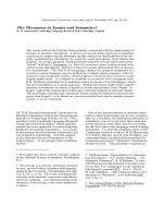
Fig. 1. C1q interaction with peripheral blood monocytes. (A) Blood
cells were stained with PE anti-CD91 (x-axis) and F-C1q (y-axis).
The red blood cells were lyzed and remaining cells were analyzed
by flow cytometry. Only monocytic cells gated by forward scatter
and side scatter is shown. (B) Peripheral blood monocytes are
highly CD91 positive. Blood cells were stained with flourophore-
conjugated antibodies recognizing CD14 and CD91 (left) or an iso-
type control (right). Red blood cells were lyzed and remaining cells
were analyzed by flow cytometry. (C) C1q binding levels for three
individuals. Blood cells were stained with F-C1q and PE anti-CD14.
The red blood cells were lyzed and remaining cells were analyzed
by flow cytometry. C1q binding levels of white blood cells are pre-
sented on the x-axis with CD14 positivity on the y-axis. (D) PBMCs
from human blood were stained with F-C1q with the addition of
the CD91 blocking protein RAP (100-fold molar excess), soluble
CD91 (10-fold molar excess) or the control protein HSA (100-fold
molar excess). After staining, C1q-positive cells were quantified by
flow cytometry. F-C1q stained cells without inhibition were set as
100%.
K. Duus et al. Direct interaction between CD91 and C1q
FEBS Journal 277 (2010) 3526–3537 ª 2010 The Authors Journal compilation ª 2010 FEBS 3527
therefore inhibition experiments were conducted with
isolated peripheral blood mononuclear cells (PBMCs).
These experiments showed that partial inhibition of
the C1q–monocyte interaction could be obtained with
soluble CD91 and the CD91 blocker, receptor-associ-
ated protein (RAP) (Fig. 1D). Similar results were
obtained by Lillis et al. [34], where only some phago-
cytotic inhibition was observed with monocytes defi-
cient for CD91. These results indicate that scavenging
of C1q complexes relies on a highly redundant recep-
tor system and that several receptors for C1q are likely
to exist.
Interaction of C1q with CD91 expressing cells
The monocytic cell line Mono Mac 6 (MM6) was used
to examine C1q interaction with monocytic cells. The
cell line has previously been described to express CD91
[35]. However, in the present study, only a low level of
CD91 was identified on the surface of MM6 cells
(approximately 7% of cells were CD91 positive; data
not shown).
To increase surface expression of CD91, the cells
were stimulated with lipopolysaccharide (LPS). As
measured by flow cytometry, approximately 40% of
the cells were found to be CD91 positive after LPS
stimulation (Fig. 2A), and the mean fluorescent inten-
sity of the cells stained with fluorescently-labelled
CD91 antibody was observed to increase. Some 85%
of the CD91 positive cells were highly CD14 positive
(data not shown).
When the stimulated MM6 cells were stained
with fluorescein isothiocyanate (FITC)-labelled C1q
(F-C1q), 1.2–9% of the cell population showed high
fluorescence intensity, indicating C1q interaction. The
percentage of positive cells could be inhibited by RAP
and unlabelled C1q (Fig. 2B and results not shown). We
used confocal scanning laser microscopy to visualize
localization at the cell surface. CD91 antibody bound
MM6 at the cell surface and some (but not complete)
co-localization was observed between the added F-C1q
and CD91 expressed by the cells (Fig. 2C), indicating
that other receptor systems are likely to exist. Staining
of the nuclei revealed no signs of apoptosis.
Cell size
Cell size
PE anti-isotype control
PE anti-CD91
0
FSC-H
45.52%
6.13%
1023
10
0
10
1
10
2
10
3
FL2-H
10
4
0
FSC-H
1023
10
0
10
1
10
2
10
3
FL2-H
10
4
2
1
Relative binding
0
300300
RAP/C1q molar ratio
(i) (ii) (iii)
A
B
C
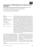
Fig. 2. CD91 expression and C1q binding
on Mono Mac 6 cells. (A) Flow cytometry of
stimulated MM6 cells showing CD91
expression. After LPS stimulation, CD91
expression was measured on MM6 cells by
the addition of PE-labelled CD91 antibody
(left) or an isotype control (right) and fluores-
cence was measured by flow cytometry.
Approximately 40% of the LPS-stimulated
cells were determined to be CD91 positive
(45.52–6.13% = 39.39%). (B) C1q interac-
tion with stimulated MM6 cells and RAP
inhibition. MM6 cells were stained with
F-C1q with or without the addition of RAP.
RAP showed inhibition of the C1q interac-
tion in 30- and 300-fold molar excess. The
results are presented with the relative bind-
ing with F-C1q without the addition of RAP
set as 1. (C) CD91 and C1q localization in
MM6 cells analyzed by confocal laser
microscopy. Cells were stained with biotiny-
lated antibodies against CD91 and streptavi-
din coupled with Alexa Fluor 546 (red
colour, i) and the addition of F-C1q (green,
ii). Images i and ii are merged to produce an
overlay plot (iii). Scale bar = 10 l m.
Direct interaction between CD91 and C1q K. Duus et al.
3528 FEBS Journal 277 (2010) 3526–3537 ª 2010 The Authors Journal compilation ª 2010 FEBS
Evidence for a direct interaction between CD91
and C1q
Evidence for a direct interaction was obtained by surface
plasmon resonance (SPR) analysis. Using C1q as the
soluble analyte and CD91 as the immobilized ligand,
several concentrations of C1q were used to reveal a time-
and concentration-dependent interaction (Fig. 3A).
Furthermore, in the reversed configuration with C1q
immobilized and CD91 as the soluble analyte, an
interaction was observed (Fig. 3B). Using an ELISA
binding assay, CD91 showed a direct interaction with
C1q coated on a polystyrene surface (Fig. 4A). At a
physiological salt concentration (0.15 m NaCl), the
interaction was time-dependent, saturable and fast,
being detectable after only a few minutes of incubation.
By contrast, increasing the NaCl concentration to
0.65 m strongly inhibited the interaction (Fig. 4A). In
the opposite configuration, with CD91 immobilized, sol-
uble C1q also bound and could be detected with C1q
specific antibodies (Fig. 4B) although some nonspecific
interaction was observed between the polyclonal anti-
body directed against C1q and CD91. The C1q–CD91
interaction was also observed when C1q was bound to
immobilized IgM, indicating that CD91 binding is possi-
ble with ligand-bound C1q (Fig. 4C).
Kinetic analysis of the SPR binding data, showed
that the best fit for the interaction between immobi-
lized CD91 and soluble C1q was provided by a two-
state reaction model (Table 1), suggesting that several
interaction phases take place. This was supported by
the ELISA, which revealed that binding of soluble
CD91 to C1q coated on the polystyrene surface was
highly sensitive to 0.65 m NaCl in the early phase of
the interaction (Fig. 4A). By contrast, if CD91 and
C1q were allowed to interact under physiological salt
concentrations before the addition of 0.5 m NaCl, then
the interaction gradually became insensitive to high
salt (Fig. 4D). This was suggestive of a two-state inter-
action with an initial ionic binding step allowing subse-
quent stable interaction.
Interaction of CD91 with the globular domain of
C1q and its collagen fragment was also analyzed by
SPR analysis, using the latter proteins as soluble
ligands. In the case of the globular domain, the two-
state binding model also provided the best fit. More-
over, the affinity of the interaction calculated by a
Langmuir 1:1 model was approximately 20-fold lower
compared to intact C1q (K
D
= 3.0 · 10
)7
m). The
C1q collagen fragment also bound immobilized CD91,
and the reaction followed a classical 1 : 1 Langmuir
model, with a K
D
in the nanomolar range (Table 1).
0 100 200
0
40
80
120
Response difference (RU)
Time (s)
50 nM
40 nM
30 nM
20 nM
10 nM
5 nM
2.5 nM
Response difference (RU)
100
–100 0
0
100
200
Time (s)
50 nM
40 nM
30 nM
20 nM
A
B
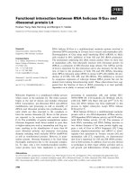
Fig. 3. Interaction of CD91 with C1q, demonstrated by SPR analy-
sis. (A) CD91 was immobilized by coupling of amino groups to a
carboxylated surface. C1q was injected for 120 s at concentrations
in the range 2.5–50 n
M. (B) CD91–C1q interaction. C1q was immo-
bilized by amine coupling to a carboxylated surface and soluble
CD91 was injected for 120 s at the indicated concentration in the
range 20–50 n
M.
Table 1. Comparison of the kinetic constants of the CD91 interaction between C1q, C1q globular heads and C1q collagen tails.
Ligand Model with best fit K
a
(K
a1
, K
a2
) K
d
(K
d1
, K
d2
)
K
D
, Langmuir
1 : 1 model Chi squared
C1q Two-state reaction with
conformational change
4.04 · 10
5
M
)1
s
)1
6.61 · 10
)10
S
)1
5.09 · 10
)3
s
)1
0.464 s
)1
– 6.14
C1q globular region Two-state reaction with
conformational change
1.94 · 10
4
M
)1
s
)1
3.28 · 10
)9
S
)1
5.97 · 10
)3
s
)1
1.87 · 10
)7
s
)1
– 5.93
C1q collagen region Langmuir 1 : 1 5.13 · 10
5
M
)1
s
)1
2.04 · 10
)3
s
)1
3.95 · 10
)9
M 5.89
K. Duus et al. Direct interaction between CD91 and C1q
FEBS Journal 277 (2010) 3526–3537 ª 2010 The Authors Journal compilation ª 2010 FEBS 3529
Several known interaction partners of CD91 and
C1q inhibit their interaction
To characterize the CD91–C1q interaction in more
detail, the ability of known ligands of either protein to
interfere with binding was investigated. RAP is a
CD91 chaperone that has previously been shown to
block the interaction of CD91 with all its known
ligands [36,37]. In agreement with this characteristic,
RAP also strongly inhibited binding of CD91 to
immobilized C1q (Fig. 5A). a-2-macroglobulin and
Pseudomonas exotoxin A are known ligands of CD91
and they also exerted some inhibition (15% and 40%
inhibition, respectively) (data not shown). Calreticulin,
a protein known to act as a ligand for the collagen
region of C1q and the proposed C1q-binding compo-
nent of the CD91 ⁄ calreticulin receptor complex, signifi-
cantly inhibited binding of CD91 to immobilized C1q
in the presence of 5 mm Ca
2+
ions, with almost 45%
inhibition at a 300 : 1 calreticulin ⁄ CD91 molar ratio
(Fig. 5B). Inhibition was also observed in the absence
of Ca
2+
, although to a lesser extent.
Serum amyloid P (SAP), another known ligand of
C1q [38,39], abolished binding of CD91 to immobi-
lized C1q in the presence of 5 mm Ca
2+
but had only
a slight inhibitory effect in the absence of Ca
2+
(Fig. 5C). A significant inhibition of the interaction by
the sulphated polysaccharide fucoidan was also
observed, with approximately 65% inhibition at a
600 : 1 fucoidan ⁄ CD91 molar excess (Fig. 5D). The
physiological partner proteases of C1q, C1r and C1s
were also tested as inhibitors, with inhibition observed
only for C1r (Fig. 5E). Inhibition was also observed
with the globular region of C1q, confirming the results
observed by the SPR data (results not shown).
Taken together, these inhibition experiments indicate
that C1q occupy known ligand-binding sites on CD91
and that CD91 presumably interacts with C1q at
0
1
2
3
4
1500
1000
5000
Time (min)
Physiological NaCl (0.15
M
)
0.5
M
additional NaCl (0.65
M
)
CD91 binding (A
405
)
A
0
0.2
0.4
0.6
0.8
1
IgG (agg)IgMIgG
b-CD91
C1q + b-CD91
CD91 binding (A
405
)
C
0
1
2
3
4
150
100
50
0
Time without NaCl (min)
CD91 binding (A
405
)
D
0
1
2
3
CD91-C1q CD91-BSA CD91
Anti-C1q
Anti-isotype control
Antibody binding (A
405
)
B
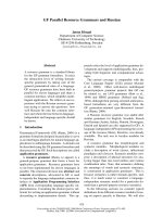
Fig. 4. Interaction between CD91 and C1q, demonstrated by
ELISA. (A) Time dependence of the association between CD91 and
C1q. Biotinylated CD91 (b-CD91) was added to microtitre plates
coated with C1q. b-CD91 was allowed to incubate for the indicated
time (from 10 min to 20 h) with a NaCl concentration of either 0.15
or 0.65
M. Bound b-CD91 was quantified by incubation with alkaline
phosphatase-conjugated streptavidin and pNPP. (B) CD91 was
immobilized on a microtitre plate and C1q or the control protein
bovine serum albumin was allowed to interact. Bound C1q was
detected with rabbit antibodies recognizing C1q and alkaline phos-
phatase-conjugated secondary antibodies. Data are presented as
the mean ± SD of two individual wells for one representative
experiment. (C) A microtitre plate was coated with the indicated
protein; either IgG, IgM or IgG aggregated [IgG(agg)] by incubation
at 60 °C for 20 min, b-CD91 was either added directly (grey bars)
or after a layer of C1q (white bars) and bound CD91 was detected
by incubation with alkaline phosphatase-conjugated streptavidin and
pNPP. (D) The time-dependent influence of 0.5
M NaCl on CD91
binding to C1q. b-CD91 was added to a microtitre plate coated with
C1q. After the indicated amounts of time, 0.5
M NaCl was added
and the incubation continued for a total incubation time of 2 h,
before the bound b-CD91 was quantified by incubation with alkaline
phosphatase-conjugated streptavidin and pNPP.
Direct interaction between CD91 and C1q K. Duus et al.
3530 FEBS Journal 277 (2010) 3526–3537 ª 2010 The Authors Journal compilation ª 2010 FEBS
several sites, with one of them being at or near the
C1r attachment site. Calreticulin was only able to inhi-
bit the interaction to 45%, despite the use of a very
large excess. Considering that calreticulin is known to
interact strongly with C1q, this suggests that only a
part of the binding sites on C1q are shared by calreti-
culin and CD91. No interaction between CD91 and
calreticulin could be observed, regardless of which of
them was immobilized (results not shown).
Discussion
It is well established that C1q initiates the clearance of
apoptotic material, although the identification of direct
C1q-receptors has remained elusive. CD91 has previ-
ously been described to function as a receptor complex
together with calreticulin. In this receptor complex,
calreticulin is suggested to act as the C1q-recognition
molecule and CD91 as the phagocytic, transmembrane
molecule.
To determine whether C1q binding could occur on
human blood cells, we first confirmed the high CD91
level on human blood monocytes. We then analyzed
C1q binding to human blood cells, confirming a mono-
cyte interaction. The interaction between C1q and
PBMCs could be partially inhibited by RAP and solu-
ble CD91. The interaction between C1q and CD91
was also investigated using the monocytic cell line
MM6 known to present CD91 at the cell surface.
Fluorescent-labelled C1q was found to bind these cells
in a manner overlapping the areas of CD91 expression.
The partial co-localization suggests that other C1q
receptors exist. This was confirmed by the inhibition
experiments where the interaction between C1q and
MM6 cells could be partially inhibited by the addition
of RAP, suggesting that only part of the C1q interac-
tion occurs specifically through CD91 or other mem-
bers of the LDL receptor superfamily.
CD91 and calreticulin have previously been
described to function as a C1q receptor complex with
calreticulin as the recognition unit of the complex.
Despite these reports, in the present study, we provide
evidence that CD91 directly recognizes C1q indepen-
0
1
2
3
4
100100
RAP/CD91 molar ratio
RAP
Ovalbumin
CD91 binding (A
405
)
0
1
2
3
4
0 30 300
Calreticulin/CD91 molar ratio
Without calcium
5 m
M calcium
CD91 binding (A
405
)CD91 binding (A
405
)
0
1
2
3
4
600600
SAP/CD91 molar ratio
Without calcium
5 m
M calcium
0
1
2
3
600600
Fucoidan/CD91 molar ratio
CD91 binding (A
405
)
0
0.5
1
1.5
500
C1s
C1r
BSA
CD91 binding (A
405
)
CD91/serine protease molar ratio
A
B
C
D
E
Fig. 5. Inhibition of the CD91–C1q interaction. Biotinylated CD91
was added to microtitre plates coated with C1q, together with the
indicated molar excess of inhibitor. (A) RAP and the control protein
ovalbumin; (B) calreticulin with and without the addition of 5 m
M
calcium; (C) SAP with and without 5 mM calcium; (D) fucoidan; and
(E) C1r and C1s. After 2 h of incubation, the amount of bound bioti-
nylated CD91 was quantified by incubation with alkaline phospha-
tase-conjugated streptavidin and pNPP. Data are the mean ± SD of
two individual wells for one representative experiment.
K. Duus et al. Direct interaction between CD91 and C1q
FEBS Journal 277 (2010) 3526–3537 ª 2010 The Authors Journal compilation ª 2010 FEBS 3531
dently of calreticulin. The CD91–C1q interaction
showed all of the signs for being specific because the
interaction was time- and concentration-dependent.
Furthermore, the interaction was inhibited by several
known ligands of either protein. Interaction was
observed both with commercially available C1q ⁄ CD91
and purified C1q ⁄ CD91. In several assays, the interac-
tion showed signs of two-state binding. First, the inter-
action appeared to be salt sensitive at the early stages
but became salt insensitive at later stages. Second,
when the interaction was investigated using SPR,
a two-state binding was indicated and, third, an inter-
action was observed both for the globular region and
with the collagen tail of C1q. A likely hypothesis is
that CD91 binds C1q first through its collagen tail in a
salt-sensitive manner and then mediates a salt-insensi-
tive interaction with the globular head or vice versa.
CD91 contains four ligand-binding clusters of which
RAP is known to block at least three [36,40]. RAP is
specific for the receptors of the ‘LDL receptor super-
family’ and blocks all tested ligands of CD91 [36].
RAP completely inhibited binding between CD91 and
C1q, indicating that the interaction occurs through
one of more of these ligand-binding clusters. Some
inhibition was also observed with the CD91 ligands
a-2-macroglobulin and exotoxin A. These ligands
occupy the CD91 ligand-binding cluster II and IV,
thereby suggesting that C1q might interact with one or
both of these sites on CD91 [41,42].
Several interaction partners of C1q were also tested
for their ability to inhibit the interaction between
CD91 and C1q. Fucoidan yielded inhibition of CD91
binding to C1q. Fucoidan was reported to bind C1q at
a site that abolishes attachment of the serine proteases
[43]. It is therefore possible that CD91 interacts with
C1q at a site near or at the serine protease binding
site. This theory was supported by inhibition as a
result of the serine protease C1r, where a 50-fold
excess inhibited the interaction between CD91 and
C1q by approximately 40%. C1r binds C1q at the col-
lagen tail at a site where the collagen tail is trimeric
without obtaining higher-order oligomeric forms
[44,45]. Complete inhibition was also observed with
SAP. SAP is known to bind C1q at sites on both the
collagen stalk and the globular region [38,39] and it is
therefore possible that CD91 interacts at one or both
of these sites. The possibility that both the globular
region and the collagen region could be involved in
this interaction was strengthened by SPR data, where
either region showed an interaction with CD91. Taken
together, these inhibition experiments imply that the
CD91–C1q interaction occurs at one or more of the
ligand-binding clusters of CD91. On the C1q molecule,
the interaction site may be near or at the serine prote-
ase attachment site, although several sites on the C1q
molecule are assumed to be involved.
Calreticulin is known to bind C1q and is a reported
co-receptor of CD91. Only 45% inhibition was
obtained in the case where calreticulin was used as an
inhibitor of the C1q–CD91 interaction. The calreticulin
inhibition indicates that only a part of the binding sites
on C1q is shared between CD91 and calreticulin. No
binding was detected between CD91 and calreticulin in
the ELISA, regardless of which of them was immobi-
lized or in solution. These results are somewhat
intriguing because calreticulin has been reported to
function as a co-receptor for CD91 in the binding of
C1q and collectins [20,22,23]. However, Donnelly et al.
[46] also were unable to detect distinct binding between
calreticulin and CD91 and Walters and Berwin [47]
reported that the uptake of calreticulin is different
from another CD91 ligand, thereby suggesting that
calreticulin is not a traditional ligand of CD91.
In conclusion, the data obtained in the present study
support the hypothesis that CD91 is a receptor for
C1q and presumably detects and internalizes ligand
bound C1q in the same way that it internalizes other
ligands. We therefore conclude that CD91 itself is a
receptor for C1q. On the basis of these findings, we
propose a binding model for CD91 scavenging of C1q
that is independent of calreticulin (Fig. 6).
Materials and methods
Proteins and chemicals
Ovalbumin, bovine serum albumin, p-nitrophenyl-phos-
phate (pNPP) substrate tablets, FITC, N-hydroxy-succinim-
idobiotin, a-2-macroglobulin, exotoxin A, fucoidan, LPS
(Pseudomonas aeruginosa) and C1q were obtained from
Sigma (St Louis, MO, USA). Alkaline phosphatase-conju-
gated streptavidin and IgM were obtained from Dako
(Glostrup, Denmark). MaxiSorp microtitre plates were
obtained from Nunc (Roskilde, Denmark). CD91 (contain-
ing RAP) and biotin-labelled CD91 antibody were obtained
from BioMac (Chamalie
`
res, France). RAP was obtained
from Innovative Research (Southfield, MI, USA). SAP was
a gift from N. H. H. Heega
˚
rd (Statens Serum Institut,
Copenhagen, Denmark). Adiponectin (trimeric form) was
obtained from Abcam (Cambridge, UK). Human IgG
and human serum albumin (HSA) were obtained from
SSI (Copenhagen, Denmark). CM-5 sensorchips, surfactant
P20, 1-ethyl-3-(3-dimethylaminopropyl)carbodiimide, N-hydro-
xysuccinimide and ethanolamine were obtained from GE
healthcare, BIAcore (Uppsala, Sweden). Excell medium
was obtained from Safc Biosciences (Hampshire, UK).
Direct interaction between CD91 and C1q K. Duus et al.
3532 FEBS Journal 277 (2010) 3526–3537 ª 2010 The Authors Journal compilation ª 2010 FEBS
Penicillin, streptomycin and glutamine were obtained from
Gibco-BRL (NY, USA). Flowbuffer, phycoerytrin (PE)
mouse anti-human CD91, PE mouse anti-human CD14,
FITC mouse anti-human CD91, FITC mouse anti-human
CD14 and corresponding isotype control antibodies were
obtained from BD Biosciences (Franklin Lakes, NJ, USA).
C1r and C1s were obtained from R&D Systems (Abingdon,
UK). Rabbit anti human C1q polyclonal antibody was
obtained from Dako. High-yield Lyse and streptavidin
labelled with Alexa Fluor 546 were obtained from Invitro-
gen (Carlsbad, CA, USA). Vectashield mounting medium
was from Vector Laboratories (Burlingame, CA, USA).
Purification of human placenta calreticulin
Human placenta calreticulin was purified using a previously
described procedure [48,49]. The purified protein showed a
single band with an apparent molecular weight of 60 kDa
by SDS ⁄ PAGE and a single peak by MALDI-TOF ⁄ TOF
mass spectrometry.
Purification of C1q and C1q-derived fragments
C1q was purified from human plasma as described previ-
ously [50]. The collagen-like fragments of C1q were
obtained by pepsin digestion and purified as described pre-
viously [51]. The fragments corresponding to the C1q glob-
ular domains were generated by treatment of C1q with
collagenase and purified by high-pressure gel filtration chro-
matography as described previously [51].
Purification of CD91
Commercially obtained CD91 (BioMac) was used when not
stated otherwise. CD91 containing no RAP was a gift from
S. K. Moestrup (Institute of Medical Biochemistry, Univer-
sity of A
˚
rhus, A
˚
rhus, Denmark), purified as described pre-
viously [52], and was used to confirm the C1q interaction.
Protein biotinylation
The proteins subjected to biotinylation were dialysed against
0.1 m NaHCO
3
(pH 9.0) at 4 °C, followed by the addition
of N-hydroxysuccinimidobiotin in dimethyl sulphoxide
(10 mgÆmL
)1
) to a final concentration of 4 mgÆ mg
)1
CD91.
The solution was incubated for 2 h at room temperature
with end-over-end agitation, and then dialysed against
NaCl ⁄ P
i
(10 mm NaH
2
PO
4
⁄ Na
2
HPO
4
, pH 7.3, 0.15 m NaCl)
at 4 °C. Biotinylated CD91 was stored at )20 °C until use.
FITC labelling
Commercially available C1q and HSA were dialysed against
50 mm sodium carbonate (pH 9.5). FITC (100 lgÆ mg
)1
pro-
tein) was added and the reaction mixture was incubated
with end-over-end agitation for 1 h in the dark. Excess fluo-
rescein was removed by dialysis against NaCl ⁄ P
i
at 4 °C.
ELISA
Unless otherwise stated, incubations and washings were
performed at room temperature on a shaking table using
100 lL per well for incubation and 200 lL per well for
washing and blocking. TTN buffer (0.025 m Tris–HCl,
0.5% Tween 20, 0.15 m NaCl, pH 7.5) was used for block-
ing, incubation and washing.
Unless otherwise stated, the C1q used was from Sigma
(St Louis, MO, USA). Proteins were coated at 1 lgÆmL
)1
onto the surface of the microtitre plates using 0.05 m
sodium carbonate (pH 9.6) as coating buffer. After coating
overnight at 4 °C, plates were washed three times for 1 min,
followed by blocking for 30 min. Subsequently, incubation
with or without biotinylated CD91 diluted to 1 lgÆmL
)1
was carried out for 2 h, followed by another three washes.
Finally, the plates were incubated for 1 h with alkaline
phosphatase-conjugated streptavidin diluted 1 : 1000. After
another three washes, bound CD91 was quantified using
pNPP (1 mgÆmL
)1
)in1m diethanolamine, 0.5 mm MgCl
2
(pH 9.8). A
405
was read with background subtraction at
Epidermal growth factor-type repeat
Ligand-binding repeat
Beta-propeller
C1q
Ligand bound
C1q
CD91
Apoptotic material
Phagocyte
Fig. 6. Proposed model for CD91 interaction with C1q. CD91 is
present on phagocytes and consists of two noncovalently bound
polypeptide chains; with the 85 kDa b-chain as the transmembrane
and the a-chain of 515 kDa with four ligand-binding clusters (blue
areas). Ligand interaction occurs through 31 similar ligand-binding
repeats (blue squares) distributed unequally between the four
ligand-binding clusters. The CD91 a-chain also consists of epider-
mal growth factor-type repeats (grey circles) and b-propellers (black
stars). Interaction occurs at several sites on C1q and possibly
through one or more ligand-binding domains at CD91. CD91 is a
phagocytic receptor and is likely to internalize C1q bound material.
K. Duus et al. Direct interaction between CD91 and C1q
FEBS Journal 277 (2010) 3526–3537 ª 2010 The Authors Journal compilation ª 2010 FEBS 3533
650 nm on a VERSAmax microplate reader (Molecular
Devices, Sunnyvale, CA, USA). All experiments were per-
formed at least twice and the results are presented as the
mean ± SD of two wells for one representative experiment.
Inhibition ELISA
Inhibition of the CD91–C1q interaction was tested by mix-
ing biotinylated CD91, diluted to a concentration of
1 lgÆmL
)1
, with varying amounts of the indicated inhibitor
for 1 h. The mixture was then added for 2 h to plates
coated overnight with C1q. Subsequently, the plates were
washed, incubated with alkaline phosphatase-conjugated
streptavidin and developed as described above.
SPR experiments
CD91 and C1q were immobilized on a BIAcore CM5 sensor-
chip in NaCl ⁄ Hepes-P20 (10 mm Hepes, 0.15 m NaCl,
3.4 mm EDTA, 0.005% surfactant P20) in accordance with
the manufacturer’s instructions, resulting in an immobiliza-
tion level of 13 000 and 17 000 relative units, respectively.
Interaction between immobilized CD91 and C1q, the C1q
globular domain and collagen fragment was investigated
on a BIAcore 3000 instrument using NaCl ⁄ Tris (10 mm
Tris–HCl, 0.15 m NaCl, pH 7.4) as running buffer. Inter-
action between immobilized C1q and soluble CD91 was
investigated on a BIAcore 1000 instrument with the same
running buffer. The analyte was injected at a flow rate of
20 lLÆmin
)1
for 120 s. Background was subtracted from a
quenched (activated ⁄ deactivated) surface showing an
absence of nonspecific binding. The surface was fully regen-
erated using low concentrations of NaOH. Kinetics were
determined using the software biaevaluation, version 3.1
(BIAcore).
Two independent experiments were performed with all
concentrations. Data are reported for one representative
experiment.
Culture of MM6 cells
MM6 was used as a monocytic cell line. Cells were cultured
at 37 °C under 5% CO
2
in Excell medium (to avoid serum)
with the addition of 1% glutamine and 1% penstrep. These
cells were stimulated twice with 10 ngÆmL
)1
LPS with a
3-day interval and harvested on day 6 or 7.
Flow cytometry analysis of MM6 cells
A sample containing 10
5
cells was centrifuged to remove
the culture medium and the cells were washed with
NaCl ⁄ P
i
. The cell pellet was resuspended in 10 lLof
NaCl ⁄ P
i
and stained by adding 2 lL of F-C1q or FITC-
labelled HSA for 20 min at room temperature. Staining
with CD91 antibody, CD14 antibody or isotype control
antibodies was performed in accordance with the manufac-
turer’s instructions. After staining, cells were washed with
NaCl ⁄ P
i
and resuspended in 1 mL of the buffer. Flow
cytometry was performed using a Calibur instrument (BD
Biosciences), counting 10 000 cells and using cellquest
(BD Biosciences) and winmdi () for
the data analysis.
Flow cytometry analysis of whole blood
Two microlitres of F-C1q, F-HSA or antibodies labelled
with PE or flourescein were added to 12.5 lL of heparin-
ized blood. After incubation for 15 min, red blood cells
were lysed by the addition of 1 mL of High-yield Lyse
(Invitrogen). Cells were analyzed on a Calibur instrument
(BD Biosciences), counting 5000 or 10 000 events.
PBMC isolation and inhibition experiments
Human peripheral blood was collected and mixed with
NaCl ⁄ P
i
(1 : 1). The mixture was overlayed on Ficoll and
centrifuged (800 g for 20 min at 4 °C). The interface of
mononuclear cells was harvested and stained with 2 l Lof
F-C1q and the described antibodies for 10 lL of PBMC,
with or without the addition of inhibitor, for 20 min at
room temperature. Cells were analyzed on a Calibur instru-
ment (BD Biosciences), counting 10 000 events.
Confocal scanning laser microscopy
MM6 cells were grown on glass cover slips for 6 days and
stimulated with LPS (10 ngÆmL
)1
) on days 1 and 3. The cells
were fixed with 4% paraformaldehyde for 20 min at 37 °C
and permeabilized with 0.1% Triton X-100 for 5 min after
three washes with NaCl ⁄ P
i
. After four washes, the cells were
stained for 30 min with biotinylated mouse anti-CD91 (dilu-
tion 1 : 25) in Hanks balanced salt solution containing 5%
inactivated fetal bovine serum and 40 mm Hepes. After four
washes with NaCl ⁄ P
i
, the cells were stained with F-C1q
(10 lgÆmL
)1
) and streptavidin-Alexa 546 (dilution 1 : 50) in
the same staining buffer. The coverslips were mounted with
4¢,6¢-diamidino-2-phenylindole-containing mounting solu-
tion and pictures were taken with a Leica, SP2 confocal
microscope (Leica Microsystems, Wetzlar, Germany). Pic-
tures were processed using image j [53].
Acknowledgements
Dorthe Tange Olsen and Anne Mortensen are thanked
for their excellent technical work. Søren Kragh Moest-
rup and Christian Jacobsen are thanked for providing
purified CD91 as well as helpful suggestions. Hans Jør-
gen Jensen and Susana Aznar are thanked for help
with the confocal microscopy.
Direct interaction between CD91 and C1q K. Duus et al.
3534 FEBS Journal 277 (2010) 3526–3537 ª 2010 The Authors Journal compilation ª 2010 FEBS
References
1 Goldfarb RD & Parrillo JE (2005) Complement. Crit
Care Med 33, S482–S484.
2 Nauta AJ, Roos A & Daha MR (2004) A regulatory
role for complement in innate immunity and autoimmu-
nity. Int Arch Allergy Immunol 134, 310–323.
3 Bottcher A, Gaipl US, Furnrohr BG, Herrmann M,
Girkontaite I, Kalden JR & Voll RE (2006) Involve-
ment of phosphatidylserine, alphavbeta3, CD14, CD36,
and complement C1q in the phagocytosis of primary
necrotic lymphocytes by macrophages. Arthritis Rheum
54, 927–938.
4 Korb LC & Ahearn JM (1997) C1q binds directly and
specifically to surface blebs of apoptotic human
keratinocytes: complement deficiency and systemic
lupus erythematosus revisited. J Immunol 158, 4525–
4528.
5 Nauta AJ, Trouw LA, Daha MR, Tijsma O, Nieuwland
R, Schwaeble WJ, Gingras AR, Mantovani A, Hack
EC & Roos A (2002) Direct binding of C1q to apopto-
tic cells and cell blebs induces complement activation.
Eur J Immunol 32, 1726–1736.
6 Navratil JS, Watkins SC, Wisnieski JJ & Ahearn JM
(2001) The globular heads of C1q specifically recognize
surface blebs of apoptotic vascular endothelial cells.
J Immunol 166, 3231–3239.
7 Pittoni V & Valesini G (2002) The clearance of apopto-
tic cells: implications for autoimmunity. Autoimmun Rev
1, 154–161.
8 Sontheimer RD, Racila E & Racila DM (2005) C1q: its
functions within the innate and adaptive immune
responses and its role in lupus autoimmunity. J Invest
Dermatol 125, 14–23.
9 Tarr J & Eggleton P (2005) Immune function of C1q
and its modulators CD91 and CD93. Crit Rev Immunol
25, 305–330.
10 Edelson BT, Stricker TP, Li Z, Dickeson SK, Shepherd
VL, Santoro SA & Zutter MM (2006) Novel collec-
tin ⁄ C1q receptor mediates mast cell activation and
innate immunity. Blood 107, 143–150.
11 Eggleton P, Reid KB & Tenner AJ (1998) C1q – how
many functions? How many receptors? Trends Cell Biol
8, 428–431.
12 Eggleton P, Tenner AJ & Reid KB (2000) C1q recep-
tors. Clin Exp Immunol 120, 406–412.
13 Ghebrehiwet B, Silvestri L & McDevitt C (1984)
Identification of the Raji cell membrane-derived C1q
inhibitor as a receptor for human C1q. Purification and
immunochemical characterization. J Exp Med 160,
1375–1389.
14 Ghiran I, Tyagi SR, Klickstein LB & Nicholson-Weller
A (2002) Expression and function of C1q receptors and
C1q binding proteins at the cell surface. Immunobiology
205, 407–420.
15 McGreal E & Gasque P (2002) Structure–function stud-
ies of the receptors for complement C1q. Biochem Soc
Trans 30, 1010–1014.
16 Sim RB, Moestrup SK, Stuart GR, Lynch NJ, Lu J,
Schwaeble WJ & Malhotra R (1998) Interaction of C1q
and the collectins with the potential receptors calreticu-
lin (cC1qR ⁄ collectin receptor) and megalin. Immunobiol-
ogy 199, 208–224.
17 Basu S, Binder RJ, Ramalingam T & Srivastava PK
(2001) CD91 is a common receptor for heat shock pro-
teins gp96, hsp90, hsp70, and calreticulin. Immunity 14,
303–313.
18 Binder RJ, Han DK & Srivastava PK (2000) CD91:
a receptor for heat shock protein gp96. Nat Immunol 1,
151–155.
19 Binder RJ, Vatner R & Srivastava P (2004) The heat-
shock protein receptors: some answers and more ques-
tions. Tissue Antigens 64, 442–451.
20 Gardai SJ, Xiao YQ, Dickinson M, Nick JA, Voelker
DR, Greene KE & Henson PM (2003) By binding
SIRPalpha or calreticulin ⁄ CD91, lung collectins act as
dual function surveillance molecules to suppress or
enhance inflammation. Cell 115, 13–23.
21 Ogden CA, deCathelineau A, Hoffmann PR, Bratton
D, Ghebrehiwet B, Fadok VA & Henson PM (2001)
C1q and mannose binding lectin engagement of cell
surface calreticulin and CD91 initiates macropinocytosis
and uptake of apoptotic cells. J Exp Med 194, 781–795.
22 Orr AW, Pedraza CE, Pallero MA, Elzie CA, Goicoe-
chea S, Strickland DK & Murphy-Ullrich JE (2003)
Low density lipoprotein receptor-related protein is a
calreticulin coreceptor that signals focal adhesion disas-
sembly. J Cell Biol 161, 1179–1189.
23 Vandivier RW, Ogden CA, Fadok VA, Hoffmann PR,
Brown KK, Botto M, Walport MJ, Fisher JH, Henson
PM & Greene KE (2002) Role of surfactant proteins A,
D, and C1q in the clearance of apoptotic cells in vivo
and in vitro: calreticulin and CD91 as a common collec-
tin receptor complex. J Immunol 169, 3978–3986.
24 Obeid M, Tesniere A, Ghiringhelli F, Fimia GM,
Apetoh L, Perfettini JL, Castedo M, Mignot G,
Panaretakis T, Casares N et al. (2007) Calreticulin
exposure dictates the immunogenicity of cancer cell
death. Nat Med 13, 54–61.
25 Gardai SJ, McPhillips KA, Frasch SC, Janssen WJ,
Starefeldt A, Murphy-Ullrich JE, Bratton DL,
Oldenborg PA, Michalak M & Henson PM (2005)
Cell-surface calreticulin initiates clearance of viable or
apoptotic cells through trans-activation of LRP on the
phagocyte. Cell 123, 321–334.
26 Herz J & Strickland DK (2001) LRP: a multifunctional
scavenger and signaling receptor. J Clin Invest 108,
779–784.
27 Krieger M & Herz J (1994) Structures and functions of
multiligand lipoprotein receptors: macrophage
K. Duus et al. Direct interaction between CD91 and C1q
FEBS Journal 277 (2010) 3526–3537 ª 2010 The Authors Journal compilation ª 2010 FEBS 3535
scavenger receptors and LDL receptor-related protein
(LRP). Annu Rev Biochem 63, 601–637.
28 Moestrup SK & Gliemann J (1991) Analysis of ligand
recognition by the purified alpha 2-macroglobulin
receptor (low density lipoprotein receptor-related
protein). Evidence that high affinity of alpha 2-macro-
globulin-proteinase complex is achieved by binding to
adjacent receptors. J Biol Chem 266, 14011–14017.
29 Stebbing J, Savage P, Patterson S & Gazzard B (2004)
All for CD91 and CD91 for all. J Antimicrob Chemo-
ther 53, 1–3.
30 Strickland DK, Ashcom JD, Williams S, Burgess WH,
Migliorini M & Argraves WS (1990) Sequence identity
between the alpha 2-macroglobulin receptor and low
density lipoprotein receptor-related protein suggests that
this molecule is a multifunctional receptor. J Biol Chem
265, 17401–17404.
31 Willnow TE, Orth K & Herz J (1994) Molecular
dissection of ligand binding sites on the low density
lipoprotein receptor-related protein. J Biol Chem 269,
15827–15832.
32 Strickland DK, Kounnas MZ & Argraves WS (1995)
LDL receptor-related protein: a multiligand receptor
for lipoprotein and proteinase catabolism. FASEB J 9,
890–898.
33 Moestrup SK, Gliemann J & Pallesen G (1992) Distri-
bution of the alpha 2-macroglobulin receptor ⁄ low den-
sity lipoprotein receptor-related protein in human
tissues. Cell Tissue Res 269, 375–382.
34 Lillis AP, Greenlee MC, Mikhailenko I, Pizzo SV,
Tenner AJ, Strickland DK & Bohlson SS (2008) Murine
low-density lipoprotein receptor-related protein 1 (LRP)
is required for phagocytosis of targets bearing
LRP ligands but is not required for C1q-triggered
enhancement of phagocytosis. J Immunol 181, 364–
373.
35 Schulz S, Birkenmeier G, Schagdarsurengin U, Wenzel
K, Muller-Werdan U, Rehfeld D, Suss T, Kabisch A,
Werdan K & Glaser C (2003) Role of LDL receptor-
related protein (LRP) in coronary atherosclerosis. Int
J Cardiol 92, 137–144.
36 Bu G (1998) Receptor-associated protein: a specialized
chaperone and antagonist for members of the LDL
receptor gene family. Curr Opin Lipidol 9, 149–155.
37 Striekland DK, Ashcom JD, Williams S, Battey F,
Behre E, McTigue K, Battey JF & Argraves WS (1991)
Primary structure of alpha 2-macroglobulin
receptor-associated protein. Human homologue of a
Heymann nephritis antigen. J Biol Chem 266, 13364–
13369.
38 Ying SC, Gewurz AT, Jiang H & Gewurz H (1993)
Human serum amyloid P component oligomers bind
and activate the classical complement pathway via resi-
dues 14-26 and 76-92 of the A chain collagen-like
region of C1q. J Immunol 150, 169–176.
39 Zahedi K (1996) Characterization of the binding of
serum amyloid P to type IV collagen. J Biol Chem 271,
14897–14902.
40 Obermoeller-McCormick LM, Li Y, Osaka H, FitzGer-
ald DJ, Schwartz AL & Bu G (2001) Dissection of
receptor folding and ligand-binding property with func-
tional minireceptors of LDL receptor-related protein.
J Cell Sci 114, 899–908.
41 Horn IR, van den Berg BM, van der Meijden PZ,
Pannekoek H & van Zonneveld AJ (1997) Molecular
analysis of ligand binding to the second cluster of com-
plement-type repeats of the low density lipoprotein
receptor-related protein. Evidence for an allosteric com-
ponent in receptor-associated protein-mediated inhibi-
tion of ligand binding. J Biol Chem 272 , 13608–13613.
42 Neels JG, van den Berg BM, Lookene A, Olivecrona G,
Pannekoek H & van Zonneveld AJ (1999) The second
and fourth cluster of class A cysteine-rich repeats of the
low density lipoprotein receptor-related protein share
ligand-binding properties. J Biol Chem 274, 31305–
31311.
43 Tissot B, Montdargent B, Chevolot L, Varenne A,
Descroix S, Gareil P & Daniel R (2003) Interaction of
fucoidan with the proteins of the complement classical
pathway. Biochim Biophys Acta 1651
, 5–16.
44 Gaboriaud C, Thielens NM, Gregory LA, Rossi V,
Fontecilla-Camps JC & Arlaud GJ (2004) Structure and
activation of the C1 complex of complement: unraveling
the puzzle. Trends Immunol 25, 368–373.
45 Gal P & Ambrus G (2001) Structure and function of
complement activating enzyme complexes: C1 and
MBL-MASPs. Curr Protein Pept Sci 2, 43–59.
46 Donnelly S, Roake W, Brown S, Young P, Naik H,
Wordsworth P, Isenberg DA, Reid KB & Eggleton P
(2006) Impaired recognition of apoptotic neutrophils by
the C1q ⁄ calreticulin and CD91 pathway in systemic
lupus erythematosus. Arthritis Rheum 54, 1543–1556.
47 Walters JJ & Berwin B (2005) Differential CD91 depen-
dence for calreticulin and Pseudomonas exotoxin-A
endocytosis. Traffic 6, 1173–1182.
48 Hojrup P, Roepstorff P & Houen G (2001) Human pla-
cental calreticulin-characterization of domain structure
and post-translational modifications. Eur J Biochem
268, 2558–2565.
49 Steino A, Jorgensen CS, Laursen I & Houen G (2004)
Interaction of C1q with the receptor calreticulin
requires a conformational change in C1q. Scand J
Immunol 59, 485–495.
50 Arlaud GJ, Reboul A, Sim RB & Colomb MG (1979)
Interaction of C1-inhibitor with the C1r and C1s sub-
components in human C1. Biochim Biophys Acta 576,
151–162.
51 Tacnet-Delorme P, Chevallier S & Arlaud GJ (2001)
Beta-amyloid fibrils activate the C1 complex of comple-
ment under physiological conditions: evidence for a
Direct interaction between CD91 and C1q K. Duus et al.
3536 FEBS Journal 277 (2010) 3526–3537 ª 2010 The Authors Journal compilation ª 2010 FEBS
binding site for A beta on the C1q globular regions.
J Immunol 167, 6374–6381.
52 Moestrup SK, Kaltoft K, Sottrup-Jensen L & Gliemann
J (1990) The human alpha 2-macroglobulin receptor
contains high affinity calcium binding sites important
for receptor conformation and ligand recognition.
J Biol Chem 265, 12623–12628.
53 Collins TJ (2007) ImageJ for microscopy. BioTechniques
43, 25–30.
K. Duus et al. Direct interaction between CD91 and C1q
FEBS Journal 277 (2010) 3526–3537 ª 2010 The Authors Journal compilation ª 2010 FEBS 3537