Báo cáo khoa học: Relaxin-3⁄ insulin-like peptide 7, a neuropeptide involved in the stress response and food intake Masaki Tanaka pptx
Bạn đang xem bản rút gọn của tài liệu. Xem và tải ngay bản đầy đủ của tài liệu tại đây (251.17 KB, 8 trang )
MINIREVIEW
Relaxin-3
⁄
insulin-like peptide 7, a neuropeptide involved
in the stress response and food intake
Masaki Tanaka
Department of Basic Geriatrics, Kyoto Prefectural University of Medicine, Japan
Introduction
Relaxin-3 ⁄ insulin-like peptide-7 (INSL7) has recently
been identified as a new member of the insulin ⁄ relaxin
family using human genomic databases [1]. The 142
amino acid human precursor polypeptide sequence is
well conserved among humans, pigs, rats and mice [2].
Structurally, this precursor polypeptide consists of sig-
nal peptides, and a B-chain, C-peptide and A-chain,
and contains the RXXXRXXI motif in the B chain
(B12–B19 in human) for binding to the relaxin recep-
tor [3]. Similar to insulin, a mature two-chain peptide
is produced after removal of the C-peptide and the for-
mation of three disulfide bonds between respective cys-
teine residues of the A-chain and B-chain [4]. An
evolutionary study showed that relaxin-3 orthologs are
present in fugu fish and zebrafish, but not in any inver-
tebrate or prokaryote, and that these orthologs show
high homology between different species in the mature
peptide region. When compared with other insu-
lin ⁄ relaxin superfamily members, relaxin-3 is con-
strained by strong purifying selection, suggesting that
this protein is an ancestral form and has a highly-con-
served function [5].
In the present minireview, the expression of relaxin-3
in the brain, and particularly its functions, including
the stress response and food intake, are described.
Expression of relaxin-3 in the brain
Relaxin-3 neurons in the brain
Examination of relaxin-3 mRNA expression by north-
ern blotting and reverse transcriptase-PCR revealed
Keywords
food intake; gene expression;
hypothalamus; nucleus incertus; RXFP3;
stress
Correspondence
M. Tanaka, Department of Basic Geriatrics,
Kyoto Prefectural University of Medicine,
Kawaramachi-Hirokoji, Kamikyo-ku, Kyoto
602-8566, Japan
Fax: +81 75 251 5797
Tel: +81 75 251 5797
E-mail:
(Received 13 June 2010, revised 26 August
2010, accepted 18 October 2010)
doi:10.1111/j.1742-4658.2010.07931.x
Relaxin-3, also known as insulin-like peptide-7, is a newly-identified
peptide of the insulin superfamily. All members of this superfamily have a
similar structure, which consists of two subunits (A-chain and B-chain)
linked by disulfide bonds. Relaxin-3 is so named because it has a motif that
can interact with the relaxin receptor. By contrast to other relaxins,
relaxin-3 is mainly expressed in the brain and testis. In rodent brain, ana-
tomical studies have revealed its predominant expression in neurons of the
nucleus incertus of the dorsal pons, and a few other regions of the brain-
stem. On the other hand, relaxin-3-expressing nerve fibers and the relaxin-3
receptors, RXFP3 and RXFP1, are widely distributed in the forebrain,
with the hypothalamus being one of the most densely-innervated regions.
Therefore, relaxin-3 is considered to exert various actions through its
ligand-receptor system. This minireview describes the expression of relaxin-
3 in the brain, as well as its functions in the hypothalamus, including the
stress response and food intake.
Abbreviations
ARC, arcuate nucleus; CRF, corticotropin-releasing factor; CRFR1, CRF type 1 receptor; GnRH, gonadotropin-releasing hormone;
HPA, hypothalamo-pituitary-adrenal; HPG, hypothalamo-pituitary-gonadal; INSL, insulin-like peptide; KO, knockout; LH, lateral hypothalamic
area; NI, nucleus incertus; NPY, neuropeptide Y; PKA, protein kinase A; PVN, paraventricular hypothalamic nucleus; SON, supraoptic nucleus.
4990 FEBS Journal 277 (2010) 4990–4997 ª 2010 The Author Journal compilation ª 2010 FEBS
that relaxin-3 is abundant in the brain, but not in
female reproductive tissue such as the ovary and uterus
[1,6]. By contrast, the expression of two other known
relaxin genes (i.e. those encoding human relaxin-1 and
-2) was detected in the ovarian corpus luteum during
pregnancy, and in the deciduas trophoblast [7–9].
Thus, the physiological function of relaxin-3 is consid-
ered to be different from that of other relaxin proteins
involved in the growth and remodeling of reproductive
and other tissues during pregnancy [10].
In the mouse and rat brain, relaxin-3 expression was
reported to be localized to the central gray matter of
the median dorsal pons near the fourth ventricle,
termed the nucleus incertus (NI) [1,6,11]. We previ-
ously reported details of relaxin-3 expression at the cel-
lular level using immunocytochemistry and in situ
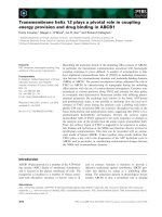
hybridization [12]. In addition to the primary site of
expression (i.e. the NI), where, in the rat, approxi-
mately 2000 relaxin-3-positive neurons are found
(Fig. 1A), a smaller number of these neurons are
scattered in the pontine raphe nucleus, the periaqu-
eductal gray matter, and the area dorsal to the
substantia nigra in the midbrain reticular formation.
By immunostaining using monoclonal antibody against
the N-terminus of the human relaxin-3 A-chain [2],
relaxin-3-immunoreactive fibers were observed to
project densely to the septum, hippocampus, lateral
hypothalamic area (LH) and intergeniculate leaflet of
the thalamus (Fig. 1B). Ultrastructural examination
revealed that relaxin-3 was localized to the dense-core
vesicles in the perikarya, and it was also observed in
the synaptic terminals of axons [12]. The NI comprises
a distinct cell group in the caudoventral region of the
pontine periventricular gray matter, adjacent to the
ventromedial border of the caudal dorsal tegmental
nucleus [13]. Studies involving neuronal tracing with
anterograde and retrograde tracers have shown that
the NI, together with the median raphe and interpe-
duncular nuclei, may form a midline behavior control
network, and many targets of the NI, such as the med-
ial septum, hippocampus, hypothalamus, mammillary
complex and amygdala, are involved in arousal mecha-
nisms, including the synchronization and desynchroni-
zation of the theta rhythm [14,15]. Recently, Ma et al.
[16] reported that relaxin-3 neurons in the NI can help
modulate spatial memory and the underlying hippo-
campal theta activity. Using immunocytochemistry
studies, relaxin-3-positive neurons in the NI have been
shown to be GABAergic and to co-express corticotro-
pin-releasing factor (CRF) type 1 receptors (CRFR1)
[12,17].
Relaxin-3 receptor
The cognate receptor for relaxin-3 is RXFP3, formally
known as GPCR135 or SALPR [6,18]. Although it can
also bind and activate RXFP1 and RXFP4, relaxin-3
binds RXPF3 with higher affinity (0.31 nm) than
RXFP1 (2.0 nm) or RXFP4 (1.1 nm) [6,19]. RXFP3
mRNA is abundant in the olfactory bulb, paraventric-
ular nucleus (PVN) and supraoptic nucleus (SON) in
the hypothalamus amygdaloid–hippocampal area, as
well as the bed nucleus stria terminalis, paraventricular
thalamus, superior colliculus and interpeduncular
nucleus in the brainstem. The distribution of RXFP3
approximately overlaps with the autoradiography
pattern, showing selective RXFP3 binding of the chi-
meric peptide, relaxin-3 B-chain ⁄ INSL5 A-chain [20].
In the brain, there is generally a close correlation
between relaxin-3-positive nerve terminals and RXFP3
expression; however, the density of expression of
ligand and receptor is not always equal. For example,
the olfactory bulb exhibits abundant RXFP3 expres-
DTg
A
B
NIc
4V
IP
Hippocampus
PAG
RSC
DB
Hypothalamus
LS
NI
DR
NId
MS
mlf
Fig. 1. (A) Relaxin-3 immunoreactivity in the NI. Relaxin-3 is
expressed in neurons of both the pars compacta (NIc) and pars dissi-
pata (NId) of the NI. DTg, dorsal tegmental nucleus; 4V, fourth ventri-
cle; mlf, medial longitudinal fasciculus. Scale bars = 100 lm. (B) A
schematic representation of the major projection of relaxin-3 in the
forebrain. DB, diagonal band; DR, dorsal raphe nucleus; IP, interpe-
duncular nucleus; LS, lateral septal nucleus; MS, medial septal
nucleus; PAG, periaqueductal gray matter; RSC, retrosplenial cortex.
M. Tanaka Relaxin-3 expression and function in the hypothalamus
FEBS Journal 277 (2010) 4990–4997 ª 2010 The Author Journal compilation ª 2010 FEBS 4991
sion, whereas it has relatively low levels of relaxin-3-
immunoreactive fibers. In the hypothalamus, relaxin-3
fibers densely innervate the lateral hypothalamic area,
although RXFP3 is strongly expressed in the PVN and
SON [12,17,21]. The structure and function of the
relaxin family peptide receptors, including RXFP3 and
RXFP4, were recently reviewed by Kong et al. [22].
Relaxin-3 expression in development and in other
species
During the development of the rat, relaxin-3 mRNA
expression appears at embryonic day 18 near the
fourth ventricle. Relaxin-3 peptide can be detected
after birth by immunocytochemistry [23]. This develop-
mental expression pattern is comparable with that of
relaxin, the rodent equivalent of human relaxin-2,
whose mRNA is not detectable in the rat brain at
embryonic day 15, although it is detectable at postna-
tal day 1 [24]. As well as rodents, the distribution of
relaxin-3 in the brain has recently been reported for
fish, monkeys and humans. In the zebrafish, the
relaxin-3 gene is expressed in two neuron clusters in
the brainstem: one is a midbrain cell cluster of the
periaqueductal gray matter and the other is in a pos-
terior region that could be homologous to the mam-
malian NI [25]. Two groups have described the
distribution of relaxin-3 in the primate brain. In the
brain of Macaca fascicularis, relaxin-3-positive cell
bodies were found to be distributed within a ventrome-
dial region of the central gray matter of the pons and
medulla, which appears to correspond to the NI in
lower species [26]. In the rhesus macaque and humans,
relaxin-3 immunostaining was predominantly observed
in the ventral and dorsal tegmental nuclei of the brain-
stem [27]. Thus, from fish to primates, this peptide is
expressed in the dorsal tegmentum of the brain stem,
corresponding to the NI in rodents.
Regulation of relaxin-3 gene expression
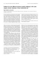
Concerning the regulation of relaxin-3 gene expression,
relaxin-3 mRNA expression in the NI is enhanced by
restraint stress or forced swim stress (Fig. 2A) [12,28].
This swim stress-induced increase in relaxin-3 tran-
script levels is blunted by the systemic administration
of CRFR1 antagonist [28]. Relaxin-3 transcript levels
are also increased after treatment with p-chlorophenyl-
alanine, a potent inhibitor of serotonin synthesis, indi-
cating that serotonin negatively regulates relaxin-3
gene expression [23]. From these results, the expression
of relaxin-3 may be observed to be dynamically altered
under different physiological conditions. We found
that relaxin-3 is expressed in a mouse neuroblastoma
cell line, Neuro2a, and investigated the intracellular
signaling that leads to activation of relaxin 3 gene
transcription in vitro [29]. Using a clone stably-trans-
fected with a relaxin-3 promoter-enhanced green fluo-
rescent protein gene, we observed that the increase in
intracellular cAMP induced by dibutyryl cAMP and
forskolin treatment increased relaxin-3 promoter activ-
ity. These increases were inhibited by pretreatment
with the protein kinase A (PKA) inhibitors, H89 and
KT5720. Moreover, the relaxin-3 promoter activity
was enhanced by CRF treatment after the expression
CRFR1
CRF
cAMP
G
s
PKA
Relaxin-3 gene
P
Transcription factor
AT P
PKA
Plasma membrane
P
Promoter
Stress
Nucleus
0
100
200
300
400
A
B
Cont
PSL
Stress
*
Cont Stress
AC
Fig. 2. (A) Relaxin-3 mRNA expression in the NI after 6 h of
restrained stress. The upper panel shows a representative image of
in situ hybridization using the [
35
S]-labeled probe. The graph below
indicates the calculated signal intensity of relaxin-3 mRNA. Data are
shown as the mean ± SD of photostimulated luminescence (PSL)
[12]. (B) A schematic representation of the intracellular signaling
that regulates relaxin-3 gene expression. Downstream of CRFR1,
the cAMP-PKA pathway is involved in the activation of relaxin-3
gene transcription.
Relaxin-3 expression and function in the hypothalamus M. Tanaka
4992 FEBS Journal 277 (2010) 4990–4997 ª 2010 The Author Journal compilation ª 2010 FEBS
of CRFR1 receptor in the cells. These results suggest
that relaxin-3 transcription in vivo is activated via the
cAMP-PKA pathway, which is downstream of CRFR1
[29] (Fig. 2B).
The function of relaxin-3 in the brain
Because relaxin-3-producing cells showed a relatively
limited distribution, predominantly in neurons of the
NI, the function of this peptide has been assessed based
upon anatomical studies of the NI at the neuronal level
[14,15]. The NI is composed of two subdivisions, the
pars compacta and pars dissipata, and relaxin-3-
positive neurons are found in both regions (Fig. 1A).
With reference to the distribution of relaxin-3-positive
nerve fibers and RXFP3 and RXFP1 expression,
several functions of relaxin-3 in the brain have been
demonstrated, including those related to neuroendo-
crine processes, stress response, water intake and spatial
memory [12,16,28,30–35]. Particularly, this peptide also
regulates food intake, as well as other hypothalamic
peptides described in this minireview series [36,37].
Stress response
The NI is a region showing abundant expression of
CRFR1, and strong c-Fos induction was observed in
the NI in response to an intracerebroventricular injec-
tion of CRF [38,39]. It is well known that CRF is
expressed in parvocellular neurons of the PVN and,
during the stress response, CRF activates the hypotha-
lamic-pituitary-adrenal (HPA) axis, acting at CRFR1
on anterior pituitary corticotropes to stimulate the
release of adrenocorticotropic hormone. There are also
extrahypothalamic CRF-expressing neurons distributed
through the brain in areas such as the neocortex and
limbic regions, including the central amygdala and
hippocampus [40,41]. The regulation of CRF expression
may be involved in setting the ‘tone’ of stress-related
behavior, including anxiety, as well as learning and
memory [42,43]. CRF exerts its actions via two major
receptors: CRFR1 and CRFR2. Both receptors belong
to the class B subtype of G protein-coupled receptors,
although they have a different distribution, suggesting
that the two receptors have different functions. CRFR1
is considered to be involved in the acute phase of the
stress response, whereas CRFR2 contributes to the
maintenance and recovery phase that involves a gradual
reduction of HPA axis activation [43,44].
In the rat NI, almost all relaxin-3-positive neurons
coexpress CRFR1 and respond to CRF intracerebro-
ventricular administration. Moreover, application of a
water-restraint stress for 2–4 h induces c-Fos expres-
sion and leads to an increase in relaxin-3 mRNA levels
in the NI [12]. On the other hand, relaxin-3-positive
neurons project fibers to the hypothalamus, and
RXFP3 is intensely expressed in the PVN where hypo-
thalamic CRF neurons exist. These results suggest that
relaxin-3-expressing neurons respond immediately to
stress and modulate the HPA axis. Recently, Banerjee
et al. [28] reported that exposure of rats to a repeated
forced swim for 10 min each time leads to a marked
increase in relaxin-3 mRNA levels in the NI at
30–60 min after the second swim. Systemic treatment
with the CRFR1 antagonist alarmin 30 min before the
second swim blunted the stress-induced effect on
relaxin-3 transcripts in the NI [28]. This supports the
idea that relaxin-3-expressing neurons in the NI (and
therefore relaxin-3) play a role in the central stress
regulating system by mutual interaction with CRF-
expressing neurons.
Food intake
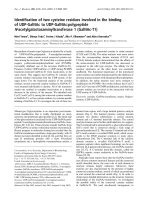
Relaxin-3 was first reported to stimulate food intake
when administered into the third ventricle or PVN of
male Wistar rats. Administration of human relaxin-3,
but not human relaxin-2, either intracerebroventricu-
larly (180 pmol) or intra-PVN (18 pmol) increased 1-h
food intake both in the early light and early dark
phase (Fig. 3) [31]. The doses of relaxin-3 required to
elicit a significant feeding response are in the picomo-
lar range and are similar to the effective doses of other
orexigenic peptides such as ghrelin (30 pmol; intra-
PVN) and neuropeptide Y (NPY) (78 pmol; intra-
PVN) [45,46]. Although RXFP3 and RXFP1 are
expressed in the PVN, relaxin (specifically, human
relaxin-2) binds RXFP1 but not RXFP3, suggesting
that this feeding-promoting action of relaxin-3 is
exerted through RXFP3 because the actions of relaxin
have not been reported to include hyperphagia, but do
include hemodynamic effects such as increasing arterial
blood pressure and vasopressin release [47], or dipso-
genesis [48]. In reverse, relaxin-3 was recently reported
to facilitate water intake as well as relaxin, suggesting
that RXFP1 was involved in this action [35]. Concern-
ing the chronic administration of relaxin-3, intracere-
broventricular injection for 14 days (600 pmolÆday
)1
)
using osmotic minipumps led to a significant increase
in food consumption and weight compared to vehicle
infusion. There was no difference in locomotor activity
between two groups either in the light phase or dark
phase, suggesting that this effect of relaxin-3 is not a
result of increased locomotor or arousal activity [34].
Chronic intra-PVN administration of human relaxin-3
(180 pmol twice a day for 7 days) also increased the
M. Tanaka Relaxin-3 expression and function in the hypothalamus
FEBS Journal 277 (2010) 4990–4997 ª 2010 The Author Journal compilation ª 2010 FEBS 4993
cumulative food intake in ad libitum-fed rats [32]. After
such chronic administration, the plasma concentration
of leptin and insulin was significantly increased [32]. In
addition to the PVN, relaxin-3-administration into the
SON or arcuate nucleus (ARC), but not into the LH,
stimulated 1-h food intake [32]. The ARC and LH are
well known as feeding centers where orexigenic
peptides such as NPY, melanin-concentrating hor-
mone and orexin are distributed. Although relaxin-
3-immunoreactive fibers are densely distributed, the
RXFP3 level is relatively low in the ARC and LH.
An electrophysiological study of neurons in these
hypothalamic nuclei may help to resolve this disparity
and clarify the hyperphagic mechanisms.
Recently, relaxin-3 gene knockout (KO) mice of
mixed background (129S5:B6) were examined in two
studies. One group reported that KO mice are smaller
and leaner than congenic controls [21], although the
results obtained by the second group indicated that
there was no genotypic difference in body weight or
motor coordination [49]. Further studies using relaxin-
3 KO mice backcrossed to C57 ⁄ B6 should help to clar-
ify the role of relaxin-3 in regulating body weight and
metabolism.
Actions of relaxin-3 at the hypothalamo-pituitary-
gonadal (HPG) axis
Recently, a role of relaxin-3 in regulation of the HPG
axis was reported in that intracerebroventricular
(5 nmol) and intra-PVN (540–1620 pmol) administra-
tion of relaxin-3 in adult male rats significantly
increased plasma luteinizing hormone levels. This effect
was inhibited by pretreatment with a peripheral gona-
dotropin-releasing hormone (GnRH) antagonist. By
contrast, the central administration of human relaxin-2
was not found to influence the plasma luteinizing hor-
mone concentration. Using hypothalamic explants and
GT1-7 cells that express RXFP1 and RXFP3, relaxin-
3 was shown to dose-dependently stimulate GnRH
release. GnRH neuronal cell bodies are found in sev-
eral forebrain regions, including the medial septum,
diagonal band, preoptic area and LH, where relaxin-3-
positive fibers and RXFP3 are moderately-to-densely
distributed [12,17,50]. These results suggest that
relaxin-3 regulates the HPG axis via hypothalamic
GnRH neurons. Thus, relaxin-3 is seen to belong to
the group of neuropeptides that regulate energy
homeostasis and reproduction (i.e. modulate both
appetite and the HPG axis). This group includes NPY,
orexin and galanin-like peptides [51–54].
Conclusions
In this minireview, relaxin-3, which is the latest mem-
ber of the insulin ⁄ relaxin family, is described in terms
of its gene transcript and peptide expression in the
brain, as well as its functional aspects that have thus
far been reported. Although relaxin-3-expressing neu-
rons show a confined distribution in the brainstem,
being particularly dense in the NI of the dorsal tegmen-
tal pons, their fibers and receptors (i.e. RXFP3 and
RXFP1) are widely distributed in the forebrain. One of
the target areas of relaxin-3 is the hypothalamus.
Relaxin-3 is considered to have various actions medi-
Fig. 3. Effect of intracerebroventricular administration of relaxin-3
in satiated male Wistar rats. (A) Effect of human relaxin-3 (H3) (18–
180 pmol) on 1-h food intake. *P < 0.05 versus vehicle in the early
light phase. (B) Effect of H3 (18–180 pmol) on cumulative food
intake over 4 h in the early light phase.
&
P < 0.05 at 18 pmol
versus vehicle; *P < 0.05 at 54 pmol versus vehicle;
#
P < 0.05
at 180 pmol versus vehicle. Reproduced with permission [31];
ª 2005, The Endocrine Society).
Relaxin-3 expression and function in the hypothalamus M. Tanaka
4994 FEBS Journal 277 (2010) 4990–4997 ª 2010 The Author Journal compilation ª 2010 FEBS
ated through receptors in the hypothalamus, including
effects on the stress response, feeding and neuroendo-
crine function.
Acknowledgements
The present work was supported by a grant (no.
21500329) to M.T. from the Ministry of Education,
Culture, Sports, Science and Technology, Japan.
References
1 Bathgate RA, Samuel CS, Burazin TC, Layfield S,
Claasz AA, Reytomas IG, Dawson NF, Zhao C, Bond
C, Summers RJ et al. (2002) Human relaxin gene 3
(H3) and the equivalent mouse relaxin (M3) gene.
Novel members of the relaxin peptide family. J Biol
Chem 277, 1148–1157.
2 Kizawa H, Nishi K, Ishibashi Y, Harada M, Asano T,
Ito Y, Suzuki N, Hinuma S, Fujisawa Y, Onda H et al.
(2003) Production of recombinant human relaxin 3 in
AtT20 cells. Regul Pept 113, 79–84.
3 Bullesbach EE, Yang S & Schwabe C (1992) The
receptor-binding site of human relaxin II. A dual prong-
binding mechanism. J Biol Chem 267, 22957–22960.
4 James R, Niall H, Kwok S & Bryand-Greenwood G
(1977) Primary structure of porcine relaxin: homology
with insulin and related growth factors. Nature 267,
544–546.
5 Wilkinson TN, Speed TP, Tregear GW & Bathgate RA
(2005) Evolution of the relaxin-like peptide family.
BMC Evol Biol 5, 14.
6 Liu C, Chen J, Sutton S, Roland B, Kuei C, Farmer N,
Sillard R & Lovenberg TW (2003) Identification of
relaxin-3 ⁄ INSL7 as a ligand for GPCR142. J Biol Chem
278, 50765–50770.
7 Hudson P, Haley J, Cronk M, Shine J & Niall H
(1981) Molecular cloning and characterization of
cDNA sequences coding for rat relaxin. Nature 291,
127–131.
8 Hudson P, John M, Crawford R, Haralambidis J,
Scanlon D, Gorman J, Tregear G, Shine J & Niall H
(1984) Relaxin gene expression in human ovaries
and the predicted structure of a human preprorelaxin
by analysis of cDNA clones. EMBO J 3, 2333–2339.
9 Hansell DJ, Bryant-Greenwood GD & Greenwood FC
(1991) Expression of the human relaxin H1 gene in the
decidua, trophoblast, and prostate. J Clin Endocrinol
Metab 72, 899–904.
10 Sherwood OD (1994) Relaxin. In The Phsiology of
Reproduction, 2nd edn (Knobil E & Neill J eds), pp.
861–1009. Raven Press, Ltd, New York.
11 Burazin TC, Bathgate RA, Macris M, Layfield S,
Gundlach AL & Tregear GW (2002) Restricted, but
abundant, expression of the novel rat gene-3 (R3)
relaxin in the dorsal tegmental region of brain. J Neuro-
chem 82, 1553–1557.
12 Tanaka M, Iijima N, Miyamoto Y, Fukusumi S, Itoh
Y, Ozawa H & Ibata Y (2005) Neurons expressing
relaxin 3 ⁄ INSL 7 in the nucleus incertus respond to
stress. Eur J Neurosci 21, 1659–1670.
13 Berman AL (1968) The brain stem of the cat: a cytoar-
chitectonic atlas with stereotaxic coordinates. University
of Wisconsin Press, Madison.
14 Goto M, Swanson LW & Canteras NS (2001) Connec-
tions of the nucleus incertus. J Comp Neurol 438, 86–
122.
15 Olucha-Bordonau FE, Teruel V, Barcia-Gonzalez J,
Ruiz-Torner A, Valverde-Navarro AA & Martinez-
Soriano F (2003) Cytoarchitecture and efferent
projections of the nucleus incertus of the rat. J Comp
Neurol 464, 62–97.
16 Ma S, Olucha-Bordonau FE, Hossain MA, Lin F,
Kuei C, Liu C, Wade JD, Sutton SW, Nunez A &
Gundlach AL (2009) Modulation of hippocampal theta
oscillations and spatial memory by relaxin-3 neurons of
the nucleus incertus. Learn Mem 16, 730–742.
17 Ma S, Bonaventure P, Ferraro T, Shen PJ, Burazin TC,
Bathgate RA, Liu C, Tregear GW, Sutton SW &
Gundlach AL (2007) Relaxin-3 in GABA projection
neurons of nucleus incertus suggests widespread
influence on forebrain circuits via G-protein-coupled
receptor-135 in the rat. Neuroscience 144, 165–190.
18 Matsumoto M, Kamohara M, Sugimoto T, Hidaka K,
Takasaki J, Saito T, Okada M, Yamaguchi T & Furui-
chi K (2000) The novel G-protein coupled receptor
SALPR shares sequence similarity with somatostatin
and angiotensin receptors. Gene 248, 183–189.
19 Liu C, Chen J, Kuei C, Sutton S, Nepomuceno D, Bon-
aventure P & Lovenberg TW (2005) Relaxin-3 ⁄ insulin-
like peptide 5 chimeric peptide, a selective ligand for G
protein-coupled receptor (GPCR)135 and GPCR142
over leucine-rich repeat-containing G protein-coupled
receptor 7. Mol Pharmacol 67, 231–240.
20 Sutton SW, Bonaventure P, Kuei C, Roland B, Chen J,
Nepomuceno D, Lovenberg TW & Liu C (2004) Distri-
bution of G-protein-coupled receptor (GPCR)135 bind-
ing sites and receptor mRNA in the rat brain suggests a
role for relaxin-3 in neuroendocrine and sensory pro-
cessing. Neuroendocrinology 80, 298–307.
21 Sutton SW, Shelton J, Smith C, Williams J, Yun S,
Motley T, Kuei C, Bonaventure P, Gundlach A, Liu C
et al. (2009) Metabolic and neuroendocrine responses to
RXFP3 modulation in the central nervous system. Ann
N Y Acad Sci 1160, 242–249.
22 Kong RC, Shilling PJ, Lobb DK, Gooley PR &
Bathgate RA (2010) Membrane receptors: structure and
function of the relaxin family peptide receptors. Mol
Cell Endocrinol 320, 1–15.
M. Tanaka Relaxin-3 expression and function in the hypothalamus
FEBS Journal 277 (2010) 4990–4997 ª 2010 The Author Journal compilation ª 2010 FEBS 4995
23 Miyamoto Y, Watanabe Y & Tanaka M (2008) Devel-
opmental expression and serotonergic regulation of
relaxin 3 ⁄ INSL7 in the nucleus incertus of rat brain.
Regul Pept 145 , 54–59.
24 Osheroff PL & Ho WH (1993) Expression of relaxin
mRNA and relaxin receptors in postnatal and adult rat
brains and hearts. Localization and developmental pat-
terns. J Biol Chem 268, 15193–15199.
25 Donizetti A, Grossi M, Pariante P, D’Aniello E,
Izzo G, Minucci S & Aniello F (2008) Two neuron
clusters in the stem of postembryonic zebrafish brain
specifically express relaxin-3 gene: first evidence of
nucleus incertus in fish. Dev Dyn 237, 3864–3869.
26 Ma S, Sang Q, Lanciego JL & Gundlach AL (2009)
Localization of relaxin-3 in brain of Macaca fascicular-
is: identification of a nucleus incertus in primate.
J Comp Neurol 517, 856–872.
27 Silvertown JD, Neschadim A, Liu HN, Shannon P,
Walia JS, Kao JC, Robertson J, Summerlee AJ &
Medin JA (2010) Relaxin-3 and receptors in the human
and rhesus brain and reproductive tissues. Regul Pept
159, 44–53.
28 Banerjee A, Shen PJ, Ma S, Bathgate RA & Gundlach
AL (2010) Swim stress excitation of nucleus incertus
and rapid induction of relaxin-3 expression via CRF1
activation. Neuropharmacology 58, 145–155.
29 Tanaka M, Watanabe Y & Yoshimoto K (2009)
Regulation of relaxin 3 gene expression via cAMP-PKA
in a neuroblastoma cell line. J Neurosci Res 87, 820–
829.
30 McGowan BM, Stanley SA, Donovan J, Thompson
EL, Patterson M, Semjonous NM, Gardiner JV, Mur-
phy KG, Ghatei MA & Bloom SR (2008) Relaxin-3
stimulates the hypothalamic-pituitary-gonadal axis. Am
J Physiol Endocrinol Metab 295, E278–E286.
31 McGowan BM, Stanley SA, Smith KL, White NE,
Connolly MM, Thompson EL, Gardiner JV, Murphy
KG, Ghatei MA & Bloom SR (2005) Central relaxin-3
administration causes hyperphagia in male Wistar rats.
Endocrinology 146, 3295–3300.
32 McGowan BM, Stanley SA, Smith KL, Minnion JS,
Donovan J, Thompson EL, Patterson M, Connolly
MM, Abbott CR, Small CJ et al. (2006) Effects of
acute and chronic relaxin-3 on food intake and energy
expenditure in rats. Regul Pept 136, 72–77.
33 McGowan BM, Stanley SA, White NE, Spangeus A,
Patterson M, Thompson EL, Smith KL, Donovan J,
Gardiner JV, Ghatei MA et al. (2007) Hypothalamic
mapping of orexigenic action and Fos-like immunore-
activity following relaxin-3 administration in male
Wistar rats. Am J Physiol Endocrinol Metab 292,
E913–E919.
34 Hida T, Takahashi E, Shikata K, Hirohashi T, Sawai
T, Seiki T, Tanaka H, Kawai T, Ito O, Arai T et al.
(2006) Chronic intracerebroventricular administration
of relaxin-3 increases body weight in rats. J Recept
Signal Transduct Res 26, 147–158.
35 Otsubo H, Onaka T, Suzuki H, Katoh A, Ohbuchi T,
Todoroki M, Kobayashi M, Fujihara H, Yokoyama T,
Matsumoto T et al. (2010) Centrally administered
relaxin-3 induces Fos expression in the osmosensitive
areas in rat brain and facilitates water intake. Peptides
31, 1124–1130.
36 Shiba K, Kageyama H, Takenoya F & Shioda S (2010)
Galanin-like peptide and the regulation of feeding
behavior and energy metabolism. FEBS J 277, 5006–
5013.
37 Takayanagi Y & Onaka T (2010) Roles of prolactin-
releasing peptide and RFamide related peptides in the
control of stress and food intake. FEBS J 277, 4998–
5005.
38 Potter E, Sutton S, Donaldson C, Chen R, Perrin M,
Lewis K, Sawchenko PE & Vale W (1994) Distribution
of corticotropin-releasing factor receptor mRNA
expression in the rat brain and pituitary. Proc Natl
Acad Sci USA 91, 8777–8781.
39 Bittencourt JC & Sawchenko PE (2000) Do centrally
administered neuropeptides access cognate receptors? an
analysis in the central corticotropin-releasing factor
system. J Neurosci 20, 1142–1156.
40 Merchenthaler I, Vigh S, Petrusz P & Schally AV
(1982) Immunocytochemical localization of corticotro-
pin-releasing factor (CRF) in the rat brain. Am J Anat
165, 385–396.
41 Swanson LW, Sawchenko PE, Rivier J & Vale WW (1983)
Organization of ovine corticotropin-releasing factor
immunoreactive cells and fibers in the rat brain: an immu-
nohistochemical study. Neuroendocrinology 36, 165–186.
42 Bale TL & Vale WW (2004) CRF and CRF receptors:
role in stress responsivity and other behaviors. Annu
Rev Pharmacol Toxicol 44, 525–557.
43 Korosi A & Baram TZ (2008) The central corticotropin
releasing factor system during development and adult-
hood. Eur J Pharmacol 583, 204–214.
44 Coste SC, Murray SE & Stenzel-Poore MP (2001)
Animal models of CRH excess and CRH receptor
deficiency display altered adaptations to stress. Peptides
22, 733–741.
45 Wren AM, Small CJ, Abbott CR, Dhillo WS, Seal LJ,
Cohen MA, Batterham RL, Taheri S, Stanley SA, Gha-
tei MA et al. (2001) Ghrelin causes hyperphagia and
obesity in rats. Diabetes 50, 2540–2547.
46 Stanley BG, Daniel DR, Chin AS & Leibowitz SF
(1985) Paraventricular nucleus injections of peptide YY
and neuropeptide Y preferentially enhance carbohydrate
ingestion. Peptides 6, 1205–1211.
47 Mumford AD, Parry LJ & Summerlee AJ (1989) Lesion
of the subfornical organ affects the haemotensive
response to centrally administered relaxin in anaesthe-
tized rats. J Endocrinol 122, 747–755.
Relaxin-3 expression and function in the hypothalamus M. Tanaka
4996 FEBS Journal 277 (2010) 4990–4997 ª 2010 The Author Journal compilation ª 2010 FEBS
48 Summerlee AJ, Hornsby DJ & Ramsey DG (1998) The
dipsogenic effects of rat relaxin: The effect of photope-
riod and the potential role of relaxin on drinking in
pregnancy. Endocrinology 139, 2322–2328.
49 Smith CM, Lawrence AJ, Sutton SW & Gundlach AL
(2009) Behavioral phenotyping of mixed background
(129S5:B6) relaxin-3 knockout mice. Ann N Y Acad Sci
1160, 236–241.
50 King JC, Tobet SA, Snavely FL & Arimura AA (1982)
LHRH immunopositive cells and their projections to
the median eminence and organum vasculosum of the
lamina terminalis. J Comp Neurol 209, 287–300.
51 Kageyama H, Takenoya F, Kita T, Hori T, Guan JL &
Shioda S (2005) Galanin-like peptide in the brain:
effects on feeding, energy metabolism and reproduction.
Regul Pept 126, 21–26.
52 Kalra SP & Kalra PS (2004) NPY and cohorts in regulat-
ing appetite, obesity and metabolic syndrome: beneficial
effects of gene therapy. Neuropeptides 38, 201–211.
53 Pu S, Jain MR, Kalra PS & Kalra SP (1998) Orexins, a
novel family of hypothalamic neuropeptides, modulate
pituitary luteinizing hormone secretion in an ovarian
steroid-dependent manner. Regul Pept 78, 133–136.
54 Seth A, Stanley S, Jethwa P, Gardiner J, Ghatei M &
Bloom S (2004) Galanin-like peptide stimulates the
release of gonadotropin-releasing hormone in vitro and
may mediate the effects of leptin on the hypothalamo-
pituitary-gonadal axis. Endocrinology 145, 743–750.
M. Tanaka Relaxin-3 expression and function in the hypothalamus
FEBS Journal 277 (2010) 4990–4997 ª 2010 The Author Journal compilation ª 2010 FEBS 4997