Báo cáo khoa học: Roles of prolactin-releasing peptide and RFamide related peptides in the control of stress and food intake potx
Bạn đang xem bản rút gọn của tài liệu. Xem và tải ngay bản đầy đủ của tài liệu tại đây (160.68 KB, 8 trang )
MINIREVIEW
Roles of prolactin-releasing peptide and RFamide related
peptides in the control of stress and food intake
Yuki Takayanagi and Tatsushi Onaka
Division of Brain and Neurophysiology, Department of Physiology, Jichi Medical University, Japan
Introduction
RFamide peptides, defined by their carboxy-terminal
arginine (R) and amidated phenylalanine (F) residues
(hence RFamide), were originally discovered in inverte-
brates [1] and have recently been identified in verte-
brates [2]. The first reported RFamide peptides in
mammals were neuropeptide FF (NPFF) and neuro-
peptide AF, which were later confirmed to be encoded
on a single gene [3]. By applying a reverse pharmaco-
logical approach, in which orphan G protein-coupled
receptor ligands were identified by detecting signal
transduction induced in cells expressing a targeted
orphan G protein-coupled receptor after stimulation,
prolactin-releasing peptide (PrRP) was identified to be
a ligand of an orphan G protein-coupled receptor,
GPR10 (hGR3 ⁄ UHR-1 ⁄ PRLHR) and to belong to the
RFamide peptide [4]. Subsequently, by utilizing DNA
databases, another gene for RFamide peptides was
identified in mammals [5,6]. The RFamide peptides
encoded by the gene were named RFamide related
peptide (RFRP)-1 ⁄ NPSF and RFRP-3 ⁄ NPVF, which
were found to be orthologs of avian peptide LPLRFa-
mide. Thus, RFRPs are allocated into the LPXRFa-
mide peptide family (X = L or Q). Several other
RFamide peptides also have been discovered by a
reverse pharmacological method and by DNA
sequence database searching, and there are now five
families of RFamide peptides known to exist in
mammals: NPFF, PrRP, LPXRFamide, kisspeptin
Keywords
dorsomedial hypothalamus; energy
consumption; energy metabolism; food
intake; nucleus tractus solitarii; oxytocin;
prolactin-releasing peptide; RFamide
peptide; RFamide-related peptide; stress
Correspondence
T. Onaka, Division of Brain and
Neurophysiology, Department of Physiology,
Jichi Medical University, Shimotsuke-shi,
Tochigi-ken 329-0498, Japan
Fax: +81 285 44 8147
Tel: +81 285 58 7318
E-mail:
(Received 13 June 2010, revised 2
September 2010, accepted 13 October
2010)
doi:10.1111/j.1742-4658.2010.07932.x
Subsequent to the isolation of the first recognized RFamide neuropeptide,
FMRFamide, from the clam, a large number of these peptides have been
identified. There are now five groups of RFamide peptides identified in
mammals. RFamide peptides show diversity with respect to their N-termi-
nal sequence and biological activity. RFamide peptides have been impli-
cated in a variety of roles, including energy metabolism, stress and pain
modulation, as well as effects in the neuroendocrine and cardiovascular
systems. In the present minireview, we focus on prolactin-releasing peptide
(PrRP) and RFamide related peptide (RFRP) with respect to their roles in
the control of energy metabolism and stress responses. Both food intake
and stressful stimuli activate PrRP neurons. The administration of PrRP
affects energy metabolism and neuroendocrine systems. PrRP-deficient or
PrRP receptor-deficient mice show abnormal energy metabolism and ⁄ or
stress responses. On the other hand, RFRP neurons are activated by stress-
ful stimuli and the administration of RFRP induces neuroendocrine
and behavioral stress responses. Taken together, these data suggests that
PrRP and RFRP neurons play a role in the control of energy metabolism
and/or stress responses.
Abbreviations
ACTH, adrenocorticotropic hormone; CCK, cholecystokinin-8; CRH, corticotropin-releasing hormone; NPFF, neuropeptide FF;
NTS, nucleus tractus solitarii; PrRP, prolactin-releasing peptide; QFRP, pyroglutamylated RFamide peptide; RFRP, RFamide related peptide.
4998 FEBS Journal 277 (2010) 4998–5005 ª 2010 The Authors Journal compilation ª 2010 FEBS
(previously known as metastin) and pyroglutamylated
RFamide peptide (QFRP) (Table 1). RFamide peptides
show diversity in their N-terminal sequence and, as a
result, a broad pattern of biological activities, includ-
ing the control of energy metabolism and stress, as
well as effects in the neuroendocrine and cardiovascu-
lar systems. In the present minireview, we focus on
PrRP and RFRP (Table 2) and review recent progress
in research investigating the roles of these two peptides
in the control of energy metabolism and stress.
PrRP
PrRP was considered to serve as a hypothalamic-
releasing factor and to act on the anterior pituitary
to stimulate prolactin release from the pituitary.
However, no PrRP immunoreactivity was found in the
external layer of the median eminence, from where
classic hypothalamic hormones are released into the
portal blood to control anterior pituitary hormone
release. Thus, PrRP is not a classic hypothalamic hor-
mone in mammals. Instead, PrRP appears to play an
important role in the control of energy metabolism
and stress [7].
Localization of PrRP and its receptors
PrRP neurons are localized mostly in the nucleus trac-
tus solitarii (NTS), with modest expression in the ven-
trolateral medulla regions of the brainstem, and slight
expression in the dorsomedial hypothalamus. In the
medulla oblongata, PrRP expression is restricted to the
Table 1. Summary of mammalian RFamide peptides and their receptors. The effects of administration of RFamide peptides upon stress
responses (hormone release) and energy metabolism are also described.
Family
name
Peptide in
mammals Receptors Stress responses
Energy metabolism
Food intake
Energy
consumption
NPFF NPFF
NPAF
GPR74 (NPFF-2, NPGPR, HLWAR77) Decrease in vasopressin
release
Decrease [51] Increase
PrRP PrRP GPR10 (hGR3, UHR-1, PRLHR) Increase in the release
of ACTH, oxytocin,
vasopressin and prolactin
Decrease Increase
LPXRF RFRP-1 (NPSF)
RFRP-3 (NPVF)
GPR147 (NPFF-1, OT7T022, RFRPR) Increase in the release
of ACTH, oxytocin and
prolactin
Increase
?
?
Kisspeptin Kiss1 (metastin) GPR54 (OT7T175, AXOR12, KISS-1R) ? No effects ?
QFRP QRFP (26 Rfa, P513) GPR103 (AQ27, SP9155) ? Increase [52,53] ?
Table 2. Amino acid sequences of PrRP and RFRP in human, rats and mice are shown. *Deduced from the cDNA sequence.
Peptide Species
Number of
of amino acids Sequence Reference
PrRP Human 20
31
TPDINPAWYASRGIRPVGRF-NH
2
SRTHRHSMEIRTPDINPAWYASRGIRPVGRF-NH
2
[4]*
[4]*
Rat 20
31
TPDINPAWYTGRGIRPVGRF-NH
2
SRAHQHSMETRTPDINPAWYTGRGIRPVGRF-NH
2
[4]*
[4]*
Mouse 20
31
TPDINPAWYTGRGIRPVGRF-NH
2
SRAHQHSMETRTPDINPAWYTGRGIRPVGRF-NH
2
*
*
RFRP-1 (NPSF) Human 37
12
SLNFEELKDWGPKNVIKMSTPAVNKMPHSFANLPLRF-NH
2
MPHSFANLPLRF-NH
2
[5]*
[5]*, [54]
Rat 37
12
SVTFQELKDWGAKKDIKMSPAPANKVPHSAANLPLRF-NH
2
VPHSAANLPLRF-NH
2
[55]
[5]*
Mouse 37
12
SVSFQELKDWGAKKDIKMSPAPANKVPHSAANLPLRF-NH
2
VPHSAANLPLRF-NH
2
[5]*
[5]*
RFRP-3 (NPVF) Human 8
17
VPNLPQRF-NH
2
NMEVSLVRRVPNLPQRF-NH
2
[5]*, [54]
[5]*
Rat 18 ANMEAGTMSHFPSLPQRF-NH
2
[56]
Mouse 17 or 18 (V)NMEAGTRSHFPSLPQRF-NH
2
[5]*
Y. Takayanagi and T. Onaka PrRP and RFRPs in metabolism and stress
FEBS Journal 277 (2010) 4998–5005 ª 2010 The Authors Journal compilation ª 2010 FEBS 4999
caudal A2 and A1 noradrenergic neurons. Immunohis-
tochemical studies have shown that PrRP-immunoreac-
tive fibers are widely distributed in the brain [8]. The
main receptor for PrRP, GPR10, is also widely
expressed in the brain (especially in the reticular
nucleus of the thalamus, bed nucleus of the stria termi-
nalis, preoptic areas, hypothalamic paraventricular
nucleus, periventricular nucleus, dorsomedial hypothal-
amus, NTS and area postrema) [9]. In addition to
GPR10, PrRP has a high affinity for NPFF receptor 2
(also known as GPR74 ⁄ NPGPR ⁄ HLWAR77). NPFF
receptor 2 is expressed in the dorsal horn of the spinal
cord, thalamus, hypothalamus and hippocampus [10].
Although the physiological functions of NPFF recep-
tor 2 activated by PrRP remain to be clarified, a recent
study has proposed that central cardiovascular effects
of PrRP are mediated via NPFF receptor 2 [11].
Role of PrRP in the control of energy metabolism:
food intake
Intracerebroventricular injection of PrRP reduces food
intake [12]. Both PrRP-deficient mice [13] and GPR10-
deficient mice [14] show hyperphagia. Acute inhibition
of endogenous PrRP signaling by injections of neutral-
izing monoclonal antibodies against PrRP also induces
hyperphagia [13]. From experiments with PrRP-defi-
cient mice or PrRP-neutralizing antibodies, PrRP has
been shown to regulate meal size rather than meal fre-
quency [13]. Meal size is regulated by satiety signals
that terminate each meal, and one important satiety sig-
nal is cholecystokinin-8 (CCK). CCK is released from
the intestine in response to meals, and acts via the
CCK
A
receptor on afferent vagal fibers that project into
the medulla oblongata, which relays information into
the hypothalamus. Food intake [13] or the administra-
tion of CCK [15] activates PrRP neurons in the NTS.
The anorectic effects of CCK are impaired in both
PrRP-deficient mice [13] and GPR10-deficient mice [16],
suggesting that PrRP relays satiety signaling of CCK.
The downstream actions of PrRP neurons remain to
be clarified. However, neurons expressing corticotro-
pin-releasing hormone (CRH) or oxytocin may relay
PrRP signaling to reduce food intake. Anatomical
studies have shown that oxytocin neurons in the hypo-
thalamus receive direct projections from PrRP neurons
in the medulla oblongata. The administration of PrRP
activates neurons expressing CRH or oxytocin in the
hypothalamus, both of which are anorexic peptides.
PrRP-induced anorexia is attenuated by a CRH recep-
tor antagonist or oxytocin receptor antagonist [16].
Furthermore, an oxytocin receptor antagonist reduces
the anorexic actions of CCK [17,18], and increases
meal size [19]. Oxytocin receptor-deficient mice show
an increased meal size [20]. Taken together, these
results suggest that the PrRP-oxytocin system plays a
pivotal role in relaying the satiety signaling of CCK.
PrRP neurons in the brainstem and hypothalamus
express leptin receptors [21]. Leptin regulates long-term
energy metabolism. Leptin induces the expression of
phosphorylated signal transducer and activator of
transcription protein 3 in PrRP neurons, especially in
the dorsomedial hypothalamus [13] (Fig. 1) and the
anorectic effects of leptin are impaired in PrRP-defi-
cient mice [13]. These data suggest that the anorectic
effects of leptin signaling are mediated, at least in part,
by PrRP.
Role of PrRP in the control of energy metabolism:
energy expenditure
PrRP has also been associated with energy expendi-
ture. An intracerebroventricular injection of PrRP
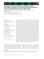
Fig. 1. Activation of PrRP and RFRP neurons in the dorsomedial
hypothalamus after stressful stimuli and leptin administration. The
number of PrRP-immunoreactive neurons expressing Fos protein
(the protein product of the immediate early gene, c-fos) or phos-
phorylated signal transducer and activator of transcription 3 (i.e. a
transcription factor downstream of leptin) after conditioned fear
stimuli or leptin is shown (top). Both conditioned fear stimuli and
leptin administration activate PrRP neurons in the dorsomedial
hypothalamus. The number of RFRP-immunoreactive neurons
expressing Fos protein after footshocks is shown (bottom). Foot-
shocks activate RFRP neurons in the dorsomedial hypothalamus.
Data are obtained from previous studies [13,27,48]. *P < 0.05,
**P < 0.01, ***P < 0.001.
PrRP and RFRPs in metabolism and stress Y. Takayanagi and T. Onaka
5000 FEBS Journal 277 (2010) 4998–5005 ª 2010 The Authors Journal compilation ª 2010 FEBS
increases body temperature and oxygen consumption
[12,22], although neither PrRP-deficient [13], nor GPR10-
deficient male mice [23] show significant difference in
oxygen consumption under basal conditions. PrRP-
deficient mice also show no significant difference in
core body temperature either at room temperature or
after cold exposure [13]. Pair-fed PrRP-deficient mice
show no obese phenotype and no significant difference
in oxygen consumption [13], suggesting that endoge-
nous PrRP is not important for regulating energy
expenditure under resting conditions. On the other
hand, obese GPR10-deficient female mice show slightly
reduced oxygen consumption [23], suggesting that
GPR10 might be important for regulating energy
expenditure in females. Obesity has been reported to
be more pronounced in female than in male GPR10-
deficient mice. It is interesting to note that PrRP neu-
rons in the brainstem express estrogen receptors and
that PrRP expression in the brainstem is higher in
female than in male rats [24]. Thus, the function of
PrRP-GPR10 system in the control of energy con-
sumption might differ between sexes.
PrRP has been suggested to be involved in energy
consumption under stressful conditions. Stressful stim-
uli increase oxygen consumption. This increase in oxy-
gen consumption after stressful stimuli is lower in
PrRP-deficient mice [25]. Stressful stimuli activate
PrRP neurons, and thus it is possible that PrRP
increases energy consumption under the conditions in
which PrRP neurons are activated.
Roles of PrRP in the control of stress responses
PrRP neurons in the medulla oblongata and ⁄ or in the
dorsomedial hypothalamus are activated by a variety
of stressful stimuli [26], including restraint of body
movement, conditioned fear [27] (Fig. 1), footshocks,
hemorrhage [28], exercise [29] and inflammatory stress
(e.g. lipopolysaccharide injection) [30]. PrRP neurons
have been suggested to be involved in neuroendocrine
responses to stress. PrRP neurons project directly to
CRH neurons and oxytocin neurons in the hypothala-
mus [31]. An intracerebroventricular injection of PrRP
activates CRH neurons and oxytocin neurons in the
hypothalamus, and facilitates adrenocorticotropic hor-
mone (ACTH) and oxytocin release into the systemic
circulation. Blockade of endogenous PrRP signaling by
the administration of PrRP-neutralizing antibodies
reduces the activation of hypothalamic paraventricular
neurons after noxious stimuli (formalin injection) [30]
or reduces oxytocin release in response to conditioned
fear [27], suggesting that endogenous PrRP has facilita-
tive roles in neuroendocrine stress responses. On the
other hand, the administration of PrRP-neutralizing
antibodies facilitates ACTH release in response to
exercise, suggesting that PrRP inhibits ACTH release
in response to exercise [29]. Corticosterone release in
response to restraint stress has been reported to be
enhanced in PrRP-deficient mice [32]. These data sug-
gest that PrRP has inhibitory effects on neuroendo-
crine responses to stress. At present, the mechanisms
underlying these apparent discrepant data remain to
be clarified. However, the roles of PrRP in the control
of stress responses may depend upon the nature of
stressful stimuli used.
Stressful stimuli affect food intake and increase
energy expenditure. On the other hand, food intake
affects stress responses [33]. PrRP-deficient mice show
a lower increase in oxygen consumption after stressful
stimuli [25], although the effects of stress on food
intake and the effects of food intake on stress
responses have not yet been examined in PrRP-defi-
cient mice. PrRP might be involved in the integration
in the control of energy metabolism and stress
responses, whereas the underlying detailed mechanisms
need further investigation.
RFRP
The mammalian members of the LPXRFamide peptide
family are RFRP-1 and RFRP-3. RFRP-1 and RFRP-
3 are derived from a single precursor protein. Immu-
nohistochemical studies have shown that single cells
contain both RFRP-1 and RFRP-3. RFRP-1 and
RFRP-3 bind with high affinity to a G protein-coupled
receptor, GPR147 (also known as OT7T022, NPFF
receptor 1 or RFRP receptor).
In birds, peptides of the LPXRFamide family are
termed gonadotropin inhibitory hormones. RFRPs
have also been reported to serve a similar function in
mammals [34]. The administration of RFRP-3 sup-
presses plasma luteinizing hormone or follicle-stimulat-
ing hormone concentrations in mammals. However,
the mechanisms are not fully understood. RFRP-3
inhibits the activity of gonadotropin-releasing hormone
neurons in the hypothalamus [35–37]. RFRP-3 has
been also reported to act on pituitary gonadotrophs to
inhibit luteinizing hormone or follicle-stimulating hor-
mone release [38–40], although it is currently a matter
of controversy as to whether RFRP-3 exerts a hypo-
physiotropic role in mammals [37,41]. RFRP mRNA
expression is not affected either by estrogen [42] or tes-
tosterone [43], whereas RFRP neurons have been
reported to express estrogen receptors [35]. The precise
physiological roles of RFRP in the mammalian repro-
ductive system need further investigation.
Y. Takayanagi and T. Onaka PrRP and RFRPs in metabolism and stress
FEBS Journal 277 (2010) 4998–5005 ª 2010 The Authors Journal compilation ª 2010 FEBS 5001
Localization of RFRPs and their receptors
The RFRP gene is expressed in the caudal hypothala-
mus, including the dorsomedial hypothalamus and the
periventricular nucleus. Immunohistochemical studies
have shown that RFRP-immunoreactive fibers are
widely distributed within the brain [44]. Expression of
the main receptor for RFRP, GPR147, is broadly dis-
tributed within the brain, including the septal areas,
amygdala, bed nucleus of the stria terminalis, hypotha-
lamic paraventricular nucleus, dorsomedial hypothala-
mus, ventromedial hypothalamus and the anterodorsal
thalamic nucleus.
Roles of RFRP in the control of food intake and
stress responses
The dorsomedial hypothalamus plays an important
role in the control of energy metabolism [45]. Indeed,
RFRPs have been suggested to contribute not only to
the control of the reproductive system, but also to the
control of energy balance. RFRP neurons project
directly to cells expressing neuropeptide Y or pro-opio-
melanocortin in the arcuate nucleus of the hypothala-
mus, both of which are key molecules in the control of
energy balance [46]. The administration of RFRPs
induces Fos protein in the arcuate nucleus, which is a
center for food intake, and stimulates food intake in
rats [36,38]. However, food restriction does not change
the expression of RFRP in Siberian hamsters [47]. The
effects of RFRP upon energy consumption or oxygen
consumption are not known. The downstream and
physiological significance of orexigenic actions by
RFRP remain to be determined.
The involvement of RFRP in the control of stress
responses has also been reported. The dorsomedial
hypothalamus where RFRP neurons exist plays an
important role in the control of stress responses as well
as food intake [45]. Subsequent to stressful stimuli, the
percentage of RFRP neurons expressing Fos protein
[48] (Fig. 1) and the expression of RFRP mRNA in
the hypothalamus are increased [49]. RFRP fibers are
observed in the hypothalamic paraventricular nucleus
and appear to project directly to cells containing CRH
or oxytocin [46] in the hypothalamus. The administra-
tion of RFRP increases Fos expression in the hypotha-
lamic paraventricular nucleus and in hypothalamic
oxytocin neurons, and facilitates the release of ACTH
and oxytocin into the peripheral circulation. Similar
patterns of Fos expression and hormone release are
observed after stressful stimuli. Furthermore, the cen-
tral application of RFRP-1 or RFRP-3 induces anxi-
ety-related behavior [48]. Taken together, these data
are consistent with the view that stressful stimuli acti-
vate RFRP neurons and that RFRP-1 and RFRP-3
are involved in neuroendocrine and behavioral
responses to stressful stimuli.
RFRP neurons express glucocorticoid receptors and
the administration of glucocorticoid increases the
expression of RFRP mRNA in the hypothalamus [49].
Fig. 2. Possible neural pathways controlling stress and food intake by PrRP and RFRP. The PrRP and RFRP systems influence energy
homeostasis and stress responses. The dorsomedial hypothalamus has direct connections to and from the limbic areas, other hypothalamic
nuclei and the brainstem, which are involved in stress and energy balance. AMY, amygdala; ARC, arcuate nucleus; BST, bed nucleus of the
stria terminalis; DMH, dorsomedial hypothalamus; LHA, lateral hypothalamic area; NTS, nucleus tractus solitarii (A2 noradrenergic region);
PBN, parabrachial nucleus; POA, preoptic area; PVN, paraventricular nucleus; SCN, suprachiasmatic nucleus; VLM, ventrolateral medulla (A1
noradrenergic region); VMH, ventromedial hypothalamus.
PrRP and RFRPs in metabolism and stress Y. Takayanagi and T. Onaka
5002 FEBS Journal 277 (2010) 4998–5005 ª 2010 The Authors Journal compilation ª 2010 FEBS
RFRP neurons also express serotonin receptors and
the number of RFRP neurons is increased after
chronic administration of a selective serotonin reup-
take inhibitor, citalopram [50]. Glucocorticoid and
serotonin play major roles in the control of food
intake and stress responses. The physiological func-
tions of these receptors expressed in RFRP neurons
remain to be determined.
Conclusions
Epidemiological studies have shown that both stress
and obesity cause deleterious effects on human health.
Obesity is caused by a positive energy balance. Stress-
ful stimuli affect neurons in the brainstem and hypo-
thalamus, and induce neuroendocrine and behavioral
responses. Food intake also activates the brainstem
and hypothalamus, resulting in the termination of
meals and the induction of energy consumption.
Energy homeostasis and stress interact with each other.
Stress affects food intake and energy expenditure. On
the other hand, energy balance conditions affect stress
responses. As described in the present minireview, neu-
rons expressing RFamide peptides receive information
concerning both internal metabolic states and environ-
mental conditions, and play a role in energy homeosta-
sis and stress responses (Fig. 2). Thus, it is interesting
to speculate that RFamide peptides are pivotal in
interactions between stress and energy metabolism.
Other neuropeptides, including GALP [57–59] and
relaxin-3 [60], also may play a role in these interac-
tions. The detailed mechanisms underlying the interac-
tions between stress and energy metabolism remain to
be clarified.
Acknowledgements
The present work was supported by KAKENHI
(20590237, 20020023, 20790194, 22120512, 22659050)
from MEXT and JSPS.
References
1 Price DA & Greenberg MJ (1977) Structure of a mol-
luscan cardioexcitatory neuropeptide. Science 197, 670–
671.
2 Fukusumi S, Fujii R & Hinuma S (2006) Recent
advances in mammalian RFamide peptides: the discov-
ery and functional analyses of PrRP, RFRPs and
QRFP. Peptides 27, 1073–1086.
3 Perry SJ, Yi-Kung Huang E, Cronk D, Bagust J, Shar-
ma R, Walker RJ, Wilson S & Burke JF (1997) A
human gene encoding morphine modulating peptides
related to NPFF and FMRFamide. FEBS Lett 409,
426–430.
4 Hinuma S, Habata Y, Fujii R, Kawamata Y, Hosoya
M, Fukusumi S, Kitada C, Masuo Y, Asano T, Mat-
sumoto H et al. (1998) A prolactin-releasing peptide in
the brain. Nature 393, 272–276.
5 Hinuma S, Shintani Y, Fukusumi S, Iijima N,
Matsumoto Y, Hosoya M, Fujii R, Watanabe T,
Kikuchi K, Terao Y et al. (2000) New neuropeptides
containing carboxy-terminal RFamide and their
receptor in mammals. Nat Cell Biol 2, 703–708.
6 Liu Q, Guan XM, Martin WJ, McDonald TP,
Clements MK, Jiang Q, Zeng Z, Jacobson M, Williams
DL Jr, Yu H et al. (2001) Identification and character-
ization of novel mammalian neuropeptide FF-like pep-
tides that attenuate morphine-induced antinociception.
J Biol Chem 276, 36961–36969.
7 Onaka T, Takayanagi Y & Leng G (2010) Metabolic
and stress-related roles of prolactin-releasing peptide.
Trends Endocrinol Metab 21, 287–293.
8 Maruyama M, Matsumoto H, Fujiwara K, Kitada C,
Hinuma S, Onda H, Fujino M & Inoue K (1999)
Immunocytochemical localization of prolactin-releasing
peptide in the rat brain. Endocrinology 140, 2326–2333.
9 Roland BL, Sutton SW, Wilson SJ, Luo L, Pyati J,
Huvar R, Erlander MG & Lovenberg TW (1999)
Anatomical distribution of prolactin-releasing peptide
and its receptor suggests additional functions in the
central nervous system and periphery. Endocrinology
140, 5736–5745.
10 Bonini JA, Jones KA, Adham N, Forray C,
Artymyshyn R, Durkin MM, Smith KE, Tamm JA,
Boteju LW, Lakhlani PP et al. (2000) Identification and
characterization of two G protein-coupled receptors for
neuropeptide FF. J Biol Chem 275, 39324–39331.
11 Ma L, MacTavish D, Simonin F, Bourguignon JJ,
Watanabe T & Jhamandas JH (2009) Prolactin-releasing
peptide effects in the rat brain are mediated through the
Neuropeptide FF receptor. Eur J Neurosci 30, 1585–
1593.
12 Lawrence CB, Celsi F, Brennand J & Luckman SM
(2000) Alternative role for prolactin-releasing peptide in
the regulation of food intake. Nat Neurosci 3, 645–646.
13 Takayanagi Y, Matsumoto H, Nakata M, Mera T,
Fukusumi S, Hinuma S, Ueta Y, Yada T, Leng G &
Onaka T (2008) Endogenous prolactin-releasing peptide
regulates food intake in rodents. J Clin Invest 118,
4014–4024.
14 Gu W, Geddes BJ, Zhang C, Foley KP & Stricker-
Krongrad A (2004) The prolactin-releasing peptide
receptor (GPR10) regulates body weight homeostasis in
mice. J Mol Neurosci 22, 93–103.
15 Lawrence CB, Ellacott KL & Luckman SM (2002)
PRL-releasing peptide reduces food intake and may
mediate satiety signaling. Endocrinology 143, 360–367.
Y. Takayanagi and T. Onaka PrRP and RFRPs in metabolism and stress
FEBS Journal 277 (2010) 4998–5005 ª 2010 The Authors Journal compilation ª 2010 FEBS 5003
16 Bechtold DA & Luckman SM (2006) Prolactin-releasing
Peptide mediates cholecystokinin-induced satiety in
mice. Endocrinology 147, 4723–4729.
17 Olson BR, Drutarosky MD, Stricker EM & Verbalis
JG (1991) Brain oxytocin receptor antagonism blunts
the effects of anorexigenic treatments in rats: evidence
for central oxytocin inhibition of food intake. Endocri-
nology 129, 785–791.
18 Blevins JE, Eakin TJ, Murphy JA, Schwartz MW &
Baskin DG (2003) Oxytocin innervation of caudal
brainstem nuclei activated by cholecystokinin. Brain
Res 993, 30–41.
19 Blouet C, Jo YH, Li X & Schwartz GJ (2009) Medioba-
sal hypothalamic leucine sensing regulates food intake
through activation of a hypothalamus-brainstem circuit.
J Neurosci 29, 8302–8311.
20 Leng G, Onaka T, Caquineau C, Sabatier N, Tobin VA
& Takayanagi Y (2008) Oxytocin and appetite. Prog
Brain Res 170, 137–151.
21 Ellacott KL, Lawrence CB, Rothwell NJ & Luckman
SM (2002) PRL-releasing peptide interacts with leptin
to reduce food intake and body weight. Endocrinology
143, 368–374.
22 Lawrence CB, Liu YL, Stock MJ & Luckman SM
(2004) Anorectic actions of prolactin-releasing peptide
are mediated by corticotropin-releasing hormone recep-
tors. Am J Physiol Regul Integr Comp Physiol 286,
R101–107.
23 Bjursell M, Lenneras M, Goransson M, Elmgren A &
Bohlooly YM (2007) GPR10 deficiency in mice results
in altered energy expenditure and obesity. Biochem Bio-
phys Res Commun 363, 633–638.
24 Toth ZE, Zelena D, Mergl Z, Kirilly E, Varnai P,
Mezey E, Makara GB & Palkovits M (2008) Chronic
repeated restraint stress increases prolactin-releasing
peptide ⁄ tyrosine-hydroxylase ratio with gender-related
differences in the rat brain. J Neurochem 104, 653–666.
25 Takayanagi Y & Onaka T (2009) Role of prolactin-
releasing peptide in stress-induced energy expenditure.
Neurosci Res 65 , S220.
26 Onaka T (2004) Neural pathways controlling central
and peripheral oxytocin release during stress. J Neuro-
endocrinol 16, 308–312.
27 Zhu LL & Onaka T (2003) Facilitative role of prolac-
tin-releasing peptide neurons in oxytocin cell activation
after conditioned-fear stimuli. Neuroscience 118, 1045–
1053.
28 Uchida K, Kobayashi D, Das G, Onaka T, Inoue K &
Itoi K (2010) Participation of the prolactin-releasing
peptide-containing neurones in caudal medulla in con-
veying haemorrhagic stress-induced signals to the parav-
entricular nucleus of the hypothalamus. J
Neuroendocrinol 22, 33–42.
29 Ohiwa N, Chang H, Saito T, Onaka T, Fujikawa T &
Soya H (2007) Possible inhibitory role of prolactin-
releasing peptide for ACTH release associated with run-
ning stress. Am J Physiol Regul Integr Comp Physiol
292, R497–504.
30 Mera T, Fujihara H, Kawasaki M, Hashimoto H, Saito
T, Shibata M, Saito J, Oka T, Tsuji S, Onaka T et al.
(2006) Prolactin-releasing peptide is a potent mediator
of stress responses in the brain through the hypotha-
lamic paraventricular nucleus. Neuroscience 141, 1069–
1086.
31 Ibata Y, Iijima N, Kataoka Y, Kakihara K, Tanaka M,
Hosoya M & Hinuma S (2000) Morphological survey
of prolactin-releasing peptide and its receptor with
special reference to their functional roles in the brain.
Neurosci Res 38, 223–230.
32 Mochiduki A, Takeda T, Kaga S & Inoue K (2010)
Stress response of prolactin-releasing peptide knockout
mice as to glucocorticoid secretion. J Neuroendocrinol
22, 576–584.
33 Kawakami A, Okada N, Rokkaku K, Honda K,
Ishibashi S & Onaka T (2008) Leptin inhibits and
ghrelin augments hypothalamic noradrenaline release
after stress. Stress 11, 363–369.
34 Smith JT & Clarke IJ (2010) Gonadotropin inhibitory
hormone function in mammals. Trends Endocrinol
Metab 21, 255–260.
35 Kriegsfeld LJ, Mei DF, Bentley GE, Ubuka T, Mason
AO, Inoue K, Ukena K, Tsutsui K & Silver R (2006)
Identification and characterization of a gonadotropin-
inhibitory system in the brains of mammals. Proc Natl
Acad Sci USA 103, 2410–2415.
36 Johnson MA, Tsutsui K & Fraley GS (2007) Rat RFa-
mide-related peptide-3 stimulates GH secretion, inhibits
LH secretion, and has variable effects on sex behavior
in the adult male rat. Horm Behav 51, 171–180.
37 Anderson GM, Relf HL, Rizwan MZ & Evans JJ
(2009) Central and peripheral effects of RFamide-
related peptide-3 on luteinizing hormone and prolactin
secretion in rats. Endocrinology 150, 1834–1840.
38 Murakami M, Matsuzaki T, Iwasa T, Yasui T,
Irahara M, Osugi T & Tsutsui K (2008) Hypo-
physiotropic role of RFamide-related peptide-3 in the
inhibition of LH secretion in female rats. J Endocrinol
199, 105–112.
39 Clarke IJ, Sari IP, Qi Y, Smith JT, Parkington HC,
Ubuka T, Iqbal J, Li Q, Tilbrook A, Morgan K et al.
(2008) Potent action of RFamide-related peptide-3 on
pituitary gonadotropes indicative of a hypophysiotropic
role in the negative regulation of gonadotropin secre-
tion. Endocrinology 149, 5811–5821.
40 Kadokawa H, Shibata M, Tanaka Y, Kojima T,
Matsumoto K, Oshima K & Yamamoto N (2009)
Bovine C-terminal octapeptide of RFamide-related
peptide-3 suppresses luteinizing hormone (LH) secretion
from the pituitary as well as pulsatile LH secretion in
bovines. Domest Anim Endocrinol 36, 219–224.
PrRP and RFRPs in metabolism and stress Y. Takayanagi and T. Onaka
5004 FEBS Journal 277 (2010) 4998–5005 ª 2010 The Authors Journal compilation ª 2010 FEBS
41 Rizwan MZ, Porteous R, Herbison AE & Anderson
GM (2009) Cells expressing RFamide-related peptide-
1 ⁄ 3, the mammalian gonadotropin-inhibitory hormone
orthologs, are not hypophysiotropic neuroendocrine
neurons in the rat. Endocrinology 150, 1413–1420.
42 Quennell JH, Rizwan MZ, Relf HL & Anderson GM
(2010) Developmental and steroidogenic effects on the
gene expression of RFamide related peptides and their
receptor in the rat brain and pituitary gland. J Neuroen-
docrinol 22, 309–316.
43 Revel FG, Saboureau M, Pevet P, Simonneaux V &
Mikkelsen JD (2008) RFamide-related peptide gene is a
melatonin-driven photoperiodic gene. Endocrinology
149, 902–912.
44 Yano T, Iijima N, Kakihara K, Hinuma S, Tanaka M
& Ibata Y (2003) Localization and neuronal response
of RFamide related peptides in the rat central nervous
system. Brain Res 982, 156–167.
45 DiMicco JA, Samuels BC, Zaretskaia MV &
Zaretsky DV (2002) The dorsomedial hypothalamus
and the response to stress: part renaissance, part
revolution. Pharmacol Biochem Behav 71, 469–480.
46 Qi Y, Oldfield BJ & Clarke IJ (2009) Projections of
RFamide-related peptide-3 neurones in the ovine hypo-
thalamus, with special reference to regions regulating
energy balance and reproduction. J Neuroendocrinol 21,
690–697.
47 Paul MJ, Pyter LM, Freeman DA, Galang J & Prender-
gast BJ (2009) Photic and nonphotic seasonal cues dif-
ferentially engage hypothalamic kisspeptin and
RFamide-related peptide mRNA expression in Siberian
hamsters. J Neuroendocrinol 21, 1007–1014.
48 Kaewwongse M, Takayanagi Y & Onaka T (2010)
Effects of RFRP-1 and RFRP-3 on oxytocin release
and anxiety-related behaviour in rats. J Neuroendocrinol
in press.
49 Kirby ED, Geraghty AC, Ubuka T, Bentley GE &
Kaufer D (2009) Stress increases putative gonadotropin
inhibitory hormone and decreases luteinizing hormone
in male rats. Proc Natl Acad Sci USA 106, 11324–11329.
50 Soga T, Wong DW, Clarke IJ & Parhar IS (2010) Cita-
lopram (antidepressant) administration causes sexual
dysfunction in male mice through RF-amide related
peptide in the dorsomedial hypothalamus. Neurophar-
macology 59, 77–85.
51 Murase T, Arima H, Kondo K & Oiso Y (1996) Neuro-
peptide FF reduces food intake in rats. Peptides 17,
353–354.
52 Chartrel N, Dujardin C, Anouar Y, Leprince J, Decker
A, Clerens S, Do-Rego JC, Vandesande F, Llorens-
Cortes C, Costentin J et al. (2003) Identification of
26RFa, a hypothalamic neuropeptide of the RFamide
peptide family with orexigenic activity. Proc Natl Acad
Sci USA 100, 15247–15252.
53 Takayasu S, Sakurai T, Iwasaki S, Teranishi H, Yama-
naka A, Williams SC, Iguchi H, Kawasawa YI, Ikeda
Y, Sakakibara I et al. (2006) A neuropeptide ligand of
the G protein-coupled receptor GPR103 regulates
feeding, behavioral arousal, and blood pressure in mice.
Proc Natl Acad Sci USA 103, 7438–7443.
54 Ukena K, Iwakoshi E, Minakata H & Tsutsui K (2002)
A novel rat hypothalamic RFamide-related peptide
identified by immunoaffinity chromatography and mass
spectrometry. FEBS Lett 512, 255–258.
55 Ubuka T, Morgan K, Pawson AJ, Osugi T,
Chowdhury VS, Minakata H, Tsutsui K, Millar RP &
Bentley GE (2009) Identification of human GnIH
homologs, RFRP-1 and RFRP-3, and the cognate
receptor, GPR147 in the human hypothalamic pituitary
axis. PLoS ONE 4, e8400.
56 Fukusumi S, Habata Y, Yoshida H, Iijima N,
Kawamata Y, Hosoya M, Fujii R, Hinuma S, Kitada
C, Shintani Y et al. (2001) Characteristics and distribu-
tion of endogenous RFamide-related peptide-1. Biochim
Biophys Acta 1540
, 221–232.
57 Shiba K, Kageyama H, Takenoya F & Shioda S (2010)
Galanin-like peptide and the regulation of feeding
behavior and energy metabolism. FEBS J 277, 5006–
5013.
58 Suzuki H, Onaka T, Dayanithi G & Ueta Y (2010)
Pathophysiological roles of galanin-like peptide in the
hypothalamus and posterior pituitary gland. Pathophys-
iology 17, 135–140.
59 Onaka T, Kuramochi M, Saito J, Ueta Y & Yada T
(2005) Galanin-like peptide stimulates vasopressin,
oxytocin and adrenocorticotropic hormone release in
rats. Neuroreport 16, 243–247.
60 Tanaka M (2010) Relaxin-3 ⁄ INSL7, a neuropeptide
involved in the stress response and food intake. FEBS J
277, 4990–4997.
Y. Takayanagi and T. Onaka PrRP and RFRPs in metabolism and stress
FEBS Journal 277 (2010) 4998–5005 ª 2010 The Authors Journal compilation ª 2010 FEBS 5005