Báo cáo khoa học: Galanin-like peptide and the regulation of feeding behavior and energy metabolism pptx
Bạn đang xem bản rút gọn của tài liệu. Xem và tải ngay bản đầy đủ của tài liệu tại đây (350.13 KB, 8 trang )
MINIREVIEW
Galanin-like peptide and the regulation of feeding
behavior and energy metabolism
Kanako Shiba
1
, Haruaki Kageyama
1
, Fumiko Takenoya
1,2
and Seiji Shioda
1
1 Department of Anatomy, Showa University School of Medicine, Tokyo, Japan
2 Department of Physical Education, Hoshi University School of Pharmacy and Pharmaceutical Science, Tokyo, Japan
Introduction
Neuropeptides of G protein-coupled receptor (GPCR)
ligands are shown to perform a range of physiological
functions. Subsequent to the discovery of leptin [1] and
ghrelin [2], a number of studies have demonstrated
structural and functional characters of appetite-regu-
lating neuropeptides, such as orexin, melanin-concen-
trating hormone (MCH), neuropeptide Y (NPY),
a-melanocyte stimulating hormone (a-MSH) derived
from pro-opiomelanocortin (POMC) [3], neuropeptide
W [4], relaxin-3 [5] and prolactin-releasing peptide [6].
Galanin is a 29 amino acid peptide that was dis-
covered by the detection of its C-terminal amide
sequence in porcine intestinal extract in 1983 [7]. The
galanin receptors (GALRs) belong to one of the
GPCR families and have three known subtypes:
GALR1, GALR2 and GALR3. Sixteen years after
the discovery of galanin, a galanin-like peptide
(GALP) that consists of 60 amino acids was isolated
from porcine hypothalamus using a binding assay for
GALRs [8]. The 9–21 amino acid sequence of GALP
is identical to that of the first 13 amino acids of gala-
nin (Fig. 1). However, galanin and GALP are
encoded by separate genes that are typically located
on separate chromosomes: the GALP gene is located
Keywords
clinical implication; feeding regulation;
galanin; GPCRs leptin; mouse; neuronal
network; obesity; rat; thermogenesis
Correspondence
S. Shioda, Department of Anatomy, Showa
University School of Medicine, 1-5-8
Hatanodai, Shinagawa-ku, Tokyo 142- 8555,
Japan
Fax: +81 3 3784 6815
Tel: +81 3 3784 8103
E-mail:
(Received 14 June 2010, revised 5
September 2010, accepted 12 October
2010)
doi:10.1111/j.1742-4658.2010.07933.x
The hypothalamic neuropeptides modulate physiological activity via G pro-
tein-coupled receptors (GPCRs). Galanin-like peptide (GALP) is a
60 amino acid neuropeptide that was originally isolated from porcine hypo-
thalamus using a binding assay for galanin receptors, which belong to the
GPCR family. GALP is mainly produced in neurons in the hypothalamic
arcuate nucleus. GALP-containing neurons form neuronal networks with
several other types of peptide-containing neurons and then regulate feeding
behavior and energy metabolism. In rats, the central injection of GALP
produces a dichotomous action that involves transient hyperphasia fol-
lowed by hypophasia and a reduction in body weight, whereas, in mice, it
has only one action that reduces both food intake and body weight. In the
present minireview, we discuss current evidence regarding the function of
GALP, particularly in relation to feeding and energy metabolism. We also
examine the effects of GALP activity on food intake, body weight and
locomotor activity after intranasal infusion, a clinically viable mode of
delivery. We conclude that GALP may be of therapeutic value for obesity
and life-style-related diseases in the near future.
Abbreviations
ARC, arcuate nuclei; a-MSH, a-melanocyte stimulating hormone; DMH, dorsomedial hypothalamus; GALP, galanin-like peptide; GALR,
galanin receptor; GPCR, G protein-coupled receptors; IL-1, interleukin-1; LH, lateral hypothalamus; MCH, melanin-concentrating hormone;
MPA, medial preoptic area; NPY, neuropeptide Y; NTS, nucleus tractus solitarii; POA, preoptic area; POMC, pro-opiomelanocortin; PVN,
paraventricular nuclei; SON, supraoptic nuclei; VMH, ventromedial hypothalamic nuclei.
5006 FEBS Journal 277 (2010) 5006–5013 ª 2010 The Authors Journal compilation ª 2010 FEBS
on chromosome 7, whereas the galanin gene is on
chromosome 19 in mice.
The primary structures of both rat and human
GALP have been deduced from the corresponding
cDNA. Mature GALP is cleaved from the precursor
protein preproGALP, which consists of 115–120 amino
acids depending on the species. The 1–24 and 41–53
amino acid sequences of GALP are highly conserved
between mice [9], rats [8], pigs [8], monkeys [10] and
humans [8]. Ligand binding assays and functional
studies show that the human GALP (3–32) fragment is
at least as potent as mature GALP [11], whereas
neither GALP (1–21), nor GALP (22–60) has any
discernible effect on the feeding response in mice [12].
This suggests that the putative fragment GALP (3–32)
might represent the strongest mediator of the peptide’s
biological activity.
GALP is involved in feeding behavior and energy
metabolism via neuronal circuits formed with sev-
eral types of appetite-regulating peptide-containing
neurons. The present minireview summarizes the neu-
ronal network involving GALP in the hypothalamus
where the appetite regulation centers are located, and
discusses the physiological actions of this peptide, par-
ticularly in relation to feeding and energy metabolism.
We also consider the therapeutic value of the intrana-
sal administration of GALP. In addition, this review
will provide an overview of a novel peptide, alarin,
generated by alternative splicing of the GALP gene.
GALP receptors
Receptor binding studies using membranes from the
Chinese hamster ovary cells transfectants expressing
rat GALR1 and rat GALR2 initially reveal that the
binding affinity of galanin for GALR1 is IC
50
=
0.097 nm and, for GALR2, is IC
50
= 0.48 nm [8]. By
contrast, porcine mature GALP has a higher affinity
for the receptor GALR2 (IC
50
= 0.24 nm) than for
GALR1 (IC
50
= 4.3 nm ) [8]. The latest studies on the
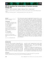
Fig. 1. The primary structure and gene structure of galanin and GALP in several species. Black shaded characters indicate the amino acid
sequences that are common to galanin and GALP. Galanin and GALP are encoded by separate genes that are typically located on separate
chromosomes: the GALP gene is located on chromosome 7, whereas the galanin gene is on chromosome 19 in mice. Galanin: the first exon
encodes the 5¢-untranslated region of preprogalanin. Cording region of galanin is present on exons 2–4. Galanin message-associated peptide
is encoded on exons 4–6 [48]. GALP: the first exon is untranslated region. The preproGALP is encoded by exons 2–6. Amino acid is repre-
sented by one letter code. EX, exon.
K. Shiba et al. GALP in feeding and energy metabolism
FEBS Journal 277 (2010) 5006–5013 ª 2010 The Authors Journal compilation ª 2010 FEBS 5007
binding affinity of GALP for GALRs have demon-
strated, using human neuroblastoma cells expressing
all three human GALRs, that GALR3 binds GALP
with the highest affinity, with the order of binding
potency of the GALRs for GALP being GALR3
(IC
50
=10nm), GALR2 (IC
50
=28nm) and GALR1
(IC
50
=77nm) [11]. In situ hybridization mapping
studies have shown that the three galanin receptor
transcripts are present throughout the hypothalamus.
High levels of expression of GALR1 are found in the
medial preoptic area (MPA), paraventricular nuclei
(PVN) and supraoptic nuclei (SON) [13]. GALR2 is
expressed in the preoptic area (POA), arcuate nuclei
(ARC), dorsomedial hypothalamus (DMH), PVN,
periventricular suprachiasmatic and mammillary nuclei
[14]. GALR3 expression is confined to the PVN,
DMH and ventromedial hypothalamic nuclei (VMH)
[15]. GALP reduces food intake and body weight in
both GALR1 and GALR2 knockout mice, similar to
the situation in wild-type mice [12]. It is therefore pos-
sible that GALR3 mediates feeding behavior. How-
ever, the central administration of a GALR2 ⁄ 3 agonist
had no effect on food intake, body weight and body
temperature in rodents [16]. In addition, other studies
have used quantitative analysis of c-Fos immunoreac-
tivity to show that, although galanin induces a signifi-
cantly greater number of c-Fos-positive nuclei in the
PVN compared to GALP, GALP induces significantly
more c-Fos-positive cells in the horizontal limb of the
diagonal band of Broca, caudal POA, ARC and med-
ian eminence [17]. These results suggest that GALP
and galanin act through different receptor-mediated
pathways to exert their effects on the regulation
of feeding. In other words, it is possible that GALP
mediates its effect via a yet-to-be-identified GALP
receptor.
In 2006, the novel 25 amino acid peptide, alarin,
was discovered as an alternate transcript of the GALP
gene [18–20]. Recently, it was shown that intracerebro-
ventricular injection of alarin increased food intake
and body weight [21]. Alarin immunoreactive cell
bodies are detected within the locus coeruleus and
locus subcoeruleus of the midbrain [21]. Alarin stimu-
lates Fos induction in the hypothalamic nuclei, such as
the PVN and nucleus tractus solitarii (NTS) [21].
Because alarin does not share any homology to gala-
nin, alarin is most unlikely to activate GALR [19,21].
In alarin, the signal sequence of the GALP precursor
peptide and the first five amino acids of the mature
GALP are followed by 20 amino acids without homol-
ogy to any other murine protein [19]. These studies
suggest that alarin is a neuromediator of food intake
and body weight via a specific receptor for alarin.
Regulation of GALP mRNA expression
GALP mRNA gradually increases between postnatal
days 8 and 14, and markedly increases between days
14 and 40, which represent the weaning and pubertal
periods in rats [22]. These findings suggest that GALP
may be associated with developmental changes such
as weaning, feeding and maturation of reproductive
function.
Fasting decreases both the number of GALP-
expressing neurons [23] and the expression of GALP
mRNA [24]. Leptin administration restores the number
of GALP-expressing cells in fasted rats [23] and leptin-
deficient ob ⁄ ob mice [9], with the expression levels of
GALP mRNA being reduced in the hypothalamus of
leptin receptor-deficient Zucker obese rats, and db ⁄ db
and ob ⁄ ob obese mice [25]. These findings clearly show
that leptin positively regulates activity of GALP
neurons in the hypothalamus. Furthermore, streptozo-
tocin-induced diabetic rats are associated with a signifi-
cant reduction in the expression of GALP mRNA,
which is reversed by treatment with either insulin or
leptin [26]. This suggests that GALP-expressing neu-
rons are direct regulatory targets not only for leptin,
but also for insulin.
Neuronal networks involving GALP-
containing neurons
Galanin is broadly distributed in the brain [27],
whereas GALP-immunoreactive neuronal cell bodies
are located in the hypothalamic ARC, being particu-
larly dense in the medial posterior section of the
nucleus [28]. In the rat brain, GALP mRNA is
expressed only in the ARC [23,29,30], with GALP-
positive fibers projecting from this nucleus to several
other hypothalamic nuclei, including the PVN, lateral
septal nucleus, bed nucleus of the stria terminalis and
MPA [28], as well as to the lateral hypothalamus
(LH) around the fornix [31]. On the basis of these
results, at least two major neural pathways involving
GALP have been proposed: one in which GALP-
containing neurons project from the ARC to the
PVN, and the other in which they project to the
MPA, bed nucleus of the stria terminalis and lateral
septal nucleus.
Central administration of GALP activates neurons
in various regions of the rat brain. Injection of GALP
into the third ventricle induces c-Fos expression, a
marker of cell activation, in the horizontal limb of the
diagonal band of Broca, POA, ARC and median emi-
nence [17], whereas injection into the lateral ventricle
activates several brain regions, including the DMH,
GALP in feeding and energy metabolism K. Shiba et al.
5008 FEBS Journal 277 (2010) 5006–5013 ª 2010 The Authors Journal compilation ª 2010 FEBS
LH, NTS of the brainstem, PVN and SON [32].
In mice, intracerebroventricular injection of GALP
into the lateral ventricle induces c-Fos expression in
the parenchyma surrounding the ventricles, the ventric-
ular ependymal cells and the meninges, but not in the
SON, DMH, LH and NTS [33], highlighting the exis-
tence of species-specific differences between rats and
mice. Additional work is therefore required to clarify
the link between GALP-induced c-Fos expression
and the neural circuitry involving GALP-containing
neurons.
Neuropeptides are divided into two groups: orexi-
genic peptides, including orexin, MCH and NPY, and
anorexigenic peptides, including a-MSH derived from
POMC [3].
GALP neurons in the ARC are innervated by orex-
inergic neurons in the LH and NPY-expressing neu-
rons in the ARC. Nine percent of GALP-positive
neurons express orexin-1 receptor [34]. GALP-positive
neurons have also been shown to express NPY Y1
receptor by double-label in situ hybridization [35], with
NPY- and orexin-containing fibers lying in close appo-
sition with GALP-containing neurons in the ARC
[34,36]. In addition, more than 85% of GALP-contain-
ing neurons express the leptin receptor [28]. However,
the GALP-containing neurons in the ARC are
reported to be different from the leptin receptor-
expressing neurons that express NPY ⁄ agouti-related
protein and galanin [30,34,36,37]. Taken together,
these morphological studies suggest that GALP-con-
taining neurons are regulated by both orexigenic and
anorexigenic signals.
With regard to the targets of GALP-containing neu-
rons in rats, morphological studies have shown that
GALP-like-immunoreactive nerve fibers make direct
contact with orexin- and MCH-like-immunoreactive
neurons in the LH [31]. At the ultrastructural level,
GALP-immunoreactive axon terminals have been
found to make synapses on orexin-immunoreactive cell
bodies and dendritic processes in the LH [38]. We have
previously reported that 3–12% of GALP-positive neu-
rons in the ARC also express a-MSH derived from
POMC [36]. These observations suggest that GALP-
containing neurons introduce feeding and ⁄ or satiety
signals. In addition, we have found that GALP-posi-
tive nerve fibers appear to make direct contact with
tyrosine hydroxylase-containing neurons in the ARC
[39], suggesting that GALP may interact with dopami-
nergic neurons in this region. GALP-positive neurons
have been shown to form circuits involving many neu-
rons. Although galanin is co-expressed with a number
of transmitters (monoamines and amino acids) and dif-
ferent peptides in neurons in various brain regions
[40], it is yet to be reported that GALP-neurons
express other neuropeptides or transmitters except a-
MSH in the ARC, indicating that GALP-expressing
neurons are unique.
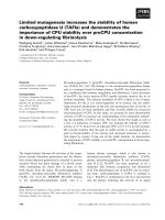
A schematic diagram summarizing the hypothalamic
neuronal networks involved in feeding regulation is
presented in Fig. 2. GALP-positive neurons are
affected by leptin, which conveys satiety signals from
the peripheral tissues, NPY and orexin. GALP regu-
lates both orexigenic (NPY and ⁄ or orexin) and anorex-
igenic (POMC) pathways in the central nervous
system.
POMC
MCH
NPY
Orexin
Leptin
Leptin
adipose tissue
adipose tissue
3V
DA
LH
VMH
ARC
NPY
DMH
GALP
Fig. 2. Distribution of GALP-producing neurons in the hypothala-
mus. GALP-induced hyperphagia is mediated via activation of orexin
neurons in the LH and NPY neurons in the DMH. GALP nerve
fibers make direct contact with MCH neurons in the LH and tyro-
sine hydroxylase-containing neurons in the ARC, although their
physiological actions are uncertain. GALP neurons in the ARC are
innervated by orexin neurons in the LH and NPY neurons in the
ARC, although their physiological actions are uncertain. More than
85% of GALP neurons express the leptin receptor. Leptin positively
regulates the activity of GALP neurons in the hypothalamus. GALP
neurons in the ARC also express a-MSH derived from POMC. 3V,
third cerebroventricle; DA, dopamine. Red arrows indicate stimula-
tory effects. Blue arrows indicate an uncertain function.
K. Shiba et al. GALP in feeding and energy metabolism
FEBS Journal 277 (2010) 5006–5013 ª 2010 The Authors Journal compilation ª 2010 FEBS 5009
Effect of GALP on feeding behavior
and energy metabolism
Galanin and biologically active fragments such as gala-
nin (1–16) stimulate food intake after acute microinjec-
tion into the PVN, LH, VMH and the central nucleus
of the amigdala, producing a rapid increase in the
feeding response and total caloric intake without alter-
ing feeding-associated behaviors such as drinking,
grooming and motor activity [20], whereas GALP has
complex actions on feeding behavior and energy bal-
ance. Intracerebroventricular injection of GALP signif-
icantly stimulates feeding during the first hour in rats
[32,41], whereas it inhibits food intake in mice [42].
The physiological significance of this behavioral differ-
ence between the rats and mice remains unclear,
although it may be a result of species differences in
neuronal circuitry.
In rats, three pathways have been demonstrated to
mediate the orexigenic effect of GALP: one via orexin-
ergic neurons in the LH; one via NPY-expressing neu-
rons in the DMH; and the third via POMC-expressing
neurons in the ARC. c-Fos immunoreactivity is
increased in orexin-immunoreactive neurons but not in
MCH-immunoreactive neurons in the LH after intra-
cerebroventricular injection of GALP [38]. Further-
more, anti-orexin IgG markedly inhibits GALP-
induced hyperphagia [38]. These results suggest that
orexin-containing neurons in the LH are targeted by
GALP, and that GALP-induced hyperphagia is medi-
ated via orexinergic neurons in the rat hypothalamus.
In addition, GALP focally injected into the DMH
stimulates food intake for 2 h after injection [43].
Intracerebroventricular injection of GALP induces
c-Fos expression in NPY-containing neurons in the
DMH. GALP also increases the cytosolic calcium con-
centration in NPY-immunoreactive neurons isolated
from the DMN. Furthermore, both anti-NPY IgG and
NPY antagonists, when preinjected, counteract the
feeding induced by GALP administration. In an in vi-
tro study of GALP-treated rat hypothalamic explants,
it was suggested that GALP-induced hyperphagica
could be mediated by an increase in NPY release [44].
These results reveal that GALP mediates a potent
short-term stimulation of food intake via activation of
NPY-containing neurons in the DMN. Moreover,
in vivo, the number of POMC mRNA-expressing cells
in the ARC of the ob ⁄ ob mouse is reduced after
chronic GALP injection [45]. These findings suggest
that GALP also promotes feeding behavior through
suppression of the anorexigenic POMC system.
GALP also increases food intake when injected into
the POA or PVN [46]. Although it is possible that the
POA and the PVN have specific roles in mediating the
orexigenic effect of GALP, the subpopulations of neu-
rons in these regions that mediate GALP-induced
overeating remain unknown.
Long-term continuous treatment with GALP causes
only transient reductions in both food intake and
body weight in wild-type mice, leading to the conclu-
sion that these animals become insensitive to contin-
ued exposure to GALP [17,42]. However, in the ob ⁄ ob
mouse, chronic GALP administration results in a sus-
tained decrease in body weight, despite a significant
recovery in food intake [42,45]. This suggests that
GALP promotes ongoing energy expenditure under
leptin-deficient conditions. Indeed, GALP promotes
thermogenesis, with intracerebroventricular injection
of GALP being shown to cause a dose-dependent
increase in core body temperature, which lasts for
6–8 h after injection. GALP-induced thermogenesis is
attenuated by peripheral administration of the cyclo-
oxygenase inhibitor, flurbiprofen, suggesting a depen-
dence on the actions of prostaglandins [47]. Astrocytes
produce prostaglandins and have been implicated in
thermogenesis in the brain, with an immunohistochem-
ical study revealing that GALP induces c-Fos expres-
sion in astrocytes but not in microglia [32]. These
findings suggest that GALP mediates the production
of fever via the prostaglandin pathway in the brain.
Recent data also suggest that GALP induces the
expression of interleukin-1 (IL-1) in the brain, and
that its anorexic and febrile actions are mediated by
this cytokine acting via the IL-1 type I receptor [48].
This indicates that IL-1 is a key mediator of inflam-
mation that acts to induce fever via the release of
prostaglandins in response to GALP in the hypothala-
mus. Brown adipose tissue innervated and activated
by the sympathetic nervous system plays an important
role in the regulation of thermogenesis. Repeated
treatment with GALP has been shown to increase
both mRNA and protein expression of uncoupling
protein-1, a key thermogenic molecule, in the brown
adipose tissue of the ob ⁄ ob mouse [45]. These findings
suggest that GALP may partly mediate energy metab-
olism through thermogenesis by long-term activation
of the sympathetic nervous system. Therefore, both
prostaglandins in the brain and uncoupling protein-1
in peripheral tissue are involved in GALP-induced
thermogenesis.
Although GALP is also present in blood [49], the
production of GALP in the peripheral organs remains
to be elucidated. Further studies are required to deter-
mine the link between the brain and peripheral tissues
involved in the regulation of feeding and energy
metabolism by GALP.
GALP in feeding and energy metabolism K. Shiba et al.
5010 FEBS Journal 277 (2010) 5006–5013 ª 2010 The Authors Journal compilation ª 2010 FEBS
Overall, these findings suggest that acute hyperpha-
gia mediated by GALP occurs via the activation of
orexin- and ⁄ or NPY-expressing neurons, and that
long-term body weight loss is a result of the promotion
of energy expenditure.
Clinical implications
To determine the potential clinical efficacy of GALP,
we investigated its intranasal delivery into the brain.
Recently, we have reported that the uptake by the
whole brain, olfactory bulb and cerebrospinal fluid
after intranasal administration is greater than that
after intravenous injection [50]. These findings indicate
that intranasal administration is an effective route of
delivery of GALP to the brain. We also studied the
effect of intranasal infusion of GALP on feeding
behavior in mice (K. Shiba, H. Kageyama, N. Non-
aka, F. Takenoya and S. Shioda, unpublished data).
Intranasal infusion of GALP significantly reduced
body weight over the course of 1 week. These results
suggest that intranasal administration of GALP repre-
sents a viable option for obese people who seek to
combat obesity and similar life-style-related diseases.
Conclusions
GALP is mainly produced in the hypothalamic ARC,
and plays important roles in the regulation of feeding
behavior and energy metabolism through complicated
neuronal networks.
The central administration of GALP produces a
short-term increase (followed by a subsequent decrease)
in food intake in rats, whereas it produces only a
decrease in mice. GALP also reduces body weight and
stimulates thermogenesis in rodents. The short-term
orexigenic actions of GALP are mediated via NPY and
the orexinergic pathway in the rat. The long-term ano-
rectic and thermogenic actions of GALP are mediated
via the pro-inflammatory pathway in rodents. The iden-
tification of a specific receptor for GALP is of consider-
able importance if the physiological functions and
mechanism of action of GALP are to be fully under-
stood. Little is known about the role of alarin, which
was discovered as an alternate transcript of the GALP
gene. Further elucidation of the function of GALP and
alarin will provide the necessary basis for the treatment
and prevention of obesity and related disorders.
Acknowledgements
The authors thank Dr Tetsuya Ohtaki from Takeda
Pharmaceutical Company. The present work was sup-
ported in part by the High-Technology Research Cen-
ter Project from the Ministry of Education, Sports,
Science and Technology and by grant-in-Aid for
Exploratory Research (#21659059).
References
1 Friedman JM & Halaas JL (1998) Leptin and the regula-
tion of body weight in mammals. Nature 395, 763–770.
2 Kageyama H, Takenoya F, Shiba K & Shioda S (2010)
Neuronal circuits involving ghrelin in the hypothala-
mus-mediated regulation of feeding. Neuropeptides 44,
133–138.
3 Shioda S, Takenoya F, Yagi M, Wang L, Hori Y &
Kageyama H (2008) Neural networks of several novel
neuropeptides involved in feeding regulation. Nutrition
24, 848–853.
4 Takenoya F, Kageyama H, Shiba K, Date Y, Nakazato
M & Shioda S (2010) Neuropeptide W: a key player in
the homeostatic regulation of feeding and energy
metabolism? Ann NY Acad Sci 1200, 162–169.
5 Tanaka M (2010) Bioactive neuropeptides: food intake
and energy balance: relaxin-3 ⁄ INSL7, a neuropeptide
involved in the stress response and food intake. FEBS J
277, 4990–4997.
6 Takayanagi Y & Onaka T (2010) Bioactive neuropep-
tides: food intake and energy balance: roles of prolac-
tin-releasing peptide and RFamide related peptides in
the control of stress and food intake. FEBS J 277,
4998–5005.
7 Tatemoto K, Rokaeus A, Jornvall H, McDonald TJ &
Mutt V (1983) Galanin – a novel biologically active
peptide from porcine intestine. FEBS Lett 164, 124–128.
8 Ohtaki T, Kumano S, Ishibashi Y, Ogi K, Matsui H,
Harada M, Kitada C, Kurokawa T, Onda H & Fujino
M (1999) Isolation and cDNA cloning of a novel gala-
nin-like peptide (GALP) from porcine hypothalamus.
J Biol Chem 274, 37041–37045.
9 Jure
´
us A, Cunningham MJ, Li D, Johnson LL,
Krasnow SM, Teklemichael DN, Clifton DK & Steiner
RA (2001) Distribution and regulation of galanin-like
peptide (GALP) in the hypothalamus of the mouse.
Endocrinology 142, 5140–5144.
10 Cunningham MJ, Scarlett JM & Steiner RA (2002)
Cloning and distribution of galanin-like peptide mRNA
in the hypothalamus and pituitary of the macaque.
Endocrinology 143, 755–763.
11 Lang R, Berger A, Santic R, Geisberger R, Hermann
A, Herzog H & Kofler B (2005) Pharmacological and
functional characterization of galanin-like peptide frag-
ments as potent galanin receptor agonists. Neuropep-
tides 39, 179–184.
12 Krasnow SM, Hohmann JG, Gragerov A, Clifton DK
& Steiner RA (2004) Analysis of the contribution of
K. Shiba et al. GALP in feeding and energy metabolism
FEBS Journal 277 (2010) 5006–5013 ª 2010 The Authors Journal compilation ª 2010 FEBS 5011
galanin receptors 1 and 2 to the central actions of gala-
nin-like peptide. Neuroendocrinology 79, 268–277.
13 Mitchell V, Habert-Ortoli E, Epelbaum J, Aubert JP &
Beauvillain JC (1997) Semiquantitative distribution of
galanin-receptor (GAL-R1) mRNA-containing cells in
the male rat hypothalamus. Neuroendocrinology 66,
160–172.
14 Mitchell V, Bouret S, Howard AD & Beauvillain JC
(1999) Expression of the galanin receptor subtype Gal-
R2 mRNA in the rat hypothalamus. J Chem Neuroanat
16, 265–277.
15 Mennicken F, Hoffert C, Pelletier M, Ahmad S &
O’Donnell D (2002) Restricted distribution of galanin
receptor 3 (GalR3) mRNA in the adult rat central ner-
vous system. J Chem Neuroanat 24, 257–268.
16 Man PS & Lawrence CB (2008) The effects of galanin-
like peptide on energy balance, body temperature and
brain activity in the mouse and rat are independent of
the GALR2 ⁄ 3 receptor. J Neuroendocrinol 20, 128–137.
17 Fraley GS, Shimada I, Baumgartner JW, Clifton DK &
Steiner RA (2003) Differential patterns of fos induction
in the hypothalamus of the rat following central injec-
tions of galanin-like peptide and galanin. Endocrinology
144, 1143–1146.
18 Santic R, Fenninger K, Graf K, Schneider R, Hauser-
Kronberger C, Schilling FH, Kogner P, Ratschek M,
Jones N, Sperl W et al. (2006) Gangliocytes in neurob-
lastic tumors express alarin, a novel peptide derived by
differential splicing of the galanin-like peptide gene.
J Mol Neurosci 29, 145–152.
19 Santic R, Schmidhuber SM, Lang R, Rauch I, Voglas
E, Eberhard N, Bauer JW, Brain SD & Kofler B (2007)
Alarin is a vasoactive peptide. Proc Natl Acad Sci USA
104, 10217–10222.
20 Lang R, Gundlach AL & Kofler B (2007) The galanin
peptide family: receptor pharmacology, pleiotropic bio-
logical actions, and implications in health and disease.
Pharmacol Ther 115, 177–207.
21 Van Der Kolk N, Madison FN, Mohr M, Eberhard N,
Kofler B & Fraley GS (2010) Alarin stimulates food
intake in male rats and LH secretion in castrated male
rats. Neuropeptides 44, 333–340.
22 Kawagoe R, Yamamoto Y, Kubo K, Dobashi K,
Asayama K, Ueta Y & Shirahata A (2008) Postnatal
development of galanin-like peptide mRNA expression
in rat hypothalamus. Regul Pept 145, 133–140.
23 Jure
´
us A, Cunningham MJ, McClain ME, Clifton DK
& Steiner RA (2000) Galanin-like peptide (GALP) is a
target for regulation by leptin in the hypothalamus of
the rat. Endocrinology 141, 2703–2706.
24 Kuramochi M, Kohno D, Onaka T, Kato S & Yada T
(2005) Galanin-like peptide and ghrelin increase cyto-
solic Ca
2+
in neurons containing growth hormone-
releasing hormone in the arcuate nucleus. Regul Pept
126, 85–89.
25 Kumano S, Matsumoto H, Takatsu Y, Noguchi J,
Kitada C & Ohtaki T (2003) Changes in hypothalamic
expression levels of galanin-like peptide in rat and
mouse models support that it is a leptin-target peptide.
Endocrinology 144, 2634–2643.
26 Fraley GS, Scarlett JM, Shimada I, Teklemichael DN,
Acohido BV, Clifton DK & Steiner RA (2004) Effects
of diabetes and insulin on the expression of galanin-like
Peptide in the hypothalamus of the rat. Diabetes 53,
1237–1242.
27 Skofitsch G & Jacobowitz DM (1985) Immunohisto-
chemical mapping of galanin-like neurons in the rat
central nervous system. Peptides 6, 509–546.
28 Takatsu Y, Matsumoto H, Ohtaki T, Kumano S,
Kitada C, Onda H, Nishimura O & Fujino M (2001)
Distribution of galanin-like peptide in the rat brain.
Endocrinology 142, 1626–1634.
29 Kerr NC, Holmes FE & Wynick D (2000) Galanin-
like peptide (GALP) is expressed in rat hypothalamus
and pituitary, but not in DRG. Neuroreport 11, 3909–
3913.
30 Larm JA & Gundlach AL (2000) Galanin-like peptide
(GALP) mRNA expression is restricted to arcuate
nucleus of hypothalamus in adult male rat brain. Neu-
roendocrinology 72, 67–71.
31 Takenoya F, Hirayama M, Kageyama H, Funahashi H,
Kita T, Matsumoto H, Ohtaki T, Katoh S, Takeuchi M
& Shioda S (2005) Neuronal interactions between gala-
nin-like-peptide- and orexin- or melanin-concentrating
hormone-containing neurons. Regul Pept 126, 79–83.
32 Lawrence CB, Williams T & Luckman SM (2003) Intra-
cerebroventricular galanin-like peptide induces different
brain activation compared with galanin. Endocrinology
144, 3977–3984.
33 Man PS & Lawrence CB (2008) Galanin-like peptide:
a role in the homeostatic regulation of energy balance?
Neuropharmacology 55 , 1–7.
34 Takenoya F, Aihara K, Funahashi H, Matsumoto H,
Ohtaki T, Tsurugano S, Yamada S, Katoh S,
Kageyama H, Takeuchi M et al. (2003) Galanin-like
peptide is target for regulation by orexin in the rat
hypothalamus. Neurosci Lett 340, 209–212.
35 Cunningham MJ, Shahab M, Grove KL, Scarlett JM,
Plant TM, Cameron JL, Smith MS, Clifton DK &
Steiner RA (2004) Galanin-like peptide as a possible
link between metabolism and reproduction in the
macaque. J Clin Endocrinol Metab 89, 1760–1766.
36 Takenoya F, Funahashi H, Matsumoto H, Ohtaki T,
Katoh S, Kageyama H, Suzuki R, Takeuchi M &
Shioda S (2002) Galanin-like peptide is co-localized
with alpha-melanocyte stimulating hormone but not
with neuropeptide Y in the rat brain. Neurosci Lett 331,
119–122.
37 Hakansson ML, Brown H, Ghilardi N, Skoda RC &
Meister B (1998) Leptin receptor immunoreactivity in
GALP in feeding and energy metabolism K. Shiba et al.
5012 FEBS Journal 277 (2010) 5006–5013 ª 2010 The Authors Journal compilation ª 2010 FEBS
chemically defined target neurons of the hypothalamus.
J Neurosci 18, 559–572.
38 Kageyama H, Kita T, Toshinai K, Guan JL, Date Y,
Takenoya F, Kato S, Matsumoto H, Ohtaki T, Nakazato
M et al. (2006) Galanin-like peptide promotes feeding
behaviour via activation of orexinergic neurones in the
rat lateral hypothalamus. J Neuroendocrinol 18, 33–41.
39 Kageyama H, Takenoya F, Hori Y, Yoshida T & Shi-
oda S (2008) Morphological interaction between gala-
nin-like peptide- and dopamine-containing neurons in
the rat arcuate nucleus. Regul Pept 145, 165–168.
40 Gundlach AL (2002) Galanin ⁄ GALP and galanin recep-
tors: role in central control of feeding, body
weight ⁄ obesity and reproduction? Eur J Pharmacol 440,
255–268.
41 Matsumoto Y, Watanabe T, Adachi Y, Itoh T, Ohtaki
T, Onda H, Kurokawa T, Nishimura O & Fujino M
(2002) Galanin-like peptide stimulates food intake in
the rat. Neurosci Lett 322, 67–69.
42 Krasnow SM, Fraley GS, Schuh SM, Baumgartner JW,
Clifton DK & Steiner RA (2003) A role for galanin-like
peptide in the integration of feeding, body weight regu-
lation, and reproduction in the mouse. Endocrinology
144, 813–822.
43 Kuramochi M, Onaka T, Kohno D, Kato S & Yada T
(2006) Galanin-like peptide stimulates food intake via
activation of neuropeptide Y neurons in the hypotha-
lamic dorsomedial nucleus of the rat. Endocrinology
147, 1744–1752.
44 Seth A, Stanley S, Dhillo W, Murphy K, Ghatei M &
Bloom S (2003) Effects of galanin-like Peptide on food
intake and the hypothalamo-pituitary-thyroid axis.
Neuroendocrinology 77, 125–131.
45 Hansen KR, Krasnow SM, Nolan MA, Fraley GS,
Baumgartner JW, Clifton DK & Steiner RA (2003)
Activation of the sympathetic nervous system by gala-
nin-like peptide-a possible link between leptin and
metabolism. Endocrinology 144, 4709–4717.
46 Patterson M, Murphy KG, Thompson EL, Smith KL,
Meeran K, Ghatei MA & Bloom SR (2006) Microinjec-
tion of galanin-like peptide into the medial preoptic
area stimulates food intake in adult male rats. J Neuro-
endocrinol 18, 742–747.
47 Lawrence CB, Baudoin FM & Luckman SM (2002)
Centrally administered galanin-like peptide modifies
food intake in the rat: a comparison with galanin.
J Neuroendocrinol 14, 853–860.
48 Man PS & Lawrence CB (2008) Interleukin-1 mediates
the anorexic and febrile actions of galanin-like Peptide.
Endocrinology 149, 5791–5802.
49 Kastin AJ, Akerstrom V & Hackler L (2001) Food
deprivation decreases blood galanin-like peptide and its
rapid entry into the brain. Neuroendocrinology 74,
423–432.
50 Nonaka N, Farr SA, Kageyama H, Shioda S & Banks
WA (2008) Delivery of galanin-like peptide to the brain:
targeting with intranasal delivery and cyclodextrins.
J Pharmacol Exp Ther 325, 513–519.
K. Shiba et al. GALP in feeding and energy metabolism
FEBS Journal 277 (2010) 5006–5013 ª 2010 The Authors Journal compilation ª 2010 FEBS 5013