Báo cáo khoa học: Mutational analysis of the preferential binding of human topoisomerase I to supercoiled DNA pot
Bạn đang xem bản rút gọn của tài liệu. Xem và tải ngay bản đầy đủ của tài liệu tại đây (802.59 KB, 14 trang )
Mutational analysis of the preferential binding of human
topoisomerase I to supercoiled DNA
Zheng Yang, James F. Carey and James J. Champoux
Department of Microbiology, School of Medicine, University of Washington, Seattle, WA, USA
Introduction
Type I DNA topoisomerases relax supercoils by intro-
ducing a transient single-strand break in the DNA.
These enzymes are classified into type IA and type IB
subfamilies based on the polarity of attachment to the
cleaved DNA [1–3]. The members of the two subfami-
lies share no sequence homology and are further dis-
tinguished by their substrate requirements and
mechanisms of relaxation. Type IA subfamily members
require a single-stranded region to bind DNA, become
attached to the 5¢ end upon cleavage, and only relax
negatively supercoiled DNA in the presence of divalent
cations such as Mg
2+
. Escherichia coli DNA topo-
isomerase I is the prototype of the type IA subfamily.
Type IB subfamily members bind double-stranded
DNA, become attached to the 3¢ end of the cleaved
strand, and relax both positive and negative supercoils.
ATP or divalent cations are not required for the
type IB enzymes, although Mg
2+
and Ca
2+
enhance
the rate of relaxation [4].
The cleavage–religation reaction catalyzed by human
DNA topoisomerase I, the prototypical type IB
enzyme, is essential for many biological processes,
Keywords
competition binding assay; DNA topology;
node binding; supercoiled DNA;
topoisomerase I
Correspondence
J. J. Champoux, Department of
Microbiology, University of Washington, Box
357242, Seattle, WA 98195-7242, USA
Fax: +1 206 543 8297
Tel: +1 206 543 8574
E-mail:
(Received 7 July 2009, revised 9 August
2009, accepted 11 August 2009)
doi:10.1111/j.1742-4658.2009.07270.x
Human topoisomerase I binds DNA in a topology-dependent fashion with
a strong preference for supercoiled DNAs of either sign over relaxed circu-
lar DNA. One hypothesis to account for this preference is that a second
DNA-binding site exists on the enzyme that mediates an association with
the nodes present in supercoiled DNA. The failure of the enzyme to dimer-
ize, even in the presence of DNA, appears to rule out the hypothesis that
two binding sites are generated by dimerization of the protein. A series of
mutant protein constructs was generated to test the hypotheses that the
homeodomain-like core subdomain II (residues 233–319) provides a second
DNA-binding site, or that the linker or basic residues in core subdo-
main III are involved in the preferential binding to supercoiled DNAs.
When putative DNA contact points within core subdomain II were altered
or the domain was removed altogether, there was no effect on the ability
of the enzyme to recognize supercoiled DNA, as measured by both a gel
shift assay and a competition binding assay. However, the preference for
supercoils was noticeably reduced for a form of the enzyme lacking the
coiled-coil linker region or when pairs of lysines were changed to glutamic
acids in core subdomain III. The results obtained implicate the linker and
solvent-exposed basic residues in core subdomain III in the preferential
binding of human topoisomerase I to supercoiled DNA.
Abbreviations
Dcap, NH
2
-terminal truncation of human topoisomerase beginning at residue 433; GST, glutathione S-transferase; topo31, a fragment of
human topoisomerase I extending from residues 175–433; topo56, COOH-terminal truncation of topo70 missing the last 126 amino acids;
topo58, COOH-terminal truncation of topo70 missing the last 106 amino acids; topo70, NH
2
-terminal truncation of human topoisomerase I
missing the first 174 amino acids; topo70DL, a form of topo70 missing linker residues 660–688.
5906 FEBS Journal 276 (2009) 5906–5919 ª 2009 The Authors Journal compilation ª 2009 FEBS
including DNA replication, transcription and recombi-
nation [2,3]. Strand cleavage is initiated by nucleophilic
attack of the O4 atom of the active site tyrosine on the
scissile phosphate in the DNA, resulting in the cova-
lent attachment of the enzyme to the 3¢ end of the
broken strand [2]. Rotation of the duplex region
downstream of the break site relieves any supercoiling
strain in the DNA prior to religation and release of
the topoisomerase [5,6].
Human DNA topoisomerase I is composed of 765
amino acids and has a molecular mass of 91 kDa. On
the basis of sequence comparisons, limited proteolysis
studies and the crystal structure of the enzyme [7,8],
four domains have been identified in the protein: an
NH
2
-terminal domain (Met1-Gly214), a core domain
(Ile215-Ala635), a linker domain (Pro636-Lys712) and
a COOH-terminal domain (Gln713-Phe765) (Fig. 1).
The NH
2
-terminal domain is unstructured, poorly con-
served, highly charged and dispensable for the DNA
relaxation activity in vitro . It contains nuclear localiza-
tion signals and was shown to interact with nucleolin,
the SV40 large T antigen, p53, and possibly certain
transcription factors [9–13]. Topo70 is a truncated
form of human topoisomerase I that lacks residues
1–174 of the NH
2
-terminal domain, yet retains full
enzymatic activity [7] and a preference for binding su-
percoiled DNA. The core domain is highly conserved
and more protease-resistant than the other domains.
The poorly-conserved linker domain is highly charged
and forms an anti-parallel coiled-coil structure that
connects the core domain to the COOH-terminal
domain. The linker protrudes from the body of the
protein and, instead of tracking with the axis of a
bound DNA helix, angles away from DNA. The
COOH-terminal domain is highly conserved and con-
tains the active site tyrosine, Tyr723. When separately
expressed, the COOH-terminal and core domains can
associate in vitro to reconstitute wild-type levels of
enzymatic activity, demonstrating that the linker
domain is not required for activity [4,7,14].
The co-crystal structure of human topoisomerase I
with bound DNA indicates that the core domain can
be further divided into three subdomains [8] (Fig. 1).
Core subdomain I (residues 215–232, 320–433) and
core subdomain II (residues 233–319) form the Cap
structure of the enzyme and cover one side of the
DNA. Core subdomain III (residues 434–635) contains
all the residues implicated in catalysis except Tyr723
and cradles the DNA on the side opposite of the Cap
[5,8]. Although there is little sequence similarity, the
fold of the subdomain II is very similar to that of a
homeodomain found in a family of DNA-binding
proteins. For example, residues 244–314 of the core
subdomain II superimpose on the POU homeodomain
of the Oct-1 transcription factor with an rmsd of only
3.0 A
˚
[8,15]. This observation suggests that core sub-
domain II, which forms part of the exposed Cap,
could represent a second DNA-binding site distinct
from the substrate binding channel observed in the
co-crystal structure. However, the conserved residues
that are involved in base-specific contacts in the POU
homeodomain are absent in core subdomain II of
human topoisomerase I, suggesting that, if sub-
domain II interacts with DNA, it does so with low
affinity and likely without sequence specificity [15,16].
It has been proposed that topoisomerases relax the
negative and positive supercoils generated by the trans-
location of an RNA polymerase along the DNA
during transcription [17]. In support of this model,
eukaryotic type IB topoisomerases have been found to
associate with transcriptionally active genes and have
been reported to interact directly with the transcription
machinery [18–26]. Eukaryotic type IB topoisomerases
have also been shown to provide the swivels for the
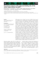
Fig. 1. Crystal structure of human topoisomerase I. Core subdo-
mains I, II and III are colored yellow, blue and red, respectively,
with the linker and C-terminal domains colored orange and green,
respectively. The Cap and Linkers regions are labeled along with
the amino acid residues that were changed in the present study.
Amino acids in core subdomain II (His266, Lys299 and Ser306) that
were changed to glutamic acid in the combinations indicated in the
text are shown in ball and stick and colored magenta. The three
amino acids in the linker (Lys650 ⁄ Lys654 ⁄ Gln657) that were
simultaneously changed to alanine are similarly depicted and
colored brown. The four amino acids in the linker (Lys679 ⁄
Lys682 ⁄ Lys687 ⁄ Lys689) that were simultaneously changed to
serine are colored gray. Surface-exposed lysine residues in core
subdomain III (Lys466 ⁄ Lys468 and Lys545 ⁄ Lys549) that were
pairwise mutated to glutamic acid are colored black.
Z. Yang et al. Supercoil binding by topoisomerase I
FEBS Journal 276 (2009) 5906–5919 ª 2009 The Authors Journal compilation ª 2009 FEBS 5907
relaxation of positive supercoils during DNA replica-
tion [20,27–31]. The mechanism for recruiting DNA
topoisomerase I to transcriptionally active and repli-
cating DNA remains unclear, although several studies
have shown that the enzyme prefers to bind super-
coiled over relaxed DNA [32–37]. Because the enzyme
binds supercoiled DNA irrespective of the sign of the
supercoils, Zechiedrich and Osheroff [36] hypothesized
that topoisomerase I specifically binds at a node where
two duplex regions of the supercoiled DNA cross and
also provided electron microscopic evidence in support
of this hypothesis [36].
The structural basis for the preferential binding of
human topoisomerase I to supercoiled DNA is
unknown but, if node recognition is important, then it
is likely that the binding involves an interaction with
two regions of DNA at the point of crossing. One
hypothesis to explain how the enzyme provides two
DNA-binding sites to stabilize an interaction with a
DNA node is to assume that it binds as a dimer
(Fig. 2A). An alternative hypothesis is that, in addition
to the substrate binding channel identified in the crys-
tal structure of the protein (Fig. 1) [8], there is a
second DNA-binding site present on the protein that
stabilizes an interaction at a DNA node (Fig. 2B). In
the present study, we performed experiments designed
to distinguish between these possible explanations for
the preference of topoisomerase I for supercoils.
Results
Human topoisomerase I does not dimerize in the
absence or presence of DNA
We previously used a gel filtration assay to demon-
strate that, although topo70DL, a mutant form of
topo70 missing a portion of the linker (i.e. linker
residues 660–688), formed dimers through a domain
swapping mechanism, no dimerization of WT topo70
was detectable under the same conditions [4,38].
Because these earlier experiments were carried out in
the absence of DNA, we wanted to test whether
dimers could form in the presence of DNA. In the
present study, we used a glutathione S-transferase
(GST) pull-down assay to determine whether topo70
that was already covalently bound to a DNA oligonu-
cleotide could dimerize. GST-topo70 was incubated
with free topo70 in the absence or presence of an oli-
gonucleotide suicide substrate, and any protein bound
to GST-topo70 was collected by adsorption to gluta-
thione S-Sepharose beads and analyzed by SDS–
PAGE. Control experiments showed that free topo70
did not bind to either GST alone or to the beads
(Fig. 3, lanes 6 and 7). Under the same conditions, no
topo70 was found associated with the bead-bound
GST-topo70 either in the absence or presence of DNA
(Fig. 3, lanes 2 and 3, respectively). The slower migrat-
ing species of the doublet observed in lane 3 in Fig. 3
is the result of suicide cleavage and shows that approx-
imately half of the GST-topo70 contained covalently
bound oligonucleotide DNA. Thus, these results con-
firm our earlier finding that topo70 does not dimerize
when free in solution and also extend the results to
show that, even when bound to DNA after suicide
cleavage, dimerization does not occur.
Fig. 2. Alternative modes for topoisomerase I binding to a DNA
node. (A) Node binding occurs through dimerization of topoisomer-
ase I. (B) Node binding is mediated by two DNA-binding sites on a
single molecule of topoisomerase I.
Fig. 3. GST pull-down experiment to test for dimerization. The indi-
cated combinations of GST-topo70, topo70 and GST were incu-
bated with and without a suicide DNA oligonucleotide and mixed
with glutathione Sepharose 4B beads (GSH beads). The beads
were collected by centrifugation, washed and the samples were
analyzed by SDS-PAGE. Lane 1, protein markers with sizes (kDa)
indicated along the left side of the gel. Lanes 4 and 5 contain GST-
topo70 and topo70 size markers, respectively. The GST protein in
lane 6 was run off the gel in this analysis. Although all of the
samples were analyzed on the same gel, lanes with unrelated data
were removed digitally at the places indicated by the thin vertical
lines.
Supercoil binding by topoisomerase I Z. Yang et al.
5908 FEBS Journal 276 (2009) 5906–5919 ª 2009 The Authors Journal compilation ª 2009 FEBS
DNA-binding properties of mutant proteins as
measured by a gel shift assay
A structural alignment of core subdomain II of human
topoisomerase I with the POU homeodomain of Oct-1
indicated that the residues making base specific
contacts with the DNA in the homeodomain are not
conserved in the core subdomain II. However, basic
residues K25, R46 and R53 of the POU homeodomain
that make hydrogen bonds with phosphates in the
bound DNA correspond to residues His266, Lys299
and Ser306 in core subdomain II of human topo-
isomerase I (Fig. 1). All three of these residues are
conserved among known eukaryotic topoisomerase I
sequences. To test whether these amino acids mediate
an interaction with DNA that accounts for node bind-
ing by the enzyme, site-directed mutagenesis was used
to replace these residues with glutamic acid in topo70.
These changes would be predicted to disrupt an inter-
action with the DNA phosphate backbone, but have a
minimal effect on the overall enzyme structure because
all three are in a solvent-exposed region. Because the
assays to detect the preferential binding to supercoiled
DNA require a catalytically inactive form of the pro-
tein [37], a mutation in the active site tyrosine (Y723F)
was also introduced into the proteins. Topo70 capKS-
E ⁄ Y723F and topo70 capHKS-E ⁄ Y723F were
expressed and purified from recombinant baculovirus-
infected insect SF-9 cells. The following proteins were
similarly purified for use in these assays: a reconsti-
tuted form of the protein lacking the linker, compris-
ing a COOH-terminal truncation of topo70 missing
the last 126 amino acids (topo56) plus the Y723F
mutant form of the COOH-terminal domain (topo6.3),
the catalytically inactive NH
2
-terminal truncation of
human topoisomerase beginning at residue 433 (Dcap)
[39] and a fragment of human topoisomerase I extend-
ing from residues 175–433 (topo31) (Fig. 4).
The various forms of the topoisomerase protein
described above were mixed with an equimolar mixture
of supercoiled, nicked circular and linear pBluescript
KSII(+) DNAs, and a gel shift assay [40–44] was
used to analyze the preference of the proteins for the
different topological forms of DNA. For the positive
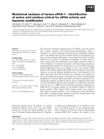
Fig. 4. Human topoisomerase I fragments
used in the DNA-binding studies. (A) The
four domains of full-length human topoisom-
erase I (topo I) are shown above the various
constructs used in the binding studies:
topo70, a 70 kDa NH
2
-terminally truncated
protein that starts with an engineered Met
upstream of Lys175; topo58, a COOH-termi-
nal deletion of topo70, ending at Ala659;
topo31, a COOH-terminal deletion of topo70
ending at Ser433; Dcap, an NH
2
-terminal
truncation starting at Ser433; topo56 ⁄ 6.3, a
reconstituted protein comprising the core
domain from Lys175 to Thr639 and the
COOH-terminal domain from Lys713 to the
COOH terminus (Phe765). (B) SDS-PAGE
analysis of 2 lg of the indicated purified pro-
teins. Lane 1, protein markers with sizes
(kDa) indicated along left side of the panel;
lane 2, topo70 Y723F; lane 3, topo70 capKS-
E ⁄ Y723F; lane 4, topo70 capHKS-E ⁄ Y723F;
lane 5, topo56 ⁄ 6.3 Y723F (6.3 kDa fragment
of topo6.3 Y723F was run off the bottom of
the gel); lane 6, Dcap; lane 7, topo31; lane
8, protein markers; lane 9, topo70 K466-
468E ⁄ Y723F; lane 10, topo70 K545-
549E ⁄ Y723F.
Z. Yang et al. Supercoil binding by topoisomerase I
FEBS Journal 276 (2009) 5906–5919 ª 2009 The Authors Journal compilation ª 2009 FEBS 5909
control protein, topo70 Y723F, the mobility of the
supercoiled DNA was reduced, with essentially no effect
on the mobility of either the nicked or linear DNAs at
the two lowest protein concentrations (Fig. 5A, com-
pare lanes 2 and 3 with lane 1). As the amount of topo70
Y723F protein was increased, the supercoiled DNA was
shifted further and, to a lesser extent, both the linear
and nicked DNA bands became shifted as well (Fig. 5A,
lanes 4 and 5). These results confirmed the earlier find-
ing that topo70 Y723F has a preference for supercoiled
over linear and nicked DNA [37]. Topo31, which corre-
sponds to the Cap region of human topoisomerase I,
provides a convenient nonspecific negative control for
this analysis. As shown in Fig. 5A, lanes 22–26, all three
forms of the plasmid DNA responded equally to
increasing concentrations of the topo31 fragment, con-
sistent with a lack of preference for one form over
another. A higher concentration of topo31 was required
to effect a gel shift, reflecting the lower affinity of the
protein for DNA compared to topo70.
Both topo70 capKS-E ⁄ Y723F and topo70 capHKS-
E ⁄ Y723F retained the preference for binding super-
coiled DNA (Fig. 5A, lanes 7–10 and 12–15), ruling
out Cap residues His266, Lys299 and Ser306 as con-
tributors to the preferential binding to supercoils. To
further test the possible involvement of the core subdo-
main II in the preferential binding to supercoiled
DNA, the Dcap mutant lacking core subdomains I and
II was also tested in the gel shift analysis (Fig. 5A,
lanes 17–20). Dcap contains core subdomain III, the
linker domain, and the COOH-terminal domain (resi-
dues 433–765) (Fig. 4A), and is catalytically inactive,
despite containing all of the residues known to be
directly involved in catalysis [39]. At the lower concen-
trations of the Dcap protein, the supercoiled DNA was
selectively shifted upon binding, although the magni-
tude of the shift was less compared to that observed
with the topo70 protein (Fig. 5A, compare lanes 17–20
with lanes 1–5). This reduction in the shift most likely
resulted from the two-fold lower affinity of the Dcap
for DNA [39] and the lower molecular weight of Dcap
(41 kDa) compared to topo70 (71 kDa). Thus, deletion
of the Cap region that includes subdomain II did not
eliminate the preference for supercoiled DNA, indicat-
ing that core subdomain II is dispensable for the
preferential binding of topoisomerase I to supercoils.
Although the band corresponding to the supercoiled
DNA was selectively shifted in the presence of topo70
Y723F and all of the mutant proteins except topo31,
we wanted to formally rule out the possibility that the
Fig. 5. DNA-binding measured by an agarose gel shift assay. (A) Two-fold serial dilutions of the indicated proteins were incubated with equal
amounts of supercoiled, linear and nicked pBluescript KSII(+) plasmid DNA and analyzed by electrophoresis in an agarose gel as described in
the Experimental procedures. The mobilities of unshifted supercoiled, linear and nicked DNAs are indicated along the right side. Lanes 1, 6,
11, 16, 21 and 27 contain DNA alone; lanes 2–5 contain 0.88, 1.75, 3.5 and 7 pmol of topo70 Y723F, respectively; lanes 7–10 contain 0.88,
1.75, 3.5 and 7 pmol of topo70 capKS-E ⁄ Y723F, respectively; lanes 12–15 contain 0.88, 1.75, 3.5 and 7 pmol of topo70 capHKS-E ⁄ Y723F,
respectively; lanes 17–20 contain 0.88, 1.75, 3.5 and 7 pmol of Dcap, respectively; and lanes 22-26 contain 0.88, 1.75, 3.5, 7 and 14 pmol of
topo31, respectively. The white spaces demarcate separate gel analyses. (B) Same experimental design as in (A) for the indicated proteins.
Lanes 1, 6 and 11 are DNA alone; lanes 2–5 contain 0.88, 1.75, 3.5 and 7 pmol of topo70 Y723F, respectively; and lanes 7–10 contain 0.88,
1.75, 3.5 and 7 pmol of topo56 ⁄ 6.3 Y723F, respectively.
Supercoil binding by topoisomerase I Z. Yang et al.
5910 FEBS Journal 276 (2009) 5906–5919 ª 2009 The Authors Journal compilation ª 2009 FEBS
proteins bound to the supercoiled, linear and nicked
DNAs equally well, but only the supercoiled DNA
shift was detected visually because of its greater initial
mobility. Therefore, the gel shift assay was repeated
using topo70 Y723F or topo70 capHKS-E ⁄ Y723F that
had been previously labeled with
32
P using protein
kinase C. The autoradiograph of the agarose gel
showed that the majority of the labeled proteins were
associated with the shifted supercoiled DNA and that
the amount of bound label correlated with the extent
of the shift (Fig. 6, lanes 2, 3, 5 and 6). Furthermore,
label was only associated with the nicked and linear
DNAs at the protein concentration where a mobility
shift of these species was also detected (Fig. 6, lanes 3
and 6). These results validated the gel shift assay and
confirmed that the selective shift of the supercoiled
DNA band results from preferential binding.
To further define the region that is involved in the
preferential binding to supercoiled DNA, we repeated
the assays using a form of human topoisomerase I
reconstituted from a mixture of topo56 and topo6.3
Y723F (Fig. 4A). This reconstituted protein contains
only the core and COOH-terminal domains and com-
pletely lacks the linker region (Fig. 1). When tested in
the gel shift assay, topo56 ⁄ 6.3 Y723F retained a pref-
erence for supercoiled DNA, although the preference
was reduced compared to that of the topo70 Y723F
(Fig. 5B). For example, although only the supercoiled
DNA was shifted by both topo70 Y723F and
topo56 ⁄ 6.3 Y723F at the lowest protein concentration
tested (Fig. 5B, lanes 2 and 7), at the higher protein
concentrations where mainly the supercoiled DNA was
shifted by topo70 Y723F, the reconstituted enzyme
shifted the linear and nicked DNAs as well (Fig. 5B,
in particular, compare lane 4 with lane 9). These
results suggest that an intact linker region is necessary
for the full manifestation of the preference for super-
coiled DNA but, in its absence, the enzyme can still
distinguish to a limited extent a supercoiled from a
nonsupercoiled DNA.
Competition binding assays
To verify these results by an independent method and
to provide a more quantitative measure for the binding
of the various proteins to supercoiled DNA, we
employed a filter binding assay similar to the one we
used previously [37]. Unlabeled nicked and supercoiled
SV40 DNAs were used separately as competitors for
the binding of
3
H-labeled nicked SV40 DNA to cata-
lytically inactive (Y723F) mutant forms of topo70. The
competition assays were carried out for topo70 cap-
HKS-E ⁄ Y723F and 4cap and the results were com-
pared with those obtained for topo70 ⁄ Y723F. For all
three proteins, the competition profile for the like com-
petitor (nicked DNA) exhibited a half-maximum at the
expected 1 : 1 ratio of competitor to labeled DNA
(Fig. 7A, closed symbols), whereas only approximately
one-tenth as much supercoiled competitor was required
to reduce the binding of the labeled nicked DNA to
the 50% level (Fig. 7A, open symbols). The competi-
tion profile of topo56 ⁄ 6.3 Y723F for the supercoiled
DNA showed that the amount of supercoiled DNA
needed to compete to the 50% level was approximately
one-third as much as for the nicked DNA (Fig. 7B).
These results are consistent with the gel shift assays
and confirm that topo70 Y723F, topo70 capHKS-
E ⁄ Y723F and Dcap have a strong preference for
supercoiled DNA over nicked DNA, whereas the
reconstituted topo56 ⁄ 6.3 Y723F lacking the linker has
a reduced ability to discriminate supercoiled from
nicked DNA.
Because the above results implicate the linker in the
preference for binding supercoiled DNA, we wanted to
investigate whether the clusters of positively-charged
amino acids in the linker region are required for this
effect. To test this possibility, we generated two
mutant forms of topo70 Y723F, each of which elimi-
nates the positive charges associated with clusters of
basic amino acids within one of the a-helices of the lin-
ker region (a18). The changes in one of the mutant
proteins were K650A ⁄ K654A ⁄ Q657A and in the
Fig. 6. Gel shift assay with
32
P labeled proteins. (A) Agarose gel
shift assay as described for Fig. 5 using
32
P labeled topo70 Y723F
and topo70 capHKS-E ⁄ Y723F. Lanes 1 and 4, DNA alone; lanes 2
and 3 contain 1.75 and 3.5 pmol of topo70 Y723F, respectively;
lanes 5 and 6 contain 1.75 and 3.5 pmol of topo70 capHKS-
E ⁄ Y723F, respectively. (B) Autoradiogram of the gel shown in (A).
The mobilities of unshifted supercoiled, linear and nicked DNAs are
indicated along the right side.
Z. Yang et al. Supercoil binding by topoisomerase I
FEBS Journal 276 (2009) 5906–5919 ª 2009 The Authors Journal compilation ª 2009 FEBS 5911
second were K679S ⁄ K682S ⁄ K687S ⁄ K689S (Fig. 1).
These proteins are referred to as topo70 linkerKKQ-
A ⁄ Y723F and topo70 linker4K-S ⁄ Y723F, respectively.
When these proteins were used in the competition
binding assay, the ratio of unlabeled supercoiled com-
petitor to labeled nicked DNA that was required for
half-maximal binding was offset from the ratio for the
nicked or like competitor by the same amount for the
mutants as for the topo70 Y723F protein (Fig. 8A).
The magnitude of this offset was slightly less for the
competition profiles in Fig. 8A compared to that
observed in Fig. 7A because the preparation of unla-
beled supercoiled competitor used in this experiment
contained a slightly higher percentage of nicked mole-
cules ( 20% compared with the previous 5%, data
not shown). On the basis of these results, we conclude
that the absence of either of these two clusters of basic
amino acid within the linker does not affect the ability
of the protein to preferentially bind supercoiled DNA.
The solvent-exposed region of the core subdo-
main III distal from the Cap represents yet another
Fig. 8. (A) Filter binding assays comparing unlabeled supercoiled
and nicked SV40 DNAs as competitors for
3
H-labeled nicked SV40
DNA-binding to topoisomerase variants containing multiple amino
acid changes in the linker domain: topo70 Y723F (nicked competi-
tor, solid squares; supercoiled competitor, open squares); topo70
linker4K-S ⁄ Y723F (nicked competitor, solid triangles; supercoiled
competitor, open triangles); and topo70 linkerKKQ-A ⁄ Y723F (nicked
competitor, solid diamonds; supercoiled competitor, open diamonds,
dashed line). (B) Filter binding assays for topoisomerase variants
containing mutations at exposed lysine residues in the core domain
of the enzyme: topo70 Y723F (nicked competitor, solid diamonds,
supercoiled competitor, open diamonds); topo70 K466-468E Y723F
(nicked competitor, solid squares, supercoiled competitor, open
squares); and topo70 K545-549E Y723F (nicked competitor, solid tri-
angles, supercoiled competitor, open triangles). For topo70 Y723F,
the values plotted are the mean of seven independent determina-
tions and, for the two mutant proteins, the values are the mean of six
independent determinations.
Fig. 7. Filter binding assays comparing unlabeled supercoiled and
nicked SV40 DNAs as competitors for
3
H-labeled nicked SV40
DNA-binding to topoisomerase I constructs. (A) The results of the
competition assay for topo70 Y723F (nicked competitor, solid
squares; supercoiled competitor, open squares), topo70 capHKS-
E ⁄ Y723F (nicked competitor, solid triangles; supercoiled competi-
tor, open triangles) and Dcap (nicked competitor, solid diamonds;
supercoiled competitor, open diamonds). (B) Results for the compe-
tition assay for topo56 ⁄ 6.3 Y723F (nicked competitor, solid circles;
supercoiled competitor, open circles).
Supercoil binding by topoisomerase I Z. Yang et al.
5912 FEBS Journal 276 (2009) 5906–5919 ª 2009 The Authors Journal compilation ª 2009 FEBS
region of the protein that might provide a binding
interface for a second DNA-binding site. To examine
this possibility, we generated mutant proteins in which
pairs of positively-charged lysine residues within core
subdomain III were changed to glutamates (Fig. 1)
and tested these proteins in the competition binding
assay. As shown in Fig. 8B, the competition profiles of
the nicked competitor DNA for the topo70 K466-
468E ⁄ Y723F and topo70 K545-549E ⁄ Y723F proteins
are identical to the profile for the control topo70
Y723F protein (Fig. 8B, closed symbols) but, impor-
tantly, the supercoiled DNA did not compete as well
for the binding to the two mutant proteins as it did
for the binding to the control topo70 Y723F protein
(Fig. 8B, compare the open squares and triangles with
the open diamonds). To be certain that these differ-
ences were significant, multiple experiments were per-
formed to determine the mean value for the ratio of
unlabeled nicked to supercoiled competitor required to
reduce binding to the 50% level. For the positive con-
trol topo70 Y723F, this ratio (±SD) was found to be
8.6 ± 3.9 (seven repeats), which is consistent with the
earlier determinations, whereas the corresponding
ratios for topo70 K466-468E ⁄ Y723F and topo70
K545-549E ⁄ Y723F were 4.1 ± 1.1 and 4.6 ± 1.7,
respectively (six repeats). Using the t-test, these differ-
ences of the ratios for the two mutant proteins from
the control are significant at P < 0.05, and thus the
mutant proteins have a reduced ability to discriminate
supercoiled from nonsupercoiled DNA.
Discussion
Although protein–protein interactions have been impli-
cated in targeting topoisomerase I to supercoiled sub-
strates in vivo [21,24–26], when given a choice of
supercoiled and relaxed substrates in the absence of
other proteins in vitro, the enzyme exhibits a prefer-
ence for binding to the supercoiled DNA [32–37].
Because this intrinsic preference for supercoils is inde-
pendent of the sign of the supercoiling [37,45], it is
likely the DNA feature being recognized by the
enzyme is a DNA node [36], a structural element that
is shared by DNAs with positive and negative super-
coils. In the absence of DNA, the topoisomerase I
protein is a bi-lobed structure that exists in an open
clamp conformation [5]. Upon binding DNA, the
clamp closes around the duplex to form a clearly-
defined channel that interacts with the DNA backbone
over a length of approximately 6 bp (Fig. 1) [8]. The
simplest model to explain node recognition by the
enzyme assumes that, in addition to this well-charac-
terized DNA-binding channel, the protein has a second
DNA-binding region that stabilizes the interaction
with a DNA crossing. Here, we consider four struc-
ture-related hypotheses that could explain node bind-
ing. First, the bent structure of a supercoiled duplex
could be a feature that is recognized by a single topo-
isomerase I protein without the need for a second
DNA-binding site. Second, a topoisomerase I homodi-
mer could provide two DNA-binding sites on the same
protein molecule (Fig. 2A). Third, core subdomain II,
which structurally resembles a homeodomain and is an
exposed feature of the Cap (Fig. 1), could constitute
a second DNA-binding site on the protein. Fourth,
clusters of basic residues in core subdomain III, and
the linker on the side of the protein distal from the
Cap, could mediate DNA-binding at a node.
For some proteins, the preference for binding to
supercoiled DNA is related to the tendency of the
proteins to cause DNA bending. For example, high-
mobility group proteins [44,46–50] and the p53 protein
[40–43,51] preferentially bind supercoiled DNA and, in
both cases, it was shown that the proteins bend DNA.
Moreover, in the case of the high-mobility group pro-
teins, the DNA bending capacity correlates with the
supercoiled DNA-binding [50]. In the crystal structure
of the human topoisomerase I-DNA complex, the
22 bp DNA substrate does not show any bending
deformation and is an almost perfect B-shaped helix
[8]. This observation suggests that the preference of
human topoisomerase I for supercoiled DNA is not
the result of an attraction of the enzyme for bent
DNA.
In a previous study [38], we showed that the
topo70DL form of human topoisomerase I missing
part of the coiled-coil linker domain could form dimers
through a domain swapping mechanism involving the
core and COOH-terminal domains of the two subunits.
We hypothesized that the shortened linker in the
mutant enzyme destabilized the interaction between
the COOH-terminal and core domains, enabling the
COOH-terminal domain of one protein to occupy its
binding site in the core domain of the other protein
and vice versa. Consistent with this suggestion, we
were unable to detect dimerization of free wild-type
enzyme containing the normal length linker [4,38].
However, these results did not rule out the possibility
that dimerization of the enzyme only occurs after the
first molecule of enzyme is already bound to DNA. In
this regard, it was shown that a molecule of topoisom-
erase I that is covalently trapped on DNA after suicide
cleavage recruits another molecule of enzyme to cleave
approximately 13 bp upstream of the trapped enzyme
[52]. Although the basis for dimerization in this case
is unknown, this interaction between two enzyme
Z. Yang et al. Supercoil binding by topoisomerase I
FEBS Journal 276 (2009) 5906–5919 ª 2009 The Authors Journal compilation ª 2009 FEBS 5913
molecules is unlikely to mediate node binding because
the second molecule of enzyme is bound to the DNA
immediately adjacent to the one already trapped on
the DNA. For our GST pull-down assay, we deliber-
ately chose an oligonucleotide that was too short to
permit this type of side-by-side contact (total duplex
length 14 bp) to assay for DNA-mediated dimeriza-
tion. Importantly, under these conditions, we show
that a topoisomerase I molecule covalently bound to
DNA after suicide cleavage does not bind another
molecule of the enzyme. These results rule against the
hypothesis that dimerization of topoisomerase I
accounts for the preference of the enzyme for super-
coiled DNA.
In previous studies [36,37] demonstrating a prefer-
ence of topoisomerase I for supercoils, the full length
enzyme was used. In the present study, we demonstrate
that topo70, a form of the enzyme missing residues
1–174 that constitute most of the N-terminal domain,
also preferentially binds supercoiled over relaxed
DNA. This observation rules out this portion of the
N-terminus as a region of the enzyme that provides a
second DNA-binding site involved in node recogni-
tion.
In the present study, we tested whether the homeo-
domain-like region within the Cap of the enzyme (core
subdomain II) constitutes a second DNA-binding site
on the enzyme that mediates the preference for super-
coils (Fig. 2B). Alignment of the sequences of human
topoisomerase I and the Oct-1 homeodomain revealed
three amino acids within core subdomain II of the Cap
that might be expected to interact with the negatively-
charged DNA backbone and form the basis for a sec-
ond DNA-binding site on the enzyme (His266, Lys299
and Ser306) (Fig. 1). Replacing all three of these resi-
dues with a glutamic acid residue or complete deletion
of the Cap region (Dcap) had no effect on the ability
of the resulting proteins to preferentially bind super-
coiled DNA when assayed by either a gel shift assay
or a competition binding assay. These results rule out
the hypothesis that an interaction with a node is medi-
ated by a second DNA-binding site localized to core
subdomain II of the enzyme.
The results obtained in the present study with
respect to topo56 ⁄ 6.3 Y723F, a reconstituted enzyme
completely missing the linker region, reveal that this
form of the enzyme has a reduced preference for
supercoiled DNA compared to the wild-type enzyme.
In a study carried out prior to the availability of the
co-crystal structure of topoisomerase I [8], we exam-
ined the substrate binding preference of topo58, a form
of the protein now known to contain the core domain
and one third of the linker region (residues 175–659)
(Fig. 4). At the time, we concluded that the binding
properties of a COOH-terminal truncation of topo70
missing the last 106 amino acids (topo58) was similar
to those of topo70 Y723F, but a re-examination of
these older data [37] reveals that, similar to the recon-
stituted topo56 ⁄ 6.3 Y723F investigated in the present
study, topo58 alone exhibits a reduced preference for
supercoiled DNA. Taken together, these observations
suggest that an intact linker region of the enzyme is
necessary for the full manifestation of the preference
for supercoils. It is noteworthy that the elimination of
either of the clusters of basic amino acids within the
linker region (Fig. 1) does not affect the preference of
the enzyme for supercoiled DNA. Our interpretation
of this finding is that the contribution of the linker to
node binding relates to how the linker influences local
protein structure rather than via the formation of a
second DNA-binding site that makes direct amino acid
side chain contacts with the DNA backbone. In this
regard, it is noteworthy that the linker region is not
only remarkably flexible [53], but also mutations that
affect its flexibility can influence the structure of the
protein at distant sites [54].
Unlike the linker where the evidence rules out a
direct interaction between basic amino acids and the
DNA in node binding, mutational studies within core
subdomain III indicate that reversing the charge on
pairs of basic, surface-exposed amino acids (K466 ⁄
K468 and K545 ⁄ K549) (Fig. 1) has a significant
impact on the preferential binding of the topoisomer-
ase to supercoiled DNA. Notably, these lysine residues
are conserved in the topoisomerase I protein in most
higher eukaryotes. (Fig. 9). These results suggest that
basic amino acids within core subdomain III contrib-
ute to node binding through direct contacts with the
DNA. The observation that the pairwise mutation of
these lysines to glutamic acid only partially eliminates
the preference for supercoiled DNA suggests that other
residues within this domain also contribute to the for-
mation of a second DNA-binding region in the pro-
tein. Taken together, the results obtained in the
present study strongly support the node binding
hypothesis to explain the preference of human topo-
isomerase I for supercoiled DNA [36].
The related type IB topoisomerase from vaccinia
virus also preferentially binds to node structures in
duplex DNA [36,55]. In a recent study, it was found
that the vaccinia topoisomerase binds cooperatively to
DNA to form long filaments in a reaction that is
nucleated by the formation of an intramolecular node
on DNA [56]. Although it is not known whether the
initial node binding event involves a monomer or
dimer of the enzyme, if a monomer is sufficient for
Supercoil binding by topoisomerase I Z. Yang et al.
5914 FEBS Journal 276 (2009) 5906–5919 ª 2009 The Authors Journal compilation ª 2009 FEBS
node binding, then a second DNA-binding region must
exist within the viral enzyme, as we have suggested
above for the human enzyme. If this were to be the
case, it is noteworthy that the structural similarity
between the human and vaccinia enzymes is confined
to the region referred to as subdomain III in the
human enzyme [57,58] and that two of the residues in
the human enzyme that we have implicated in node
binding (Lys466 and Lys549) are conserved in the viral
enzyme (Fig. 9). Thus, it is conceivable that the struc-
tural basis for node binding by the two enzymes is
similar.
Experimental procedures
Generation of baculovirus constructs expressing
mutant proteins
pFASTBAC1-topo70 K299E ⁄ S306E, pFASTBAC1-topo70
K299E ⁄ S306E ⁄ Y723F, pFASTBAC1-topo70 H266E ⁄
K299E ⁄ S306E and pFASTBAC1-topo70 H266E ⁄ K299E ⁄ -
S306E ⁄ Y723F were generated as follows. The plasmid
pGEX-topo70 [14] was the template for making site-directed
mutations using the QuickChangeÔ mutagenesis kit from
Stratagene (La Jolla, CA, USA). A pair of oligonucleotides
containing the nucleotide changes for replacing Lys299 and
Ser306 with glutamic acid was used to generate pGEX-topo70
K299E ⁄ S306E. The resulting plasmid and another set of
oligonucleotides that changed His266 to glutamic acid were
similarly used to generate pGEX-topo70 H266E ⁄ K299E ⁄
S306E. Both pGEX-topo70 K299E ⁄ S306E and pGEX-
topo70 H266E ⁄ K299E ⁄ S306E were digested with NdeI and
NheI and the fragments that contain the point mutations were
purified and used to replace the corresponding fragments in
NdeI and NheI digested pFASTBAC1-topo70 [59]. The result-
ing constructs, pFASTBAC1-topo70 K299E ⁄ S306E and
pFASTBAC1-topo70 H266E ⁄ K299E⁄ S306E, were used to
generate baculoviruses with the Bac-to-Bac system (Invitro-
gen, Carlsbad, CA, USA) in accordance with the manufac-
turer’s instructions. Recombinant baculovirus infection of Sf9
cells was used to produce proteins referred to as topo70 cap-
KS-E and topo70 capHKS-E, respectively. These same two
pFASTBAC1 constructs were also digested with NdeI and
PpuMI and the fragments containing the mutations were puri-
fied by gel electrophoresis. The isolated fragments were used
to replace the corresponding fragment of pFASTBAC1-
topo70 Y723F [59] that had been digested with the same two
restriction enzymes to generate pFASTBAC1-topo70 K299E ⁄
S306E ⁄ Y723F and pFASTBAC1-topo70 H266E ⁄ K299E ⁄
S306E ⁄ Y723F. The catalytically inactive proteins expressed
in baculoviruses from these two constructs are referred to as
topo70 capKS-E ⁄ Y723F and topo70 capHKS-E ⁄ Y723F,
respectively.
Starting from pFASTBAC1-topo70, two sets of oligonu-
cleotide pairs were used to introduce clustered mutations in
the linker-coding region to produce pFASTBAC1-topo70
K650A ⁄ K654A ⁄ Q657A and pFASTBAC1-topo70 K679S ⁄
K682S ⁄ K687S ⁄ K689S using the QuickChange method
Fig. 9. Sequence alignment within core subdomain III of representative eukaryotic members of the type IB subfamily of topoisomerases.
Human, Drosophila, Saccharomyces cerevisiae and vaccinia virus topoisomerase I sequences were aligned using
CLUSTALW2 software avail-
able online from the European Bioinformatics Institute ( The homology of the bacterial type IB
enzymes to these eukaryotic members of the family was too weak for them to be included in the alignment. The key conserved active site
residues Arg488 and Lys532 (human numbering) are marked with closed circles. The open circles identify the residues in the human enzyme
(Lys466, Lys468, Lys545 and Lys549) that are implicated in the preferential binding to supercoils.
Z. Yang et al. Supercoil binding by topoisomerase I
FEBS Journal 276 (2009) 5906–5919 ª 2009 The Authors Journal compilation ª 2009 FEBS 5915
described above. Starting with these two pFASTBAC1 con-
structs, an oligonucleotide containing the codon change that
generates the Y723F mutation was subsequently used to pro-
duce pFASTBAC1-topo70 K650A ⁄ K654A ⁄ Q657A ⁄ Y723F
and pFASTBAC1-topo70 K679S ⁄ K682S ⁄ K687S ⁄ K689S ⁄
Y723F. The catalytically inactive versions of the two proteins
produced in baculoviruses by these constructs are referred to
as topo70 linkerKKQ-A ⁄ Y723F and topo70 linker4K-S ⁄
Y723F, respectively. Similarly, starting with pFASTBAC1-
topo70 ⁄ Y723F, oligonucleotide based mutagenesis was used
to introduce two pairs of clustered mutations into the coding
region for the core domain of topoisomerase I to produce
pFASTBAC1-topo70 K466E ⁄ K468E ⁄ Y723F and pFAST-
BAC1-topo70 K545E ⁄ K549E ⁄ Y723F. The mutant proteins
produced in baculoviruses by these constructs are referred to
as topo70 K466-468E ⁄ Y723F and topo70 K545-549E ⁄
Y723F, respectively. A construct of the topo70 K466E ⁄
K468E mutant containing the wild-type Tyr723 codon
(pFASTBAC1-topo70 K466E ⁄ K468E) was produced by
replacing a NheI-EcoRI fragment with the corresponding
fragment from a wild-type clone. The Tyr723-containing ver-
sion of pFASTBAC1-topo70 K545E ⁄ K549E was toxic to
E. coli and therefore baculoviruses expressing this particular
mutant protein could not be obtained.
Site-directed mutagenesis, as described above, was used
to introduce a stop codon after residue 639 in pGEX-
topo70. The resulting plasmid was digested with Eco0109I
and NheI. The fragment containing the stop codon was
purified and used to replace the corresponding fragment of
pFASTBAC1-topo70 that had been digested with the same
two restriction enzymes to generate pFASTBAC1-topo56.
All the mutants were confirmed by dideoxy sequencing.
The generation of Dcap and topo31 has been described pre-
viously [39]. The structures of the constructs described
above are illustrated in Fig. 4A.
Expression and purification of proteins
Expression and purification of GST-topo70 has been
described previously [14]. Topo70, topo70 capKS-E, topo70
capHKS-E, topo70 capKS-E ⁄ Y723F, topo70 capHKS-
E ⁄ Y723F, topo70 linkerKKQ-A, topo70 linker4K-S,
topo70 linkerKKQ-A ⁄ Y723F, topo70 linker4K-S ⁄ Y723F,
topo70 K466-468E, topo70 K466-468E ⁄ Y723F, topo70
K545-549E ⁄ Y723F and Dcap were expressed and purified
as described previously for topo70 [7]. Topo56 ⁄ 6.3 Y723F
was purified by the procedure described previously for
topo58 ⁄ 6.3 [14]. The purification of top31 has been
described previously [39]. SDS-PAGE analysis of the puri-
fied proteins is shown in Fig. 4B. The DNA-binding assays
were carried out with the various mutant proteins contain-
ing the Y723F inactivating mutation but, to ensure that the
mutations did not affect the overall fold of the protein, the
mutant proteins containing the active site Tyr723 were also
purified and assayed for plasmid relaxation activity. In all
cases tested, the proteins containing Tyr723 were found to
retain almost full wild-type activity. As explained above, we
were unable to obtain the topo70 K545E ⁄ K549E mutant
protein to test whether it was enzymatically active.
GST pull-down assay for dimerization
Fifteen picomols of purified GST-topo70 were incubated in
the absence or presence of a two-fold molar excess of an oligo-
nucleotide suicide substrate CP14 ⁄ CL25 (5¢-GAAAAAAGA
CTTAG ⁄ 5¢TAAAAATTTTTCTAAGTCTTTTTTC-3¢) [60]
in 10 mm Tris-HCl, pH 7.5, 100 mm KCl, 1 mm EDTA,
0.1 mgÆmL
)1
BSA for 60 min at 23 °C. An equimolar amount
of purified topo70 was added to the samples and the mixtures
were incubated at 23 °C for 30 min. Each reaction was added
to a 15 l L packed volume of glutathione Sepharose 4B beads
(Amersham Biosciences Corp., Piscataway, NJ, USA) and
mixed by rotation for 2 h at 23 °C. As a negative control,
topo70 alone was incubated with either purified GST or the
Sepharose beads. Reactions were centrifuged for 2 min at
8000 g, and the supernatant containing unbound protein was
discarded. The pelleted beads were washed one time with
1 mL of the same buffer, pelleted and resuspended in SDS gel
loading buffer, and boiled. The samples were fractionated
with size standards by 8% SDS–PAGE as described previ-
ously [39]. Proteins were visualized by Coomassie blue stain-
ing and photographed using the AlphImager IS-2200 (Alpha
Innotech, San Leandro, CA, USA) digital imager.
Generation of nicked and linear DNA
Unlabeled and
3
H-labeled supercoiled SV40 DNA, pre-
pared as described previously [37], and pBluescript
KSII(+) DNA were relaxed by topo70 in relaxation buffer
(150 mm KCl, 1 mm EDTA, 1 mm dithiothreitol, 10 mm
Tris-HCl, pH 7.5, 50 lgÆmL
)1
BSA), and phenol ⁄ chloro-
form extracted before ethanol precipitation. The relaxed
DNAs (100 lgÆmL
)1
) were treated with BamH1 (1000
unitsÆmL
)1
) in a buffer containing 150 mm NaCl, 10 mm
Tris–HCl, pH 7.9, 10 mm MgCl
2
,1mm dithiothreitol,
100 lgÆmL
)1
BSA, and 100 lgÆmL
)1
ethidium bromide.
After incubation at 37 °C for 1 h, the majority of the DNA
was nicked by the restriction enzyme under these conditions
[61]. The reaction was phenol ⁄ chloroform extracted, etha-
nol precipitated, and stored in TE buffer (10 mm Tris-HCl,
pH 8.0, 1 mm EDTA) at 4 °C. Linear DNAs were gener-
ated by digestion with BamH1 under the same conditions
as above, without the addition of ethidium bromide.
Labeling proteins with protein kinase C
Fifty picomols of the indicated mutant topoisomerase I
proteins were incubated with 20 ng of protein kinase C
(Upstate Biotechnology, Inc., Lake Placid, NY, USA) and
Supercoil binding by topoisomerase I Z. Yang et al.
5916 FEBS Journal 276 (2009) 5906–5919 ª 2009 The Authors Journal compilation ª 2009 FEBS
20 lCi of [c-
32
P]ATP (3000 CiÆmmol
)1
)in20lL of labeling
buffer (20 mm Hepes, pH 7.4, 10 mm MgCl
2
, 0.5 mm
CaCl
2
, 50 ng of phosphatidylserine, 2 lL of diacylglycerol).
The reactions were incubated at 30 °C for 30 min in amber
Eppendorf tubes because of the light sensitivity of both
phosphatidylserine and diacylglycerol. Labeled proteins
were stored at 4 °C.
Agarose gel shift assay
Equal molar amounts of supercoiled, linear, or nicked
pBluescript KSII(+) DNA (0.08 pmol of each) were incu-
bated with the indicated amounts of the various protein
constructs in 10 lL of reaction buffer (10 mm Tris–HCl,
pH 7.5, 50 mm KCl, 1 mm EDTA, 1 mm dithiothreitol) at
23 °C for 20 min, and 2.5 lL of 50% glycerol was added to
the reaction before loading on a 1% agarose gel. The gel
was run with 0.5 · TBE buffer (45 mm Tris base, 45 mm
boric acid, 1 mm EDTA) at 4 °C. Bands were visualized
with a UV illuminator after staining with ethidium bro-
mide. For the gel shift assay with labeled proteins, the gel
was stained with ethidium bromide, visualized under a UV
illuminator, and then dried before exposure to film.
Filter binding assay
Filter binding assays were carried out by incubating
0.08 pmol of nicked
3
H-labeled SV40 DNA with 0.32 pmol
of topo70 Y723F, topo70 capKS-E ⁄ Y723F, topo70
capHKS-E ⁄ Y723F, topo70 linkerKKQ-A ⁄ Y723F, topo70
linker4K-S ⁄ Y723F or 0.64 pmol of Dcap in 10 lL of reac-
tion buffer at 23 °C for 20 min. Ten microliters of reaction
buffer containing the indicated amounts of either unlabeled
supercoiled or unlabeled nicked SV40 DNA was added to
the reaction as competitor DNAs. The reactions were fur-
ther incubated at 23 °C for 30 min. The 20 lL reactions
were then applied to 0.45 lm, 13 mm nitrocellulose filter
papers (D25AGR; Whatman, Maidstone, UK), which had
been soaked in H
2
O, positioned on a suction apparatus and
filtered at the rate of 3–4 mLÆmin
)1
. The filters were imme-
diately washed with 0.8 mL of buffer containing 10 mm
Tris–HCl, pH 7.5, 50 mm KCl, dried and immersed in
5 mL of toluene ⁄ Omnifluor (Perkin Elmer, Boston, MA,
USA) (4 gÆL
)1
) scintillation fluid before counting in a
Beckman LS 3801 scintillation counter (Beckman Coulter,
Fullerton, CA, USA).
Acknowledgements
This work was supported by Grants GM60330 and
GM49156 from the National Institutes of Health. We
thank Matthew Redinbo and Wim Hol for their assis-
tance with the structural comparison of core subdo-
main II of human topoisomerase I with homeodomains.
We gratefully acknowledge Sharon Schultz and Heidrun
Interthal for critically reading the manuscript.
References
1 Wang JC (1996) DNA topoisomerases. Annu Rev
Biochem 65, 635–692.
2 Champoux JJ (2001) DNA topoisomerases: structure,
function, and mechanism. Annu Rev Biochem 70, 369–
413.
3 Wang JC (2002) Cellular roles of DNA topoisomerases:
a molecular perspective. Nat Rev Mol Cell Biol 3, 430–
440.
4 Stewart L, Ireton GC, Parker LH, Madden KR &
Champoux JJ (1996) Biochemical and biophysical anal-
yses of recombinant forms of human topoisomerase I.
J Biol Chem 271, 7593–7601.
5 Stewart L, Redinbo MR, Qiu X, Hol WG & Champoux
JJ (1998) A model for the mechanism of human
topoisomerase I. Science 279, 1534–1541.
6 Koster DA, Croquette V, Dekker C, Shuman S &
Dekker NH (2005) Friction and torque govern the
relaxation of DNA supercoils by eukaryotic
topoisomerase IB. Nature 434, 671–674.
7 Stewart L, Ireton GC & Champoux JJ (1996) The
domain organization of human topoisomerase I. J Biol
Chem 271, 7602–7608.
8 Redinbo MR, Stewart L, Kuhn P, Champoux JJ & Hol
WG (1998) Crystal structures of human topoisomerase I
in covalent and noncovalent complexes with DNA.
Science 279, 1504–1513.
9 Alsner J, Svejstrup JQ, Kjeldsen E, Sorensen BS &
Westergaard O (1992) Identification of an N-terminal
domain of eukaryotic DNA topoisomerase I dispens-
able for catalytic activity but essential for in vivo
function. J Biol Chem 267, 12408–12411.
10 Bharti AK, Olson MO, Kufe DW & Rubin EH (1996)
Identification of a nucleolin binding site in human topo-
isomerase I. J Biol Chem 271, 1993–1997.
11 Simons DT (1996) Simian virus 40 large T antigen
binds to topoisomerase I. Virol 222, 365–374.
12 Haluska P Jr, Saleem A, Rasheed Z, Ahmed F, Su
EW, Liu LF & Rubin EH (1999) Interaction between
human topoisomerase I and a novel RING
finger ⁄ arginine-serine protein. Nucleic Acids Res 27,
2538–2544.
13 Soe K, Hartmann H, Schlott B, Stevnsner T &
Grosse F (2002) The tumor suppressor protein p53
stimulates the formation of the human topoisomer-
ase I double cleavage complex in vitro. Oncogene 21,
6614–6623.
14 Stewart L, Ireton GC & Champoux JJ (1997) Reconsti-
tution of human topoisomerase I by fragment comple-
mentation. J Mol Biol 269, 355–372.
Z. Yang et al. Supercoil binding by topoisomerase I
FEBS Journal 276 (2009) 5906–5919 ª 2009 The Authors Journal compilation ª 2009 FEBS 5917
15 Klemm JD, Rould MA, Aurora R, Herr W & Pabo CO
(1994) Crystal structure of the Oct-1 POU domain
bound to an octamer site: DNA recognition with teth-
ered DNA-binding modules. Cell 77, 21–32.
16 Pomerantz JL & Sharp PA (1994) Homeodomain deter-
minants of major groove recognition. Biochemistry 33,
10851–10858.
17 Liu LF & Wang JC (1987) Supercoiling of the DNA
template during transcription. Proc Natl Acad Sci USA
84, 7024–7027.
18 Fleischmann G, Pflugfelder G, Steiner EK, Javaherian
K, Howard GC, Wang JC & Elgin SC (1984) Drosoph-
ila DNA topoisomerase I is associated with transcrip-
tionally active regions of the genome. Proc Natl Acad
Sci USA 81, 6958–6962.
19 Gilmour DS, Pflugfelder G, Wang JC & Lis JT (1986)
Topoisomerase I interacts with transcribed regions in
Drosophila cells. Cell 44, 401–407.
20 Brill SJ, DiNardo S, Voelkel-Meiman K & Sternglanz
R (1987) DNA topoisomerase activity is required as a
swivel for DNA replication and for ribosomal RNA
transcription. NCI Monogr 4, 11–15.
21 Rose KM, Szopa J, Han FS, Cheng YC, Richter A &
Scheer U (1988) Association of DNA topoisomerase I
and RNA polymerase I: a possible role for topoisomer-
ase I in ribosomal gene transcription. Chromosoma 96,
411–416.
22 Zhang H, Wang JC & Liu LF (1988) Involvement of
DNA topoisomerase I in transcription of human ribo-
somal RNA genes. Proc Natl Acad Sci USA 85, 1060–
1064.
23 Kroeger PE & Rowe TC (1989) Interaction of topo-
isomerase 1 with the transcribed region of the Drosoph-
ila HSP 70 heat shock gene. Nucleic Acids Res 17,
8495–8509.
24 Stewart AF, Herrera RE & Nordheim A (1990) Rapid
induction of c-fos transcription reveals quantitative
linkage of RNA polymerase II and DNA topoisomer-
ase I enzyme activities. Cell 60, 141–149.
25 Kretzschmar M, Meisterernst M & Roeder RG (1993)
Identification of human DNA topoisomerase I as a
cofactor for activator-dependent transcription by
RNA polymerase II. Proc Natl Acad Sci USA 90,
11508–11512.
26 Merino A, Madden KR, Lane WS, Champoux JJ &
Reinberg D (1993) DNA topoisomerase I is involved in
both repression and activation of transcription. Nature
365, 227–232.
27 Snapka RM (1986) Topoisomerase Inhibitors can selec-
tively interfere with different stages of simian virus 40
DNA replication. Mol Cell Biol 6, 4221–4227.
28 Yang L, Wold MS, Li JJ, Kelly TJ & Liu LF (1987)
Roles of DNA topoisomerases in simian virus 40 DNA
replication in vitro. Proc Natl Acad Sci USA 84, 950–
954.
29 Avemann K, Knippers R, Koller T & Sogo JM (1988)
Camptothecin, a specific inhibitor of type I DNA topo-
isomerase, induces DNA breakage at replication forks.
Mol Cell Biol 8 , 3026–3034.
30 Kim RA & Wang JC (1989) Function of DNA topoi-
somerases as replication swivels in Saccharomyces cere-
visiae. J Mol Biol 208, 257–267.
31 Champoux JJ (1992) Topoisomerase I is preferentially
associated with normal SV40 replicative intermediates,
but is associated with both replicating and nonreplicat-
ing SV40 DNAs which are deficient in histones. Nucleic
Acids Res 20, 3347–3352.
32 Muller MT (1985) Quantitation of eukaryotic topoisom-
erase I reactivity with DNA. Preferential cleavage of
supercoiled DNA. Biochim Biophys Acta 824, 263–267.
33 Camilloni G, Di Martino E, Caserta M & di Mauro E
(1988) Eukaryotic DNA topoisomerase I reaction is
topology dependent. Nucleic Acids Res 16, 7071–7085.
34 Caserta M, Amadei A, Di Mauro E & Camilloni G
(1989) In vitro preferential topoisomerization of bent
DNA. Nucleic Acids Res 17, 8463–8474.
35 Caserta M, Amadei A, Camilloni G & Di Mauro E
(1990) Regulation of the function of eukaryotic DNA
topoisomerase I: analysis of the binding step and of the
catalytic constants of topoisomerization as a function of
DNA topology. Biochemistry 29, 8152–8157.
36 Zechiedrich EL & Osheroff N (1990) Eukaryotic topoi-
somerases recognize nucleic acid topology by preferen-
tially interacting with DNA crossovers. EMBO J 9,
4555–4562.
37 Madden KR, Stewart L & Champoux JJ (1995) Prefer-
ential binding of human topoisomerase I to superhelical
DNA. EMBO J 14, 5399–5409.
38 Ireton GC, Stewart L, Parker LH & Champoux JJ
(2000) Expression of human topoisomerase I with a
partial deletion of the linker region yields monomeric
and dimeric enzymes that respond differently to
camptothecin. J Biol Chem 275, 25820–25830.
39 Yang Z & Champoux JJ (2002) Reconstitution of enzy-
matic activity by the association of the cap and catalytic
domains of human topoisomerase I. J Biol Chem 19,
19.
40 Palecek E, Vlk D, Stankova V, Brazda V, Vojtesek B,
Hupp TR, Schaper A & Jovin TM (1997) Tumor sup-
pressor protein p53 binds preferentially to supercoiled
DNA. Oncogene 15, 2201–2209.
41 Mazur SJ, Sakaguchi K, Appella E, Wang XW, Harris
CC & Bohr VA (1999) Preferential binding of tumor
suppressor p53 to positively or negatively supercoiled
DNA involves the C-terminal domain. J Mol Biol 292,
241–249.
42 Palecek E, Brazdova M, Cernocka H, Vlk D, Brazda V
& Vojtesek B (1999) Effect of transition metals on
binding of p53 protein to supercoiled DNA and to
Supercoil binding by topoisomerase I Z. Yang et al.
5918 FEBS Journal 276 (2009) 5906–5919 ª 2009 The Authors Journal compilation ª 2009 FEBS
consensus sequence in DNA fragments. Oncogene 18,
3617–3625.
43 Palecek E, Brazdova M, Brazda V, Palecek J, Billova S,
Subramaniam V & Jovin TM (2001) Binding of p53
and its core domain to supercoiled DNA. Eur J Bio-
chem 268, 573–581.
44 Stros M (2001) Two mutations of basic residues within
the N-terminus of HMG-1 B domain with different
effects on DNA supercoiling and binding to bent DNA.
Biochemistry 40, 4769–4779.
45 Stewart L & Champoux JJ (2001) Assaying DNA topo-
isomerase I relaxation activity. Methods Mol Biol 95,
1–11.
46 Butler AP (1986) Supercoil-dependent recognition of
specific DNA sites by chromosomal protein HMG 2.
Biochem Biophys Res Commun 138, 910–916.
47 Sheflin LG, Fucile NW & Spaulding SW (1993) The
specific interactions of HMG 1 and 2 with negatively
supercoiled DNA are modulated by their acidic C-ter-
minal domains and involve cysteine residues in their
HMG 1 ⁄ 2 boxes. Biochemistry 32, 3238–3248.
48 Payet D & Travers A (1997) The acidic tail of the high
mobility group protein HMG-D modulates the struc-
tural selectivity of DNA binding. J Mol Biol 266,
66–75.
49 Grasser KD, Teo SH, Lee KB, Broadhurst RW, Rees
C, Hardman CH & Thomas JO (1998) DNA-binding
properties of the tandem HMG boxes of high-mobil-
ity-group protein 1 (HMG1). Eur J Biochem 253,
787–795.
50 Stros M & Muselikova E (2000) A role of basic residues
and the putative intercalating phenylalanine of the
HMG-1 box B in DNA supercoiling and binding to
four-way DNA junctions. J Biol Chem 275, 35699–
35707.
51 Jett SD, Cherny DI, Subramaniam V & Jovin TM
(2000) Scanning force microscopy of the complexes of
p53 core domain with supercoiled DNA. J Mol Biol
299, 585–592.
52 Soe K, Dianov G, Nasheuer HP, Bohr VA, Grosse F &
Stevnsner T (2001) A human topoisomerase I cleavage
complex is recognized by an additional human topisom-
erase I molecule in vitro. Nucleic Acids Res 29, 3195–
3203.
53 Redinbo MR, Stewart L, Champoux JJ & Hol WG
(1999) Structural flexibility in human topoisomerase I
revealed in multiple non-isomorphous crystal structures.
J Mol Biol 292, 685–696.
54 Losasso C, Cretaio E, Palle K, Pattarello L, Bjornsti
MA & Benedetti P (2007) Alterations in linker flexibility
suppress DNA topoisomerase I mutant-induced cell
lethality. J Biol Chem 282, 9855–9864.
55 Shuman S, Bear DG & Sekiguchi J (1997) Intramolecu-
lar synapsis of duplex DNA by vaccinia topoisomerase.
EMBO J 16, 6584–6589.
56 Moreno-Herrero F, Holtzer L, Koster DA, Shuman S,
Dekker C & Dekker NH (2005) Atomic force micros-
copy shows that vaccinia topoisomerase IB generates fil-
aments on DNA in a cooperative fashion. Nucleic Acids
Res 33, 5945–5953.
57 Cheng C, Kussie P, Pavletich N & Shuman S (1998)
Conservation of structure and mechanism between
eukaryotic topoisomerase I and site-specific recombinas-
es. Cell 92, 841–850.
58 Redinbo MR, Champoux JJ & Hol WG (1999) Struc-
tural insights into the function of type IB topoisomeras-
es. Curr Opin Struct Biol 9, 29–36.
59 Yang Z and Champoux JJ (2001) The role of histidine
632 in catalysis by human topoisomerase I. J Biol Chem
276, 677–685.
60 Interthal H, Quigley PM, Hol WG & Champoux JJ
(2004) The role of lysine 532 in the catalytic mechanism
of human topoisomerase I. J Biol Chem 279, 2984–
2992.
61 Parker RC, Watson RM & Vinograd J (1977) Mapping
of closed circular DNAs by cleavage with restriction
endonucleases and calibration by agarose gel electro-
phoresis. Proc Natl Acad Sci USA 74, 851–855.
Z. Yang et al. Supercoil binding by topoisomerase I
FEBS Journal 276 (2009) 5906–5919 ª 2009 The Authors Journal compilation ª 2009 FEBS 5919