Báo cáo khoa học: Comparison of the inward- and outward-open homology models and ligand binding of human P-glycoprotein potx
Bạn đang xem bản rút gọn của tài liệu. Xem và tải ngay bản đầy đủ của tài liệu tại đây (811.8 KB, 11 trang )
Comparison of the inward- and outward-open homology
models and ligand binding of human P-glycoprotein
Ilza K. Pajeva
1,2,
*, Christoph Globisch
1,
* and Michael Wiese
1
1 Pharmaceutical Institute, University of Bonn, Germany
2 Center of Biomedical Engineering, Bulgarian Academy of Science, Sofia, Bulgaria
Introduction
Since its discovery in 1976 [1], P-glycoprotein (P-gp)
continues to be the main focus of research interest. In
addition to its involvement in cancer multidrug resis-
tance (MDR) [2], the protein acts as a protector of nor-
mal tissues against xenobiotics, and is a significant
factor for the absorption, distribution, metabolism and
excretion of drugs [3]. These observations explain the
strong research interest in P-gp, currently making it the
most studied ABC transporter. A number of experimen-
tal studies have been reported to date attempting to
explain its structure–function relationships, broad sub-
strate specificity and interactions of its ligands [4–15].
Investigations of P-gp by computational methods
have developed over the years as a function of the data
available for modeling. In recent years, a number of
three-dimensional structures of related MDR trans-
porters have become available and these data have
stimulated P-gp homology modeling. Several models of
the protein have been published based on the struc-
tures of bacterial MDR transporters [16–19].
Keywords
binding sites; homology model; ligand
interactions; multidrug resistance;
P-glycoprotein
Correspondence
M. Wiese, Pharmaceutical Institute,
University of Bonn, An der Immenburg 4,
53121 Bonn, Germany
Fax: +49 228 737929
Tel: +49 228 735213
E-mail:
*These authors contributed equally to this
work
(Received 7 August 2009, revised 21
September 2009, accepted 29 September
2009)
doi:10.1111/j.1742-4658.2009.07415.x
An homology model of human P-glycoprotein, based on the X-ray struc-
ture of the recently resolved mouse P-glycoprotein, is presented. The
model corresponds to the inward-facing conformation competent for drug
binding. From the model, the residues involved in the protein-binding cav-
ity are identified and compared with those in the outward-facing confor-
mation of human P-glycoprotein developed previously based on the
Sav1866 structure. A detailed analysis of the interactions of the cyclic pep-
tides QZ59-RRR and QZ59-SSS is presented in both the X-ray structures
of mouse P-glycoprotein and the human P-glycoprotein model generated
by ligand docking. The results confirm the functional role of transmem-
brane domains TM4, TM6, TM10 and TM12 as entrance gates to the
protein cavity, and also imply differences in their functions. The analysis
of the cavities in both models suggests that the ligands remain bound to
the same residues during the transition from the inward- to the outward-
facing conformations. The analysis of the ligand–protein interactions in
the X-ray complexes shows differences in the residues involved, as well as
in the specific interactions performed by the same ligand within the same
protein. This observation is supported by docking of the QZ59 ligands
into human P-glycoprotein, thus aiding in the understanding of the com-
plex behavior of P-glycoprotein substrates and inhibitors. The results con-
firm the possibility for multispecific drug interactions of the protein, and
are important for elucidating the P-glycoprotein function and ligand
interactions.
Abbreviations
HB, hydrogen bond; MDR, multidrug resistance; MOE, Molecular Operating Environment; P-gp, P-glycoprotein; TM, transmembrane.
7016 FEBS Journal 276 (2009) 7016–7026 ª 2009 The Authors Journal compilation ª 2009 FEBS
Very recently, the X-ray structure of mouse P-gp,
with 87% sequence identity to human P-gp, has been
refined to 3.8 A
˚
resolution [20]. The apo and drug-
bound structures have been obtained that are open to
the cytoplasm, thus corresponding to the inward-facing
conformation of P-gp. This conformation is considered
to represent the initial stage of the transport cycle
competent for drug binding. The large internal cavity,
formed by the bundles of the transmembrane (TM)
helices, can accommodate more than one compound
simultaneously, and implies a common mechanism of
polyspecific drug recognition. The experimental data
obtained so far on the similarities [21,22] and differ-
ences [23] in substrate specificity between mouse and
human P-gp, despite the strong similarity of their
primary structures, raise questions regarding the way
in which the co-crystallized ligands may interact with
human P-gp [24].
In this article, we describe a homology model of
human P-gp for the inward-facing conformation of the
protein using the reported three-dimensional structure
of mouse P-gp [20]. We further compare our model
with the previously published homology model of P-gp
[18] for the open to the extracellular space or outward-
facing conformation based on the Sav1866 structure
[25,26]. The comparison has been performed in rela-
tion to the residues exposed to the binding cavity of
the protein in both conformations. The same algorithm
for the identification of the binding site residues has
been applied. The analysis confirms the functional role
of TM4, TM6, TM10 and TM12 for the entrance gates
(portals) to the cavity, in agreement with the X-ray
data, and also suggests differences in their functions.
Next, the interactions of the QZ59 compounds in the
X-ray structures of mouse P-gp were analyzed in
detail. The analysis reveals differences in the specific
interactions of each ligand with the protein. The dock-
ing of the QZ59 stereoisomers into the human P-gp
model, and the subsequent analysis of the ligand
interactions, confirms the possibility for multispecific
interactions of the ligands with the protein.
Results
Homology modeling of human P-gp
In the template structure of mouse P-gp (PDB ID
code: 3G61 chain A), the missing residues (982–1000)
in the disrupted helix TM12 were replaced with the
homologous part of TM6 (amino acids 339–357) by
superposing the backbone atoms of the three terminal
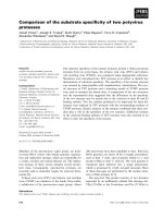
amino acids (Fig. S1). In Fig. 1, the distribution of the
residues, identified as outliers in the Ramachandran
plot (Fig. S2), are shown. In total, 95 outliers were
identified, depicted in a space-filled rendering mode in
dark yellow (Fig. 1).
The homology model of the template structure was
minimized using the Amber 99 force field with the
ligands as environment. One hundred models were
generated using the best intermediate option with
medium minimization, including the prevention of
clashes, to stay as close as possible to the initial
structure. The final template model was selected
according to the best score of the Molecular Operat-
ing Environment (MOE) scoring function. A multise-
quence alignment was performed by the ‘Align’ tool
in MOE (see Materials and methods), including the
template (PDB ID: 3G61 chain A), Swissprot data-
base human MDR1 (code P08183) and closest relative
to the human P-gp [hamster MDR1 (code P21448)]
sequences.
To obtain the final homology model, 100 structures
were generated using the best intermediate option and
Fig. 1. X-Ray structure of mouse P-gp (PDB ID code 3G61): distri-
bution of the amino acid outliers (95 residues) identified from the
Ramachandran plot (Fig. S2); the outliers are rendered in dark-
yellow, space-filled mode.
I. K. Pajeva et al. Comparison of open and closed models of human P-gp
FEBS Journal 276 (2009) 7016–7026 ª 2009 The Authors Journal compilation ª 2009 FEBS 7017
medium minimization with the Amber 99 force field
for each model to remove the bad contacts. The best
model was selected according to the MOE scoring
function and was investigated by the protein report
function in MOE. No outliers in the TM domains of
the protein were found that were important in estimat-
ing the drug-binding competency. The deviating amino
acids, located mostly within the loop regions of the
nucleotide-binding domains (31 outliers, data not
shown), were then minimized, together with the adja-
cent residues, keeping the remaining protein fixed.
After minimization, the outliers in the Ramachandran
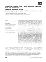
plot (Fig. S3) were reduced to 20. In Fig. 2, the loca-
tions of the outliers within the final model of human
P-gp are shown in a space-filled rendering mode in
dark yellow. The rmsd of all a-carbon atoms between
the homology model after minimization of the
Ramachandran plot outliers and the template with the
modified TM12 was 0.188 A
˚
.
The protonation state of the model was assigned by
the protonate 3D module in MOE, which considers
the solvent accessibility and regional neighboring of
the amino acids.
Comparison of the cavities in the inward- and
outward-facing conformations of the human
P-gp models
In Table 1, the residues involved in the binding
cavity in the inward- and outward-facing conforma-
tions of the homology model of human P-gp are
shown. The residues reported for the outward-facing
conformation have been identified in a previous
study in which a homology model of P-gp was
developed on the basis of the crystal structure of the
multidrug transporter Sav1866 (see table 9 in ref.
[18]). To have an equal basis for comparison, the
same approach was applied for the identification of
the binding pockets in the cavity of the inward-
facing conformation of P-gp (the module ‘Site
Finder’ in MOE, see Materials and methods). In
Table 1, the residues related to the binding of the
substrates dibromobimane (d), verapamil (v) and
rhodamine (r) are marked, according to the experi-
mental findings of Loo and coworkers [4,5,7,8,11].
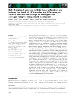
Figure 3 illustrates the cavity and binding pockets
identified. In Fig. 3A, the general view of the cavity
and, in Fig. 3B, a closer view (from the inside) are
shown. Clearly outlined are the large hydrophobic
spheres occupying the entrances (portals) from the
inner leaflet of the membrane to the protein cavity: the
first formed by TM4 (light green) and TM6 (light
magenta), and the second by TM10 (dark green) and
TM12 (dark magenta).
Comparing the residues reported in Table 1 for the
inward- and outward-facing models, differences can be
seen in the residues of the TMs exposed to the cavity
in both conformations.
Amino acid residues involved in the interactions
with the QZ59 compounds in mouse P-gp
The binding sites of the cyclic peptides QZ59 were
analyzed in the X-ray structures using the ‘Ligand
Interactions’ module in MOE (see Materials and meth-
ods). In Table 1, for each TM, the residues involved in
the interactions of the QZ59 ligands are denoted by ‘q’
in the table row ‘sub’.
For the same ligand, different amino acids involved
in the interactions were identified as a function of the
protein molecule in the asymmetric cell unit of the
crystal. For the stereoisomer QZ59-RRR (PDB ID
3G60), 11 common residues were found for both P-gp
molecules (A and B) in the unit cell; mouse Tyr303
(human Tyr307), Leu335 (human L339) and Ser725
(human Ala729) were also involved in the case of
molecule A (Fig. 4A), and Met68 (human Met69),
Fig. 2. Homology model of human P-gp: distribution of the amino
acid outliers (20 residues) identified from the Ramachandran plot
(Fig. S3); outliers are rendered in dark-yellow, space-filled mode.
Comparison of open and closed models of human P-gp I. K. Pajeva et al.
7018 FEBS Journal 276 (2009) 7016–7026 ª 2009 The Authors Journal compilation ª 2009 FEBS
Table 1. Amino acids involved in the binding cavity of the inward (‘in’) and outward (‘out’)-facing conformations of human P-gp; the numbers
and letters of the residues correspond to human P-gp; sub, residues related to substrate binding; grey color, light (in), dark (out).
TM1
54: G T L A A I I H G A G L P L M M L V F G E M
in
a
xx x x
out
sub
b
TM2
117: Y Y S G I G A G V L V A A Y I Q V S F W
in x
out
sub
TM3
190: I G M F F Q S M A T F F T G F I V
in
out
sub
TM4
219: L A I S P V L G L S A A V W A K I L S
in
out
c
sub v
d
TM5
296: N I S I G A A F L L I Y A S Y A L A F W
in x x x
out
sub q d q
TM6
330: Q V L T V F F S V L I G A F S V G Q A S P
in x x x
out
sub q q
v
d
q
rv
d
q
r
TM7
718: A I I N G G L Q P A F A
d
I I F S K I I G V F
in x x x x
out
sub q q q q
TM8
762: L G I I S F I T F F L Q G F T F
in x
out
sub q
TM9
831: S R L A V I T Q N I A N L G T G I I I S F I Y
in
out
sub r
TM10
857: L T L L L L A I V P I I A I A G V V E
in
sub v
d
d
TM11
938: F G I T F S F T Q A M M Y F S Y A G C F
in x x x
out
sub v
d
v
d
q q
TM12
974: V L L V F S A V
d
V F G A M A V G Q V S S F
in x x x x x
out
sub q
r
d
qq q
r
q
r
d
v
q
d
qqqq
a
x, residues involved in the binding pocket of the QZ59 ligands (identified by docking, see Materials and methods).
b
d, dibromobimane
[4,5,7,8]; q, QZ59 (RRR and SSS); r, rhodamine [7,11]; v, verapamil [7,8].
c
no residue involved in the outward-facing conformation.
d
A729
(mouse S725); V981 (mouse A977).
I. K. Pajeva et al. Comparison of open and closed models of human P-gp
FEBS Journal 276 (2009) 7016–7026 ª 2009 The Authors Journal compilation ª 2009 FEBS 7019
Phe71 (human Phe72) and Leu971 (human Leu975) in
molecule B (Fig. 4B). Arene–arene interactions were
identified with Phe332 (human Phe336) and Phe974
(human Phe978) for molecules A and B, respectively.
Compared with the residues reported for QZ59-RRR
in [20, fig. 3], mouse Ser725 (human Ala729) and
Ala981 (human Ala985) for the ligand in molecule A,
and Phe71 (human Phe72), Leu971 (human Leu975)
and Ala981 (human Ala985) for the ligand in mole-
cule B, were also identified, with Ala981 (human
A
B
Fig. 3. Binding cavity in the inward-facing conformation of the
homology model of human P-gp: (A) general view (face); (B) closer
view (from inside). The pockets are filled with alpha spheres; grey
spheres indicate hydrophobic atoms and red spheres hydrophilic
atoms. The protein backbone is colored by the secondary structure;
colors used for the portal TMs: light green (TM4), magenta (TM6),
dark green (TM10), purple (TM12).
A
B
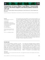
Fig. 4. The interaction panel of the ligand QZ59-RRR in the X-ray
structure of mouse P-gp (PDB ID code 3G60): (A) ligand 1, mole-
cule A; (B) ligand 2, molecule B. The receptor exposure is shown
by the size and intensity (the darker the color, the more exposed
the residue) of the disks drawn behind some of the residues to
denote the difference in their solvent exposure as a result of the
presence of the ligand; the ligand solvent exposure is shown by
smudges drawn behind some of the ligand atoms to denote the
extent of solvent exposure. Symbols:
, arene–arene interactions;
, proximity contour; , ligand exposure; , receptor exposure.
Comparison of open and closed models of human P-gp I. K. Pajeva et al.
7020 FEBS Journal 276 (2009) 7016–7026 ª 2009 The Authors Journal compilation ª 2009 FEBS
Ala985) being well exposed according to the size and
intensity of the disk drawn around the residue
(Fig. 4A,B).
Figure 5 illustrates the interactions for the
QZ59-SSS stereoisomer (PDB ID 3G61). Similar to
QZ59-RRR, differences have been recorded in the
interactions of the ligand in the different molecules
in the unit cell. For ligand 1 in molecule A, only
nonspecific van der Waals’ interactions were exhib-
ited (Fig. 5A), whereas, for ligand 3 in molecule B
(Fig. 5C), a hydrogen bond (HB) was formed with
residue Tyr303 (human Tyr307). Ligand 2 (mole-
cule A) and ligand 4 (molecule B) were not fully
resolved and the analyses of the ligand interactions
thus involved only parts of the structures. For
ligand 2 (Fig. 5B), arene–arene interactions occurred
with Phe974 (human Phe978). No specific interac-
tions were recorded for ligand 4 (Fig. 5D). Obvi-
ously, the same ligand can occupy different positions
within the protein-binding pocket.
Compared with the residues reported in ref. [20,
fig. 3] for QZ59-SSS, mouse Phe71, Phe728, Leu971
AC
BD
Fig. 5. The interaction panel of the ligand QZ59-SSS in the X-ray structure of mouse P-gp (PDB ID code 3G61): (A) ligand 1, molecule A; (B)
ligand 2, molecule A; (C) ligand 3, molecule B; (D) ligand 4, molecule B. The receptor exposure is shown by the size and intensity (the darker
the color, the more exposed the residue) of the disks drawn behind some of the residues to denote the difference in their solvent exposure
as a result of the presence of the ligand; the ligand solvent exposure is shown by smudges drawn behind some of the ligand atoms
to denote the extent of solvent exposure. Symbols:
, arene–arene interactions; , proximity contour; , ligand exposure; , receptor
exposure.
I. K. Pajeva et al. Comparison of open and closed models of human P-gp
FEBS Journal 276 (2009) 7016–7026 ª 2009 The Authors Journal compilation ª 2009 FEBS 7021
and Ile977 (Fig. 5B) were also identified, with Tyr303
performing a specific HB interaction (Fig. 5C).
Amino acid residues involved in the interactions
with QZ59 compounds in the homology model of
human P-gp
The analysis of the residues involved in the interac-
tions was performed in two ways: (a) using the QZ59-
SSS ligands as incorporated from the X-ray sources;
and (b) using docking (by GOLD, see Materials and
methods) to better explore the binding possibilities of
the ligands. In the homology model of human P-gp,
the same amino acids as in the X-ray structures inter-
acted with the QZ59-SSS ligand, and two additional
residues were identified: human Gln725 and Met986
(data not shown).
The docking of the QZ59 stereoisomers yielded sta-
ble and similar solutions. Several runs were performed
using either one or two ligands simultaneously to
define the binding pocket. The poses were fully repro-
ducible with close GoldScore values. The top score
poses were used for further analysis. In Table 1, the
residues involved in the binding sites of the QZ59
ligands identified in the best docking poses in the
human P-gp inward-open model are marked by ‘x’.
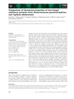
Figure 6 visualizes the interaction of QZ59-RRR
and QZ59-SSS with the residues in the human P-gp
model. Figure 6A shows the general view of the best
docked poses of both stereoisomers in the two binding
sites superimposed on the P-gp backbone. Similar to
the SSS enantiomer, the RRR enantiomer could poten-
tially occupy the two binding sites. Figure 6B shows a
closer view of the interactions of the two peptides in
the lower located binding site, and Fig. 6C in the
upper site. Comparing the residues comprising the
binding sites of the ligands in human and mouse P-gp
(Table 1: ‘q’ with ‘x’; Figs 4 and 5 with Fig. 6), simi-
larities and differences in relation to the particular
amino acids involved and their specific interactions can
be outlined. The residues in the ligand-binding sites in
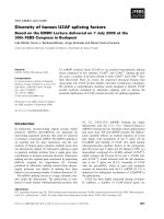
Fig. 6. (A) A front overview of the P-gp binding cavity with the
inhibitors QZ59-RRR and QZ59-SSS (in space-filling form) docked
into the two binding sites and superimposed on the P-gp backbone
(grey line). (B) Closer view (from the bottom) of the protein–ligand
interactions and the surface of the binding site in the lower binding
site. (C) Closer view (from the bottom) of the protein–ligand interac-
tions and the surface of the binding site in the upper binding site.
The Gaussian contact surface is presented with the following color
coding: green, hydrophobic; magenta, HB; blue, mildly polar. The
HB distance and score are colored in magenta; the residues are col-
ored in green; the structures of the ligands are rendered in stick
form and colored according to the atom types; the HB atoms are
shown as balls and the distance between (=O) of QZ59-RRR and
(–N<) of Gln725 is 2.84 A
˚
(64% score, see Materials and methods).
A
B
C
Comparison of open and closed models of human P-gp I. K. Pajeva et al.
7022 FEBS Journal 276 (2009) 7016–7026 ª 2009 The Authors Journal compilation ª 2009 FEBS
the X-ray structures of mouse P-gp partially overlap
with those in the docked poses in the human P-gp
model. Although Tyr303 (human Tyr307) performs an
HB interaction with QZ59-SSS in the X-ray structure
(Fig. 5C), human Gln725 is involved in an HB–donor
interaction in human P-gp, and Phe343 can perform
arene–arene interactions with QZ59-RRR (Fig. 6B).
Thus, it can be proposed that the QZ59 ligands will
bind to human P-gp in a similar, but not identical,
manner as to mouse P-gp, interacting specifically with
different residues from the same environment. Such a
result is not surprising considering the observed differ-
ences mentioned above in the interactions of the same
ligand with two different molecules of the same protein
in the X-ray structures (Figs 4 and 5). Notably, the
hydrophobic residues with the human P-gp codes
Phe336, Phe343, Phe728, Phe978 and Val982 are the
most involved amino acids in both the X-ray and
docked poses of the ligands. The same residues have
been proven experimentally to relate to other P-gp
substrates [4,7,8,10,11,14].
Discussion
For the different TMs, differences between the residues
exposed to the cavity in both the open and closed con-
formations have been observed. The most striking dif-
ference is the absence of amino acids of TM4 and
TM10 facing the cavity. Although residues of TM4
and TM10 are broadly accessible in the inward-open
conformation, they are fully buried in the outward-
open form (Table 1). At the same time, the same
residues of TM6 and TM12 face the cavity in both
conformations and, in addition, these domains possess
the highest number of experimentally proven amino
acids involved in drug binding. Among them are a
number of hydrophobic residues, such as human
Phe336, Phe339, Phe340, Phe343, Phe978, Val981,
Val982, Ala985, identified in the binding sites of the
QZ59 ligands; moreover, some have been found to per-
form specific (arene–arene) interactions, such as
Phe336 (mouse Phe332, Fig. 4A), Phe978 (mouse
Phe974, Figs 4B and 5B) and Phe343 (Fig. 6B). TM6
and TM12 are also the most involved domains in
interactions with other P-gp substrates, such as verapa-
mil, rhodamine and dibromobimane, proven by
drug-binding experiments (Table 1, row ‘sub’). In the
most recent study, Loo et al. [5] also found the largest
number of mutations in TM6 and TM12 in arginine-
scanning mutagenesis experiments. Considering that
these TMs form the two portals (TM4–TM6 and
TM10–TM12) for entering the cavity from the inner
leaflet of the membrane, the differences observed
above also suggest differences in their role for the
function of protein and ligand binding. TM4 and
TM10, being parts of the portals, could be involved in
weaker interactions with the entering ligands that are
lost during the transport cycle. In Table 1, three resi-
dues only are reported as belonging to these domains,
related to interactions with P-gp substrates: human
Ser222 (TM4), Ile868 and Gly872 (TM10) (Table 1).
Figuratively, TM4 and TM10 could function as ‘portal
keepers’, preventing the substances that enter the cav-
ity from the inner leaflet of the membrane escaping
back. In contrast, TM6 and TM12 could be regarded
as the ‘portal carriers’, being mainly responsible for
ligand interactions. Interestingly, the TM6 and T12
residues mostly perform hydrophobic interactions,
whereas the more specific HB-type interactions could
be related to other domains, such as, for example,
Tyr307 (mouse Tyr303) of TM5 (Fig. 5C) and Gln725
of TM7 (Fig. 6A). Studying the interactions of specific
P-gp inhibitors, representatives of the third-generation
MDR modulators, we found specific HB interactions
with Tyr117 (TM2), Tyr307 (TM5) and an arene–
arene-type interaction with Tyr953 (TM11) [27], thus
outlining, in addition, the role of TM2, TM5, TM7
and TM11 in ligand interactions. It is worth noting
that these residues also face the cavity in both confor-
mations of the protein (Table 1).
From the above analysis, it is most likely that the
ligands remain bound to the same residues during the
transition from the inward- to the outward-facing con-
formation of the protein, suggesting that the ligand is
not flipped.
Next, the detailed analyses of the ligand interactions
with the protein, as recorded here for the QZ59 com-
pounds (Figs 4 and 5), show differences in the residues
involved, as well as in the specific interactions of the
same ligand with the same protein. For QZ59-RRR,
Phe332 (human Phe336) is in a favorable position for
arene–arene interactions in molecule A, whereas
Phe974 (human Phe978) is involved in such interac-
tions in molecule B (Fig. 4A,B); Tyr303 (human
Tyr307) forms an HB interaction with ligand 3 in mol-
ecule B (Fig. 5C); at the same time, no such interac-
tion is recorded for ligand 1 in molecule A (Fig. 5A).
Whether these differences can be related to the experi-
mental conditions under which the ligands were
co-crystallized, or reflect the possibility for different
binding locations and orientations of the same ligand
in the same protein environment, remains to be
proven. The docking of QZ59 ligands into the human
P-gp binding cavity supports the latter suggestion. The
results confirm the possibility for binding of the same
ligand in two different binding sites, as shown for
I. K. Pajeva et al. Comparison of open and closed models of human P-gp
FEBS Journal 276 (2009) 7016–7026 ª 2009 The Authors Journal compilation ª 2009 FEBS 7023
QZ59-RRR in Fig. 6B,C. This observation, illustrated
here by the analysis of the X-ray complexes and sup-
ported by binding simulation, could help, for example,
to better understand the complex behavior of P-gp
substrates and inhibitors in functional assays.
In conclusion, the results of this study confirm the
possibility for multispecific interactions of the protein
with its ligands, and aid in the elucidation of P-gp
function and drug interactions.
Materials and methods
Homology modeling
Chain A from the crystal structure of mouse P-gp (PDB ID:
3G61 [20]) was used as template for the homology model.
The Swissprot database sequences of human P-gp (P08183)
and hamster P-gp (P21448) were used for alignment [28].
The alignment was obtained with the ‘Align’ tool in
MOE [29], using the default tree-based approach with the
blosum62 substitution matrix and increased values for the
gap penalty of 15 and gap extension penalty of 2.
The homology model was calculated by the ‘Homology
Model’ method in MOE using a rotamer library and loop
dictionary derived from the X-ray structures to predict the
coordinates of deviating residues. The best intermediate
approach with the medium minimization option and the
Amber 99 force field was used to derive the model. The co-
crystallized ligand structures were defined as environment
and kept fixed. The best model (out of 100) was selected
according to the GB ⁄ VI scoring function (Coulomb and
generalized Born interaction energies of the model and
environment). The stereochemical quality of the model was
inspected by the protein report of MOE. The MOE-ProSu-
perpose module was used to calculate the rmsd values.
Identification of the binding pockets
The ‘Site Finder’ tool in MOE [29] was employed for the
identification of the binding sites in the inward-open con-
formation of the P-gp homology model. The program is
based on the methodology of convex hulls which produces
pockets invariant to rotation of the atomic coordinates. It
treats the set of three-dimensional points by triangulation
and associates each resulting simplex with a sphere, coded
as ‘alpha sphere’. The radius of the sphere is proportional
to the convex hull of the point set. Each sphere is classified
as either ‘hydrophobic’ or ‘hydrophilic’ depending on
whether the sphere is a good HB point in the receptor.
Hydrophilic spheres not close to hydrophobic ones
are eliminated as they generally correspond to water sites.
The generated pockets consist of one or more alpha
spheres, and at least one is hydrophobic. The following
settings were used: radius of a hydrophilic HB sphere,
1.4 A
˚
; radius of a hydrophobic sphere, 1.8 A
˚
; isolate
donor–acceptor distance, 3 A
˚
; connection distance between
clusters of alpha spheres, 2.5 A
˚
; minimum site size, 3;
minimum site radius, 2 A
˚
.
Docking of the QZ59 ligands into human P-gp
models
The QZ59 ligands were prepared in Sybyl [30]. QZ59-RRR
was extracted from its complex 3G60 and QZ59-SSS from
3G61. The chirality and atom types of the compounds were
checked; the missing hydrogen atoms were added, and the
geometries were optimized with the Tripos force field and
Gasteiger–Hueckel charges. The minimized structures were
subsequently exported as mol2 files for docking with the
GOLD Suite [31,32], which applied a genetic optimization
algorithm for docking flexible ligands into protein-binding
sites. The binding pocket was defined on the basis of the
co-crystallized ligands in the X-ray structure. Considering
the substantial volume of the binding cavity (around
6000 A
˚
3
[20]), the pocket was extended by 10 A
˚
and out-
ward-facing amino acids were deselected. The default
setting for the genetic algorithm and the original GoldScore
function were used to rank the ligand poses. Docking was
performed by the ‘slow (most accurate)’ option for balance
between speed and accuracy.
Analysis of the ligand interactions
The X-ray complexes (mouse P-gp) and docked poses
(human P-gp) of the QZ59 compounds were analyzed by
the ‘Ligand Interactions’ tool in MOE. The method imple-
mented is fully described in [33]. The information content
displayed in the ligand interactions panel consists of the
selected ligand and the receptor-interacting entities, namely
HB residues, close but non-bonded residues (approaching
the ligand but not having any strong interactions, i.e. HBs),
solvent molecules and ions. The solvent-accessible surface
area and the ligand proximity outline were also estimated.
HBs were assigned to each pair of heavy atoms from the
ligand and receptor according to probability criteria derived
from a large training set [34,35]. The HB scores were
expressed as percentages and the HB directionality was
noted. The ligand and residue solvent accessibility metrics
were estimated by measuring the exposed surface area once
each of the atoms had been assigned a van der Waals’-like
radius of +1.4 A
˚
(water solvent). The solvent exposure of
receptor residues was calculated by examining the difference
between the solvent-exposed surface area of the receptor
with and without the presence of the ligand. For the
ligands, the surface accessibility calculation was carried out
on the ligand + receptor complex. The default settings
were applied for the definition of hydrogen-bonded and
proximity interactions.
Comparison of open and closed models of human P-gp I. K. Pajeva et al.
7024 FEBS Journal 276 (2009) 7016–7026 ª 2009 The Authors Journal compilation ª 2009 FEBS
Acknowledgements
I.P. and M.W. gratefully acknowledge the generous
support by the Alexander von Humboldt Foundation,
Germany. I.P also thanks the National Science Foun-
dation of Bulgaria.
References
1 Juliano RL & Ling V (1976) A surface glycoprotein
modulating drug permeability in Chinese hamster ovary
cell mutants. Biochim Biophys Acta 455, 152–162.
2 Sharom FJ (2008) ABC multidrug transporters: struc-
ture, function and role in chemoresistance. Pharmacoge-
nomics 9, 105–127.
3 Glavinas H, Krajcsi P, Cserepes J & Sarkadi B (2004)
The role of ABC transporters in drug resistance, metab-
olism and toxicity. Curr Drug Deliv 1, 27–42.
4 Loo TW & Clarke DM (1997) Identification of residues
in the drug-binding site of human P-glycoprotein using
a thiol-reactive substrate. J Biol Chem 272, 31945–
31948.
5 Loo TW & Clarke DM (2000) Identification of residues
within the drug-binding domain of the human multi-
drug resistance P-glycoprotein by cysteine-scanning
mutagenesis and reaction with dibromobimane. J Biol
Chem 275, 39272–39278.
6 Martin C, Berridge G, Higgins CF, Mistry P, Charlton
P & Callaghan R (2000) Communication between
multiple drug binding sites on P-glycoprotein. Mol
Pharmacol 58, 624–632.
7 Loo TW & Clarke DM (2001) Defining the drug-bind-
ing site in the human multidrug resistance P-glycopro-
tein using a methanethiosulfonate analog of verapamil,
MTS-verapamil. J Biol Chem 276, 14972–14979.
8 Loo TW & Clarke DM (2002) Location of the rhoda-
mine-binding site in the human multidrug resistance
P-glycoprotein. J Biol Chem 277, 44332–44338.
9 Ecker GF, Csaszar E, Kopp S, Plagens B, Holzer W,
Ernst W & Chiba P (2002) Identification of ligand-bind-
ing regions of P-glycoprotein by activated-pharmaco-
phore photoaffinity labeling and matrix-assisted laser
desorption ⁄ ionization-time-of-flight mass spectrometry.
Mol Pharmacol 61, 637–648.
10 Loo TW, Bartlett MC & Clarke DM (2003) Simulta-
neous binding of two different drugs in the binding
pocket of the human multidrug resistance P-glycopro-
tein. J Biol Chem 278, 39706–39710.
11 Loo TW, Bartlett MC & Clarke DM (2003) Methan-
ethiosulfonate derivatives of rhodamine and verapamil
activate human P-glycoprotein at different sites. J Biol
Chem 278, 50136–50141.
12 Lugo MR & Sharom FJ (2005) Interaction of LDS-751
and rhodamine 123 with P-glycoprotein: evidence for
simultaneous binding of both drugs. Biochemistry 44,
14020–14029.
13 Gatlik-Landwojtowicz E, Aa
¨
nismaa P & Seelig A (2006)
Quantification and characterization of P-glycoprotein–
substrate interactions. Biochemistry 45, 3020–3032.
14 Loo TW, Bartlett MC & Clarke DM (2006) Transmem-
brane segment 7 of human P-glycoprotein forms part of
the drug-binding pocket. Biochem J 399, 351–359.
15 Loo TW, Bartlett MC & Clarke DM (2009) Identifica-
tion of residues in the drug-translocation pathway of
the human multidrug resistance P-glycoprotein by
arginine mutagenesis. J Biol Chem 284, 24074–24087.
16 Stenham DR, Campbell JD, Sansom MS, Higgins CF,
Kerr ID & Linton KJ (2003) An atomic detail model
for the human ATP binding cassette transporter P-gly-
coprotein derived from disulfide cross-linking and
homology modeling. FASEB J 17, 2287–2289.
17 O’Mara ML & Tieleman DP (2007) P-glycoprotein
models of the apo and ATP-bound states based on
homology with Sav1866 and MalK. FEBS Lett 581,
4217–4222.
18 Globisch C, Pajeva IK & Wiese M (2008) Identification
of putative binding sites of P-glycoprotein based on its
homology model. ChemMedChem 3, 280–295.
19 Becker JP, Depret G, Van Bambeke F, Tulkens PM &
Pre
´
vost M (2009) Molecular models of human P-glyco-
protein in two different catalytic states. BMC Struct
Biol 9,3.
20 Aller SG, Yu J, Ward A, Weng Y, Chittaboina S, Zhuo
R, Harrell PM, Trinh YT, Zhang Q, Urbatsch IL et al.
(2009) Structure of P-glycoprotein reveals a molecular
basis for poly-specific drug binding. Science 323, 1718–
1722.
21 Tombline G, Holt JJ, Gannon MK, Donnelly DJ,
Wetzel B, Sawada GA, Raub TJ & Detty MR (2008)
ATP occlusion by P-glycoprotein as a surrogate
measure for drug coupling. Biochemistry 47, 3294–3307.
22 Gannon MK 2nd, Holt JJ, Bennett SM, Wetzel BR, Loo
TW, Bartlett MC, Clarke DM, Sawada GA, Higgins JW,
Tombline G et al. (2009) Rhodamine inhibitors of
P-glycoprotein: an amide ⁄ thioamide ‘switch’ for ATPase
activity. J Med Chem 52, 3328–3341.
23 Tang-Wai DF, Kajiji S, DiCapua F, de Graaf D,
Roninson IB & Gros P (1995) Human (MDR1) and
mouse (mdr1, mdr3) P-glycoproteins can be distin-
guished by their respective drug resistance profiles and
sensitivity to modulators. Biochemistry 34, 32–39.
24 Gottesman MM, Ambudkar SV & Xia D (2009)
Structure of a multidrug transporter. Nat Biotechnol 27,
546–547.
25 Dawson RJ & Locher KP (2006) Structure of a bacte-
rial multidrug ABC transporter. Nature 443, 180–185.
26 Dawson RJ & Locher KP (2007) Structure of the multi-
drug ABC transporter Sav1866 from Staphylococcus
I. K. Pajeva et al. Comparison of open and closed models of human P-gp
FEBS Journal 276 (2009) 7016–7026 ª 2009 The Authors Journal compilation ª 2009 FEBS 7025
aureus in complex with AMP-PNP. FEBS Lett 581,
935–938.
27 Pajeva IK, Globisch C & Wiese M (2009)
Combined pharmacophore, docking and
3D QSAR study of inhibitors of ABCB1 and
ABCC1 transporters. ChemMedChem 4, 1883–
1896.
28 Gasteiger E, Gattiker A, Hoogland C, Ivanyi I, Appel
RD & Bairoch A (2003) ExPASy: the proteomics server
for in-depth protein knowledge and analysis. Nucleic
Acids Res 31, 3784–3788.
29 MOE 2008.10 (Molecular Operating Environment).
Chemical Computing Group, Montreal, Quebec.
30 SYBYL 8.1. Tripos Inc., St Louis, MO.
31 GOLD Version 4.0.1. The Cambridge Crystallographic
Data Centre, Cambridge, UK.
32 Jones G, Willett P, Glen RC, Leach AR & Taylor R
(1997) Development and validation of a genetic
algorithm for flexible docking. J Mol Biol 267,
727–748.
33 Clark AM & Labute P (2007) 2D depiction of
protein–ligand complexes. J Chem Inf Model 47,
1933–1944.
34 Labute P, Williams C, Feher M, Sourial E & Schmidt
JM (2001) Flexible alignment of small molecules. J Med
Chem 44, 1483–1490.
35 Labute P (2001) Probabilistic receptor potentials. J Chem
Comput Group, />htm [accessed on 12 October 2009].
Supporting information
The following supplementary material is available:
Fig. S1. Backbone of the TMs in the X-ray structure
of mouse P-gp (PDB ID code 3G61) with the dis-
rupted helix in TM12 and the superposed part of TM6
(blue, on the left of the cavity) used as a template for
the modified TM12.
Fig. S2. Ramachandran plot of the X-ray structure of
chain A (PDB ID code 3G61).
Fig. S3. Ramachandran plot of the homology model
of human P-gp.
This supplementary material can be found in the
online version of this article.
Please note: As a service to our authors and readers,
this journal provides supporting information supplied
by the authors. Such materials are peer-reviewed and
may be re-organized for online delivery, but are not
copy-edited or typeset. Technical support issues arising
from supporting information (other than missing files)
should be addressed to the authors.
Comparison of open and closed models of human P-gp I. K. Pajeva et al.
7026 FEBS Journal 276 (2009) 7016–7026 ª 2009 The Authors Journal compilation ª 2009 FEBS