Báo cáo khoa học: The in vitro nuclear aggregates of polyamines pot
Bạn đang xem bản rút gọn của tài liệu. Xem và tải ngay bản đầy đủ của tài liệu tại đây (566.82 KB, 12 trang )
The in vitro nuclear aggregates of polyamines
Aldo Di Luccia
1,2,
*, Gianluca Picariello
1,
*, Giuseppe Iacomino
1,
*, Annarita Formisano
1
,
Luigi Paduano
3
and Luciano D’Agostino
1,4
1 Institute of Food Sciences, National Research Council (CNR), Avellino, Italy
2 Department PROGESA, University of Bari, Italy
3 Department of Chemistry, University of Naples ‘Federico II’, Italy
4 Department of Clinical and Experimental Medicine, University of Naples ‘Federico II’, Italy
Self-assembly of polyamines – putrescine (Put), spermi-
dine (Spd), and spermine (Spm) – with phosphate ions
was previously described by our group [1]: the interca-
lation of a phosphate anion between the N-terminal
ends of two polyamines determines, by electrostatic
interaction, the formation of basic cyclical structures
that further aggregate into supramolecular complexes
[2] by means of hydrogen bonds, thus producing three
different structural classes of molecular aggregates that
interact with the genomic DNA [1,3,4]. These com-
pounds were named nuclear aggregates of polyamines
(NAPs). Interestingly, other authors have described the
phosphate-induced self-assembly of polyamines in a
different biological setting [5].
Polyamine and phosphate self-aggregation is reputed
to be an important phenomenon in directing DNA orga-
nization and functions [1]. In our earlier studies, Caco-2
cells were used to assess the biological properties of
NAPs, but investigations concerning NAPs extracted
from nuclei of many different cell types have also been
described [1,3]. However, only preliminary observations
concerning the in vitro production of these compounds
have been reported [1,3,4]. In addition, the mecha-
nism(s) regulating the supramolecular self-aggregation
of polyamines and phosphates and the cooperative
action of NAP–DNA aggregates have yet to be defined.
For this reason, we determined the conditions neces-
sary for the aggregation of polyamines in a simplified
Keywords
DNA interactions; nanostructures;
polyamines; self-assembly; supramolecular
chemistry
Correspondence
L. D’Agostino, Department of Clinical and
Experimental Medicine, University of Naples
‘Federico II’ Ed. 6, Via S. Pansini, 5, 80131
Naples, Italy
Fax: +39 081 7462707
Tel: +39 081 7462707
E-mail:
*These authors contributed equally to this
work
(Received 16 September 2008, revised 9
February 2009, accepted 11 February 2009)
doi:10.1111/j.1742-4658.2009.06960.x
Natural polyamines (putrescine, spermidine, and spermine) self-assemble in
a simulated physiological environment (50 mm sodium phosphate buffer,
pH 7.2), generating in vitro nuclear aggregates of polyamines (ivNAPs).
These supramolecular compounds are similar in structure and molecular
mass to naturally occurring cellular nuclear aggregates of polyamines, and
they share the ability of NAPs to interact with and protect the genomic
DNA against nuclease degradation. Three main ivNAP compounds were
separated by gel permeation chromatography. Their elution was carried
out with 50 mm sodium phosphate buffer supplemented with 150 mm
NaCl. Freezing and thawing of selected chromatographic fractions
obtained by GPC runs in which the mobile phase was sodium phosphate
buffer not supplemented with NaCl yielded three different microcrystallites,
specifically corresponding to the ivNAPs, all of which were able to bind
DNA. In this study, we demonstrated that in vitro self-assembly of polyam-
ines and phosphates is a spontaneous, reproducible and inexpensive event,
and provided the indications for the production of the ivNAPs as a new
tool for manipulating the genomic DNA machinery.
Abbreviations
DLS, dynamic light scattering; EtBr, ethidium bromide; GPC, gel permeation chromatography; ivNAP, in vitro nuclear aggregate of
polyamines; NAP, nuclear aggregate of polyamines; Put, putrescine; Spd, spermidine; Spm, spermine.
2324 FEBS Journal 276 (2009) 2324–2335 ª 2009 The Authors Journal compilation ª 2009 FEBS
in vitro model in order to investigate some of the fea-
tures of the polyamine–phosphate interactions. Specifi-
cally, we examined the role played by each polyamine
in the self-assembly of in vitro NAPs (ivNAPs) and
their ability to interact with genomic DNA. Further
aims of the present study were to investigate the mech-
anisms that regulate the interactions among polyam-
ines and phosphate ions that induce the assembly of
these supramolecular structures, and to gather addi-
tional conceptual elements for molecular modeling and
determination of NAP functions.
In this article, we report findings indicating struc-
tural and functional analogies among extractive and
synthetic NAPs: therefore, according to their mole-
cular masses, and in keeping with the terminology of
natural NAPs [3], we named the synthetic compounds
l-ivNAP, m-ivNAP, and s-ivNAP (in vitro large,
medium and small), respectively. Furthermore, for the
first time, we show images of crystallized aggregates of
polyamines and phosphates interacting with genomic
DNA.
Results and Discussion
Gel permeation chromatography (GPC) analysis
of ivNAPs
In vitro aggregation of polyamines and phosphate ions
generated supramolecular compounds, the ivNAPs,
characterized by an extended electronic delocalization
detectable by a distinctive absorbance peak at
k = 280 nm in the UV spectrum, which is completely
absent for unassembled polyamines (data not shown).
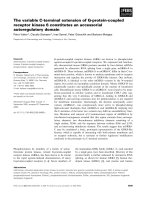
Representative GPC profiles of ivNAPs are shown
in Fig. 1, where it is also possible to analyze GPC
chromatogram modifications by varying the concentra-
tion of one of the three polyamines at a time (range
5–48 mm), while keeping the concentrations of the
other two constant (24 mm).
Three main peaks with different intensities and esti-
mated molecular masses of 8000, 5000 and 1000 Da,
according to increasing elution time and corresponding
to l-ivNAP, m-ivNAP and s-ivNAP, respectively, were
detected. Although polyamine concentrations of 24 lm
were able to produce detectable GPC peaks [1], we
noted that the peak variations were more appreciable
when a 24 mm polyamine solution was used. The GPC
profiles and the estimated molecular masses of the
ivNAPs were similar to those of naturally occurring
NAPs, particularly those found in the nuclei of the
cells at the top of their replication phase [1].
Attempts to assemble ivNAPs in phosphate-
free buffers failed. In fact, no GPC peaks were
detected at k = 280 nm when polyamines were
dissolved in 100 mm Tris ⁄ HCl pH 7.2 buffer (data
not shown).
Fig. 1. Self-assembly of polyamines assayed by GPC with detec-
tion at k = 280 nm. Chromatograms were obtained by progres-
sively increasing (in the range 5–48 m
M) the concentration of (A)
Spm, (B) Spd and (C) Put, keeping the concentration of the remain-
ing two polyamines constant at 24 m
M.
A. Di Luccia et al. ivNAPs
FEBS Journal 276 (2009) 2324–2335 ª 2009 The Authors Journal compilation ª 2009 FEBS 2325
The ivNAP chromatographic peak areas as a func-
tion of the stepwise change of polyamine concentra-
tions are reported in Table 1. In all three sets of
experiments, the peak area corresponding to m-ivNAP
remained the most prominent. The peak area of
m-ivNAP – 50.3 min retention time – was only slightly
affected by the polyamine concentration. The increase
in concentration of the three polyamines caused a pro-
gressive decrement in l- ivNAP areas (retention time:
54.3 min), whereas only minor fluctuations were
observed for the s-ivNAP areas (retention time:
44.6 min). Another interesting feature of this kind of
polyamine assembly was the complete fusion of the
l-ivNAP peak with that of m-ivNAP (Fig. 1A),
recorded at 48 mm Spm.
Self-assembly is a process by which molecular
subunits spatially organize in well-defined supra-
molecular structures through noncovalent interactions.
The structures generated in molecular self-assembly
are usually in equilibrium states (or at least in metasta-
ble states). Self-assembled molecular compounds have
been recognized in biological systems [1,3–6], and
designed for the generation of advanced materials [7]
by means of the aggregation of nanoparticles. At the
moment, self-assembly is the most general strategy uti-
lized for generating nanostructures [7].
Self-assembly of polyamines and phosphates is, in
our case, substantiated by the detection at 280 nm of a
discrete set of aggregates with estimated molecular
masses ranging from 1000 to 8000 Da, arising from
low molecular mass species, and by the absence of
covalent interactions in the aggregates. Furthermore,
the appearance of the absorbance band around
280 nm, missing in single polyamine solutions (data
not shown), is the demonstration that the aggregation
of the single components determines an impressive
change in their electronic properties. The absorbance
band at 280 nm is due to the establishment of an
electron delocalization favored by the electrophilic
properties of the polyamines and the cyclic structure of
the unimers.
Surprisingly, whatever the polyamine concentrations
– assayed in the range 24 lm to 48 mm – used, the for-
mation of three ivNAP compounds was observed, and
these compounds had estimated molecular masses very
close to those of the ‘biological’ aggregates. This spe-
cial chemical–physical behavior indicates that some
sort of molecular mass set point regulates polyamine–
phosphate ion self-assembly. Thus, the formation of
these complexes can be attributed to an existing chemi-
cal and thermodynamic equilibrium between reagents
(polyamines and phosphates) and products (ivNAPs)
[8]. Furthermore, our data suggest that self-structuring
of polyamines and phosphate ions occurs within well-
defined ratios, as predicted [1,3,4], indicating that this
kind of aggregation is a finely self-regulated chemical–
physical event.
One of the principles of self-organization is the tran-
sition from a disordered to an ordered state by sponta-
neous symmetry breaking. The transition from a
disordered into an ordered phase takes place through
changes in thermodynamic or physical field strengths.
Such changes may be of temperature and chemical
potential (concentration, pH value, salt addition), of
mechanical fields (pressure, shear, extension, ultrason-
ics), or of electric and magnetic fields. In our case, it
seems that the increase in polyamine concentration,
the sole variable, functioned as an ‘actuator’ and ‘sta-
bilizer’ of symmetry, producing an ordered state. This
last condition is characterized by the facts that individ-
ual molecules are located at restricted three-dimen-
sional regions, and that a localization is always
accompanied by a decrease in the number of realizable
states and, hence, a loss of entropy.
Furthermore, in phosphate-buffered solution or in a
phosphate ion-rich environment (in vivo), enthalpy
Table 1. Percentage distribution of ivNAPs. Relative amounts of
ivNAPs were estimated by integrating the peak area of the GPC
chromatograms (Fig. 1) obtained from the separation of polyamine
solutions prepared by changing the concentration of a single poly-
amine and keeping the concentrations of the other two constant
(24 m
M). In the case of variation of Spm concentration, the
mean ± standard deviation (SD) values were calculated from three
observations (at 5, 10 or 24 m
M), as the m-ivNAP and l-ivNAP areas
fused at 48 m
M. ND, not detected.
Polyamine
concentration
l-ivNAP
(% relative)
m-ivNAP
(% relative)
s-ivNAP
(% relative)
Put (m
M)
5 34.9 50.4 14.7
10 13.2 63.7 23.l
24 12.9 64.7 22.4
48 11.4 62.1 26.5
Mean ± SD 18.1 ± 11.2 60.2 ± 6.6 21.7 ± 5.0
Spd (m
M)
5 18.2 59.6 22.2
10 16.6 64.6 18.8
24 13.5 66.7 19.8
48 5.1 68.7 26.3
Mean ± SD 13.3 ± 5.8 64.9 ± 3.9 21.8 ± 3.3
Spm (m
M)
5 30.8 49.5 19.7
10 23 58.6 18.4
24 12 64.4 23.4
48 ND 82.8 17.2
Mean ± SD 21.9 ± 9.4 57.5 ± 7.5 20.5 ± 2.6
ivNAPs A. Di Luccia et al.
2326 FEBS Journal 276 (2009) 2324–2335 ª 2009 The Authors Journal compilation ª 2009 FEBS
changes are due to cooperating short-range attractive
and long-range repulsive forces established by charged
polyamines [9]. All of these principles can be evoked
to give a possible explanation for the exclusive aggre-
gation of the polyamines and phosphates into three
molecular complexes.
Another intriguing point is the relationship existing
between the three-dimensional arrangement of these
structures and the regular production of only three
main compounds, whatever the solute (polyamine)
concentration was. We are persuaded that the number
of hydrogen bonds is crucial in defining both the
three-dimensional outlines and the molecular masses.
In our previous papers [3,4], we proposed a hierar-
chical process of supramolecular polymerization based
on the assembly of polyamines and phosphates (the
extractive NAPs). The initial step is the self-arrange-
ment of polyamines in disk-like unimers by means of
their terminal interactions with the phosphate groups.
The formation of ring-like unimers can be attributed
to the low equilibrium constant for isodesmic polymer-
ization [10], which characterizes the system, whereas
the successive formation of the medium and large
assemblies is an expression of a ring stabilization pro-
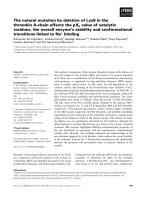
cess. A clear example of this multistep process of
supramolecular assembly is m-NAP, which in solution
– unbound to the DNA – was depicted as structured
in a two-dimensional planar (not columnar) disk-like
arrangement resulting from the oligomeric aggregation
of five s-NAP unimers [3] (Fig. 2). Our modeling
should be considered in line with an isodesmic supra-
molecular polymerization [10] for the further reason
that, since this theory predicts the production of only
oligomers and a preferential disposition of the unimers
in a linear chain, rather than their columnar stacking,
if the hydrogen bonds are single and arranged in a
chain [3]. The data reported here concerning the
ivNAPs support this belief, as a linear chain-type
assembly fits better with the constant and reproducible
detection of low molecular mass aggregates (oligomers)
than with a columnar stacking of disks (polymers)
that, by means of the serial aggregation of available
disk-like monomers, should ultimately generate com-
pounds with greater molecular masses.
However, it is interesting to note that, in the case of
their interaction with the DNA, the assembly of these
supramolecular structures can be imagined, without
contradiction, to be in a columnar form. In fact, the
establishment of two or more hydrogen bonds among
adjacent disk-like unimers can ultimately lead to the
formation of supramolecular nanotubes enveloping the
entire DNA [4]. The process of interaction and colum-
nar disposition of the unimers along the DNA grooves
is probably driven by the phosphates of the DNA,
which can in part replace (two for each ring) the phos-
phates terminally linking the polyamines [4] (Fig. 2). A
similar mechanism, based on the recognition of specific
helically distributed chemical groups, has been already
described in biological systems, e.g. for the assembly
of the protein capsid of tobacco mosaic virus along
the polynucleotide chain. Namely, it is well established
that in the helical columnar assembly of the tobacco
mosaic virus protein coat, the viral RNA acts as a
template and provides additional stability to the
columnar aggregate after formation. However, infor-
mation governing the hierarchical self-assembly process
is, for the most part, encoded within the protein com-
ponents, as, under certain pH conditions, the capsid
subunits are able to self-assemble in the absence of the
RNA strand. In this biologically occurring example of
strict self-assembly, as well as in our case, the com-
ponents spontaneously aggregate without external
guidance into ordered structures [11].
A
B
Fig. 2. Proposed model for polyamine and phosphate group assem-
bly. (A) A multistep process of supramolecular assembly occurs in
solution. The electrostatic interactions between the amine termini
of polyamines and the phosphate groups generate cyclic ivNAP uni-
mers, which further aggregate to form disk-like supramolecular
compounds. (B) The interaction of these compounds with the DNA
and ⁄ or their in loco aggregation produces the DNA shielding, and
promotes and assists the DNA conformational changes. The ulti-
mate result of the hierarchical self-assembly is the formation of
organized polyamine–phosphate nanotubes that wrap but do not
constrict the double helix.
A. Di Luccia et al. ivNAPs
FEBS Journal 276 (2009) 2324–2335 ª 2009 The Authors Journal compilation ª 2009 FEBS 2327
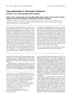
Composition of GPC peaks
To determine the relative ratios among the individual
polyamines forming ivNAPs, collected GPC fractions
were derivatized with dansyl chloride and analyzed by
RP-HPLC (Fig. 3).
Table 2 shows the concentration of the polyamines
constituting the ivNAPs. Spm was the major com-
ponent in both l-ivNAP and m-ivNAP, Spd was pre-
dominant in s-ivNAP, and Put was completely absent
in l-ivNAP.
Total recovery values, also reported in Table 2, were
87.7% for Spm, 68.3% for Spd, and 16.5% for Put.
Recovery was not quantitative, indicating that a frac-
tion of polyamines escaped detection at k = 280 nm,
probably because they did not aggregate in cyclic
structures.
The recovery values for Put were generally lower
than those for Spd and Spm, and the highest percent-
ages of Put were found in s-ivNAP. Recovery of Spm,
the major constituent of l-ivNAP, progressively
increased with the ivNAP size. In contrast, recovery of
Put and Spd followed an inverse trend.
Put recovery was significantly lower than that of the
other polyamines. The differences in recoveries
reported in Table 2 could be indicative of a thermo-
dynamic equilibrium among the free polyamines and
the supramolecular aggregates, which depends not only
on the different concentrations of the solutes but also
on the electrostatic interactions in the solution.
Molecular masses estimated by GPC (Table 2) are
quite similar to those reported for NAPs extracted
from cell nuclei [1,3]. Our data, however, do not per-
mit the definition of simplest formulas, as self-assem-
bled compounds present in broad GPC peaks have to
be considered as resulting from a Gaussian distribution
Fig. 3. Quantitative determination of polyamine in ivNAPs by RP-HPLC analysis of dansyl chloride derivatives. Chromatograms of the deriva-
tized polyamines from (A) l-ivNAP, (B) m-ivNAP, and (C) s-ivNAP.
Table 2. Relative concentrations and recoveries of polyamines in
ivNAPs. Polyamines were quantified by RP-HPLC after derivatiza-
tion with dansyl chloride. In vitro NAPs were in this case obtained
by pooling 48 m
M polyamines in 50 mM phosphate buffer solutions
(pH 7.2). A typical GPC chromatogram is shown. Concentrations of
polyamines in the ivNAPs are expressed as m
M. ND, not detected.
Putrescine
(% recovery)
Spermidine
(% recovery)
Spermine
(% recovery)
Estimated
molecular
mass (Da)
l-ivNAP ND 0.75 (4.1) 10 (55.0) 8000
m-ivNAP 0.23 (2.3) 8.3 (18.6) 10.4 (23.5) 5000
s-ivNAP 1.9 (14.2) 6.1 (45.6) 1.5 (9.3) 1000
Total
recovery
16.5 68.3 87.8
ivNAPs A. Di Luccia et al.
2328 FEBS Journal 276 (2009) 2324–2335 ª 2009 The Authors Journal compilation ª 2009 FEBS
of the molecular masses of several coeluting
compounds. On the other hand, attempts to confirm the
proposed molecular formulas by means of ‘soft’ MS
techniques (MALDI-TOF and ESI-MS, in appropriate
conditions for detecting noncovalent interactions) were
unsuccessful, most likely because ivNAPs ⁄ NAPs were
destructured in the ionization because of the weakness
of the interactions involved.
Influence of NaCl on ivNAP stability
In vitro NAPs were separated by GPC in the presence
or absence of 150 mm NaCl in 50 mm phosphate buf-
fer (pH 7.2) as mobile phase. Even though the yield of
ivNAPs was significantly increased in the presence of
NaCl, chromatographic patterns were only slightly
affected by ionic strength. However, extraphysiological
modifications of salt concentration and ⁄ or pH destabi-
lize the supramolecular assembly, making the com-
pounds undetectable by GPC analysis.
In vitro NAPs isolated in NaCl-enriched sodium
phosphate buffer were freeze–thaw stable (Fig. 4A).
Conversely, ivNAPs isolated in sodium phosphate buf-
fer not supplemented with NaCl contained macro-
scopic precipitates (Fig. 4A). Figure 4B–D clearly
illustrates that the precipitates were due to the forma-
tion of crystallites. The crystallite shapes from s-ivNAP
and m-ivNAP solutions were similar, and showed
mainly tetragonal forms, whereas l-ivNAP crystallites
had a more complex dendritic–broad-branched appear-
ance (Fig. 4). Interestingly, isolated polyamines did not
give rise to precipitates if frozen and thawed in sodium
phosphate buffer not supplemented with NaCl.
In order to determine the presence of polyamines in
the crystallites, we resolubilized them and repeated the
RP-HPLC analysis, obtaining chromatograms of the
derivatized polyamines similar to those reported in
Fig. 3 (data not shown). These analyses showed
the presence of distinct polyamine patterns in the
crystallites.
We have taken into account the possibility of
cocrystallization in the genesis of the crystallites.
Cocrystallization of polyamines and phosphates seems
to be less probable than crystallization of ivNAPs, on
the basis of the following experimental observations: (a)
precipitation of the sole phosphates was easily excluded,
as polyamines were recovered in the crystallites – fur-
thermore, previously reported data [12] showed that
NaH
2
PO
4
did not precipitate at all under freeze–thaw
conditions, even at high concentrations (0.5–1 m); (b)
formation of crystallites is a property of the NAPs only,
as it was not observed at all for single polyamines dis-
solved in phosphate buffer (with or without NaCl), even
after several freeze–thaw cycles; and (c) crystallites, in
microscopy analysis, assume distinct shapes for each
one of the three ivNAPs. For all of these reasons, we
are inclined to believe that each ivNAP crystallizes with
conservation of its supramolecular assembly. However,
we think that a definite answer to this question will be
given by X-ray diffraction studies.
Defrosted ivNAPs I-ivNAP
m-ivNAP s-ivNAP
AB
CD
Fig. 4. In vitro NAP crystallization. (A) The
defrosted ivNAPs solution obtained by GPC
in which the mobile phase was sodium
phosphate buffer not supplemented with
NaCl exhibits turbidity if compared to the
unfrozen control. (B–D) Crystallites of the
ivNAPs were clearly distinguishable in these
defrosted GPC fractions (l-ivNAP, m-ivNAP,
or s-ivNAP). Images were acquired by phase
contrast microscopy at · 400 magnification.
The scale bars correspond to 20 lm.
A. Di Luccia et al. ivNAPs
FEBS Journal 276 (2009) 2324–2335 ª 2009 The Authors Journal compilation ª 2009 FEBS 2329
The role of NaCl as a phase separator factor in our
experimental conditions is supported by studies con-
cerning silica precipitation [5,13]. These studies
describe: (a) the mechanisms by which long-chain poly-
amines, consisting of 15–21 repeating units of N-meth-
ylpropyleneimine attached to Put, undergo phase
separation and form microemulsions in the presence of
either phosphate or other polyanions; and (b) the abil-
ity of polyamines (with molecular masses ranging from
1000 to 1250 Da) to promptly precipitate silica nano-
spheres from a silicic acid solution. This occurrence is
strictly dependent on the presence of phosphate ions
and on ionic strength. In our case, the phase separa-
tion observed after freezing of soluble and natural
(small-sized) polyamines, in the presence of phosphate
ions and in an environment lacking NaCl, is a surpris-
ing phenomenon that signifies the reassembly of small
structures (ivNAPs) into larger and insoluble supra-
molecules.
The role played by NaCl can be also be satisfacto-
rily explained by referring to the theory of polyampho-
lytes [14]: in the absence of salt, the attraction of the
fixed charges leads to molecular collapse in globular
forms and to consequent insolubility; with low salt, as
in our system, the charge shielding of the molecules by
mobile ions prevents their globularization, thus leading
to solubility and increasing molecular network swell-
ing; with high salt, salting-out effects lead again to
insolubility or association. Similar effects occur even
under nonisoelectric conditions.
Furthermore, when saline solutions are cooled to
subzero temperatures, H
2
O freezes as pure ice, and ions
are ejected into the unfrozen part of the system. This
event occurs only when the solution temperature over-
comes the eutectic point of a given salt [15,16] (in our
system, )21.1 °C for NaCl and )9.9 °C for NaH
2
PO
4
⁄
Na
2
HPO
4
buffer). As the freezing process progresses, a
salt concentration gradient, as well as a temperature
gradient (due to latent heat release), establishes across
the freezing front. This leads to the occurrence of mac-
roscopic instabilities due to the formation of pockets of
unfrozen salt-concentrated brine [17,18]. Therefore,
considering that the saline bonds are at the basis of
NAP ⁄ ivNAP formation, it can be inferred that, in
NaCl-free solutions, polyamine–phosphate salt precipi-
tation occurs more easily in a crystalline form than in
an amorphous one [16]. In our case, in these pockets of
unfrozen salt-concentrated brine, greater suprastruc-
tures assembled and finally precipitated, forming
crystallites as a consequence of the increased concentra-
tions of polyamines and phosphate salts [16,19].
We are persuaded that the influence of NaCl in
determining the size and shape of the aggregates is
quite delicate, and needs to be investigated in detail.
Dynamic light scattering (DLS) measurements can be
useful for clarifying this matter. Preliminary DLS data
indicate that, in the absence of NaCl, ivNAP solutions
have a natural tendency to form large aggregates. At
room temperature, the process is time-dependent: a
sample left for several hours on the bench becomes
opalescent. Low temperatures or freeze–thaw processes
speed up the superaggregation of ‘NaCl-free’ ivNAPs.
Every way, the aggregation produces micrometer-sized
particles that, for their dimension, are outside the DLS
detection range. In contrast, 150 mm NaCl l-ivNAP,
m-ivNAP or s-ivNAP solutions remained clear in all of
the above-mentioned conditions. DLS measurements
performed on these solutions after a freeze–thaw cycle
gave reproducible and fitting results about the hydro-
dynamic size of the superaggregates, the radii of which
ranged between 200 and 500 nm. These dimensions
could be ascribed to both large hydration shells and
shape effects of the compounds. However, to obtain
information on these aggregates at the mesostructural
and microstructural scales, a specific study based on
DLS and small-angle neutron scattering measurements
would be required. In any case, the analysis of both
the correlation function and the corresponding distri-
bution function of the hydrodynamic radii revealed a
quite small polydispersity in size of the complexes
(Fig. 5).
These data indicate that ivNAPs can remain struc-
turally stable in appropriate saline conditions. It is
likely that the presence of ions in the hydration sphere
of ivNAPs induces an orientation of the electric water
dipoles and ⁄ or repulsion among the charges that stabi-
lizes the aggregates and restrains their further growth
into macrocomplexes. Further studies are also needed
to provide an understanding of these underlying chem-
ical and physical mechanisms. However, it is clear that,
in our systems, fusion phenomena are drastically
depressed by the presence of NaCl in the solutions.
The role played by NaCl in conferring stability on
these supramolecular aggregates is a rough indication
of the degree of difference in complexity between the
in vitro and in vivo nuclear settings. For instance, it is
easy to suppose that both the presence of many other
ions in the cell and the complicated system of regula-
tion of polyamine metabolism [20] modulate their
formation and functions.
ivNAP–DNA interaction
The interaction of ivNAPs with genomic DNA was
studied using ivNAPs obtained from equimolar 48 mm
polyamines in 50 mm sodium phosphate (pH 7.2)
ivNAPs A. Di Luccia et al.
2330 FEBS Journal 276 (2009) 2324–2335 ª 2009 The Authors Journal compilation ª 2009 FEBS
solutions and separated by GPC with NaCl-free
50 mm sodium phosphate buffer, in order to prevent
the influence of NaCl on DNase I activity [21,22]. As
reported in Fig. 6A, the three ivNAPs protected geno-
mic DNA from DNase I degradation more efficiently
than did single polyamines (Fig. 6B), which were coas-
sayed as controls at the highest concentrations found
in the chromatographic fractions of ivNAPs (Table 2).
This suggests that the interaction of ivNAPs with the
genomic DNA leads to shielding of the phosphodiester
bonds, so protecting the DNA against hydrolytic
attack. The three ivNAPs exhibited comparable protec-
tive abilities in preventing DNA degradation, as shown
by absorbance analysis (Fig. 6). Furthermore, the
detection of ivNAP crystallites in phoshate buffer not
supplemented with NaCl prompted us to verify their
2.4
A
B
C
10
0
0.9
I-ivNAP
m-ivNAP
s-ivNAP
100% R = 443 nm
100% R = 265 nm
100% R = 447 nm
0.7
0.5
0.3
0.1
10
1
10
2
10
3
nm
10
0
10
1
10
2
10
3
nm
10
0
10
1
10
2
10
3
nm
0.9
0.7
0.5
0.3
0.1
0.9
0.7
0.5
0.3
0.1
2.2
2.0
1.8
1.6
1.4
1.2
1.0
1E5 1E4 1E3 0.01 0.1
t(ms)
g
2
(t)
–1
2.4
2.2
2.0
1.8
1.6
1.4
1.2
1.0
2.0
1.8
1.6
1.4
1.2
1.0
g
2
(t)
–1
g
2
(t)
–1
1
1E5 1E4 1E3 0.01 0.1
t(ms)
1
1E5 1E4 1E3 0.01 0.1
t(ms)
1
Fig. 5. DLS features of ivNAPs in 150 mM NaCl phosphate buffer
solution. The correlation function and the corresponding distribution
function of the hydrodynamic radius (insets) for l-ivNAP, m-ivNAP
or s-ivNAP are shown. The narrow hydrodynamic radius distribution
functions indicate low polydispersity of the systems. Average
hydrodynamic radius measured values are also reported.
A
B
Fig. 6. In vitro NAPs protect genomic DNA against DNase I degra-
dation and influence the DNA conformation. The electrophoretic
migration at 37 °C of genomic DNA preincubated with ivNAPs and
exposed to DNase I. Whole genomic DNA and DNA fully digested
by DNase I were used as controls. (A) Lane 1: DNA + DNase I +
l-ivNAP (11 lL). Lane 2: DNA + DNase I + m-ivNAP (11 lL).
Lane 3: DNA + DNase I + s-ivNAP (11 lL). Lane 4: DNA + DNase
I + sodium phosphate buffer (11 lL). Lane 5: DNA + sodium
phosphate buffer (11 lL). Lane 6: DNA + DNase I + H
2
O (11 lL).
(B) Incubation of genomic DNA with DNase I in the presence of
single polyamines. Lane 7: DNA + DNase I + Spm (10 m
M).
Lane 8: DNA + DNase I + Spd (6.1 m
M). Lane 9: DNA + DNase
I + Put (2 m
M). Lane 10: DNA + DNase I + sodium phosphate
buffer (11 lL). Lane 11: DNA + sodium phosphate buffer (11 lL).
Lane 12: DNA + DNase I + H
2
O (11 lL).
A. Di Luccia et al. ivNAPs
FEBS Journal 276 (2009) 2324–2335 ª 2009 The Authors Journal compilation ª 2009 FEBS 2331
potential ability to interact with the genomic DNA.
DNA localization was determined by ethidium bro-
mide (EtBr) staining and microscopy analysis, carried
out on the same field of view both with fluorescence
and with bright field light.
The images (Fig. 7A,B) clearly show that fluorescent
DNA labeling perfectly corresponds to l-ivNAP,
m-ivNAP or s-ivNAP crystallites observed in bright
field light (Fig. 7A). No fluorescence was detectable
when the acquisition of images was performed in the
absence of DNA (Fig. 7B).
It is noteworthy that, despite their morphological
diversities, the three kinds of crystallites are all able to
interact with genomic DNA. In Fig. 7, we show, for
the first time, microscopic images of genomic DNA
wrapping the polyamine–phosphate superaggregates.
As revealed by the EtBr staining in comparison with
bright field light microscopy, fluorescence localized
precisely, and exclusively, on crystallite structures, thus
confirming the ability of ivNAPs to interact with geno-
mic DNA. Therefore, our data indicate that: (a) the
latter is a typical attribute of both NAPs and their
in vitro equivalents; and (b) the ivNAPs, similarly to
the cellular analogs, are able to protect genomic DNA
from DNase I digestion. Finally, the images illustrat-
ing the genomic DNA–ivNAP crystallite interaction
suggest that other important aspects of DNA physiol-
ogy, such as conformation and packaging, can be
exploited by these supramolecular aggregates, as
already proposed [3,4].
Structural and functional features
All NAP functions were proposed by us to be per-
formed by tunnel-like supramolecular structures,
entirely enveloping the genomic DNA [3,4], of the
helical face-to-face rosette nanotube type [23]. The
basic modules, formed by the intercalation of a phos-
phate anion between the N-terminal ends of two
polyamines and arranged in macro(poly)cyclic struc-
tures, were further assembled by the hydrogen bonds
into a polymeric supramolecular system [24]. Such a
molecular organization, which has structural properties
that are considered to be favorable for maximizing
and optimizing the functional DNA machinery [2],
recently found support in a crystallographic study by
Ohishi et al., showing a water–polyamine nanowire
compound that was able to bind DNA minor grooves
[25].
Even though in vitro and ‘natural’ NAPs share a
series of structural characteristics, in the present article
we are describing the in vitro assembly of polyamines
and phosphates in conditions that are different from
those present in the biological setting. Explicitly, in
this work we demonstrate that the self-assembly hap-
pens under conditions of thermodynamic equilibrium
and independently of the presence of the DNA
template. However, our data clearly indicate that it is
possible, by mimicking in vitro the physiological con-
text (pH and ionic strength), to obtain supramolecular
compounds similar to the extractive ones.
200x
Fluorescence
A
NAP + DNA + EtBr
Brightfield
NAP + EtBr
Fluorescence
B
Bright field
Fig. 7. DNA interaction with crystals of
ivNAPs demonstrated by EtBr staining and
fluorescence microscopy analysis (· 200
magnification). (A) Fluorescence detection of
DNA–EtBr complex after incubation with
ivNAP crystallites. The images can be
matched with those acquired by bright field
light microscopy. Fluorescent DNA exactly
corresponds to the ivNAP crystallite shapes.
(B) No fluorescence was detectable when
ivNAPs were incubated with EtBr in the
absence of DNA.
ivNAPs A. Di Luccia et al.
2332 FEBS Journal 276 (2009) 2324–2335 ª 2009 The Authors Journal compilation ª 2009 FEBS
Altogether, our data concerning the ivNAPs do not
contradict the NAP model, but indicate that the stabil-
ity and formation of the ‘natural’ supramolecular
structures has to be ascribed to more complex mecha-
nisms. For instance, the concentrations that we used in
order to obtain comparable protective effects on geno-
mic DNA were in the millimolar range (about 1000
times higher than the physiological concentration).
Thus, it is possible to infer from our results that NAPs
are more efficient, as well as more stable, than the
ivNAPs, and that polyamine introduction into the
complexes could be, at least for Put, which is a nones-
sential component of l-ivNAP, actively regulated in the
cell nuclear environment. Hence, although we believe
that the thermodynamic forces involved in the assem-
bly of ivNAPs are basically the same as those involved
in the production of the biological analogs, additional
regulatory processes should be investigated in the cell
setting.
This kind of molecular aggregation seems to be
more effective than other types of polyamine aggrega-
tion; in fact, polyamine dendrimers, which also interact
with dsDNA, barely protect it from DNase I [26].
Nevertheless, all the known types of polyamine aggre-
gate are more effective than single polyamines in the
carrying out of the crucial functions of the dsDNA
protection and conformation, thus indicating that
polyamine aggregation is a prerequisite for their inter-
action with the DNA. It is not surprising, then, that
the functions of one supramolecular structure, DNA,
are regulated by others, the NAPs–ivNAPs, as the
hierarchical organization of supramolecules is consid-
ered to be fundamental for the integrated function of
biochemical structures [27].
Conclusions
Our data indicate that ivNAPs can be produced by
means of an easy, fast, reproducible and inexpensive
synthetic method. The products are stable if the GPC
separation is performed in the presence of NaCl, are
able to interact with the genomic DNA and, conse-
quently, are potentially utilizable in many fields of
research in which polyamines are involved [4]. Further-
more, starting from individual polyamine–phosphate
aggregates, we produced definite crystallized forms that
were able to imprint the genomic DNA.
It is our conviction that the ivNAPs, which mimic
naturally occurring NAPs, are components of a new
class of biologically relevant supramolecular com-
pounds and that they represent an excellent example of
the fundamental working strategy of nature: to achieve
great results with the simplest and cheapest tools.
Experimental procedures
Polyamines (Put, Spd, and Spm) and reagents were pur-
chased from Sigma-Aldrich (Milan, Italy). All chemicals
and reagents used in the study were of analytical grade.
HPLC-grade acetonitrile was obtained from Baker
(J. T. Baker, Deventer, the Netherlands). Milli-Q water,
obtained through a Millipore filter system (Millipore Co.,
Bedford, MA, USA) with conductivity < 18.2 lSÆcm
)1
,
was used throughout to prepare aqueous buffers. Human
genomic DNA was isolated from peripheral blood leuko-
cytes. DNA was extracted and purified using a standard
phenol ⁄ chloroform procedure, and then resuspended in
Tris ⁄ EDTA buffer.
The in vitro self-assembly was performed at room tem-
perature by incubating polyamines (Put, Spd, and Spm) in
50 mm sodium phosphate buffer (pH 7.2) for 10–15 min.
The concentration of each polyamine was independently
varied (5, 10, 24 or 48 mm), keeping constant the concen-
tration of the other two (24 mm). GPC-HPLC separation
of ivNAPs was carried out on a Gilson modular chroma-
tographer, model 152 A (Gilson Inc., Middleton, WI,
USA), equipped with a Superose 12 prepacked HR 10 ⁄ 30
column (GE Healthcare, Uppsala, Sweden), which has an
optimum for separation in the range 1–300 kDa. The col-
umn was equilibrated with 50 mm sodium phosphate buffer
containing 150 mm NaCl (pH 7.2), and calibration was car-
ried out using a protein standard mixture according to the
instructions of the column manufacturer. Fifty microliters
of polyamine–phosphate solution was diluted in an equal
volume of equilibration buffer and loaded onto the column.
Elution was performed with the same buffer at a constant
flow rate of 0.4 mLÆmin
)1
, and effluents were monitored at
280 nm. The GPC peaks (the ivNAPs) were manually col-
lected and stored at 4 °C until being used.
To quantify the polyamines that formed the ivNAPs, RP-
HPLC peak areas of derivatized polyamines with dansyl
chloride (Sigma) were referred to calibration curves
obtained by derivatizing the single standard polyamines
(aliquots ranging between 0.125 and 0.5 lg for Put and
Spd, and between 0.5 and 3 lg for Spm). Each standard
sample was run in triplicate, and the mean value was used.
Derivatization was carried out on ivNAPs obtained from
48 mm solutions of polyamines by adapting protocols
already described [28]. Aliquots (125 lL) of GPC eluted
peaks (the ivNAPs) or aliquots of the standard polyamine
solution (1 mgÆmL
)1
) were diluted to 250 lL with a 50 mm
sodium phosphate solution, previously filtered. After sam-
ple alkalinization, performed by vigorous vortexing with
40 lLof2m NaOH and 60 lL of saturated NaHCO
3
solu-
tion, 250 lLof10mgÆmL
)1
dansyl chloride in acetone was
added. Derivatization was left to proceed for 15 min at
room temperature, and finally stopped with 20 lL of 33%
NH
4
OH. The reaction mixture was diluted with 380 lLof
0.1 m sodium acetate containing 50% (v ⁄ v) acetonitrile.
A. Di Luccia et al. ivNAPs
FEBS Journal 276 (2009) 2324–2335 ª 2009 The Authors Journal compilation ª 2009 FEBS 2333
Fifty microliters of the resulting solution was injected onto
a Beckman System Gold HPLC column (Beckmann, Fuller-
ton, CA, USA), using a Reodyne valve equipped with a
50 lL injection loop.
Chromatographic separation of both derivatized single
polyamines and ivNAPs was carried out on a C
18
reversed-
phase Vydac column (Vydac, Hesperia, CA, USA)
(250 · 4.6 mm internal diameter) with 5 lm stationary
phase particles; elution was performed at a constant flow
rate of 1 mLÆmin
)1
by application of a linear gradient of
solvent A (50–90% in 30 min), where solvent A was aceto-
nitrile and solvent B was 0.1 m sodium acetate. The effluent
was monitored by UV detection at k = 254 nm. Chromato-
graphic peaks were integrated using the software provided
with the HPLC instrument.
The determination of polyamine concentrations in the
GPC peaks allowed us to calculate the chromatographic
recovery in 1 mL of equimolar 48 mm polyamines dissolved
in 50 mm sodium phosphate buffer solution.
To study the protective effect of ivNAPs on the genomic
DNA against DNase I digestion, highly concentrated and
freshly prepared ivNAPs were used. GPC peaks were col-
lected from equimolar 48 mm single polyamines in 50 mm
sodium phosphate (pH 7.2) solutions eluted with 50 mm
sodium phosphate buffer not supplemented with NaCl.
This kind of GPC was performed in order to obtain as
‘pure’ as possible ivNAPs and thus to prevent a possible
inhibition of DNase I by NaCl [18,19]. Human genomic
DNA (1.25 lg) was incubated with 11 lL of either
l-ivNAP, m-ivNAP, s-ivNAP or single polyamines (10 mm
Spd, 6.1 mm Spm, or 2 mm Put), as control, in a 12.25 lL
final volume of phosphate buffer (50 mm, pH 7.2) for
6 min at 37 °C. These polyamine concentrations were used
since they correspond to the highest polyamine concentra-
tion values found in the ivNAPs. The protective effect of
ivNAPs on genomic DNA was tested, as previously
described [1,3], in the presence of DNase I (RQ1RNase-free
DNase; Promega). Briefly, DNase I (0.06 UÆlg
)1
DNA)
was added to the reaction buffer solution (400 mm
Tris ⁄ HCl, pH 8, 100 mm MgSO
4
,10mm CaCl
2
) and mixed
with the genomic DNA (1.25 lg) preincubated with each
ivNAP or polyamine solution in a final volume of 16 lL,
and then incubated at 37 °C for 30 min. The enzymatic
reaction was stopped by adding 1.6 lLof20mm EDTA
(pH 8). Electrophoresis of digested genomic DNA or
kHINDIII molecular weight marker (Sigma-Aldrich) was
carried out in a Sub GT system (Bio-Rad Laboratories,
Inc., Hercules, CA, USA) at a constant temperature of
37 °C, with application of an electric field strength of
10 VÆcm
)1
in Tris ⁄ borate ⁄ EDTA buffer for 45–60 min.
EtBr solution (0.5 lgÆmL
)1
) was added to 1.5% molecular
biology Agarose (Bio-Rad Laboratories). Images were digi-
tized on the GelDoc 200 Instrument (Bio-Rad Laborato-
ries), and densitometric analysis was performed with
quantity one software (Bio-Rad Laboratories).
As the ivNAPs obtained by GPC in which the mobile
phase was sodium phosphate buffer not supplemented with
NaCl generated visible precipitates when frozen and
thawed, the influence of NaCl on the stability of ivNAPs
was investigated. Namely, ivNAPs were produced by dis-
solving 24 mm polyamines in 50 mm phosphate buffer
(pH 7.2), and isolated by GPC using the same mobile
phase. The isolated GPC fractions formed cloudy precipi-
tates as consequence of their freezing and defrosting. To
determine whether the chemical composition of precipitates
was ascribable to ivNAPs, precipitates were redissolved,
derivatized, and analyzed by RP-HPLC. Samples were cen-
trifuged at 14 800 g at 4 °C, for 20 min (Microfuge, Herae-
us Instruments, Germany). The collected precipitates were
washed twice with 50 mm phosphate buffer at 4 °C, and
redissolved in 250 lLof50mm sodium phosphate buffer
containing 40 lLof2m NaOH and 60 lL of a saturated
solution of NaHCO
3
. Polyamines were derivatized with
dansyl chloride as previously described, and separated by
RP-HPLC. Derivatization and analysis were also performed
on the supernatants.
Crystallites present in defrosted fractions obtained by
GPC runs in which the mobile phase was sodium phos-
phate buffer not supplemented with NaCl and correspond-
ing to l-ivNAPs, m-ivNAPs, or s-ivNAPs, were analyzed
under a Zeiss AxioVert 200 inverted epifluorescence micro-
scope (Gottingen, Germany) equipped with a standard UV
filter set and an AxioCam HRc color CCD camera. Images
were acquired by phase contrast microscopy at · 400 mag-
nification.
The ivNAP crystallite–DNA interaction was evaluated by
fluorescence microscopy. Briefly, 10 lL of the single
defrosted crystallized fractions (l-ivNAP, m-ivNAP, or
s-ivNAP) were preincubated with 1.25 lg of genomic DNA
and H
2
O in a final volume of 20 lL for 10 min at 37 °C. Sub-
sequently, 100 ng of EtBr was added, and the solution was
gently vortexed and incubated in the dark for 5 min. Samples
were quickly imaged by both bright field light microscopy
and fluorescence microscopy, using an excitation filter set at
365 nm. Images were acquired at · 200 magnification.
DLS measurements were carried out as previously
reported by Accardo et al. [29]. The light-scattering device
was built using the following main components: 50 mW, a
green laser of wavelength 532 nm (Laser Quantum Ltd,
UK), a manual goniometer and thermostat (Photocor
Instruments Inc., MD, USA), and a Flex03 multiple tau
correlator (Correlator.com, NJ, USA). The scattered light
was collected with a monomodal fiber with dedicated soft-
ware (PD4042; Precision Detectors Inc., MA, USA).
Acknowledgements
The authors are grateful to S. D’Agostino for his con-
tribution to artwork production, to A. Malorni, Direc-
tor of the Institute of Food Science – CNR (Avellino)
ivNAPs A. Di Luccia et al.
2334 FEBS Journal 276 (2009) 2324–2335 ª 2009 The Authors Journal compilation ª 2009 FEBS
for advice and support, and to H. Ohishi for his
suggestions regarding ivNAP crystallite image interpre-
tation. They also thank American Journal Experts
(web site ) for text revi-
sion. This research was supported by a research grant
from the Campania Region.
References
1 D’Agostino L & Di Luccia A (2002) Polyamines inter-
act with the DNA as molecular aggregates. Eur J Bio-
chem 269, 4317–4325.
2 Lehn JM (1987) Supramolecular chemistry – scope and
perspectives molecules – supermolecules – and molecu-
lar devices. Nobel Lecture.
3 D’Agostino L, di Pietro M & Di Luccia A (2005)
Nuclear aggregates of polyamines are supramolecular
compounds that play a crucial role in genomic DNA
protection and conformation. FEBS J 272, 3777–3787.
4 D’Agostino L, di Pietro M & Di Luccia A (2006)
Nuclear aggregates of polyamines. IUBMB Life 58,
75–82.
5 Sumper M, Lorenz S & Brunner E (2003) Biomimetic
control of size in the polyamine-directed formation of
silica nanospheres. Angew Chem Int Ed 42, 5192–5195.
6 Sarikaya M, Tamerler C, Jen AK-Y, Schulten K &
Baneyx F (2003) Molecular biomimetics: nanotechno-
logy through biology. Nat Mater 9, 577–585.
7 Whitesides GM & Boncheva M (2002) Beyond mole-
cules: self-assembly of mesoscopic and macroscopic
components. Proc Natl Acad Sci USA 99, 4769–4774.
8 Smith EB (2004) Basic Chemical Thermodynamics, 5th
edn. Imperial College Press, London.
9 Forster S & Plantenberg T (2002) From self-organizing
polymers to nanohybrid and biomaterials. Angew Chem
Int Ed 41, 688–714.
10 Ciferri A (2005) Supramolecular Polymers, 2nd edn.
CRC Press, Boca Raton, FL.
11 Keizer HM & Sijbesma RP (2005) Hierarchical self-
assembly of columnar aggregate. Chem Soc Rev, 34,
226–234.
12 Murase N & Franks F (1989) Salt precipitation during
the freeze–concentration of phosphate buffer solutions.
Biophys Chem, 34, 293–300.
13 Brunner E, Lutz K & Sumper M (2004) Biomimetic
synthesis of silica nanospheres depends on the aggrega-
tion and phase separation of polyamines in aqueous
solution. Phys Chem Chem Phys 6, 854–857.
14 Ciferri A & Kudaibergenov S (2007) Natural and syn-
thetic polyampholytes, Part.1: Theory and basic struc-
tures. Macromol Rapid Commun 28, 1953–1968.
15 Dickerson RE (1969) Molecular Thermodynamics,
Benjamin/Cummings Pub. Co., Menlo Park, CA.
16 Franks F & Murase N (1992) Nucleation and crystalli-
zation in aqueous systems during drying: theory and
practice. Pure Appl Chem 64, 1667–1672.
17 Rubinsky B (1983) Solidification processes in saline
solutions. J Cryst Growth 62, 513–522.
18 Vrbka L & Jungwirth P (2005) Brine rejection from
freezing salt solutions: a molecular dynamics study.
Phys Rev Lett 95, e148501, doi: 10.1103/PhysRev
Lett.95.148501.
19 Genceli FE, Gartner R & Witkamp GJ (2005) Eutectic
freeze crystallization of a 2nd generation cooled disc
column crystallizer for MgSO
4
.H
2
O system. J Cryst
Growth 275, 1369–1372.
20 Ja
¨
nne J, Po
¨
so
¨
H & Raina A (1978) Polyamines in
rapid growth and cancer. Biochim Biophys Acta 473,
241–293.
21 Hartikka J, Bozoukova V, Jones D, Mahajan R, Wloch
MK, Sawdey M, Buchner C, Sukhu L, Barnhart KM,
Abai AM et al. (2000) Sodium phosphate enhances
plasmid DNA expression in vivo. Gene Ther 7 , 1171–
1182.
22 Dwyer MA, Huang AJ, Pan CQ & Lazarus RA (1999)
Expression and characterization of a DNase I–Fc fusion
enzyme. J Biol Chem 274, 9738–9743.
23 Fenniri H, Deng BL, Ribbe AE, Hallenga K, Jacob J &
Thiyagarajan P (2002) Entropically driven self-assembly
of multichannel rosette nanotubes. Proc Natl Acad Sci
USA 99, 6487–6492.
24 Ciferri A (2002) Supramolecular polymerizations. Mac-
romol Rapid Commun 23, 511–529.
25 Ohishi H, Odoko M, Zhou DY, Tozuka Y, Okabe N,
Nakatani K, Ishida T & Grzeskowiak K (2008) The
crystallographic study of left-handed Z-DNA
d(CGCGCG)2 and thermine complexes crystallized
at various temperatures and at various concentration
of cations. Biochem Biophys Res Commun 368, 382–
387.
26 Orberg M-L, Schillen K & Nylander T (2007) Dynamic
light scattering and fluorescence study of the interaction
between double-stranded DNA and poly(amido-amine)
dendrimers. Biomacromolecules 8, 1557–1563.
27 Lehn JM (2002) Toward complex matter: supramolecu-
lar chemistry and self-organization. Proc Natl Acad Sci
USA 99, 4763–4768.
28 Bontemps J, Etienne A, Kadri M, van Cutsem J-L,
Dandrifosse G & Forget P-Ph (1984) High-speed analy-
sis of dansyl derivatives of polyamines. Chromatogra-
phia 18, 525–527.
29 Accardo A, Tesauro D, Aloj L, Tarallo L, Arra C,
Mangiapia G, Vaccaio M, Pedone C, Paduano L &
Morelli G (2008) Peptide-containing aggregates as selec-
tive nanocarriers for therapeutics. ChemMedChem 3,
594–602.
A. Di Luccia et al. ivNAPs
FEBS Journal 276 (2009) 2324–2335 ª 2009 The Authors Journal compilation ª 2009 FEBS 2335