Báo cáo khoa học: Adaptation to G93Asuperoxide dismutase 1 in a motor neuron cell line model of amyotrophic lateral sclerosis The role of glutathione doc
Bạn đang xem bản rút gọn của tài liệu. Xem và tải ngay bản đầy đủ của tài liệu tại đây (917.23 KB, 14 trang )
Adaptation to G93Asuperoxide dismutase 1 in a motor
neuron cell line model of amyotrophic lateral sclerosis
The role of glutathione
Silvia Tartari
1
, Giuseppina D’Alessandro
1
, Elisabetta Babetto
1,
*, Milena Rizzardini
1
,
Laura Conforti
2,
* and Lavinia Cantoni
1
1 Department of Molecular Biochemistry and Pharmacology, Istituto di Ricerche Farmacologiche Mario Negri, Milan, Italy
2 Department of Neuroscience, Istituto di Ricerche Farmacologiche Mario Negri, Milan, Italy
Amyotrophic lateral sclerosis (ALS) is a fatal disease
that manifests with progressive paralysis caused by the
degeneration and death of large motor neurons of the
spinal cord, brainstem and motor cortex. Extensive
oxidative damage to neuronal tissue is found in spo-
radic and familial forms of ALS (SALS and FALS)
[1], but the molecular mechanisms leading to these
changes remain unknown.
Mutations in the gene coding for Cu,Zn superoxide
dismutase (SOD1) cause 2–5% of ALS cases (FALS1)
[2]. SOD1 is one of the three mammalian SOD iso-
zymes that catalyse the dismutation of superoxide to
hydrogen peroxide (H
2
O
2
) and water, and provide
defence against oxidative stress. Extensive studies in
FALS1 models showed that mutations confer new
toxic properties on SOD1 rather than simply reducing
the clearance of superoxide radicals [3].
One explanation proposed for this ‘gain of toxic
function’ is that mutant SOD1 has enhanced or differ-
ent oxidative activities from wild-type SOD1 (wtSOD1)
Keywords
amyotrophic lateral sclerosis; Cu,Zn
superoxide dismutase; glutamate cysteine
ligase; glutathione; motor neuron
Correspondence
L. Cantoni, Laboratory of Molecular
Pathology, Department of Molecular
Biochemistry and Pharmacology, Istituto di
Ricerche Farmacologiche Mario Negri, Via
G. La Masa 19, 20156 Milan, Italy
Fax: +39 02 354 6277
Tel: +39 02 3901 4423
E-mail:
*Present address
Babraham Institute, Cambridge, UK
(Received 22 December 2008, revised 17
February 2009, accepted 18 March 2009)
doi:10.1111/j.1742-4658.2009.07010.x
Motor neuron degeneration in amyotrophic lateral sclerosis involves oxida-
tive damage. Glutathione (GSH) is critical as an antioxidant and a redox
modulator. We used a motor neuronal cell line (NSC-34) to investigate
whether wild-type and familial amyotrophic lateral sclerosis-linked G93A
mutant Cu,Zn superoxide dismutase (wt ⁄ G93ASOD1) modified the GSH
pool and glutamate cysteine ligase (GCL), the rate-limiting enzyme for
GSH synthesis. We studied the effect of various G93ASOD1 levels and
exposure times. Mutant Cu,Zn superoxide dismutase induced an adaptive
process involving the upregulation of GSH synthesis, even at very low
expression levels. However, cells with a high level of G93ASOD1 cultured
for 10 weeks showed GSH depletion and a decrease in expression of the
modulatory subunit of GCL. These cells also had lower levels of GSH and
GCL activity was not induced after treatment with the pro-oxidant tert-
butylhydroquinone. Cells with a low level of G93ASOD1 maintained
higher GSH levels and GCL activity, showing that the exposure time and
the level of the mutant protein modulate GSH synthesis. We conclude that
failure of the regulation of the GSH pathway caused by G93ASOD1 may
contribute to motor neuron vulnerability and we identify this pathway as a
target for therapeutic intervention.
Abbreviations
ALS, amyotrophic lateral sclerosis; dox, doxycycline; EGFP, enhanced green fluorescent protein; FALS, familial amyotrophic lateral sclerosis;
FALS1, mutant SOD1-linked familial amyotrophic lateral sclerosis; GCL, glutamate cysteine ligase; GCLC, catalytic subunit of GCL; GCLM,
modulatory subunit of GCL; GR, glutathione reductase; GSH, glutathione; GSSG, glutathione disulfide; GST, glutathione S-transferase; Nrf2,
nuclear factor erythroid 2-related factor 2; SALS, sporadic amyotrophic lateral sclerosis; SOD1, Cu,Zn superoxide dismutase; t-BHQ, tert-
butylhydroquinone; wtSOD1, wild-type Cu,Zn superoxide dismutase.
FEBS Journal 276 (2009) 2861–2874 ª 2009 The Authors Journal compilation ª 2009 FEBS 2861
[4]. Therefore, chronic exposure to mutant SOD1
might lead to the impairment of enzymatic or non-
enzymatic antioxidant systems.
Neuronal antioxidant defences rely mainly on cellu-
lar levels of glutathione (GSH) which enable cells to
function during extended periods of oxidative stress
[5,6]. GSH also has a major role in maintaining the
cellular thiol-disulfide redox status under reducing con-
ditions, which is important for key cell functions [7].
In the adaptive response to oxidative stress, cells
increase their GSH content by activating de novo
synthesis [8].
GSH is synthesized by the sequential action of gluta-
mate cysteine ligase (GCL; EC 6.3.2.2) and glutathione
synthetase. GSH is a feedback inhibitor of GCL activ-
ity. GCL catalyses the rate-limiting step and produces
c-glutamylcysteine using glutamate and cysteine in an
ATP-dependent reaction [9]. In higher eukaryotes,
GCL is a heterodimer composed of a catalytic (GCLC)
and a modulatory (GCLM) subunit encoded by evolu-
tionarily unrelated genes on different chromosomes
[10]. Regulation of GCL activity is multifaceted and
can result from transcriptional, post-transcriptional
and ⁄ or post-translational mechanisms [11].
Information on GSH status in ALS is very scarce.
Antioxidant enzymes such as glutathione S-transferase
(GST) show low activity in ALS [12,13], suggesting
that normal handling of GSH may be altered. How-
ever, GSH binding sites in the spinal cord and GSH
levels in cerebrospinal fluid were high in SALS patients
[14,15], possibly because of a long-term response to
chronic oxidative stress. Mice overexpressing human
mutant G93ASOD1, a widely used in vivo ALS model
[16], had low GSH levels in the lumbar spinal cord
during disease progression and high glutathione disul-
fide (GSSG) at disease onset [17]. However, GSH and
GSSG levels in transgenic mice expressing comparable
amounts of human wtSOD1 protein were not studied.
Wild-type and G93ASOD1 have different toxicity on
motor neurons. Highly overexpressed wtSOD1 also
has injurious effects, but only transgenic mice express-
ing mutant SOD1s develop paralysis [18].
The aim of this study was to characterize the adap-
tive response of the GSH pool in motor neuronal cells
exposed to wtSOD1 or to its mutant form G93A, and
how this response is related to modulation of the activ-
ity and ⁄ or expression of GCL. Knowledge of the strat-
egies by which cells expressing wtSOD1 limit their
damage may help improve our ability to counteract
the toxicity of the mutant forms of SOD1.
We developed a conditional and a constitutive cell
model for FALS1. We used the murine motor neuron-
like cell line NSC-34, a well-characterized in vitro
system for motor neuron biology and pathology,
expressing wild-type and G93ASOD1. Both our condi-
tional and constitutive model have previously been
shown to reproduce aspects of the oxidative and mito-
chondrial toxicity of mutant SOD1 [19–21]. In this
study, clones with different levels of expression of
G93ASOD1 – lower or higher than murine SOD1 –
were used to determine whether they differently modi-
fied the GSH pool and ⁄ or synthesis. Because FALS1
patients have only one mutant allele, clones expressing
lower levels of G93ASOD1 might be a better model of
motor neurons in the disease in terms of expression
level. However, cells expressing a higher level of
G93ASOD1 might mimic more closely the higher
expression of transgenic mice, which have a high copy
number of the mutant gene.
Results
Validation of the conditional FALS1 model
The SOD1 level of the conditional cell lines at their
fourth passage is shown in Fig. 1B. As described in
Materials and methods, wild-type and G93ASOD1
reached full expression in cells cultured without doxy-
cycline (dox)) after dox removal between the second
and third passage (Fig. 1A). Dox (1 lgÆmL
)1
) perma-
nently added to the culture medium very efficiently
blocked the expression of wild-type and G93ASOD1
proteins (Fig. 1A,B). However, even in the presence of
dox, a very small amount of the transfected SOD1 was
expressed (< 5% of that in dox) culture by densito-
metric analysis). Levels of wild-type and G93ASOD1
remained fairly constant in the tTA cell lines in culture
without dox (Fig. 1C) and were reproducible in cul-
tures from different aliquots of frozen cells (data not
shown). Under dox) culture conditions, human SOD1
in the lowG93A-tTA cell line was slightly lower than
murine SOD1, although it was higher in the
highG93A-tTA or highWT-tTA cell line (Fig. 1B,C).
Differences in the expression levels of wild-type or
G93ASOD1 among the various clones were confirmed
in western blots performed with different amounts of
cell proteins or using different exposure times for the
films (data not shown).
The time course of the inhibition of expression of
SOD1 and enhanced green fluorescent protein (EGFP)
after addition of 1 lgÆmL
)1
of dox to fully expressing
tTA cell lines was also determined. In our system, the
level of SOD1 protein was greatly reduced from 24 h
after addition of dox, and EGFP and SOD1 protein
expression decreased in parallel, showing their core-
gulation (see Fig. S1 and Doc. S1).
Glutathione in adaptation to wt ⁄ G93ASOD1 S. Tartari et al.
2862 FEBS Journal 276 (2009) 2861–2874 ª 2009 The Authors Journal compilation ª 2009 FEBS
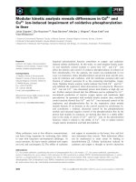
Both wild-type and G93ASOD1 increase GSH
in the conditional FALS1 model
Total GSH, GSH and GSSG were determined in the
tTA-40, highWT-tTA and high ⁄ lowG93A-tTA cell
lines at their fourth passage. In cells cultured without
dox, this time point represents the first adaptive
response to the increase in wt ⁄ G93ASOD1 expression
caused by the removal of dox, whereas in cells
cultured with dox, with their very low residual SOD1
expression, it represents the adaptation to constant,
very low levels of wt ⁄ G93ASOD1. All the SOD1-
transfected cell lines (dox)) had significantly higher
total GSH than seen in tTA-40 cells and the profile
of GSH content mirrored that of total GSH
(Fig. 2A,B). A robust threefold increase was seen in
highG93A-tTA cells. Comparable total GSH increases
were also observed in dox+ cells (Fig. 2A), suggest-
ing that this initial change takes place even with a
very small extra amount of wild-type or mutant
SOD1, and also that a low SOD1 expression level is
somehow more effective. GSSG was also significantly
increased by wild-type and G93ASOD1 overexpres-
sion, but it remained a very small percentage of GSH
( 1%) (Fig. 2C).
GSH : GSSG ratios, E
hGSH
⁄
GSSG
values and
glutathione reductase, GST activities in the
conditional FALS1 model
Because the redox equilibrium of cells affects several
aspects of cell homeostasis, the GSH : GSSG ratios
and E
hGSH ⁄ GSSG
for cells cultured without dox were
obtained (Fig. 2D,E). There was a sharp contrast in
the effect of wild-type and mutant SOD1, with a
significant increase in the GSH : GSSG ratio in
highG93A-tTAcells. This was accompanied by a shift
to a more negative value in E
hGSH ⁄ GSSG
(Fig. 2E),
reinforcing the evidence of a more reduced thiol oxida-
tion state in these cells. This did not occur in highWT-
tTA cells despite the fact that both highWT- and
highG93A-tTA cells had to adapt the GSH pool to
overexpression of a comparably high level of human
SOD1.
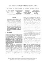
We next determined the specific activity of glutathi-
one reductase (GR), essential for maintenance of the
GSH : GSSG ratio. GR was no different in highWT-
tTA and tTA-40 cells, but it was lower in highG93A-
tTA cells than in the other cell lines (Fig. 3A). Thus
increased GSSG recycling cannot explain the relative
abundance of GSH over GSSG in highG93A-tTA
cells. We also measured the activity of GST (Fig. 3B),
a large group of proteins that use GSH to detoxify
harmful products of oxidative stress. GST activity was
unchanged in highWT-tTA cells, although it was lower
in highG93A-tTA than in all other cell lines. This
might cause lower GSH consumption in highG93A-
tTA cells, therefore contributing to maintaining the
high GSH levels.
In the lowG93A-tTA cell line (dox)), the
GSH : GSSG ratio and E
hGSH ⁄ GSSG
did not differ
from control tTA-40 or highWT-tTA cells (Fig. 2D,E).
A
B
C
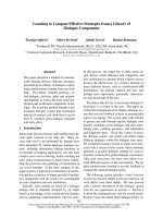
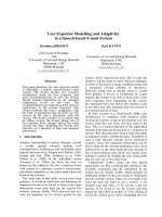
Fig. 1. Expression of wild-type or G93ASOD1 in the conditional
FALS1 model. (A) Culture system and sample collection times for
the conditional cell lines. Western blotting shows that removal of
dox (between passages 2 and 3, as described in Materials and
methods) fully induced expression of the human SOD1 (hSOD1)
after 96 h (highWT-tTA cell line). (B) Expression of human wild-type
or G93ASOD1 (hSOD1) evaluated by western blot of highWT-tTA,
highG93A-tTA and lowG93A-tTA cell lines cultured with (+)
(1 lgÆmL
)1
) or without ()) dox at the fourth passage. The control
tTA-40 cell line contained only murine SOD1 (mSOD1). (C) The
level of wt ⁄ G93ASOD1 was constant at different passage numbers
(4 and 14) in the dox) culture. Representative western blots of
total cell lysates exposed together are shown.
S. Tartari et al. Glutathione in adaptation to wt ⁄ G93ASOD1
FEBS Journal 276 (2009) 2861–2874 ª 2009 The Authors Journal compilation ª 2009 FEBS 2863
We found an increase in GR (34%, P < 0.01) and
GST (14%) activities in comparison with tTA-40 cells
(Fig. 3A,B) suggesting that when cells initially adapted
themselves to overexpression of a small amount of
mutant protein, they maintained the redox equilibrium
changing several enzymatic activities.
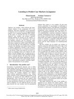
Wild-type and G93ASOD1 affect the levels
of GCLC and GCLM proteins differently
The increase in GSH in SOD1-transfected cell lines
might result from increased synthesis. This may be
because of an upregulation of the expression of GCL.
We used western blotting to analyse the expression of
the GCL subunits GCLC and GCLM in the dox)
cultured cell lines at their fourth passage (Fig. 4). In
highWT-tTA cells, GCLC remained constant, whereas
GCLM showed a 34% increase over the tTA-40
value, although this change did not reach statistical
significance. In lowG93A-tTA cells, both GCLM and
GCLC increased significantly (95% and 90%),
whereas in highG93A-tTA cells there were no signifi-
cant changes, but only a small increase (15%) in
GCLC. Thus, the mutant form of SOD1, more than
the wild-type, modified the expression of the GCL
subunits. In addition, on comparing low- and high-
G93ASOD1 cells, it was evident that the induction of
GCL subunits was inversely related to the expression
of G93ASOD1.
In lowG93A-tTA cells (dox)), the involvement of
GCL in the increase in GSH was further confirmed by
measuring GCL activity, which was 16.44 ± 0.31 nmolÆ
min
)1
Æmg
)1
of protein, i.e. 20% higher (P < 0.01
by Student’s t-test) than that of tTA-40 cells
(13.96 ± 0.32 nmolÆmin
)1
Æmg
)1
of protein; mean ±
SEM of four independent samples from two experi-
ments).
We then treated the tTA-40 and lowG93A-tTA cell
lines, both dox), with the GCL inhibitor buthionine
sulfoximine (250 lm). After 24 h, total GSH was
2% of baseline (i.e. for tTA-40 and lowG93A-tTA
cells, 3.35 ± 0.26 and 4.90 ± 0.30 ngÆlg
)1
protein;
mean ± SE of six independent samples from two
experiments, P < 0.01 by Student’s t-test) indicating
that, in both cell lines, GCL activity was responsible
for the GSH level.
A
B
D
E
C
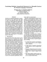
Fig. 2. GSH levels, GSH : GSSG ratio and
E
hGSH ⁄ GSSG
values in the conditional FALS1
model. Levels of (A) total GSH, (B) GSH and
(C) GSSG, (D) the GSH : GSSG ratio and (E)
E
hGSH ⁄ GSSG
were measured in the condi-
tional cell lines, cultured with (+) (1 lgÆmL
)1
)
or without ()) dox, at the fourth passage.
Values are given as mean ± SEM of four
independent experiments. DP < 0.05,
DDP < 0.01, DDDP < 0.001 versus tTA-40
(dox)). sP < 0.05, sssP < 0.001 versus
tTA-40 (dox +).
P < 0.05, P < 0.01
versus highWT-tTA (dox)). hP < 0.05,
hhP < 0.01 versus lowG93A-tTA (dox)).
P < 0.01 versus lowG93A-tTA (dox +).
(One-way ANOVA with Newman–Keuls
multiple comparison post-test).
Glutathione in adaptation to wt ⁄ G93ASOD1 S. Tartari et al.
2864 FEBS Journal 276 (2009) 2861–2874 ª 2009 The Authors Journal compilation ª 2009 FEBS
Effect of wild-type or G93ASOD1 on the GSH and
protein level of GCL subunits in the constitutive
FALS1 model
To confirm that the increase in GSH and expression
of GCL protein subunits did not derive from some
peculiarity of the conditional system, we analysed a
constitutive FALS1 model, an even simpler in vitro
system in which motor neuronal cells were never
exposed to dox, did not require hygromycin B during
culture and did not express EGFP. The expression lev-
els of wild-type and G93ASOD1 in the WT-NSC and
G93A-NSC cell lines resembled those of the
WT ⁄ G93A-tTA cell lines cultured with dox (Fig. 5A),
i.e. much lower than in the WT⁄ G93A-tTA cell lines
in dox) culture (Fig. 1B).
Total GSH was higher in both WT-NSC (57%) and
G93A-NSC (66%) than in the control NSC-34 cells at
their fourth passage (Fig. 5B). These increases were
accompanied by significant increases in GCLC and
GCLM (37% and 52%) in G93A-NSC cells only
(Fig. 6A,B). Therefore, the constitutive and the condi-
tional models responded identically, reflecting the
amount and form of transfected SOD1, either wild-
type or mutant.
Fig. 3. GR and GST activity in the conditional FALS1 model. (A) GR
and (B) GST activity were evaluated in the conditional cell lines
cultured without ()) dox at the fourth passage. Values are given
as mean ± SEM of three independent experiments. DP < 0.05,
DDP < 0.01 versus tTA-40.
P < 0.05, P < 0.01, P < 0.001
versus highWT-tTA. hh P < 0.01, hhhP < 0.001 versus lowG93A-
tTA. (One-way ANOVA with Newman–Keuls multiple comparison
post-test).
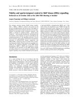
A
B
C
(1) tTA-40
(2) HighWT-tTA
(3) HighG93A-tTA
(4) LowG93A-tTA
Fig. 4. Expression of GCLC and GCLM in the conditional FALS1
model. (A) GCLC and GCLM expression of the conditional cell lines
cultured without ()) dox at the fourth passage. A representative
western blot is shown for each protein. (B, C) GCLC and GLCM
levels normalized for actin. Values are given as mean ± SEM of
three independent experiments. DDP < 0.01 versus tTA-40.
P < 0.05, P < 0.01 versus highWT-tTA. hhP < 0.01 versus
lowG93A-tTA (one-way ANOVA with Newman–Keuls multiple
comparison post-test).
S. Tartari et al. Glutathione in adaptation to wt ⁄ G93ASOD1
FEBS Journal 276 (2009) 2861–2874 ª 2009 The Authors Journal compilation ª 2009 FEBS 2865
Time of exposure to wild-type or G93ASOD1
influences the GSH pool, GCL subunit protein
levels and GCL activity in the conditional FALS1
model
Because FALS1 patients have long-term exposure to
G93ASOD1, the effect of constant expression of wild-
type and G93ASOD1 on GSH synthesis was deter-
mined at the 14th passage of the conditional cell lines
in dox) culture (Fig. 7A). Total GSH in highWT-
tTA cells did not differ from that in tTA-40 cells.
However, it was significantly lower in highG93A-tTA
cells compared with all other cell lines ( 30% com-
pared with tTA-40 cells). Only lowG93A-tTA cells
maintained a significant increase in the GSH pool
(30% over the tTA-40 and highWT-tTA and 60%
over the highG93A-tTA cells). Thus, the adaptive
process of motor neuronal cells to wt ⁄ G93ASOD1
appeared to be at least biphasic, with an initial
marked increase in GSH common to all the cell lines,
whereas, with longer exposure, the type of SOD1
(either wild-type or G93A) and the G93ASOD1 level
made the difference.
The effects of SOD1 modulation on GSH level –
typical of each wild-type or G93A-tTA cell line – were
reproducible in cultures from different frozen aliquots
of the same clone, irrespective of the fact that over the
course of the study GSH values varied slightly in the
different experiments, likely reflecting subtle differences
in growth and confluency of the cell cultures [22].
Levels of GCLM protein expression changed only in
cells expressing the mutant protein. Thus, GCLM
expression in the highG93A-tTA cell line was signifi-
cantly lower than in the tTA-40, highWT-tTA and
lowG93A-tTA cell lines, but was higher in lowG93A-
tTA cells (20% more than tTA-40 and highWT-tTA
cells), although this increase did not reach significance
(Fig. 7B,C).
The activity of GCL was also measured at the same
time point (Fig. 7D). In the lowG93A-tTA cell line,
GCL activity was higher than in the other lines. The
GCL activity in the highWT-tTA cells did not differ
from the highG93A-tTA cells even though the two
lines had significantly different total GSH (Fig. 7A).
Effect of tert-butylhydroquinone, an inducer of
GSH and GCL activity, in the conditional FALS1
model
Total GSH in the highWT-tTA, highG93A-tTA and
lowG93A-tTA cells (dox) cultured) was analysed 24 h
after treatment with tert-butylhydroquinone (t-BHQ).
All cells were at the 14th passage, the time point con-
sidered more representative of the response of cells
chronically exposed to wild-type or G93ASOD1. In all
the cell lines, t-BHQ significantly increased total GSH,
but the level in highG93A-tTA cells was significantly
lower than in highWT-tTA cells under basal conditions
and after t-BHQ treatment (Fig. 8A), indicating that
highG93A-tTA cells had a lower antioxidant capacity
than those expressing a comparable level of wtSOD1.
In lowG93A-tTA cells, total GSH after t-BHQ treat-
ment was significantly higher than in the highG93A-
tTA line and not significantly different from that of
highWT-tTA cells (Fig. 8A).
We determined the activity of GCL under the same
experimental conditions. t-BHQ significantly increased
GCL activity only in highWT-tTA cells (Fig. 8B).
Discussion
In the context of evidence of oxidative damage to
motor neurons typical of SALS and FALS [1], this
study focused on the effects of wild-type and
G93ASOD1 on GSH and GCL in an in vitro model
for FALS1. This is an important data because a
A
B
Fig. 5. Expression of wild-type or G93ASOD1 and GSH levels in
the constitutive FALS1 model. (A) Expression of human wild-type
or G93ASOD1 (hSOD1) in WT-NSC and G93A-NSC compared with
the conditional cell lines cultured with (+) dox, determined by wes-
tern blot. Thirty micrograms of protein (rather than 20 lgasin
Fig. 1B for the conditional lines) were loaded for each cell line. (B)
Total GSH levels of the NSC-34 and WT-NSC or G93A-NSC cell
lines at the fourth passage. Values are given as mean ± SEM of
four independent experiments. DDDP < 0.001 versus NSC-34 (one-
way ANOVA with Newman–Keuls multiple comparison post-test).
Glutathione in adaptation to wt ⁄ G93ASOD1 S. Tartari et al.
2866 FEBS Journal 276 (2009) 2861–2874 ª 2009 The Authors Journal compilation ª 2009 FEBS
primary decrease in GCL activity causing GSH to
decrease might be sufficient to cause spontaneous neu-
ronal death [23].
In motor neuronal cells expressing a low mutant
SOD1 content, the response led to increased GSH and
GCL activity. By contrast, with high levels of mutant
protein, a condition of subtle chronic GSH depletion
was established in comparison with controls or
wtSOD1 cells. These results highlighted the role of the
level of mutant protein in the response of the GSH
pathway. In agreement with this, in transgenic
G93ASOD1 mice, expressing very high levels of
mutant protein, a decrease in mRNA levels of both
GCL subunits in the spinal cord was reported as early
as at the embryonic stage [24]. In this mouse model,
the decrease in GSH in the spinal cord might account,
at least in part, for the toxicity of the mutant forms of
SOD1 [17]. However, transgenic mice, the unique
in vivo model available to test the effect of potential
therapies, differ from FALS1 patients in terms of the
expression level of mutant SOD1 because this is much
higher than in patients. Taking into account the results
of our in vitro model, it is tempting to suggest that the
different effects on the GSH pool and ⁄ or synthesis
accompanying different G93ASOD1 levels might
underlie some of the differences existing between the
mouse models and patients, for example, in response
to some of the therapies that have been tested [25,26].
This might apply in particular to therapies with anti-
oxidants, which may behave differently in the context
of altered redox regulation or oxidative stress [27].
Different antioxidants are available which may also
act as GSH precursors or not. Our preliminary data
suggest that the level of total GSH after acute treat-
ment with N-acetylcysteine is modulated by the level
and type of SOD1, either wild-type or G93A, whereas
it is not influenced by vitamin E (S. Tartari and
L. Cantoni, unpublished results).
Our model appears to also provide a tool to inves-
tigate the effects of chronic exposure to a small
amount of G93ASOD1, as seen in the motor neurons
of FALS1 patients. To explain the different amounts
of GSH in cells with varying levels of G93ASOD1, we
provide evidence of an effect on the expression level of
the GCL subunits GCLM and GCLC.
These two subunits contribute differently to the for-
mation of c-glutamylcysteine, the precursor of GSH.
GCLC possesses the catalytic capacity for c-glutam-
ylcysteine synthesis [28] and its upregulation supports
high levels of GSH [23,29].
In our FALS1 models, GCLC increased in the
G93A-NSC and lowG93A-tTA cells at the first time
point. This might represent the initial response of
cells expressing a low level of G93ASOD1, which is
possibly more complex because cell homeostasis is less
compromised, as suggested by the induction of GR
A
B
Fig. 6. Expression of GCLC and GCLM in
the constitutive FALS1 model. (A) GCLC and
(B) GCLM expression of the NSC-34,
WT-NSC, G93A-NSC cell lines at their fourth
passage. A representative western blot is
shown for each protein. The histograms
show GCLC and GCLM levels normalized
for actin. Values are given as mean ± SEM
of four independent experiments.
DP < 0.05, DDP < 0.01 versus NSC-34.
P < 0.05, P < 0.01 versus WT-NSC
(one-way ANOVA with Newman–Keuls
multiple comparison post-test).
S. Tartari et al. Glutathione in adaptation to wt ⁄ G93ASOD1
FEBS Journal 276 (2009) 2861–2874 ª 2009 The Authors Journal compilation ª 2009 FEBS 2867
and the lack of a decrease in GST. However, the effect
of longer exposure of cells to even a low level of
mutant protein, studied in the conditional model, was
to cancel induction of GCLC, eliminating a factor
contributing to the increase in GSH level and GCL
activity.
GCLM greatly improves the catalytic efficiency of
the holoenzyme GCL [30]. The amount of GCLM is
usually lower than the amount of GCLC and limits
GSH synthesis [31,32]. Accordingly, there are experi-
mental models showing that overexpression of GCLM
increased GSH [29,33], whereas knocking down
GCLM lowered it [23,31].
In our model, G93ASOD1 overexpression appeared
to affect GCLM more than GCLC. The effects of
these modifications are in agreement with reports from
the literature on the role of GCLM because the
increase in GCLM seemed a convenient way for the
G93ASOD1 cells to increase their GSH, whereas
the decrease in this subunit – as in highG93A-tTA cells
with prolonged exposure to the mutant protein – was
concomitant with a decrease in GSH.
This sequential inducing ⁄ inhibitory effect of
G93ASOD1 on the levels of GCLM and GSH might
markedly influence the toxicity of mutant SOD1. In
another cell model for FALS1, the high GSH level
afforded protection against S-nitroso-glutathione
toxicity and this was abolished by blocking GSH
synthesis [34]. Although GCLM is not essential for
viability [31], in contrast to GCLC [35], the lack or
disruption of GCLM alone was sufficient to increase
cell susceptibility to oxidative stress and nitric oxide
[23,31,36], whereas its overexpression rendered cells
resistant to oxidative stress [33]. Neurons are especially
vulnerable to nitric oxide-mediated mitochondrial
damage and neurotoxicity [37,38], and in ALS there is
ample evidence that nitric oxide is involved in motor
neuron degeneration [39,40]. The increase in GSH
also appears essential for adaptation to ER stress
[41], which was associated with G93ASOD1 toxicity
[42].
A major function attributed to GCLM is to improve
the GSH synthesis capacity of the cells [31,32] and this
correlates with resistance ⁄ recovery from an oxidative
A
B
D
C
Fig. 7. Effect of time on GSH, GCL activity, GCLC and GCLM expression in the conditional FALS1 model. (A) Total GSH levels of tTA-40,
highWT-tTA, highG93A-tTA and lowG93A-tTA cells at the 14th passage. The total GSH level of the tTA-40 cell line (6.73 ± 0.291 lgÆmg
)1
of
protein) was taken as 100%. Values are given as mean ± SEM of five independent experiments. (B) GCL activity was measured as in (A).
The value of the tTA-40 cell line (12.13 ± 0.218 nmolÆmin
)1
Æmg
)1
protein) was taken as 100%. Histograms present the mean ± SEM of six
independent experiments. (C) GCLC and (D) GCLM expression of the conditional cell lines at the 14th passage. A representative western
blot is shown for each protein. GCLC and GCLM levels were normalized for actin. The values of the tTA-40 cell line were taken as 100%.
Values are given as mean ± SEM of three independent experiments. nP < 0.05, nnP < 0.01 versus tTA-40;
P < 0.05, P < 0.001
versus highWT-tTA; hhP < 0.01, hhhP < 0.001 versus lowG93A-tTA (one-way ANOVA with Newman–Keuls multiple comparison post-test).
Glutathione in adaptation to wt ⁄ G93ASOD1 S. Tartari et al.
2868 FEBS Journal 276 (2009) 2861–2874 ª 2009 The Authors Journal compilation ª 2009 FEBS
insult even more than GSH level per se [43,44]. In our
study, GCL activity was not increased in G93ASOD1
cells after t-BHQ. In lowG93A-tTA cells, this effect
might be explained by the high basal GCL activity
[45], whereas in highG93A-tTA cells it suggests a fail-
ure of t-BHQ to induce GCL. The increase in GSH
after t-BHQ in this latter cell line may derive from a
combination of cytoprotective effects of this treatment
[36], however, the increase in highG93A-tTA cells was
not comparable with that in highWT-tTA cells. This
result reproduced the effect of t-BHQ on GSH in cells
lacking GCLM [36], further suggesting that the
decrease in GCLM in highG93A-tTA cells might play
a primary role in the differing toxicity of G93ASOD1
and wtSOD1.
As long as GCL activity and GCLM are elevated,
as in lowG93A-tTA cells, motor neuronal cells main-
tain some antioxidant capacity. For all these reasons,
defining the mechanism(s) governing the response of
GCLC and GCLM to G93ASOD1 might offer some
therapeutic possibilities.
In highWT-tTA cells, the increase in GSH at the
early time point may have represented the transient
adaptation of cells to the overexpression of wtSOD1
[46], a contributing factor perhaps being the expression
of a human protein in a murine cell line. Higher than
normal levels of wtSOD1 can alter ROS homeostasis
[47], a stimulus that can increase GSH [48]. At least at
the level of expression of wtSOD1 in our cells, this
increase was not accompanied by significant changes in
GCLC and GCLM or GCL activity, and may result
from a broad spectrum of changes including the acti-
vation of other enzymatic activities [49]. Factors that
stimulate cysteine uptake or attenuate GSH feedback
inhibition [9] would generally boost the intracellular
GSH concentration and might also have a role at the
late time point when the total GSH level was higher in
highWT-tTA cells than in highG93A-tTA cells. These
mechanisms need to be investigated further.
The increase in GSH was long-lasting in lowG93A-
tTA cells, coupled with higher GCL activity. In addi-
tion to the increased expression of GCL subunits, the
GCL activity can also be affected by phosphorylation
or nitrosation [9]. Inducers of GCL subunits are envi-
ronmental or endogenous compounds that cause oxi-
dative stress, but also other stresses [8,22,50,51].
Mutant forms of SOD1 are believed to have aberrant
oxidative activities [4]. We have previously reported an
increase in ROS formation under basal conditions in
the G93A-NSC cells over controls and WT-NSC cells
[19]. In this study, induction of GR activity in
lowG93A-tTA cells, and the shift to a higher
GSH ⁄ GSSG ratio in highG93A-tTA cells suggest
chronic oxidative stress in cells expressing the mutant
protein [6,7]. However, our experimental evidence
argues against a mechanism simply implying that
increased oxidation of GSH relative to the whole cell
is the signal triggering GSH induction, but rather sug-
gests more subtle roles for oxidant species potentially
formed in G93A-tTA cells.
The two GCL subunits, GR and GST, are part of
the family of the nuclear factor erythroid 2-related fac-
A
B
Fig. 8. Effect of t -BHQ on GSH and GCL activity in the conditional
FALS1 model. The highWT-tTA, highG93A-tTA and lowG93A-tTA
cell lines were compared for their response to t-BHQ (20 l
M). (A)
Total GSH and (B) GCL activity were determined 24 h after treat-
ment. No overt toxicity was observed. Cells grown in flasks for
6 days before treatment were at their 14th passage. Results are
shown as percentages of the untreated highWT-tTA cells
(5.68 lgÆmg
)1
protein for total GSH; 12.69 nmolÆmin
)1
Æmg
)1
protein
for GCL activity). Values are given as mean ± SEM of six indepen-
dent experiments. For both parameters, statistical significance of
differences was assessed by one-way ANOVA with Newman–
Keuls multiple comparison post test, comparing the basal levels of
the various cell lines (
P < 0.01, P < 0.001) or the effect
of t-BHQ in each cell line (**P < 0.01, ***P < 0.001) and in the dif-
ferent cell lines (dP < 0.05, ddP < 0.01, dddP < 0.001).
S. Tartari et al. Glutathione in adaptation to wt ⁄ G93ASOD1
FEBS Journal 276 (2009) 2861–2874 ª 2009 The Authors Journal compilation ª 2009 FEBS 2869
tor 2 (Nrf2)-regulated phase II detoxification enzymes
and their regulatory sequence is the anti-oxidant
response element (also known as electrophile-response
element) [52]. The lack of an increase in GCLC and
the decreases in GCLM, GST and GR in highG93A-
tTA cells are in agreement with the deficiency in Nrf2-
regulated genes in motor neurons from ALS patients
and in experimental models of FALS1 [24,53],
although the molecular mechanisms behind this finding
are yet to be defined. Our results indicated that the
enzymes were downregulated with different time
courses, suggesting a fine-tuning of their dependency
on Nrf2. Nrf2 is a redox-sensitive transcription factor
[52]. Induction of GST activity appears to be coupled
to a shift in E
hGSH ⁄ GSSG
towards a more oxidized
value [54], whereas in highG93A-tTA cells the opposite
tendency corresponded to a decrease in GST activity.
Studies are now underway in our laboratory to assess
the functional links between changes in the redox state
of GSH ⁄ GSSG and the expression of GST and GCL
subunits in G93ASOD1cells. In conclusion, this study
provides new information in the field of antioxidant
status in ALS, which might be useful in designing
effective therapies.
Materials and methods
Materials
The following materials and reagents were used: flasks
and plates (Corning Inc., Corning, NY, USA); opti-MEM
reduced serum medium, LipofectAMINE 2000, geneticin
(G418 sulfate) and hygromycin B (Invitrogen Life Technol-
ogies, Paisley, UK); high-glucose Dulbecco’s modified
Eagle’s medium (Cambrex, Verviers, Belgium); fetal bovine
serum (Hyclone, Logan, UT, USA); tet-screened fetal
bovine serum, pTK-Hyg and pBI-EGFP (Clontech, Palo
Alto, CA, USA). All other chemicals and enzymes were
purchased from Sigma-Aldrich (St Louis, MO, USA) and
Roche (Mannheim, Germany).
Constitutive FALS1 model
The NSC-34 cell line (a kind gift from N. R. Cashman,
University of British Columbia, Vancouver, Canada) was
used to obtain lines stably expressing human wtSOD1
(WT-NSC) or G93ASOD1 (G93A-NSC) [19].
NSC-34 cells were grown in high-glucose Dulbecco’s
modified Eagle’s medium supplemented with 5% heat-inac-
tivated fetal bovine serum, 1 mm glutamine, 1 mm pyruvate
and antibiotics (100 IUÆmL
)1
penicillin and 100 lgÆmL
)1
streptomycin). WT-NSC and G93A-NSC cell lines were
maintained in the presence of 0.5 mgÆmL
)1
G418. The cell
lines were subcultured in parallel every 7 days so they were
all at the same passage number for the experiments.
Conditional FALS1 model
From the NSC-34 cells we obtained the NSC-34 tTA-40
(tTA-40) cell line stably expressing the tetracycline-con-
trolled transactivator protein tTA and permitting tetracy-
cline-regulated gene expression [55]. In our tet-off system,
expression of the responsive protein is repressed by the
addition of the tetracycline analogue dox to the culture
medium. tTA-40 cells were stably co-transfected, following
the LipofectAMINE 2000 reagent protocol with pBI-EGFP
containing human wild-type or G93ASOD1 cDNA and
pTK-Hyg to obtain conditional clones (WT-tTA
and G93A-tTA) expressing hygromycin resistance and the
two forms of SOD1 [21,55]. Multiple WT-tTA or G93A-
tTA clones were isolated after 4 weeks’ selection with hy-
gromycin B (0.2 mgÆmL
)1
) and maintained in culture with
dox (2 lgÆmL
)1
). Cells of each clone were detached using
NaCl ⁄ P
i
–EDTA, pelletted by centrifugation, washed again
with NaCl ⁄ P
i
while in suspension and plated with or with-
out dox (dox+ ⁄ dox)) in the culture medium. After 48 h,
when the medium was changed, dox) cells were again
washed with NaCl ⁄ P
i
to remove dox released by cells and
allow rapid transgene expression [56]. All cells were col-
lected 96 h after plating and screened by western blot for
the level of the transfected SOD1 in dox+ ⁄ dox) culture
conditions. After this screening, only dox+ cultured cells
were stored in liquid nitrogen. The following cell lines were
used: tTA-40 (control), cells with a high level of wtSOD1
(highWT-tTA) and cells with a high or a low level of
G93ASOD1 (high and lowG93A-tTA respectively).
tTA-40 cells were cultured in the same way as NSC-34
cells except that tet-screened heat-inactivated fetal bovine
serum (5%) was used and G418 sulfate (0.5 mgÆmL
)1
) was
added. Hygromycin (0.2 mgÆmL
)1
) was added to the med-
ium for WT-tTA and G93A-tTA cells. In the dox+ culture
1 lgÆmL
)1
dox was added every 2 days while changing the
culture medium.
Samples for the determination of GSH, SOD1
and GCL subunit levels and GCL activity
Samples of the conditional cell lines were thawed (time 0)
and cultured with dox (Fig. 1A). At the end of the second
week of culture (second passage), each cell line was split
into two flasks, which were then cultured in parallel so that
they were all at the same passage number for the experi-
ments. One flask continued receiving dox (dox+), whereas
in the other dox was removed (dox)) using the procedure
described above, to allow full expression of the transfected
SOD1. In the dox) cells SOD1 was fully expressed from
96 h after the second passage (Fig. 1A). Cells were collected
Glutathione in adaptation to wt ⁄ G93ASOD1 S. Tartari et al.
2870 FEBS Journal 276 (2009) 2861–2874 ª 2009 The Authors Journal compilation ª 2009 FEBS
at the fourth passage, corresponding to 4 weeks’ culture,
for analysis relative to the first time point and at the 14th
passage. The growth curves of the conditional cell lines did
not significantly differ (data not shown).
NSC-34, WT-NSC and G93A-NSC cell lines were
thawed and cultured under standard conditions. Cells were
collected after 4 weeks’ culture (fourth passage). As previ-
ously reported, these cell lines did not differ in their prolif-
eration [19].
GSH measurements
Seven days before each selected time point, cells (plated at
6850 cellsÆcm
)2
in T25 flasks) were allowed to grow under
standard conditions (dox) ⁄ dox+ for the conditional cell
lines). Cells were collected and washed twice by centrifuga-
tion with Dulbecco’s NaCl ⁄ P
i
; the final pellet was resus-
pended with 5% sulfosalicylic acid (120 lL), incubated for
1 h on ice and centrifuged at 14 000 g for 10 min. The
supernatant was used to determine total GSH and GSSG
following the 5,5¢-dithiobis (2-nitrobenzoic acid) GR recy-
cling assay [57]. Total GSH was measured spectrophoto-
metrically at 30 °C as GSH equivalents (GSH + 2 GSSG).
Supernatant (25 lL) was added to an assay mixture consist-
ing of 0.7 mL NADPH (0.3 mm) dissolved in sodium phos-
phate (125 mm), pH 7.5, containing EDTA (6.3 mm),
0.1 mL 5,5¢-dithiobis (2-nitrobenzoic acid) (6 mm) dissolved
in sodium phosphate ⁄ EDTA and water to 1 mL. After
2 min preincubation, 0.6 U GR was added to the 1-mL
assay mixture and the change in absorbance at 412 nm was
measured over 3 min. Standard curves were generated using
GSH solutions in 5% sulfosalicylic acid.
To measure GSSG, 70 lL of supernatant was mixed with
4.2 lL of triethanolamine and 1.4 lL of 2-vinylpyridine,
which reacts with GSH masking it to GR, and the reaction
mixture was incubated at room temperature for 60 min.
Samples were then assayed as described above for total GSH,
but with double the amount of GR. Standard curves were
generated with GSSG solutions and the addition of 2-vinyl-
pyridine to the assay mixture. GSSG concentrations in the
cell extracts were in the middle of the range of the standard
curve (0–0.20 pmol). The amount of GSH was calculated by
subtracting twice the amount of GSSG from the total.
The protein pellet was resuspended in 1 m NaOH and
used to determine the protein content of the sample with a
bicinchoninic acid assay kit (Pierce, Rockford, IL, USA) to
normalize values for total GSH, GSH and GSSG.
GSH : GSSG ratio and E
hGSH
⁄
GSSG
in the
conditional cell lines
Two different parameters were used to indicate the redox
state of the cell [7,54]. The first was the ratio of GSH to
GSSG, which takes into account especially mechanisms
of S-thiylation for protein control. The second was the
reduction potential of the GSH ⁄ GSSG couple (E
hGSH ⁄ GSSG
),
which takes into account mechanisms of oxidation reduction
of dithiol motifs for protein control, calculated using the
Nernst equation as described by Jones [58] and Halvey et al.
[59]. Redox potentials are presented as millivolts (mV).
SDS
⁄
PAGE and western blot
To analyse SOD1 expression, cells grown in T25 flasks were
collected and washed with Dulbecco’s NaCl ⁄ P
i
. The cell
pellet was lysed for 10 min at 4 °Cin50mm Tris ⁄ HCl
(pH 8.0) containing 150 mm NaCl, 1% SDS and a protease
inhibitor cocktail (Sigma-Aldrich). The sample was then
boiled at 95 °C for 5 min and a whole-cell lysate was
obtained. The procedure described by Diaz-Hernandez
et al. [23] was used to analyse GCLC and GCLM expres-
sion. Cells were lysed for 20 min at 4 °C in Tris ⁄ HCl
(20 mm, pH 8.0), containing 1% Nonidet-P40, 5 mm
EDTA, 2 mm EGTA, 137 mm NaCl, 10% glycerol, 1 mm
Na
3
VO
4
,50mm NaF and a protease inhibitor cocktail
(Sigma-Aldrich). Extracts were centrifuged at 13 000 g for
20 min at 4 °C and aliquots of the supernatant were used.
Proteins (10–30 lg) were separated by electrophoresis on
12% or 10% polyacrylamide gels, respectively for SOD1 or
GCLC and GCLM determination. Nitrocellulose mem-
branes were probed with the following primary antibodies:
humanSOD1 (sheep polyclonal; Calbiochem, EMD Bio-
sciences, Inc. La Jolla, CA, USA), actin (mouse monoclonal;
Chemicon International Inc., Temecula, CA, USA) [19,55],
GCLC (1 : 1600; rabbit polyclonal, Lab Vision Corporation,
Fremont, CA, USA) or GCLM (1 : 10 000; rabbit poly-
clonal, a kind gift from T. J. Kavanagh, University of
Washington, Seattle, WA, USA). GCLC and GCLM antibo-
dies were used coupled to a secondary antibody to rabbit
raised in goat (1 : 2000). Protein bands were detected with
the ECL detection system (Amersham Biosciences, Little
Chalfont, UK). Films were scanned and band intensities
obtained with an AIS Image Analyser (Imaging Research
Inc., St Catharine’s, Canada).
Enzymatic activities
Cells (plated in T25 flasks, 6850 cellsÆcm
)2
) were allowed to
grow for 7 days under standard conditions. Cells were then
collected and washed twice by centrifugation with Dul-
becco’s NaCl ⁄ P
i
; the final pellet from each flask was resus-
pended in 0.55 mL of buffer (50 mm potassium phosphate,
pH 7.5, with 1 mm EDTA), sonicated and centrifuged at
12 000 g for 30 min. The supernatants were used to mea-
sure the enzymatic activities after determining the protein
content with the bicinchoninic acid assay.
GCL activity was determined as described by Zhou &
Freed [60]. The reaction mixture (final volume 200 lL) con-
tained 100 mm Tris ⁄ HCl (pH 8.2), 20 mm MgCl
2
, 150 mm
KCl, 10 mml-glutamate, 10 mml-cysteine, 5 mm ATP,
S. Tartari et al. Glutathione in adaptation to wt ⁄ G93ASOD1
FEBS Journal 276 (2009) 2861–2874 ª 2009 The Authors Journal compilation ª 2009 FEBS 2871
2mm EDTA, 0.2 mm NADH, 2 mm phosphoenolpyruvate,
pyruvate kinase (2 U) and lactate dehydrogenase (2 U).
The reaction was started by adding 100 lg protein and the
decrease in absorbance at 340 nm in a 96-well plate was
followed for 5 min at 25 °C. Specific activity was expressed
in UÆmg
)1
protein and then as a percentage of control.
GST activity was measured as described by Habig et al.
[61]. The reaction mixture (final volume 300 lL) contained
100 mm potassium phosphate (pH 6.5), 1 mm EDTA,
1mm 1-chloro-2,4-dinitrobenzene and 2 mm GSH. The
reaction was started by adding 30 lg of protein. The
increase in absorbance at 340 nm in a 96-well plate was
followed for 5 min at 25 °C after 5 min preincubation.
GR activity was measured as described by Allen et al.
[13] except that the concentration of GSSG was 1 mm. The
other components of the reaction mixture (final volume
300 lL) were: 50 mm Hepes ⁄ KOH (pH 8.0), 0.1 mm EDTA
and 30 lg of protein. The reaction was started by adding
NADPH (0.1 mm). The decrease in absorbance at 340 nm
was followed for 5 min at 25 °C in a 96-well plate after
5 min preincubation.
Treatment with tert-butylhydroquinone (t-BHQ)
Cells (6850 cellsÆcm
)2
) were grown under standard dox)
conditions in T25 flasks for 6 days and then treated with
t-BHQ (20 lm final concentration) for 24 h.
Statistical analysis
One-way analysis of variance (ANOVA), followed by
Newman–Keuls multiple comparison post-test was used for
statistical analysis.
Acknowledgements
We are grateful to Dr N. Cashman for providing the
original NSC-34 cell line and Prof. T. J. Kavanagh
for the antibody against GCLM. We thank
Dr M. P. Coleman for critically reading the manu-
script. Financial support was provided by MIUR,
FIRB, Protocol RBIN04J58W_000.
References
1 Barber SC, Mead RJ & Shaw PJ (2006) Oxidative stress
in ALS: a mechanism of neurodegeneration and a ther-
apeutic target. Biochim Biophys Acta 1762, 1051–1067.
2 Valentine JS, Doucette PA & Zittin Potter S (2005)
Copper–zinc superoxide dismutase and amyotrophic
lateral sclerosis. Annu Rev Biochem 74, 563–593.
3 Boille
´
e S, Vande Velde C & Cleveland DW (2006) ALS:
a disease of motor neurons and their nonneuronal
neighbors. Neuron 52, 39–59.
4 Liochev SI & Fridovich I (2003) Mutant Cu,Zn super-
oxide dismutases and familial amyotrophic lateral scle-
rosis: evaluation of oxidative hypotheses. Free Radic
Biol Med 34 , 1383–1389.
5 Bains JS & Shaw CA (1997) Neurodegenerative disorders
in humans: the role of glutathione in oxidative stress-
mediated neuronal death. Brain Res Rev 25, 335–358.
6 Aguirre P, Valde
´
s P, Aracena-Parks P, Tapia V &
Nu` nez MT (2007) Upregulation of {gamma}-gluta-
mate–cysteine ligase as part of the long-term adaptation
process to iron accumulation in neuronal SH-SY5Y
cells. Am J Physiol Cell Physiol 292, 2197–2203.
7 Schafer FQ & Buettner GR (2001) Redox environment
of the cell as viewed through the redox state of the glu-
tathione disulfide ⁄ glutathione couple. Free Radic Biol
Med 30, 1191–1212.
8 Krzywanski DM, Dickinson DA, Iles KE, Wigley AF,
Franklin CC, Liu R, Kavanagh TJ & Forman HJ
(2004) Variable regulation of glutamate cysteine ligase
subunit proteins affects glutathione biosynthesis in
response to oxidative stress. Arch Biochem Biophys 423,
116–125.
9 Griffith OW (1999) Biologic and pharmacologic regula-
tion of mammalian glutathione synthesis. Free Radic
Biol Med 27 , 922–935.
10 Tsuchiya K, Mulcahy RT, Reid LL, Disteche CM &
Kavanagh TJ (1995) Mapping of the glutamate–cysteine
ligase catalytic subunit gene (GLCLC) to human chro-
mosome 6p12 and mouse chromosome 9D-E and of the
regulatory subunit gene (GLCLR) to human chromo-
some 1p21-p22 and mouse chromosome 3H1-3. Genom-
ics 30, 630–632.
11 Soltaninassab SR, Sekhar JR, Meredith MJ & Freeman
ML (2000) Multi-faceted regulation of c-glutamylcyste-
ine synthetase. J Cell Physiol 182, 163–170.
12 Kuzma M, Jamrozik Z & Baranczyk-Kuzma A (2006)
Activity and expression of glutathione S-transferase pi
in patients with amyotrophic lateral sclerosis. Clin Chim
Acta 364, 217–221.
13 Allen S, Heath PR, Kirby J, Wharton SB, Cookson
MR, Menzies FM, Banks RE & Shaw PJ (2003) Analy-
sis of the cytosolic proteome in a cell culture model of
familial amyotrophic lateral sclerosis reveals alterations
to the proteasome, antioxidant defenses, and nitric
oxide synthetic pathways. J Biol Chem 278, 6371–6383.
14 Lanius RA, Krieger C, Wagey R & Shaw CA (1993)
Increased [
35
S] glutathione binding sites in spinal cords
from patients with sporadic amyotrophic lateral sclero-
sis. Neurosci Lett 163, 89–92.
15 Tohgi H, Abe T, Yamazaki K, Murata T, Ishizaki E &
Isobe C (1999) Increase in oxidized NO products and
reduction in oxidized glutathione in cerebrospinal fluid
from patients with sporadic form of amyotrophic lateral
sclerosis. Neurosci Lett 260, 204–206.
Glutathione in adaptation to wt ⁄ G93ASOD1 S. Tartari et al.
2872 FEBS Journal 276 (2009) 2861–2874 ª 2009 The Authors Journal compilation ª 2009 FEBS
16 Julien JP & Kriz J (2006) Transgenic mouse models of
amyotrophic lateral sclerosis. Biochim Biophys Acta
1762, 1013–1024.
17 Chi L, Ke Y, Luo C, Gozal D & Liu R (2007) Depletion
of reduced glutathione enhances motor neuron degener-
ation in vitro and in vivo. Neuroscience 144, 991–1003.
18 Dal Canto MC & Gurney ME (1995) Neuropathological
changes in two lines of mice carrying a transgene for
mutant human Cu,Zn SOD, and in mice overexpressing
wild type human SOD: a model of familial amyotrophic
lateral sclerosis (FALS). Brain Res 676, 25–40.
19 Rizzardini M, Mangolini A, Lupi M, Ubezio P, Bendotti
C & Cantoni L (2005) Low levels of ALS-linked Cu⁄ Zn
superoxide dismutase increase the production of reactive
oxygen species and cause mitochondrial damage and
death in motor neuron-like cells. J Neurol Sci 232, 95–103.
20 Rizzardini M, Lupi M, Mangolini A, Babetto E, Ubezio
P & Cantoni L (2006) Neurodegeneration induced by
complex I inhibition in a cellular model of familial
amyotrophic lateral sclerosis. Brain Res Bull 69, 465–474.
21 Raimondi A, Mangolini A, Rizzardini M, Tartari S,
Massari S, Bendotti C, Francolini M, Borgese N,
Cantoni L & Pietrini G (2006) Cell culture models to
investigate the selective vulnerability of motoneuronal
mitochondria to familial ALS-linked G93ASOD1. Eur J
Neurosci 24, 387–399.
22 Moellering D, McAndrew J, Patel RP, Cornwell T,
Lincoln T, Cao X, Messina JL, Forman HJ, Jo H &
Darley-Usmar VM (1998) Nitric oxide-dependent
induction of glutathione synthesis through increased
expression of c-glutamylcysteine synthetase. Arch
Biochem Biophys 358, 74–82.
23 Diaz-Hernandez JI, Almeida A, Delgado-Esteban M,
Fernandez E & Bolanos JP (2005) Knockdown of gluta-
mate–cysteine ligase by small hairpin RNA reveals that
both catalytic and modulatory subunits are essential for
the survival of primary neurons. J Biol Chem 280,
38992–39001.
24 Pehar M, Vargas MR, Robinson KM, Cassina P,
Diaz-Amarilla PJ, Hagen TM, Radi R, Barbeito L &
Beckman JS (2007) Mitochondrial superoxide
production and nuclear factor erythroid 2-related fac-
tor 2 activation in p75 neurotrophin receptor-induced
motor neuron apoptosis. J Neurosci 27, 7777–7785.
25 Nirmalananthan N & Greensmith L (2005) Amyotrophic
lateral sclerosis: recent advances and future therapies.
Curr Opin Neurol 18, 712–719.
26 Benatar M (2007) Lost in translation: treatment trials
in the SOD1 mouse and in human ALS. Neurobiol Dis
26, 1–13.
27 Ghezzi P & Di Simplicio P (2007) Glutathionylation path-
ways in drug response. Curr Opin Pharmacol 7, 398–403.
28 Shi ZZ, Osei-Frimpong J, Kala G, Kala SV, Barrios
RJ, Habib GM, Lukin DJ, Danney CM, Matzuk MM
& Lieberman MW (2000) Glutathione synthesis is
essential for mouse development but not for cell growth
in culture. Proc Natl Acad Sci USA 97, 5101–5106.
29 Mulcahy RT, Bailey HH & Gipp JJ (1995) Transfection
of complementary DNAs for the heavy and light sub-
units of human c-glutamylcysteine synthetase results in
an elevation of intracellular glutathione and resistance
to melphalan. Cancer Res 55, 4771–4775.
30 Yang Y, Chen Y, Johansson E, Schneider SN, Shertzer
HG, Nebert DW & Dalton TP (2007) Interaction
between the catalytic and modifier subunits of gluta-
mate–cysteine ligase. Biochem Pharmacol 74, 372–381.
31 Yang Y, Dieter MZ, Chen Y, Shertzer HG, Nebert
DW & Dalton TP (2002) Initial characterization of the
glutamate–cysteine ligase modifier subunit Gclm ()
⁄ ))
knockout mouse. Novel model system for a severely
compromised oxidative stress response. J Biol Chem
277, 49446–49452.
32 Chen Y, Shertzer HG, Schneider SN, Nebert DW &
Dalton TP (2005) Glutamate cysteine ligase catalysis.
Dependence on ATP and modifier subunit for regula-
tion of tissue glutathione levels. J Biol Chem 280,
33766–33774.
33 Tipnis SR, Blake DG, Shepherd AG & McLellan LI
(1999) Overexpression of the regulatory subunit of
gamma-glutamylcysteine synthetase in HeLa cells
increases gamma-glutamylcysteine synthetase activity
and confers drug resistence. Biochem J 337, 559–
566.
34 Ciriolo MR, Aquilano K, De Martino A, Carrı
`
MT &
Rotilio G (2001) Differential role of superoxide and glu-
tathione in S-nitrosoglutathione-mediated apoptosis: a
rationale for mild forms of familial amyotrophic lateral
sclerosis associated with less active Cu,Zn superoxide
dismutase mutants. J Neurochem 77, 1433–1443.
35 Dalton TP, Dieter MZ, Yang Y, Shetzer HG & Nebert
D (2000) Knockout of the mouse glutamate cysteine
ligase catalytic subunit (Gclc) gene: embryonic letal
when homozygous, and proposed model for moderate
glutathione deficiency when heterozygous. Biochem
Biophys Res Commun 279, 324–329.
36 Kann S, Estes C, Reichard JF, Huang M, Sartor MA,
Schwemberger S, Chen Y, Dalton TP, Shertzer HG,
Xia Y et al. (2005) Buthylhydroquinone protects cells
genetically deficient in glutathione biosynthesis from
arsenite-induced apoptosis without significantly chang-
ing their prooxidant status. Toxicol Sci 87, 365–384.
37 Almeida A & Bolanos JP (2001) A transient inhibition
of mitochondrial ATP synthesis by nitric oxide synthase
activation triggered apoptosis in primary cortical
neurons. J Neurochem 77, 676–690.
38 Almeida A, Almeida J, Bolanos JP & Moncada S
(2001) Different responses of astrocytes and neurons to
nitric oxide: the role of glycolytically generated ATP in
astrocyte protection. Proc Natl Acad Sci USA 98,
15294–15299.
S. Tartari et al. Glutathione in adaptation to wt ⁄ G93ASOD1
FEBS Journal 276 (2009) 2861–2874 ª 2009 The Authors Journal compilation ª 2009 FEBS 2873
39 Urishitani M & Shimohama S (2001) The role of nitric
oxide in amyotrophic lateral sclerosis. Amyotroph
Lateral Scler Other Motor Neuron Disord 2, 71–81.
40 Barbeito LH, Pehar M, Cassina P, Vargas MR, Peluffo
H, Viera L, Estevez AG & Beckman JS (2004) A role
for astrocytes in motor neuron loss in amyotrophic lat-
eral sclerosis. Brain Res Brain Res Rev 47, 263–274.
41 Chakravarthi S, Jessop CE & Bulleid NJ (2006) The
role of glutathione in disulphide bond formation and
endoplasmic-reticulum-generated oxidative stress.
EMBO Rep 7, 271–275.
42 Oh YK, Shin KS, Yuan J & Kang SJ (2008) Superoxide
dismutase mutants related to amyotrophic lateral sclero-
sis induce endoplasmic stress in neuro2a cells. J Neuro-
chem 104, 993–1005.
43 Woods JS, Kavanagh TJ, Corral J, Reese AW, Diaz D
& Ellis ME (1999) The role of glutathione in chronic
adaptation to oxidative stress: studies in a normal rat
kidney epithelial (NRK52E) cell model of sustained
upregulation of glutathione biosynthesis. Toxicol Appl
Pharmacol 160, 207–216.
44 Kenchappa RS & Ravindranath V (2003) c -Glutamyl
cysteine synthetase is up-regulated during recovery of
brain mitochondrial complex I following neurotoxic
insult in mice. Neurosci Lett 350, 51–55.
45 Dahl EL & Mulcahy T (2001) Cell-type specific
differences in glutamate cysteine ligase transcriptional
regulation demonstrate independent subunit control.
Toxicol Sci 61, 265–272.
46 Pias EK, Ekshyyan OY, Rhoads CA, Fuseler J, Harrison
L & Aw TY (2003) Differential effects of superoxide
dismutase isoform expression on hydroperoxide-induced
apoptosis in PC-12 cells. J Biol Chem 278, 13294–13301.
47 Liochev SI & Fridovich I (2007) The effects of super-
oxide dismutase on H
2
O
2
formation. Free Radic Biol
Med 42, 1465–1469.
48 Dickinson DA & Forman HJ (2002) Cellular glutathi-
one and thiols metabolism. Biochem Pharmacol 64,
1019–1026.
49 Amstad P, Moret R & Cerutti P (1994) Glutathione
peroxidase compensates for the hypersensitivity of
Cu,Zn-superoxide dismutase overproducers to oxidant
stress. J Biol Chem 269, 1606–1609.
50 Lee J, Kang J & Stipanuk MK (2006) Differential regu-
lation of glutamate–cysteine ligase subunit expression
and increased holoenzyme formation in response to cys-
teine deprivation. Biochem J 393, 181–190.
51 Dasgupta A, Das S & Sarkar PK (2007) Thyroid
hormone promotes glutathione synthesis in astrocytes
by upregulation of glutamate cysteine ligase through
differential stimulation of its catalytic and modulatory
subunit mRNAs. Free Radic Biol Med 42, 617–
626.
52 Nguyen T, Sherratt PJ & Pickett CB (2003) Regulatory
mechanisms controlling gene expression mediated by
the antioxidant response element. Annu Rev Pharmacol
Toxicol 43, 233–260.
53 Kirby J, Halligan E, Baptista MJ, Allen S, Heath PR,
Holden H, Barber SC, Loynes CA, Wood-Allum CA,
Lunec J et al. (2005) Mutant SOD1 alters the motor
neuronal transcriptome: implications for familial ALS.
Brain 128, 1686–1706.
54 Kirlin WG, Cai J, Thompson SA, Diaz D, Kavanagh
TJ & Jones DP (1999) Glutathione redox potential in
response to differentiation and enzyme inducers. Free
Radic Biol Med 27, 1208–1218.
55 Babetto E, Mangolini A, Rizzardini M, Lupi M,
Conforti L, Rusmini P, Poletti A & Cantoni L (2005)
Tetracycline-regulated gene expression in the NSC-34-
tTA cell line for investigation of motor neuron diseases.
Brain Res Mol Brain Res 140, 63–72.
56 Rennel E & Gerwins P (2002) How to make tetracy-
cline-regulated transgene expression go on and off. Anal
Biochem 309, 79–84.
57 Griffith OW (1980) Determination of glutathione and
glutathione disulfide using glutathione reductase and
2-vinylpyridine. Anal Biochem 106, 207–212.
58 Jones DP (2002) Redox potential of GSH ⁄ GSSG cou-
ple: assay and biological significance. Methods Enzymol
348, 93–112.
59 Halvey PJ, Watson WH, Hansen JM, Go Y, Samali A
& Jones DP (2005) Compartmental oxidation of
thiol-disulphide redox couples during epidermal growth
factors signalling. Biochem J 386, 215–219.
60 Zhou W & Freed CR (2005) DJ-1 up-regulates glutathi-
one synthesis during oxidative stress and inhibits A53T
a-synuclein toxicity. J Biol Chem 280, 43150–43158.
61 Habig WH, Pabst MJ & Jakoby WB (1974) Glutathi-
one S-transferases. The first enzymatic step in mercap-
turic acid formation. J Biol Chem 249, 7130–7139.
Supporting information
The following supplementary material is available:
Fig. S1. Effect of different times of exposure to doxy-
cycline (dox) on expression of enhanced green fluores-
cent protein (EGFP) and Cu,Zn superoxide dismutase
(SOD1).
Doc. S1. Additional method. Repression of Cu,Zn
superoxide dismutase (SOD1) and enhanced green fluo-
rescent protein (EGFP) expression by doxycycline
(dox).
This supplementary material can be found in the
online version of this article.
Please note: Wiley-Blackwell is not responsible for
the content or functionality of any supplementary
materials supplied by the authors. Any queries (other
than missing material) should be directed to the corre-
sponding author for the article.
Glutathione in adaptation to wt ⁄ G93ASOD1 S. Tartari et al.
2874 FEBS Journal 276 (2009) 2861–2874 ª 2009 The Authors Journal compilation ª 2009 FEBS