Báo cáo khoa học: Crystal structures of open and closed forms of cyclo⁄maltodextrin-binding protein pptx
Bạn đang xem bản rút gọn của tài liệu. Xem và tải ngay bản đầy đủ của tài liệu tại đây (1.3 MB, 12 trang )
Crystal structures of open and closed forms of
cyclo
⁄
maltodextrin-binding protein
Naoki Matsumoto
1
, Mitsugu Yamada
2,
*, Yuma Kurakata
1
, Hiromi Yoshida
2,3
, Shigehiro Kamitori
2,3
,
Atsushi Nishikawa
1
and Takashi Tonozuka
1
1 Department of Applied Biological Science, Tokyo University of Agriculture and Technology, Japan
2 Graduate School of Medicine, Kagawa University, Japan
3 Life Science Research Center, Kagawa University, Japan
Cyclodextrins (CDs) are torus-shaped molecules made
up of six [a-cyclodextrin (a-CD)], seven [b-cyclodextrin
(b-CD)] or eight [c-cyclodextrin (c-CD)] glucose resi-
dues. The structure of CDs resembles a hollow, trun-
cated cone with hydrophilic rims and a central
hydrophobic cavity capable of hosting a large number
Keywords
crystal structure; cyclodextrin; maltodextrin-
binding protein; sugar transporter;
Thermoactinomyces vulgaris
Correspondence
T. Tonozuka, Department of Applied
Biological Science, Tokyo University of
Agriculture and Technology, 3-5-8
Saiwai-cho, Fuchu, Tokyo 183-8509, Japan
Fax: +81 42 3675705
Tel: +81 42 3675702
E-mail:
*Present address
Research Unit for Quantum Beam Life
Science Initiative, Japan Atomic Energy
Agency, 1233 Watanuki, Takasaki, Gunma
370-1292, Japan
Database
The coordinates and structure factors of
TvuCMBP–a-CD, TvuCMBP–b-CD and
TvuCMBP–G4 have been deposited in the
Protein Data Bank under the accession
codes 2ZYM, 2ZYN, and 2ZYO, respectively.
The revised coordinate of TvuCMBP–c-CD
has been deposited in the Protein Data
Bank under the accession code 2ZYK
(Received 1 February 2009, revised 17
March 2009, accepted 23 March 2009)
doi:10.1111/j.1742-4658.2009.07020.x
The crystal structures of Thermoactinomyces vulgaris cyclo ⁄ maltodextrin-
binding protein (TvuCMBP) complexed with a-cyclodextrin (a-CD),
b-cyclodextrin (b-CD) and maltotetraose (G4) have been determined. A
common functional conformational change among all solute-binding pro-
teins involves switching from an open form to a closed form, which facili-
tates transporter binding. Escherichia coli maltodextrin-binding protein
(EcoMBP), which is structurally homologous to TvuCMBP, has been deter-
mined to adopt the open form when complexed with b-CD and the closed
form when bound to G4. Here, we show that, unlike EcoMBP, TvuCMBP–
a-CD and TvuCMBP–b-CD adopt the closed form when complexed,
whereas TvuCMBP–G4 adopts the open form. Only two glucose residues
are evident in the TvuCMBP–G4 structure, and these bind to the C-domain
of TvuCMBP in a manner similar to the way in which maltose binds to the
C-domain of EcoMBP. The superposition of TvuCMBP–a-CD,
TvuCMBP–b-CD and TvuCMBP–c-CD shows that the positions and the
orientations of three glucose residues in the cyclodextrin molecules overlay
remarkably well. In addition, most of the amino acid residues interacting
with these three glucose residues also participate in interactions with the
two glucose residues in TvuCMBP–G4, regardless of whether the protein is
in the closed or open form. Our results suggest that the mechanisms by
which TvuCMBP changes from the open to the closed conformation and
maintains the closed form appear to be different from those of EcoMBP,
despite the fact that the amino acid residues responsible for the initial bind-
ing of the ligands are well conserved between TvuCMBP and EcoMBP.
Abbreviations
CD, cyclodextrin; EcoALBP, Escherichia coli
D-allose-binding protein; EcoMBP, Escherichia coli maltodextrin-binding protein; G4,
maltotetraose; TvuCMBP, Thermoactinomyces vulgaris cyclo ⁄ maltodextrin-binding protein; a-CD, a-cyclodextrin; b -CD, b-cyclodextrin; c-CD,
c-cyclodextrin.
3008 FEBS Journal 276 (2009) 3008–3019 ª 2009 The Authors Journal compilation ª 2009 FEBS
of insoluble chemicals through hydrophobic interac-
tions [1,2]. We have studied the proteins involved in
the CD metabolism of a thermophilic actinomycete,
Thermoactinomyces vulgaris R-47 [3–9], and deter-
mined the crystal structures of two enzymes, TVA I
[4,5] and TVA II [6,7], both of which hydrolyze CDs
and a polysaccharide pullulan. We have also deter-
mined the crystal structure of a solute-binding protein
from T. vulgaris, and evaluated its binding affinities
for various sugars, using fluorescence measurements
[8,9]. The protein showed high affinities for both CDs
and maltodextrins, and thus was designated T. vulgaris
cyclo ⁄ maltodextrin-binding protein ( TvuCMBP).
Many bacterial solute-binding proteins bound to a
variety of sugars have been reported, and their crystal
structures have been compared [10–21]. Although the
proteins are categorized into three classes, they consist
of two similar globular domains linked by a two-
stranded or three-stranded hinge region. A common
drastic conformational change is found in this protein
family, which involves switching from an open to a
closed form through a large-scale hinge-bending
motion. TvuCMBP structurally resembles maltodex-
trin-binding proteins from Escherichia coli [18–20],
Thermococcus litoralis [22], Pyrococcus furiosus [23],
and Alicyclobacillus acidocaldarius [24], and E. coli
maltodextrin-binding protein (Eco MBP) is one of the
best characterized solute-binding proteins. EcoMBP
binds to a membrane-bound MalFGK
2
translocation
complex comprising two permease domains, MalF and
MalG, and two copies of the ATPase subunits, MalK
[25–29]. The maltodextrin transport is driven by the
energy provided from ATP hydrolysis by MalK, and
induced by the complementary binding of the malto-
dextrin-loaded EcoMBP to the external face of the
transmembrane subunits of MalF and MalG. Large
conformational changes of the EcoMBP—MalFGK
2
complex are induced, and maltodextrin permeates from
the binding site of EcoMBP to the cytoplasm through
the translocation pathway [25–29]. Unlike linear malto-
dextrins, CDs are nonphysiological ligands for E. coli,
and EcoMBP reportedly adopts an open form when
complexed with b-CD [19].
We have previously reported the crystal structure of
TvuCMBP complexed with c-CD at a moderate 2.5 A
˚
resolution [8]. Although the overall structure of
TvuCMBP resembles that of EcoMBP (the amino acid
sequence identity is 30.1%), TvuCMBP–c-CD has been
determined as the closed form. Here, we present the
crystal structures of TvuCMBP complexed with a-CD,
b-CD and maltotetraose (G4) at higher resolutions.
We also re-refined the coordinates of TvuCMBP–
c-CD, and compared them, to make clear the mecha-
nism of the ligand binding. It is interesting to note that,
unlike EcoMBP, TvuCMBP–a-CD and TvuCMBP–
b-CD were determined to be in the closed form,
whereas TvuCMBP–G4 adopted the open form.
Results and Discussion
Overall structures of TvuCMBP–ligand complexes
The crystal structures of TvuCMBP liganded with
a-CD, b-CD and G4 have been determined at 1.7, 1.8
and 1.55 A
˚
resolutions, respectively (Table 1). The crys-
tals of TvuCMBP in complex with a-CD and b-CD
belong to the monoclinic system with space group C2,
the unit cell parameters of which are similar to those of
selenomethionine-substituted TvuCMBP–c-CD (the cell
dimensions are a = 85.3 A
˚
, b = 49.3 A
˚
, c = 87.6 A
˚
,
and b = 94.9°) [8], and contain one molecule in an
asymmetric unit. On the other hand, the crystal of
TvuCMBP–G4 belongs to space group P2
1
2
1
2
1
, with
one molecule in an asymmetric unit. In Ramachandran
plots, 91.1% (TvuCMBP–a-CD), 91.1% (TvuCMBP–
b-CD) and 93.0% (TvuCMBP–G4) of residues were
shown to be in the most favored regions, and no residue
was in the generously allowed regions or disallowed
regions, as calculated by the program procheck of ccp4
[30]. The electron density (2F
o
) F
c
) maps for the three
complex structures contoured at 1r show continuous
density for almost all main chain atoms, but the N-termi-
nal segments (residues 1–16, a-CD complex; 1–14, b-CD
complex; 1–12, G4 complex) are not visible, as previ-
ously described in a report on TvuCMBP–c-CD [8].
The overall structures of the TvuCMBP–ligand com-
plexes are shown in Fig. 1A,B. Like many other bacte-
rial sugar-binding proteins, TvuCMBP consists of two
globular domains, an N-domain and a C-domain, linked
by three hinge regions. The structures of TvuCMBP–
a-CD and TvuCMBP–b-CD are almost identical to that
of TvuCMBP–c-CD. In contrast, TvuCMBP–G4 exhib-
its a markedly different conformation from the
TvuCMBP–CD complexes (Fig. 1C). Specifically,
the N-domain and C-domain of TvuCMBP–G4 are
far apart, and the cleft is opened up to the solvent. We
have reported that TvuCMBP–c-CD structurally resem-
bles the closed form of EcoMBP [8]. Superposition of
TvuCMBP–G4 and the open form of EcoMBP shows
that the conformations of these two structures are
very similar (Fig. 1D). These results indicate that
TvuCMBP–a-CD and TvuCMBP–b-CD adopt the
closed form, whereas TvuCMBP–G4 adopts the open
form. In the crystal structures of EcoMBP complexed
with reduced maltooligosaccharides (maltotriitol and
maltotetraitol), P2
1
crystals (determined as the open
N. Matsumoto et al. Open ⁄ closed cyclo ⁄ maltodextrin-binding protein
FEBS Journal 276 (2009) 3008–3019 ª 2009 The Authors Journal compilation ª 2009 FEBS 3009
form) and C2 crystals (determined as the closed form)
were obtained at 4 and 23 °C, respectively [21], but, in
this study, both of the P2
1
2
1
2
1
(TvuCMBP–G4) and C2
(TvuCMBP–a-CD and TvuCMBP–b-CD) crystals were
grown at 20 °C under almost the same buffer conditions.
Although the overall conformations of TvuCMBP–G4
and TvuCMBP–a-CD are distinct, the rmsd of their
N-domains (except for an a-helix of residues 322–333,
i.e. residues 17–127 and 282–321) is 0.531 A
˚
, and that of
their C-domains (except for a loop of residues 191–198,
i.e. residues 131–190, 199–278, and 338–397) is 0.516 A
˚
,
indicating that the open and the closed forms of each
domain are almost identical, despite differences in the
overall structures. This result suggests that the confor-
mational change is attributable to the flexible hinge
region connecting the two rigid domains.
Structure of TvuCMBP–G4
Although the whole map of TvuCMBP–G4 is well
defined, the electron density map for the ligand, G4,
was weak, and only two glucose residues were identi-
fied in the omit map (Fig. 2A). The positions of elec-
tron density, however, appear to be similar to those of
two glucose residues, consisting of c-CD bound to the
C-domain of TvuCMBP–c-CD (Fig. 2B). The glucose
residues of c-CD have been previously numbered from
Glc-1(c-CD) to Glc-8(c-CD) from the comparison with
EcoMBP–G4 (Fig. 2C) [8]. Therefore, we used both of
these proteins in our comparative analysis. Superposi-
tion of the C-domains of TvuCMBP–G4 and
EcoMBP–G4 indicated that the third and fourth glu-
cose residues from the nonreducing end are located
close to the electron density for the ligand in the
TvuCMBP–G4 structure. On the basis of Glc-3(c-CD)
and Glc-4(c-CD) of TvuCMBP–c-CD (Fig. 2B), the
model for two glucose residues of G4 was readily
placed. According to the numbering of EcoMBP–G4
and TvuCMBP–c-CD, the two glucose residues are
labeled Glc-(a) and Glc-(b) (Fig. 2A–C). It is unclear
whether Glc-(a) and Glc-(b) exactly match with the
third and fourth glucose residues, respectively, of G4,
Table 1. Data collection and refinement statistics.
Complex
G4 a-CD b-CD c-CD
Protein Data Bank ID 2ZYO 2ZYM 2ZYN 2ZYK
Data collection
Beamline PF BL5A PF BL6A PF-AR NW12 PF-AR NW12
a
Space group P2
1
2
1
2
1
C2 C2 C2
a
Cell dimensions
a(A
˚
) 48.3 83.2 82.4 167.4
a
b(A
˚
) 79.8 46.3 46.5 95.3
a
c(A
˚
) 90.5 85.6 85.4 117.1
a
b (°) 90 94.3 94.1 131.6
a
Resolution range (A
˚
) 50–1.55 (1.61–1.55)
b
50–1.8 (1.86–1.80)
b
50–1.7 (1.76–1.70)
b
50–2.5 (2.66–2.50)
a,b
Measured reflections 298 866 88 067 100 796 181 528
a
Unique reflections 49 866 28 257 33 896 47 691
a
Completeness (%) 96.6 (78.8)
b
91.9 (83.6)
b
94.5 (97.2)
b
100.0 (100.0)
a,b
I ⁄ r(I) 33.6 (3.1)
b
34.6 (6.5)
b
25.2 (3.4)
b
24.0 (8.6)
a,b
R
merge
0.062 (0.267)
b
0.040 (0.245)
b
0.081 (0.390)
b
0.069 (0.217)
a,b
Refinement statistics
R
work
0.192 0.216 0.216 0.222
R
free
0.221 0.257 0.254 0.285
rmsd
Bond lengths (A
˚
) 0.006 0.006 0.006 0.008
Bond angles (°) 0.9 1.0 1.0 1.2
Number of atoms
Protein 2992 2964 2977 11 856
Ligand 23 66 77 352
Water 597 422 389 538
a
Values are from [8].
b
The values for the highest-resolution shells are given in parentheses.
Open ⁄ closed cyclo ⁄ maltodextrin-binding protein N. Matsumoto et al.
3010 FEBS Journal 276 (2009) 3008–3019 ª 2009 The Authors Journal compilation ª 2009 FEBS
and, unlike for the open form of TvuCMBP–G4, clear
electron density for all the four glucose residues has
been identified in the closed form of EcoMBP–G4 [20].
A possible explanation is that there could be multiple
binding patterns in TvuCMBP–G4.
The C-domain side of the sugar-binding cleft has
been categorized into four regions, C-I (residues 170–
177), C-II (residues 227–232), C-III (residues 248–251),
and C-IV (residues 359–365) [8]. Aromatic side chains
of Trp360 (at region C-IV) and Tyr175 (at region C-I)
stack with Glc-(a) and Glc-(b), respectively (Fig. 2A).
A
B
C
D
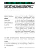
Fig. 1. Overall structures of TvuCMBP in complexes with
ligands. Complex structures with a-CD and G4 are shown in (A)
and (B), respectively. The overall structure of TvuCMBP–b-CD is
essentially identical to that of TvuCMBP–a-CD. N-domain,
C-domain, hinge regions and ligands are shown in yellow, blue,
red and green, respectively. Only two glucose residues are seen
in TvuCMBP–G4. (C) The Ca backbones of TvuCMBP in
complexes with a-CD (blue), b-CD (magenta), c-CD (green), and
G4 (orange). C-domains of their structures are superposed.
Ligands are represented as stick models. (D) Comparison of the
Ca backbones of TvuCMBP–G4 (orange) and EcoMBP–b-CD
(cyan).
A
B
C
D
Fig. 2. Stereo views of the C-domain involved in G4 binding in
TvuCMBP and related structures. Regions C-I, C-II, C-III and C-IV
are in blue, yellow, magenta and red, respectively. The ligands are
shown in gray. Three aromatic residues (two Trp and one Tyr)
stacking with the glucose residues are indicated. (A) TvuCMBP–G4.
An F
o
) F
c
omit map at the 2.0r contoured level is in purple, and
only two glucose residues, labeled (a) and (b), are seen in the map.
(B) TvuCMBP–c-CD. The glucose residues of c-CD are labeled from
1 to 8. (C) EcoMBP–G4 (Protein Data Bank ID: 4MBP). The glucose
residues of G4 are labeled from 1 to 4. (D) EcoMBP–maltose
(Protein Data Bank ID: 1ANF).
N. Matsumoto et al. Open ⁄ closed cyclo ⁄ maltodextrin-binding protein
FEBS Journal 276 (2009) 3008–3019 ª 2009 The Authors Journal compilation ª 2009 FEBS 3011
Comparison of the C-domain of TvuCMBP–G4 with
EcoMBP–maltose reveals that the two glucose residues,
Glc-(a) and Glc-(b), of TvuCMBP–G4 and the maltose
molecule bound to EcoMBP are located at structurally
identical positions, and Trp340 and Tyr155 of
EcoMBP are structurally identical to Trp360 and
Tyr175 of TvuCMBP (Fig. 2A,D). These results sug-
gest that Trp360 and Tyr175 play the key role in
anchoring the G4 molecule.
A schematic representation of the interaction
between TvuCMBP and G4 is presented in Fig. 3A.
There are numerous water molecules in the cavity
formed by the N-domain and C-domain. Although the
G4 molecule is located much closer to the C-domain
than to the N-domain, Glc-(a) and Glc-(b) interact
with Asp83 and Arg84 from the N-domain, and
Glu129 (hinge-1), directly or through the water mole-
cules (Figs 3A and 4A). The N-domain side of the
sugar-binding cleft consists of three loops, regions N-I
(residues 25–33), N-II (residues 56–61), and N-III (resi-
dues 80–85), and Arg83 and Arg84 are located in
region N-III. In EcoMBP, the ligand-induced move-
ment of Glu111 has been proposed to be the triggering
mechanism for the motion that enables the other
domain to participate in the ligand binding [18]. In
TvuCMBP, Glu129 in hinge-1 is identified as the corre-
sponding residue, and may be responsible for trigger-
ing the hinge-bending motion, together with Arg83
and Arg84 located in region N-III of the N-domain.
We have reported that Leu59 (at region N-II) inter-
acts with the central cavity of c-CD and appears to
play the key role in binding to the sugar among the
residues of the N-domain [8]. In TvuCMBP–G4, how-
ever, Leu59 seems not to interact with the G4 mole-
cule, as the closest distance between G4 [O2 of Glc-(a)]
and Leu59 (atom CD2) is 7.6 A
˚
(Fig. 4A). In the
closed form of EcoMBP, Trp62 (at region N-III) is
found at the center of the inner curvature of the oligo-
saccharides, suggesting that Trp62 of EcoMBP func-
tions similarly to Leu59 of TvuCMBP. However, Trp62
of EcoMBP can interact with the oligosaccharides
A
B
C
D
Fig. 3. Schematic drawing of the amino acid residues interacting
with the glucose residues [labeled (a) and (b)] of G4 (A), the struc-
turally shared three glucose residues (labeled 2, 3, and 4) of CDs
(B), and the structurally nonconserved glucose residues of a-CD
(labeled 5, 6, and 1) (C) and b-CD (labeled 5, 6, 7, and 1) (D). The
figures are based on a cartoon generated by the program
LIGPLOT
[33]. The C-domain and N-domain sides are categorized into four
(C-I–C-IV) and three (N-I–N-III) regions, respectively. Symbols: open
circle, oxygen atom; closed circle, carbon atom; gray circle,
nitrogen atom; dashed line, hydrogen bond. Residues involved in
hydrophobic interactions are illustrated.
Open ⁄ closed cyclo ⁄ maltodextrin-binding protein N. Matsumoto et al.
3012 FEBS Journal 276 (2009) 3008–3019 ª 2009 The Authors Journal compilation ª 2009 FEBS
through the water molecules even in the open form
EcoMBP–b-CD (Fig. 4B) [19], and this seems to be the
most important difference between the open forms of
TvuCMBP and EcoMBP.
Structures of TvuCMBP–a-CD and TvuCMBP–b-CD
In both TvuCMBP–a-CD and TvuCMBP–b-CD, omit
maps show clear density for the CD molecules
(Fig. 5A,B). The bound a-CD and b-CD are ellipsoidal
torus-shaped molecules. Crystallographic studies of
CDs alone have shown that the CD rings of a-CD [31]
and b-CD [32] are distorted because of conformational
strain, and a-CD and b-CD bound to TvuCMBP
appear to have conformations similar to the CDs
described in these reports. The structures of
TvuCMBP–a-CD, TvuCMBP–b-CD and TvuCMBP–
c-CD were superposed, and three glucose residues of
all the CD molecules overlaid remarkably well
(Fig. 5C). The glucose residues of TvuCMBP–c-CD
have been numbered according to the numbering for
EcoMBP–G4 [20], and here the glucose residues of
a-CD and b-CD were labeled in the same manner. The
three structurally homologous glucose residues of the
CDs were numbered Glc-2–Glc-4, and the glucose
residues of a-CD, b-CD and c-CD were designated
Glc-1(a-CD)–Glc-6(a-CD), Glc-1(b-CD)–Glc-7(b-CD),
and Glc-1(c-CD)–Glc-8(c-CD), respectively, as shown
in Fig. 5A,B. The angles C4–O4¢–C1¢ produced by two
glucose residues were calculated with the program
geomcalc of ccp
4 (Table 2). The angles between the
structurally shared glucose residues, Glc-2–Glc-3 and
Glc-3–Glc-4, are close to the mean angles of each CD
molecule; for example, those of Glc-2(a-CD)–Glc-
3(a-CD) and Glc-3(a-CD)–Glc-4(a-CD) are 153.0° and
152.6°, respectively, similar to the mean angle of
a-CD, 151.7°. In contrast, the angles of Glc-1(a-CD)–
Glc-2(a-CD) (146.6°), Glc-4(a-CD)–Glc-5(a-CD)
(144.2°) and Glc-5(b-CD)–Glc-6(b-CD) (144.1°) are
much smaller than the others (150–170°), resulting in
distortion of the CD molecules.
The interaction between the structurally shared three
glucose residues, Glc-2(CD), Glc-3(CD), and Glc-
4(CD), was analyzed with the program ligplot
(Fig. 3B) [33]. The amino acid residues that interact
with Glc-2(CD), Glc-3(CD) and Glc-4(CD) are almost
identical to those that interact with Glc-(a) and Glc-
(b) (Fig. 3A), regardless of whether the conformation
is the closed or open form. The nature of the interac-
tions between the two is, however, different. In the
open form, Arg84, Asp83 and Glu129 form hydrogen
bonds with O3 of Glc-(a), O3 of Glc-(b), and O2 of
Glc-(b), respectively, directly or through the water
molecules, but in the closed form, Arg84, Asp83 and
Glu129 form hydrogen bonds with different atoms,
O2 of Glc-2(CD), O3 of Glc-3(CD), and O3 of
A
B
Fig. 4. Stereo views of N-domains involved
in ligand binding in the open forms of
TvuCMBP [TvuCMBP–G4 (A)] and EcoMBP
[EcoMBP–b-CD, Protein Data Bank ID:
1DMB (B)]. Regions N-I, N-II, N-III and
hinge-1 are in green, orange, cyan and pink,
respectively. The ligands are shown in gray.
Hydrogen bonds and water molecules
hydrogen-bonded to the ligands or amino
acid residues are indicated in blue.
N. Matsumoto et al. Open ⁄ closed cyclo ⁄ maltodextrin-binding protein
FEBS Journal 276 (2009) 3008–3019 ª 2009 The Authors Journal compilation ª 2009 FEBS 3013
Glc-4(CD), respectively. In addition, Leu59 interacts
with the ligand molecule when only TvuCMBP adopts
the closed form, as described in the previous section.
The structurally nonconserved glucose residues, Glc-
5(a-CD)–Glc-1(a-CD) and Glc-5(b-CD)–Glc-1(b-CD),
were also analyzed with the program ligplot
(Fig. 3C,D). Many of the amino acid residues (Asn26,
Leu59, Glu170, Ala230, Asn234, Asn247, and Trp250)
participate in the interaction with both a-CD and
b-CD. The structures of TvuCMBP–a-CD and
TvuCMBP–b-CD are, however, essentially identical,
and the side chains of these amino acid residues are
oriented in the same directions (Fig. 5C). All of the
hydrogen bonds between TvuCMBP and the noncon-
served glucose residues are mediated through the water
molecules. These results suggest that the arrangement
A
B
C
Fig. 5. CD-binding sites in TvuCMBP. (A)
Stereo view of the N-domain involved in
a-CD binding. Regions N-I, N-II and N-III
are shown in green, orange and cyan,
respectively. The ligands (gray) and an
F
o
) F
c
omit map at the 2.0r contoured
level (purple) are shown. The amino acid
residues chosen on the basis of analysis
with the program
LIGPLOT [33] are indicated.
The glucose residues Glc-1(a-CD)–Glc-6
(a-CD) are labeled 1–6. (B) Stereo view of
the N-domain involved in b-CD binding.
Color representations are as in (A). The
glucose residues Glc-1(b-CD)–Glc-7(b-CD)
are labeled 1–7. (C) Superposition of
TvuCMBP in complexes with a-CD (blue),
b-CD (magenta), and c-CD (green). Amino
acid residues interacting with the CDs are
illustrated as stick models. Three aromatic
residues, Tyr175, Trp250, and Trp360, and
some amino acid residues shown in
Fig. 3C,D are illustrated as stick models.
The glucose residues Glc-2(CD), Glc-3(CD)
and Glc-4(CD) are labeled 2, 3 and 4,
respectively.
Table 3. Differences in Ca-torsion angles between TvuCMBP–G4
and TvuCMBP–a-CD in the vicinity of the hinge regions. Differ-
ence ={ [/(TvuCMBP–G4) ) /(TvuCMBP–a-CD)]
2
+[w(Tv uCMBP–G4) )
(TvuCMBP–a-CD)]
2
}
1 ⁄ 2
.
Hinge-1 Hinge-2 Hinge-3
Residue
number
Difference
(°)
Residue
number
Difference
(°)
Residue
number
Difference
(°)
127 9.4 278 5.4 331 7.6
128 15.1 279 17.1 332 20.1
129 10.4 280 11.0 333 11.0
130 14.3 281 7.2 334 32.0
131 5.5 282 5.2 335 21.7
– – – – 336 75.6
– – – – 337 61.7
– – – – 338 3.4
Table 2. Angles C4–O4¢–C1¢ produced by two glucose residues in the CD molecules. –, not applicable.
Numbers of the two glucose residues
1–2 2–3 3–4 4–5 5–6 6–1 or 6–7 7–1 or 7–8 8–1 Mean
a-CD (°) 146.6 153.0 152.6 144.2 157.7 156.2 – – 151.7
b-CD (°) 153.8 158.7 163.2 162.3 144.1 163.3 165.8 – 158.7
c-CD (°) 168.3 164.2 161.1 165.2 167.1 159.7 172.3 159.8 164.7
Open ⁄ closed cyclo ⁄ maltodextrin-binding protein N. Matsumoto et al.
3014 FEBS Journal 276 (2009) 3008–3019 ª 2009 The Authors Journal compilation ª 2009 FEBS
of the water molecules is important for binding to the
ligands.
Hinge bending
We determined the range of the hinge region bending
by comparing the open and closed conformations of
TvuCMBP and superposing the N-domains or
C-domains (Fig. 6). The critical conformational change
seems to be centered around Ile128–Ser130 (hinge-1)
and Lys279–Val281 (hinge-2). Hinge-1 and hinge-2
form a part of the b-strands (Fig. 6A,B), whereas
hinge-3 (Asn334–Val337) seems to be a turn linking
two a-helices (Fig. 6C,D). The interpretation of the
bending motion of hinge-3 is, however, very compli-
cated, and a superposition of individual domains indi-
cated that the conformational changes are found in an
a-helix (residues 322–333) and a loop (residues 191–
198), both of which are located near Asn334–Val337.
Differences in the Ca-torsion angles of hinge-1 and
hinge-2 between TvuCMBP–G4 and TvuCMBP–a-CD
were calculated using the program cns (Table 3) [34].
Between the two complexes, the differences in hinge-1
and hinge-2 are only 7.2–17.1°, but these values are
enough to convert the entire conformation, because
hinge-1 and hinge-2 are located in the center of
TvuCMBP. Many similarities can be seen between
unliganded EcoMBP [18] and TvuCMBP–G4. As with
EcoMBP, upon hinge bending, not only are the two
domains of the TvuCMBP apart from each other, but
the N-domain is twisted anticlockwise around the
hinge region relative to the C-domain. In measuring
the hinge bend, the dihedral angle was defined by four
atoms, Tyr175–Ca (C-domain), Phe278–Ca (hinge-2),
Ser130–Ca (hinge-1), and Leu59–Ca (N-domain), and
the difference in the dihedral angles between
TvuCMBP–G4 and TvuCMBP–a-CD was calculated.
The twist angle was also defined by the difference
in the dihedral angles of four atoms, Leu58–Ca
(N-domain), Gly280–Ca (hinge-2), Lys282–Ca
(C-domain), and Arg84–Ca (N-domain). The bend and
twist angles of TvuCMBP are 24° and 7°, respectively.
Similar measurements have been reported for
EcoMBP, and the bending and twist angles (35° and
A
B
C
D
E
Fig. 6. Structures of the hinge regions of TvuCMBP–a-CD and
TvuCMBP–G4. (A, B) Comparison of the residues in and around
hinge-1 and hinge-2. Colors: blue, TvuCMBP–a-CD (closed form);
orange, TvuCMBP–G4 (open form). N-domains (A) or C-domains (B)
of TvuCMBP–a-CD and TvuCMBP–G4 were superposed. Water
molecules hydrogen-bonded to hinge-1 or hinge-2 are shown in ball
representation. (C, D) Comparison of the residues in and around
hinge-3. N-domains (C) or C-domains (D) of TvuCMBP–a-CD and
TvuCMBP–G4 were superposed, and hinge-3 (residues 334–337)
and Ca backbones of two a-helices (residues 322–333 and 338–
348) are illustrated. Color representations are as described in (A)
and (B). (E) Alignment of the primary structures of hinge-1 and
hinge-2 of TvuCMBP and related solute-binding proteins. PfuMBP,
P. furiosus maltodextrin-binding protein (amino acid sequence iden-
tity = 33.3%) [23]; TliTMBP, Th. litoralis trehalose ⁄ maltose-binding
protein (26.3%) [22]; AacMBP, A. acidocaldarius maltose ⁄ maltodex-
trin-binding protein (30.2%) [24]; TthGBP, Thermus thermophilus
glucose-binding protein (23.9%) [35]. The numbering of the amino
acid sequences is given. Residues that are identical to those in
TvuCMBP are shown in red.
N. Matsumoto et al. Open ⁄ closed cyclo ⁄ maltodextrin-binding protein
FEBS Journal 276 (2009) 3008–3019 ª 2009 The Authors Journal compilation ª 2009 FEBS 3015
8°, respectively) are roughly comparable to those of
TvuCMBP. The structures of TvuCMBP-G4 and unli-
ganded EcoMBP are almost identical, with a 1.84 A
˚
rmsd (Fig. 1D).
The open and closed forms of several solute-binding
proteins have been previously determined, and their
hinge-bending motions have been analyzed [14,15,18–
21]. In E. coli d-allose-binding protein (EcoALBP),
two water molecules embedded in the hinge appear to
act as ballbearings, and residues with hydrophobic side
chains located at the terminus of the hinge regions are
proposed to function as grease [15]. The hinge regions
of EcoALBP and TvuCMBP share similar structural
features, although EcoALBP is categorized as a class I
solute-binding protein [11,12], whereas TvuCMBP is a
class II solute-binding protein, and the primary struc-
tures of their hinge regions have virtually no similarity.
In TvuCMBP, many water molecules are identified in
the vicinity of Glu129 at hinge-1, and amino acid resi-
dues with hydrophobic side chains, Ala127, Ile128,
Phe278, Ile279, and Val281, are found at the termini
of hinge-1 and hinge-2 (Fig. 6A,B). The primary struc-
tures of hinge-1 and hinge-2 of TvuCMBP and related
proteins [18–23,35] were aligned on the basis of struc-
tural comparison, and EcoMBP appears to be the most
similar to TvuCMBP (Fig. 6E).
Insights into the conformational change of
TvuCMBP
Comparing and contrasting the EcoMBP and
TvuCMBP structures has provided some interesting
mechanistic insights. The crystal structures of EcoMBP
indicated that EcoMBP–b-CD adopts the open form
[19], whereas the complexes with linear maltodextrins,
such as maltose, maltotriose, and G4, adopt the closed
form [18,20]. The reason why EcoMBP–b-CD adopts
the open form might be simple. In the closed form, the
cleft of the sugar-binding site of EcoMBP is very nar-
row, and CDs may not fit as readily as linear maltodext-
rins, which are half-buried when EcoMBP adopts the
closed form [8,20]. These observations suggest that the
closed form of EcoMBP may cause steric hindrance with
b-CD. In contrast, TvuCMBP–a-CD, TvuCMBP–b-CD
and Tvu CMBP–c-CD have a closed conformation,
whereas TvuCMBP–G4 remains in the open conforma-
tion. It is, however, impossible to use steric hindrance as
the explanation for the open conformation of
TvuCMBP-G4, because the CD molecules are larger
than the G4 molecule. The study of EcoMBP using
paramagnetic NMR indicated that the predominantly
open form coexists in rapid equilibrium with a minor
closed species in the absence of ligand [36], indicating
that the conformation of the solute-binding protein
could be determined by the ratio of the open fi closed
rate to the closed fi open rate.
Comparison of the open forms of TvuCMBP and
EcoMBP has revealed a possible explanation for the
shifting of the equilibrium. Many of the residues inter-
acting with Glc-(a) and Glc-(b) in the open form of
TvuCMBP–G4 are conserved in EcoMBP. A report on
EcoMBP has shown that Tyr155, Trp230 and Trp340
form the key nonpolar stacking interaction between
aromatic residues [19], and these residues are strictly
conserved in TvuCMBP as Tyr175, Trp250, and
Trp360, respectively. We previously evaluated the
affinities between TvuCMBP and various oligosaccha-
rides by measuring the fluorescence intensities, indi-
cating that the K
d
values for CDs and linear
maltodextrins are almost identical [8]. Moreover, the
amino acid sequences of hinge-1 and hinge-2 of
TvuCMBP are very similar to those of EcoMBP
(Fig. 6E), suggesting that these proteins’ mechanisms
for initial ligand binding and hinge bending could be
similar. In EcoMBP, Glu111 is has been reported to be
significant in the ligand-induced hinge–twist interdo-
main motion [20], and Trp62 appears to be important
for the open fi closed conformational change. The
functionally equivalent residue to Glu111 has been
identified as Glu129, whereas no equivalent residue to
Trp62 is found in TvuCMBP (Fig. 4). These observa-
tions suggest that the mechanism of the open fi -
closed conformational change in TvuCMBP is largely
similar to that of EcoMBP, but short linear maltodext-
rins might not affect the open fi closed conforma-
tional change as readily in TvuCMBP as they do in
EcoMBP.
By comparison, the mechanisms of the closed fi
open conformational changes seem to differ between
these two proteins. In the closed form of EcoMBP
(EcoMBP–G4; Protein Data Bank ID: 4MBP), five
hydrogen bonds, Glu45 OE2–Tyr341 OH, Ala96
O–Asn332 ND2, Arg98 O–Asn332 ND2, Asp65 OD2–
Trp340 NE1, and Ser233 OG–Asp296 O, form directly
between the N-domain and C-domain, and appear to
lock and stabilize the closed form of EcoMBP. On the
other hand, the ligand-binding cleft of the closed form
of TvuCMBP is much wider than that of EcoMBP (a
surface model of TvuCMBP–c-CD has been illustrated
previously [8]), and no direct hydrogen bond is
observed in the interdomain interactions. To maintain
the closed form of TvuCMBP, glucose residues of
CDs, especially Glc-5(CD), Glc-6(CD), Glc-7(CD),
Glc-8(CD), and Glc-1(CD), contribute to the forma-
tion of many hydrogen bonds and may function as
glue between the N-domain and C-domain.
Open ⁄ closed cyclo ⁄ maltodextrin-binding protein N. Matsumoto et al.
3016 FEBS Journal 276 (2009) 3008–3019 ª 2009 The Authors Journal compilation ª 2009 FEBS
Some bacteria and archaea have been proposed to
have a specific uptake mechanism for CDs and to uti-
lize CDs as a carbon source [37–39]. A bacterium,
Klebsiella oxytoca, has been reported to have a CD
metabolic pathway [39,40], and the identity of the pri-
mary structures between the solute-binding protein,
CymE, from K. oxytoca and TvuCMBP (34.6%) is
higher than that between EcoMBP and TvuCMBP
(30.1%), although the residue corresponding to Leu59
of TvuCMBP is not conserved in K. oxytoca CymE.
Pajatsch et al. [41] investigated the properties of a
CD-specific porin, CymA, from K. oxytoca. A mutant
of K. oxytoca with a lesion of the LamB maltoporin
gene was still able to grow on linear maltodextrins,
and they concluded that CymA has a role in the
uptake of both CDs and linear maltodextrins. The
channel properties of CymA were measured from titra-
tion experiments of the membrane conductance with
sugars, indicating that CymA binds both CDs and
linear maltodextrins, but the stability constant range
for binding of linear maltodextrins (K = 310–
32 000 m
–1
) is much smaller than the range for binding
CDs (K = 20–91 m
)1
) [42]. It is likely that TvuCMBP
possesses porins similar to CymA, and a high concen-
tration of linear maltodextrins may allow TvuCMBP
to adopt the closed form.
Experimental procedures
Expression and purification of TvuCMBP
The expression, purification and preparation of TvuCMBP in
complex with a-CD, b-CD or G4 was performed using the
same procedure as for TvuCMBP–c-CD [8,9]. Briefly, in the
step of purification with amylose resin (New England Biolabs,
Ipswich, MA, USA), TvuCMBP was eluted with 5 mm a-CD,
b-CD or G4 instead of c-CD. The eluted TvuCMBP was
bound to the ligands, and allowed us to obtain the a-CD,
b-CD and G4 complexes. The samples were further purified
using cation exchange chromatography (Hiload SP-Sepharose
HR 16 ⁄ 10 column, 1.6 · 10 cm; GE Healthcare, Chalfont St
Giles, UK) with elution by an NaCl gradient. Protein concen-
trations were determined by the measurement of absorbance
at 280 nm, as previously described [8].
Crystallization and data collection
TvuCMBP–a-CD, TvuCMBP–b-CD and TvuCMBP–G4
were crystallized at 20 °C, using the hanging drop vapor
diffusion method with the same conditions, where 1 lLof
TvuCMBP–ligand solution (8 mgÆmL
)1
)in50mm Mes ⁄
NaOH (pH 6.0) was mixed with the same volume of well
solution (20% polyethylene glycol 6000, 50 mm Mes,
pH 6.2). The obtained crystals were transferred to a solution
consisting of 34% polyethylene glycol 6000 and 50 mm Mes
(pH 6.0), and frozen in a 100 K nitrogen stream. Their dif-
fraction data were collected at beamlines PF BL5A, PF
BL6A and PF-AR NW12 (Photon Factory, Tsukuba,
Japan). The data were processed and scaled with the
program hkl2000 (Table 1) [43].
Model building and structure refinement
The structures of TvuCMBP–a-CD, TvuCMBP–b-CD, and
TvuCMBP–G4 were solved by molecular replacement with
the program molrep in the ccp4 suite [30], and a model of
TvuCMBP–c-CD (Protein Data Bank ID: 2DFZ) was
employed as a probe model. To solve the open TvuCMBP–
G4 structure, the probe model was divided into the
N-domain and C-domain, and an adequate result was
computed. The refinement was carried out using the program
cns [34], and manual adjustment and rebuilding of the model
were carried out with the programs xfit [44] and coot [45].
Superpositioning of TvuCMBP and other protein structures
and the calculation of the rmsd were carried out with the pro-
gram superpose in the ccp4 suite. Figures were generated
using pymol ( Sequence identities
were calculated using the program sim on the ExPASy server
( Based on the
coordinates of TvuCMBP–a-CD and TvuCMBP–b-CD,
incorrect rotamer assignments (especially Leu59) were
discovered in the previous coordinate of TvuCMBP–c-CD
(Protein Data Bank ID: 2DFZ, 2.5 A
˚
resolution) and were
rebuilt with the program coot. The R
work
and R
free
values
were converged at 0.222 and 0.285, respectively, and in a
Ramachandran plot calculated with the program procheck
of ccp4, no residues were present in the disallowed regions
or the generously allowed regions, and 90.5% of residues
were in the most favored region (Table 1).
Acknowledgements
We thank Hayashibara Biochemical Laboratories Inc.
and Ensuiko Sugar Refining Co., Ltd for providing
various sugars. This work was supported by a grant-
in-aid for Scientific Research (20570103) from the
Ministry of Education, Culture, Sports, Science and
Technology of Japan. This research was performed
with the approval of the Photon Factory Advisory
Committee (2007G010 and 2008G013), the National
Laboratory for High Energy Physics, Tsukuba, Japan.
References
1 Davis ME & Brewster ME (2004) Cyclodextrin-based
pharmaceutics: past, present and future. Nat Rev Drug
Discov 3, 1023–1035.
N. Matsumoto et al. Open ⁄ closed cyclo ⁄ maltodextrin-binding protein
FEBS Journal 276 (2009) 3008–3019 ª 2009 The Authors Journal compilation ª 2009 FEBS 3017
2 Aachmann FL, Otzen DE, Laesen KL & Wimmer R
(2003) Structural background of cyclodextrin–protein
interactions. Protein Eng 16, 905–912.
3 Ichikawa K, Tonozuka T, Uotsu-Tomita R, Mizuno M,
Nishikawa A & Sakano Y (2005) Bacterial and archaeal
enzymes homologous to glucoamylase: characterization
and subsite affinities of a glucoamylase from Thermoac-
tinomyces vulgaris R-47. Biologia Bratisl 60 (Suppl. 11),
161–165.
4 Kamitori S, Abe A, Ohtaki A, Kaji A, Tonozuka T &
Sakano Y (2002) Crystal structures and structural com-
parison of Thermoactinomyces vulgaris R-47 a-amylase
I (TVAI) at 1.6 A
˚
resolution and a-amylase II (TVAII)
at 2.3 A
˚
resolution. J Mol Biol 26, 443–453.
5 Abe A, Tonozuka T, Sakano Y & Kamitori S (2004)
Complex structures of Thermoactinomyces vulgaris
R-47 a-amylase 1 with malto-oligosaccharides demon-
strate the role of domain N acting as a starch-binding
domain. J Mol Biol 335, 811–822.
6 Kamitori S, Kondo S, Okuyama K, Yokota T, Shimura
Y, Tonozuka T & Sakano Y (1999) Crystal structure of
Thermoactinomyces vulgaris R-47 a-amylase II (TVA
II) hydrolyzing cyclodextrins and pullulan at 2.6 A
˚
resolution. J Mol Biol 287, 907–921.
7 Ohtaki A, Mizuno M, Tonozuka T, Sakano Y & Kami-
tori S (2004) Complex structures of Thermoactinomyces
vulgaris R-47 a-amylase 2 with acarbose and cyclodext-
rins demonstrate the multiple substrate recognition
mechanism. J Biol Chem 279, 31033–31040.
8 Tonozuka T, Sogawa A, Yamada M, Matsumoto N,
Yoshida H, Kamitori S, Ichikawa K, Mizuno M,
Nishikawa A & Sakano Y (2007) Structural basis for
cyclodextrin recognition by Thermoactinomyces vulgaris
cyclo ⁄ maltodextrin-binding protein. FEBS J 274,
2109–2120.
9 Yopi, Tonozuka T, Sakai H & Sakano Y (2002)
Cloning of a gene cluster for dextrin utilization from
Thermoactinomyces vulgaris R-47 and characterization
of the cyclodextrin-binding protein. J Appl Glycosci 49,
107–114.
10 Vyas NK, Vyas MN & Quiocho FA (1991) Comparison
of the periplasmic receptors for l-arabinose, d-glu-
cose ⁄ d-galactose, and d-ribose. Structural and func-
tional similarity. J Biol Chem 266, 226–237.
11 Fukami-Kobayashi K, Tateno Y & Nishikawa K (1999)
Domain dislocation: a change of core structure in
periplasmic binding proteins in their evolutionary
history. J Mol Biol 286, 279–290.
12 Dwyer MA & Hellinga HW (2004) Periplasmic binding
proteins: a versatile superfamily for protein engineering.
Curr Opin Struct Biol 14, 495–504.
13 Suzuki R, Wada J, Katayama T, Fushinobu S, Wakagi
T, Shoun H, Sugimoto H, Tanaka A, Kumagai H,
Ashida H et al. (2008) Structural and thermodynamic
analyses of solute-binding protein from Bifidobacterium
longum specific for core 1 disaccharide and lacto-
N-biose I. J Biol Chem 283, 13165–13173.
14 Bjo
¨
rkman AJ & Mowbray SL (1998) Multiple open
forms of ribose-binding protein trace the path of its
conformational change. J Mol Biol 279, 651–664.
15 Magnusson U, Chaudhuri BN, Ko J, Park C, Jones TA
& Mowbray SL (2002) Hinge-bending motion of
d-allose-binding protein from Escherichia coli:
three open conformations. J Biol Chem 277 ,
14077–14084.
16 Nikaido H & Hall JA (1998) Overview of bacterial
ABC transporters. Methods Enzymol 292, 3–20.
17 Lee SJ, Bo
¨
hm A, Krug M & Boos W (2007) The ABC
of binding-protein-dependent transport in Archaea.
Trends Microbiol 15, 389–397.
18 Sharff AJ, Rodseth LE, Spurlino JC & Quiocho FA
(1992) Crystallographic evidence of a large ligand-
induced hinge–twist motion between the two domains
of the maltodextrin binding protein involved in active
transport and chemotaxis. Biochemistry 31, 10657–
10663.
19 Sharff AJ, Rodseth LE & Quiocho FA (1993) Refined
1.8-A
˚
structure reveals the mode of binding of b-cyclo-
dextrin to the maltodextrin binding protein. Biochemis-
try 32, 10553–10559.
20 Quiocho FA, Spurlino JC & Rodseth LE (1997)
Extensive features of tight oligosaccharide binding
revealed in high-resolution structures of the
maltodextrin transport ⁄ chemosensory receptor.
Structure 5 , 997–1015.
21 Duan X, Hall JA, Nikaido H & Quiocho FA (2001)
Crystal structures of the maltodextrin ⁄ maltose-binding
protein complexed with reduced oligosaccharides: flexi-
bility of tertiary structure and ligand binding. J Mol
Biol 306, 1115–1126.
22 Diez J, Diederichs K, Greller G, Horlacher R, Boos W
& Welte W (2001) The crystal structure of a liganded
trehalose ⁄ maltose-binding protein from the hypertherm-
ophilic Archaeon Thermococcus litoralis at 1.85 A
˚
.
J Mol Biol 305, 905–915.
23 Evdokimov AG, Anderson DE, Routzahn KM &
Waugh DS (2001) Structural basis for oligosaccharide
recognition by Pyrococcus furiosus maltodextrin-bind-
ing protein. J Mol Biol 305, 891–904.
24 Scha
¨
fer K, Magnusson U, Scheffel F, Schiefner A,
Sandgren MO, Diederichs K, Welte W, Hulsmann A,
Schneider E & Mowbray SL (2004) X-ray structures of
the maltose-maltodextrin-binding protein of the thermo-
acidophilic bacterium Alicyclobacillus acidocaldarius
provide insight into acid stability of proteins. J Mol
Biol 335, 261–274.
25 Hall JA, Ganesan AK, Chen J & Nikaido H (1997)
Two modes of ligand binding in maltose-binding
protein of Escherichia coli. Functional significance in
active transport. J Biol Chem 272, 17615–17622.
Open ⁄ closed cyclo ⁄ maltodextrin-binding protein N. Matsumoto et al.
3018 FEBS Journal 276 (2009) 3008–3019 ª 2009 The Authors Journal compilation ª 2009 FEBS
26 Boos W & Shuman H (1998) Maltose ⁄ maltodextrin
system of Escherichia coli: transport, metabolism, and
regulation. Microbiol Mol Biol Rev 62, 204–229.
27 Chen J, Lu G, Lin J, Davidson AL & Quiocho FA
(2003) A tweezers-like motion of the ATP-binding
cassette dimer in an ABC transport cycle. Mol Cell 12,
651–661.
28 Oldham ML, Khare D, Quiocho FA, Davidson AL &
Chen J (2007) Crystal structure of a catalytic intermedi-
ate of the maltose transporter. Nature 450, 515–521.
29 Oldham ML, Davidson AL & Chen J (2008) Structural
insights into ABC transporter mechanism. Curr Opin
Struct Biol 18, 726–733.
30 Collaborative Computational Project (1994) The CCP4
suite: programs for protein crystallography. Acta Crys-
tallogr D Biol Crystallogr 50, 760–763.
31 Manor PC & Saenger W (1972) Water molecule
in hydrophobic surroundings: structure of
a-cyclodextrin-hexahydrate (C
6
H
10
O
5
)
6
.6H
2
O. Nature
237, 392–393.
32 Steiner T, Koellner G, Ali S, Zakim D & Saenger W
(1992) Crystalline b-cyclodextrinÆ12H
2
O reversibly
dehydrates to b-cyclodextrinÆ10.5H
2
O under ambient
conditions. Biochem Biophys Res Commun 188,
1060–1066.
33 Wallace AC, Laskowski RA & Thornton JM (1995)
LIGPLOT: a program to generate schematic diagrams
of protein–ligand interactions. Protein Eng 8, 127–134.
34 Bru
¨
nger AT, Adams PD, Clore GM, DeLano WL,
Gros P, Grosse-Kunstleve RW, Jiang JS, Kuszewski J,
Nilges M, Pannu NS et al. (1998) Crystallography &
NMR system: a new software suite for macromolecular
structure determination. Acta Crystallogr D Biol
Crystallogr 54, 905–921.
35 Cuneo MJ, Changela A, Warren JJ, Beese LS &
Hellinga HW (2006) The crystal structure of a thermo-
philic glucose binding protein reveals adaptations that
interconvert mono and di-saccharide binding sites. J
Mol Biol 362, 259–270.
36 Tang C, Schwieters CD & Clore GM (2007) Open-to-
closed transition in apo maltose-binding protein
observed by paramagnetic NMR. Nature 449,
1078–1082.
37 Hashimoto Y, Yamamoto T, Fujiwara S, Takagi M &
Imanaka T (2001) Extracellular synthesis, specific recog-
nition, and intracellular degradation of cyclomaltodext-
rins by the hyperthermophilic archaeon Thermococcus
sp. strain B1001. J Bacteriol 183, 5050–5057.
38 Labes A & Scho
¨
nheit P (2007) Unusual starch degrada-
tion pathway via cyclodextrins in the hyperthermophilic
sulfate-reducing archaeon Archaeoglobus fulgidus strain
7324. J Bacteriol 189, 8901–8913.
39 Fiedler G, Pajatsch M & Bo
¨
ck A (1996) Genetics of a
novel starch utilisation pathway present in Klebsiella
oxytoca. J Mol Biol 256, 279–291.
40 Pajatsch M, Gerhart M, Peist R, Horlacher R, Boos W
&Bo
¨
ck A (1998) The periplasmic cyclodextrin binding
protein CymE from Klebsiella oxytoca and its role in
maltodextrin and cyclodextrin transport. J Bacteriol
180, 2630–2635.
41 Pajatsch M, Andersen C, Mathes A, Bo
¨
ck A, Benz R &
Engelhardt H (1999) Properties of a cyclodextrin-
specific, unusual porin from Klebsiella oxytoca. J Biol
Chem 274, 25159–25166.
42 Orlik F, Andersen C, Danelon C, Winterhalter M,
Pajatsch M, Bo
¨
ck A & Benz R (2003) CymA of
Klebsiella oxytoca outer membrane: binding of cyclo-
dextrins and study of the current noise of the open
channel. Biophys J 85, 876–885.
43 Otwinowski Z & Minor W (1997) Processing of X-ray
diffraction data collected in oscillation mode. Methods
Enzymol 276, 307–326.
44 McRee D (1992) XtalView: a visual protein crystallo-
graphic software system for XII ⁄ XView. J Mol Graph
10, 44–47.
45 Emsley P & Cowtan K (2004) Coot: model-building
tools for molecular graphics. Acta Crystallogr D Biol
Crystallogr 60, 2126–2132.
N. Matsumoto et al. Open ⁄ closed cyclo ⁄ maltodextrin-binding protein
FEBS Journal 276 (2009) 3008–3019 ª 2009 The Authors Journal compilation ª 2009 FEBS 3019