Báo cáo khoa học: Human liver mitochondrial cytochrome P450 2D6 – individual variations and implications in drug metabolism docx
Bạn đang xem bản rút gọn của tài liệu. Xem và tải ngay bản đầy đủ của tài liệu tại đây (456.25 KB, 14 trang )
Human liver mitochondrial cytochrome P450
2D6 – individual variations and implications in
drug metabolism
Michelle Cook Sangar
1
, Hindupur K. Anandatheerthavarada
1
, Weigang Tang
1
, Subbuswamy K.
Prabu
1
, Martha V. Martin
2
, Miroslav Dostalek
2,
*, F. Peter Guengerich
2
and Narayan G. Avadhani
1
1 Department of Animal Biology, School of Veterinary Medicine, University of Pennsylvania, Philadelphia, PA, USA
2 Department of Biochemistry and Center in Molecular Toxicology, Vanderbilt University, Nashville, TN, USA
Cytochrome P450 2D6 (CYP2D6; EC 1.14.14.1) is a
constitutively expressed enzyme in hepatic and brain
tissues and accounts for the metabolism of 20–25% of
all drugs in clinical use [1]. This enzyme is of particular
interest because it shows a high degree of inter-individ-
ual variability as a result of the extensive genetic poly-
morphism that influences both its expression and
function. The substrates of CYP2D6 include a wide
spectrum of anti-arrhythmics, antihypertensives,
antidepressants, antipsychotics, analgesics and b-adren-
ergic blocking agents, in addition to some physiologi-
cal substrates [2,3]. Subsequent to its discovery as a
polymorphic enzyme, at least 112 allelic variants have
been described ( />Keywords
bimodal targeting signal; bufuralol
1¢-hydroxylase; human CYP2D6; liver
mitochondrial CYP2D6 content;
mitochondrial targeting
Correspondence
N. G. Avadhani, University of Pennsylvania,
School of Veterinary Medicine, 3800 Spruce
Street, Room 189E, Philadelphia, PA 19104,
USA
Fax: +1 215 573 6651
Tel: +1 215 898 8819
E-mail:
*Present address
Department of Clinical Pharmacology,
University Hospital and Faculty of Health
Studies, Ostrava University, Czech Republic
(Received 27 February 2009, revised 16
April 2009, accepted 20 April 2009)
doi:10.1111/j.1742-4658.2009.07067.x
Constitutively expressed human cytochrome P450 2D6 (CYP2D6; EC
1.14.14.1) is responsible for the metabolism of approximately 25% of drugs
in common clinical use. It is widely accepted that CYP2D6 is localized in
the endoplasmic reticulum of cells; however, we have identified this enzyme
in the mitochondria of human liver samples and found that extensive inter-
individual variability exists with respect to the level of the mitochondrial
enzyme. Metabolic assays using 7-methoxy-4-aminomethylcoumarin as a
substrate show that the human liver mitochondrial enzyme is capable of
oxidizing this substrate and that the catalytic activity is supported by mito-
chondrial electron transfer proteins. In the present study, we show that
CYP2D6 contains an N-terminal chimeric signal that mediates its bimodal
targeting to the endoplasmic reticulum and mitochondria. In vitro mito-
chondrial import studies using both N-terminal deletions and point muta-
tions suggest that the mitochondrial targeting signal is localized between
residues 23–33 and that the positively-charged residues at positions 24, 25,
26, 28 and 32 are required for mitochondrial targeting. The importance of
the positively-charged residues was confirmed by transient transfection of a
CYP2D6 mitochondrial targeting signal mutant in COS-7 cells. Both the
mitochondria and the microsomes from a CYP2D6 stable expression cell
line contain the enzyme and both fractions exhibit bufuralol 1¢-hydroxyl-
ation activity, which is completely inhibited by CYP2D6 inhibitory anti-
body. Overall, these results suggest that the targeting of CYP2D6 to
mitochondria could be an important physiological process that has signifi-
cance in xenobiotic metabolism.
Abbreviations
Adx, adrenodoxin; AdxR, adrenodoxin reductase; CCCP, carbonyl cyanide m-chlorophenylhydrazone; CYP, cytochrome P450; CYPR, NADPH-
cytochrome P450 reductase; DHFR, dihydrofolate reductase; DOX, doxycycline; ER, endoplasmic reticulum; Fdr, ferredoxin reductase; HL,
human liver sample; MAMC, 7-methoxy-4-(aminomethyl)coumarin; mtTFA, mitochondrial transcription factor A; PKA, protein kinase A; RRL,
rabbit reticulocyte lysate; TOM20, translocase of outer mitochondrial membrane 20; WT, wild-type.
3440 FEBS Journal 276 (2009) 3440–3453 ª 2009 The Authors Journal compilation ª 2009 FEBS
cyp2d6.htm) and individuals can be categorized into
four general phenotypes: poor metabolizers, who lack
the functional enzyme; intermediate metabolizers, who
are heterozygous for one deficient allele or have two
alleles causing reduced activity; extensive metabolizers,
who have two normal alleles; and ultrarapid metabo-
lizers, who have multiple gene copies that are inherited
in a dominant manner [4].
Many pharmacogenetic studies suggest that poly-
morphisms in CYP2D6 can significantly affect the
activity of the enzyme, and therefore serve as an
important guideline for determining the dose of anti-
depressant drugs and preventing drug-induced toxicity
[2–6]. A large majority of studies on the biochemical
and genetic properties, pharmacological and toxicolog-
ical roles, and clinical relevance of CYP2D6 are based
on the steady-state levels and activity of the enzyme
associated with the microsomal fraction of liver and
brain tissues [7,8].
Recent studies conducted in our laboratory have
shown that a number of xenobiotic inducible CYPs,
including CYP1A1, 2B1 and 2E1, are bimodally tar-
geted to both the microsomal and mitochondrial frac-
tions of hepatic, brain and lung tissues, and also in
cultured cells induced to express these proteins [9–13].
These studies gave rise to the concept of a new family
of N-terminal targeting signals, termed ‘chimeric sig-
nals’, which facilitate the bimodal targeting of the pro-
tein. The chimeric signals consist of a cryptic
mitochondrial targeting signal immediately adjacent to
the endoplasmic reticulum (ER) targeting and trans-
membrane domains of the apoproteins. The results
obtained in our laboratory also demonstrated that the
cryptic mitochondrial targeting signals require activa-
tion either by endoproteolytic processing by a cytosolic
protease, as in the case of CYP1A1 [9,14], or protein
kinase A (PKA; EC 2.7.11.11)-mediated protein phos-
phorylation at serine residues located approximately
100 amino acids downstream of the cryptic mitochon-
drial targeting signal, as in the case of CYP2B1 and
2E1 [11,13]. The mitochondrial targeted CYPs physi-
cally and functionally associate with adrenodoxin
(Adx) and adrenodoxin reductase (AdxR), the compo-
nents of the mitochondrial matrix electron transport
system, and efficiently catalyze drug metabolism
[10,15,16]. Some of the mitochondrial targeted forms
exhibit altered substrate specificity compared to the
microsomal enzymes. P450 MT2 (N-terminal truncated
CYP1A1) has been shown to catalyze the N-demethy-
lation of erythromycin, lidocaine, morphine and vari-
ous other neuroactive drugs [17]. Interestingly, these
reactions are not catalyzed by the microsome-associ-
ated intact CYP1A1 in reactions supported by micro-
somal NADPH-cytochrome P450 reductase (CYPR;
EC 1.6.2.4) [10,18].
In the present study, we show that CYP2D6 is pres-
ent in the mitochondria of human liver samples and
that mitochondria isolated from the liver samples are
active in the metabolism of 7-methoxy-4-(amino-
methyl)coumarin (MAMC), a substrate for microsomal
CYP2D6. We also demonstrate that CYP2D6 is tar-
geted to the mitochondrial compartment in isolated
mitochondria and in COS-7 cells transiently or stably
expressing the human protein. Mutation of the puta-
tive mitochondrial targeting signal eliminates this tar-
geting mechanism in vitro. Mitochondria isolated from
the stable expression cell line are active in the
1¢-hydroxylation of bufuralol, a probe substrate for
the microsomal CYP2D6. This activity is inhibited by
CYP2D6 inhibitory antibody. These results suggest
that the mitochondrial localization of CYP2D6 may be
an important physiological process with a possible role
in drug metabolism and drug-induced toxicity.
Results
Localization of CYP2D6 in human liver
mitochondria
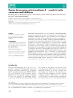
Mitoplast and microsomal isolates from 20 human
liver samples were analyzed by immunoblot analysis
using polyclonal antibody to human CYP2D6. The
blots were also co-developed with antibody to a mito-
chondrial specific marker protein, mitochondrial tran-
scription factor A (mtTFA), and a microsome specific
marker protein, CYPR. Representative immunoblot
profiles for eight such samples are presented in
Fig. 1A. The microsomal isolates from six human liver
samples (HL132, 134, 136, 137, 139 and 140) contained
a relatively high CYP2D6 content, whereas two sam-
ples (HL131 and 141) demonstrated moderate levels of
CYP2D6, as indicated by the intensity of the 50 kDa
antibody reactive band (Fig. 1A). The mitoplasts, on
the other hand, showed a marked variability in
CYP2D6 content, ranging from a relatively high level
in HL134 and 137 to a moderate level in HL136, low
levels in HL132, 139 and 140, and almost undetectable
levels in HL131 and 141 (Fig. 1A). Densitometry mea-
surements were used to calculate the subcellular distri-
bution of the CYP2D6 protein in the microsomal and
mitochondrial fractions (Fig. 1A). HL134 had almost
equal levels of CYP2D6 in mitochondria and micro-
somes, whereas almost all (97–99%) of the CYP2D6 in
HL131 and 141 was associated with the microsomal
fraction (Fig. 1A). HL137 and HL136 had 34% and
20% of the protein, respectively, in the mitochondrial
M. Cook Sangar et al. Mitochondrial targeting of human CYP2D6
FEBS Journal 276 (2009) 3440–3453 ª 2009 The Authors Journal compilation ª 2009 FEBS 3441
fraction (Fig. 1A). The immunoblots also showed that
the 78 kDa CYPR protein was detectable in the micro-
somal isolates but not significantly in the mitochon-
drial membrane isolates. Similarly, the 29 kDa mtTFA
protein was seen mostly in the mitochondrial isolates
but sparingly in the microsomal isolates. As in our pre-
vious studies [9,10,17], mitochondrial isolates were rou-
tinely analyzed for microsomal contamination by
assaying for rotenone insensitive NADPH-cytochrome
c reductase. Using this marker assay, we found that
the mitochondrial isolates contained < 1% micro-
somal contamination (data not shown).
The immunoblot (Fig. 1B) shows the results of a
control experiment that assessed the relative resistance
or sensitivity of human liver microsome- and mito-
chondria-associated CYP2D6 to limited digestion with
trypsin. Proteins localized in the mitochondrial matrix
or intermembrane space are expected to be resistant to
limited trypsin treatment under these conditions,
whereas those adventitiously adhering to the outer
mitochondrial membrane and microsomal fragments
should be sensitive. In all three microsomal isolates
tested (HL126, 130 and 141), the antibody-reactive
CYP2D6 was sensitive to trypsin treatment. By con-
trast, mitochondria-associated CYP2D6 in samples
HL126 and 130 was resistant to trypsin treatment.
This result suggests that CYP2D6 is localized within
the mitochondrial membrane compartment. Similarly,
in sample HL141, which contained no significant mito-
chondrial CYP2D6, the trypsin-treated mitochondria
did not show detectable antibody reactive protein.
Metabolic activity of mitochondrial CYP2D6
The ability of mitochondrial CYP2D6 to metabolize
substrates was investigated using MAMC, a known
substrate of microsomal CYP2D6 (Fig. 2A,B). Mito-
plasts from five randomly selected human liver samples
were tested for their ability to oxidize MAMC.
Because of the known ability of other CYPs, especially
CYP1A2, to oxidize this compound, various inhibitors
were used to assess the activity mediated by mitochon-
A
B
131
132
134 136
137
139
140
141
Mc.
Mt.
Mc.
Mt.
Mc.
Mt.
Mc.
Mt.
Mc.
Mt.
Mc.
Mt.
Mc.
Mt.
Mc.
Mt.
78 kDa
CYPR
CYP2D6
mtTFA
50 kDa
29 kDa
8
90
100
50
60
70
80
20
30
40
% distribution
0
10
131 132 134 136 137 139 140 141
Micro
Mito
Micro
Mito
Micro
Mito
Micro
Mito
Micro
Mito
Micro
Mito
Micro
Mito
Micro
Mito
HL 126
HL 130
HL 141
CYP2D6
Mc. Mc. Mt. Mt. Mc. Mc. Mt. Mt. Mc. Mc. Mt. Mt.
50 kDa
Trypsin
+
– +
–
+
– +
– +
–
+ –
Fig. 1. Localization of CYP2D6 in the mitochondria of human liver samples. (A) Immunoblot analysis of mitoplast and microsome (50 lg pro-
tein each) fractions isolated from human liver samples. Mc, microsomal fraction; Mt, mitoplast fraction. Densitometric analysis was per-
formed to determine the distribution of CYP2D6 between mitochondria and microsomes in each liver sample analyzed. (B) Immunonlot
analysis of human liver mitochondria and microsomes subjected to limited trypsin digestion (150 lgÆmg
)1
protein, 20 min on ice). Blots were
developed with polyclonal antibodies to CYP2D6 (1 : 1000) and mtTFA (1 : 3000) and monoclonal antibody to CYPR (1 : 1500).
Mitochondrial targeting of human CYP2D6 M. Cook Sangar et al.
3442 FEBS Journal 276 (2009) 3440–3453 ª 2009 The Authors Journal compilation ª 2009 FEBS
drial CYP2D6 (Fig. 2A). All five samples tested
yielded varying activity, ranging from moderate (sam-
ples HL139 and HL140) to high (HL129, HL111 and
HL130) activity for MAMC O-demethylation. The
activities of both HL129 and HL111 were inhibited by
approximately 53% and 50%, respectively, by the
addition of 10 lm quinidine, a CYP2D6 specific inhibi-
tor. (Note that a concentration of 1 lm quinidine is
generally sufficient to inhibit CYP2D6 in a system
using purified microsomes; however, the sensitivity of
CYP2D6 to quinidine within the mitochondrial com-
partment is unknown.) When these mitoplasts were
pre-incubated with antibody to Adx, an essential pro-
tein in the mitochondrial electron transfer system, the
activity was reduced by 83% and approximately
100%, respectively. The activities of HL139 and 140
liver mitochondria were reduced by 94% and 84%,
respectively, after incubation with Adx antibody. A
CYP2D6 specific inhibitory antibody was also used to
further investigate the role of CYP2D6 in this activity.
Samples HL139 and 140 both showed a considerable
reduction in metabolic activity after pre-incubation
with CYP2D6 antibody. The activity was reduced by
75% and 94%, respectively. Sample HL127 had a
moderately high activity, which was reduced by
approximately 52% after addition of CYP2D6 anti-
body. MAMC is known to be oxidized by both
CYP2D6 and CYP1A2 [19–21] and an inhibitory anti-
body to CYP1A2 inhibited the activity of HL127 liver
mitochondria by approximately 52%. The specificity
of the antibody inhibition was tested by incubating
HL130 mitochondria with either nonspecific mouse
IgG or specific CYP2D6 inhibitory antibody. The non-
specific IgG had virtually no effect on the MAMC
metabolizing activity, whereas the CYP2D6 inhibitory
antibody reduced the activity by approximately 62%.
Finally, a general P450 inhibitor, SKF-525A, reduced
the activity by 94% and 100%, respectively, in mito-
chondria from HL129 and 111 livers (Fig. 2A). The
remaining human liver sample mitoplasts were capable
A
B
8
6
7
4
5
1
2
3
0
1
Specific activity (nmol HAMC·mg
–1
·min
–1
)
Control
1m
M SKF525A
CYP2D6 Ab
10 µ
M Quinidine
Adrenodoxin Ab
Control
Control
1m
M SKF525A
10 µ
M Quinidine
Adrenodoxin Ab
Adrenodoxin Ab
CYP2D6 Ab
Control
CYP2D6 Ab
CYP1A2 Ab
Control
Adrenodoxin Ab
HL129 HL111 HL139 HL140 HL127
5
6
3
4
2
3
0
1
Specific activity (nmol HAMC·mg
–1
·min
–1
)
Control
Mouse IgG
CYP2D6 Ab
HL130
7
8
5
6
3
4
1
2
Specific activity (nmol HAMC·mg
–1
·min
–1
)
0
108 109 112 113
114 123 126 128 131 132 134 136 137 141
Human liver sam
p
le mitochondria
Fig. 2. Metabolic activity of human liver
mitochondrial CYP2D6. Mitoplasts isolated
from human liver samples were assayed for
O-demethylation activity using the substrate
MAMC. Assays were performed as
described in the Experimental procedures.
(A) Mitoplasts from five human liver sam-
ples were tested for MAMC oxidizing activ-
ity and various inhibitors were used to
establish whether the activity is mediated
by mitochondrial CYP2D6. Mitochondria
were pre-incubated with inhibitors as
described in the Experimental procedures.
Control refers to activity in the absence of
any inhibitors. The control activity for sam-
ple HL140 represents the mean ± SEM of
three separate estimates. The control activi-
ties for samples HL129, 139 and 127 repre-
sent the mean of two separate estimates.
All other values represent single assay
points. (B) MAMC O-demethylation activity
was compared between mitoplasts isolated
from the remaining fourteen human liver
samples using the protocol described in the
Experimental procedures. The activities in all
cases represent the mean ± SEM from
three separate estimates.
M. Cook Sangar et al. Mitochondrial targeting of human CYP2D6
FEBS Journal 276 (2009) 3440–3453 ª 2009 The Authors Journal compilation ª 2009 FEBS 3443
of oxidizing MAMC; however, there were significant
inter-individual differences in the level of activity
(Fig. 2B).
Characterization of mitochondrial targeting signal
of CYP2D6
The N-terminal signal sequence and the phosphory-
lation domains of CYP2B1 and 2E1 were compared
with the amino acid sequence of human CYP2D6
(Fig. 3A). The N-terminal amino acid sequence of
CYP2D6 bears resemblance to the chimeric signal
sequences identified in CYP2B1 and CYP2E1. The
sequence contains a 22 amino acid region with a
hydrophobic helical structure that is considered to act
as both an ER targeting and membrane anchor
domain [22,23]. There is an immediately adjacent puta-
tive mitochondrial targeting signal composed of a
A
B
C
D E
WT WT, CCCP WT, Oligo.
C T C T C T
50 kDa
Su9-DHFR DHFR
In
C
T
In
C T
18 kDa
34 kDa
27 kDa
P450 2B1: MEPTILLLLALL VGFLLLL VRGHPKSRGNFPPGPRPLP …………RRFSL
20
30
128
P450 2E1: MA VLGITIAL LV W V A TLL VISIWKKIYNSWNLPPGPFPL P …… RRFSL
P450 2D6:
MGLEA LV PL AV IV AIFLLL VDLMHRRQ RW AAR YPPGPLPL … RRFSVSTLR N
135
129
ER target/Transmembrane
Mito target
Proline rich PKA PKC
WT 2D 6 :
MGLEA LV PLA VIV AIFLLL VDLMHRRQ RW AAR YPPGPLPL………RRFSVSTLRNL
MAPPGPLPL………RRFSVSTLRNL
MGLG………RRFSVSTLRNL
+ 34/2D6:
+ 40/2D6:
WT 2D 6 + 34/2D6
+ 40/2D6
In In
C T
C T C T
In
50 kDa
0.8
0.9
1
0.3
0.4
0.5
0.6
0.7
0
0.1
0.2
WT 2D6 + 34/2D6 + 40/2D6
Relative import
WT 2D6:
MGLEA LV P L AV IV AIFLLL VDLMHRRQ RW AAR YPPGPLPL RRFSVSTLRNL
ArgM 2D6: MGLEA LV PLA VIV AIFLLL VDLMH NN NN Q N N WA AR YPPGPLPL………RRFSVSTLRNL
MitoM 2D6: MGLEA LV PL AV IV AIFLLL VDL M
AAA AAA Q A WA A A YPPGPLPL RRFSVSTLRNL
WT 2D6:
MGLEA LV PLA VIV AIFLLL VDLMHRRQ RW AAR YPPGPLPL ………. RRFSVSTLRNL
MitoM 2D6: MGLEA LV PL AV IV AIFLLL VDL M AAA AAA Q A WA A A YPPGPLPL …… RRFSVSTLRNL
WT 2D6 ArgM 2D6
MitoM 2D6
50 kDa
C T
In
C T
In
C T
In
0.9
1
0.4
0.3
0.5
0.6
0.7
0.8
0
0.1
0.2
WT ArgM MitoM
Relative import
Fig. 3. Localization of mitochondrial targeting signal of CYP2D6. (A) Alignment of CYP2D6 N-terminal sequence with chimeric signal
sequences of CYP2B1 and CYP2E1. (B–D) In vitro import of [
35
S]-labeled translation products in isolated rat liver mitochondria. (B, C, E)
CYP2D6 WT; (B) N-terminal truncation mutants; and (C) mitochondrial targeting signal mutants were generated in the RRL system. (D) Su-9
DHFR, in which the pre-sequence of subunit 9 of N. crassa F
0
F
1
-ATPase has been fused to DHFR, and DHFR were translated in RRL and
used as positive and negative controls respectively. (E) Mitochondria were pre-incubated with CCCP (50 l
M) or oligomycin (oligo; 50 lM) for
20 min at 37 °C prior to initiating the import reaction. In all experiments, trypsin digestion (150 lgÆ mL
)1
) of mitochondria was performed for
20 min on ice. Proteins (200 lg each) were subjected to SDS ⁄ PAGE and fluorography. C, control experiments in which total protein bound
and imported into mitochondria is present; T, trypsin-treated mitochondria in which only the protein imported into mitochondria is present. In
the lanes marked ‘In’, 20% of the counts used as input for the import reactions were loaded. (B, C) Densitometric analysis was performed
to analyze the level of import for each construct after trypsin treatment. The level of import of the WT protein was considered to be 1 when
calculating the relative import of various deletion and point mutations.
Mitochondrial targeting of human CYP2D6 M. Cook Sangar et al.
3444 FEBS Journal 276 (2009) 3440–3453 ª 2009 The Authors Journal compilation ª 2009 FEBS
stretch of positively-charged residues, including a His
at position 24 and Arg residues at positions 25, 26, 28
and 32, followed by the Pro-rich domain beginning at
position 34 and a potential PKA target phosphoryla-
tion site at Ser135, similar to those reported for
CYP2B1 and CYP2E1. The putative signal domain of
CYP2D6 contains five positively-charged residues com-
pared to two positively-charged residues in CYP2E1
and four in CYP2B1. CYP2D6 also has a putative
PKC phosphorylation site adjacent to the PKA target
site.
To map the mitochondrial targeting signal domain
of CYP2D6, a series of constructs were generated with
N-terminal truncations and point mutations in the
putative mitochondrial targeting signal and used for
in vitro import into isolated mitochondria. Intact wild-
type CYP2D6 (WT 2D6) was imported at a moderate
level into mitochondria (Fig. 3B,C). Deletion of two
N-terminal domains [i.e. the ER targeting domain and
the mitochondrial targeting signal (+34 ⁄ 2D6)] or all
three N-terminal domains [i.e. the ER targeting signal,
mitochondrial targeting signal, and the Pro-rich
domain (+40 ⁄ 2D6)] reduced import by approximately
95% compared to the WT protein (Fig. 3B). Further-
more, point mutations in the putative mitochondrial
targeting domain also significantly disrupted the mito-
chondrial import of CYP2D6 (Fig. 3C). Substitution
of Arg at positions 25, 26 and 28 with neutral Asn
(ArgM 2D6) in the putative mitochondrial targeting
signal reduced the level of mitochondrial import by
approximately 50% compared to the WT protein.
Additionally, mutation of all five positively-charged
residues in the putative mitochondrial targeting signal
to Ala residues (MitoM 2D6) reduced the mitochon-
drial import of CYP2D6 by approximately 90% com-
pared to the WT protein (Fig. 3C).
Su-9 dihydrofolate reductase (DHFR; EC 1.5.1.3)
was used as a positive control for the in vitro import
experiments (Fig. 3D). In this construct, the pre-
sequence of subunit 9 of Neurospora crassa F
0
F
1
-AT-
Pase has been fused to DHFR. This is a classic
mitochondrial targeting signal that is cleaved after
entry into mitochondria [24]. In this in vitro system,
only the cleaved protein (27 kDa) is present after
import and trypsin treatment (Fig. 3D). DHFR, a
cytosolic protein, was used as a negative control for
these experiments. There was no detectable entry of
this protein into mitochondria (Fig. 3D). Additional
controls were performed to determine whether the
import of WT CYP2D6 into mitochondria is energy
dependent. Mitochondria were incubated with
carbonyl cyanide m-chlorophenylhydrazone (CCCP),
which disrupts the mitochondrial membrane potential,
and oligomycin, which disrupts the mitochondrial ATP
pool, prior to import. The level of import of WT
CYP2D6 into mitochondria was significantly reduced
by incubation with both CCCP and oligomycin
(Fig. 3E). The relatively lower level of binding and
import of WT CYP2D6 in Fig. 3C,E compared to
Fig. 3B probably reflects natural variation in
mitochondrial activity between different rat livers.
Mitochondrial targeting of CYP2D6 in transiently
transfected COS-7 cells
Mitochondrial and microsomal fractions isolated from
cells transiently transfected with WT CYP2D6 demon-
strate almost equal levels of CYP2D6 in mitochondria
and microsomes (Fig. 4A). By contrast, when cells
were transfected with ArgM CYP2D6, the level of
mutant CYP2D6 in microsomes was two-fold higher
than that in mitochondria (Fig. 4A). Limited trypsin
digestion eliminated both WT and ArgM CYP2D6
from the microsomal fraction, but the mitochondria
associated CYP2D6 was resistant to trypsin treatment,
suggesting that the protein is localized inside the mito-
chondrial membranes (Fig. 4B). As expected, the level
of translocase of outer mitochondrial membrane 20
(TOM20) was markedly reduced by trypsin digestion
(Fig. 4B). COS cells had a low level of endogenous
CYP2D6 in the microsomal fraction that was sensitive
to trypsin digestion, whereas there was no detectable
CYP2D6 in mitochondria (Fig. 4A,B).
Role of PKA-mediated phosphorylation in
mitochondrial targeting of CYP2D6
Our previous studies have shown that mitochondrial
targeting of CYP2E1 and 2B1 was facilitated by PKA-
mediated phosphorylation at Ser129 and Ser128 of the
protein, respectively [11,13]. Analysis of CYP2D6
using netphosk 1.0 [25], which predicts phosphory-
lation sites, revealed the presence of a high consensus
(score = 0.85) PKA site (RRFSV) at Ser135 in addi-
tion to two other lower consensus sites at Ser148 and
Ser217. In addition, a recent report showed that
CYP2D6 is phosphorylated at Ser135 using mass spec-
trometry [26]. The Ser135 site is positionally similar to
the Ser128 and Ser129 PKA sites of CYP2B1 and
CYP2E1, which were shown to be functionally impor-
tant for mitochondrial import [11,13]. Therefore, we
tested the importance of the Ser135 PKA site for the
mitochondrial import of CYP2D6 by mutagenesis at
this site (Fig. 5A) and in vitro import of the protein.
In vitro import of WT CYP2D6 increases by approxi-
mately 23% when the nascent protein is pre-incubated
M. Cook Sangar et al. Mitochondrial targeting of human CYP2D6
FEBS Journal 276 (2009) 3440–3453 ª 2009 The Authors Journal compilation ª 2009 FEBS 3445
with PKA and ATP (Fig. 5B). Interestingly, the PKA
phosphorylation site mutant (PKAM2D6) was
imported at a much lower level than WT protein under
basal conditions (Fig. 5B). Pretreatment with PKA
and ATP increased the import of the mutant protein;
however, the overall level of increase was almost half
that of the WT protein subjected to PKA treatment
(Fig. 5B). These results suggest that PKA phosphory-
lation contributes to the mitochondrial transport of
human CYP2D6. The precise reason for the PKA-
mediated increase in the import of mutant PKAM2D6
remains unclear. It is likely, however, that other puta-
tive PKA sites (Ser148 and Ser217) also contribute to
mitochondrial import and that mutation in the S135
site only partly affects protein import.
Mitochondrial localization of human CYP2D6
in a stable expression cell line
To assess the role of mitochondrial CYP2D6 in drug
metabolism, we generated cell lines expressing human
CYP2D6 under the regulation of a doxycycline (DOX)
inducible promoter. Mitochondria and microsomes iso-
lated from DOX induced cells were analyzed using
immunoblot analysis (Fig. 6). CYP2D6 was present in
both the mitochondria and the microsomes after
induction with DOX, although the level in mitochon-
dria was significantly lower than that in the micro-
somes. There was no expression of CYP2D6 in the
absence of DOX induction. The immunoblots were
co-developed with CYPR and TOM20 antibodies,
demonstrating that there is minimal cross-contamina-
tion between the two subcellular fractions. Addition-
ally, analysis of CO difference spectra indicated that
the P450 concentration is 172 pmolÆmg
)1
protein in
microsomes and 146 pmolÆmg
)1
protein in mitochon-
dria in this cell line.
A
B
Mt
WT
ArgM
Mt
COS
Mt Mt. Mc. Std. Mt. Mc. Mt. Mc.
CYP2D6
TOM20
Mt. Std.
WT
ArgM COS
CYP2D6
TOM20
Mc. Mt. Mc. Mt. Mc.
70
60
50
% distribution
40
30
20
10
0
Mito Micro Mito Micro
WT ArgM
Fig. 4. Role of Arg residues from the putative signal region for the
mitochondrial targeting of CYP2D6 in COS-7 cells. Immunoblot
analysis of mitochondria and microsomes isolated from COS-7 cells
transiently transfected for 48 h with WT and ArgM CYP2D6 cDNA.
(A) Mitochondria and microsome fractions before trypsin treatment.
(B) Mitochondria and microsome fractions after limited trypsin
digestion (100 lgÆmg
)1
protein, 30 min on ice). Blots were
co-developed with polyclonal antibodies to CYP2D6 (1 : 1000) and
TOM20 (1 : 1000). (A) Densitometric analysis was performed and
the percentage distribution in the mitochondrial and microsomal
fractions was based on aggregate values (mitochondria + micro-
some) that were considered to be 100%.
WT 2D6:
MGLEALVPLAVIVAIFLLLVDLMHRRQRWAARYPPGPLPL………RRFSVSTLRNL
MGLEALVPLAVIVAIFLLLVDLMHRRQRWAARYPPGPLPL………RRF
AA
VSTLRNL
PKAM 2D6:
WT CYP2D6
PKAM CYP2D6
PKA
–– –+ +– – –+ +
T C T
PKA
50 kDa
T
CCInCIn
T
A
B
60
50
% of input
40
30
20
10
0
Basal PKA Basal PKA
PKAMWT
Fig. 5. Role of PKA-mediated phosphorylation in mitochondrial
targeting of CYP2D6. (A) Comparison of WT CYP2D6 N-terminal
sequence with PKAM 2D6 sequence, in which Ser135 has been
mutated to Ala. (B) In vitro import of [
35
S]-labeled translation pro-
ducts in rat liver mitochondria. CYP2D6 WT and PKAM constructs
were translated in the RRL system in the presence of [
35
S]Met. In
some cases, translation products were pre-incubated with PKA and
ATP for 30 min at 37 °C, prior to import. Labeled proteins were
imported into isolated mitochondria as described in the Experimen-
tal procedures. C, control experiments in which total protein bound
and imported into mitochondria is present; T, trypsin-treated mito-
chondria in which only the protein imported into mitochondria is
present. In the lanes marked ‘In’, 20% of the counts used as input
for the import reactions were loaded. Densitometric analysis was
performed to determine the extent of import after trypsin treat-
ment for each construct in the presence and absence of phosphor-
ylation. The values were expressed as the percentage of input of
each WT and mutant protein.
Mitochondrial targeting of human CYP2D6 M. Cook Sangar et al.
3446 FEBS Journal 276 (2009) 3440–3453 ª 2009 The Authors Journal compilation ª 2009 FEBS
Bufuralol 1¢-hydroxylation activity of
mitochondrial CYP2D6
Mitochondria and microsomes isolated from the stable
cell line were assayed for their bufuralol 1¢-hydroxy-
lation activity (Fig. 7). Bufuralol is a classic probe
substrate for CYP2D6 activity [27,28]. Mitochondria
and microsomes were both active in the 1¢-hydroxyl-
ation of bufuralol. Mitochondrial CYP2D6 oxidized
bufuralol at a rate of 30.2 ± 0.53 pmolÆmin
)1
Ænmol
)1
P450, whereas the microsomal rate was 27.7 ± 0.73
pmolÆmin
)1
Ænmol
)1
P450. Pre-incubation of both mito-
chondria and microsomes with CYP2D6 inhibitory
antibody almost completely eliminated the oxidation
of bufuralol (Fig. 7). These results confirm that mito-
chondria-localized CYP2D6 is active in bufuralol
metabolism.
Discussion
We reported previously that a number of CYPs,
including CYP1A1, 2B1 and 2E1, are bimodally tar-
geted to mitochondria in addition to their well-estab-
lished ER destination. In the case of CYP1A1,
endoprotease-mediated processing at the N-terminus of
the nascent protein activates the mitochondrial target-
ing signal [9,14]. By contrast, intact CYP2B1 and 2E1
are targeted to mitochondria. In the present study, we
investigated the mitochondrial targeting of constitu-
tively expressed CYP2D6 and found that it is also tar-
geted to mitochondria. We show not only the presence
of CYP2D6 in human liver mitochondria, but also that
a marked inter-individual variation exists in the mito-
chondrial content of this protein. Furthermore, we
have mapped the mitochondrial targeting signal
domain of human CYP2D6 and demonstrate meta-
bolic activity of the mitochondrial enzyme. Immuno-
blot analysis identified CYP2D6 in both the
mitochondria and microsomes of human liver samples
and also indicated that the level of the mitochondrial
enzyme varies significantly among individuals
(Fig. 1A). The mitochondrial enzyme was relatively
resistant to trypsin digestion, indicating localization
inside the mitochondrial membranes, as opposed to
the high sensitivity of microsomal CYP2D6 (Fig. 1B).
Many CYP2D6 substrates contain a basic nitrogen
atom, an aromatic moiety, and an oxidation site sepa-
rated by 5–7 A
˚
from the basic nitrogen atom [28–32],
with some exceptions [33]. The highly hydrophobic
nature of these substrates permits their entry into
mitochondria and metabolism by mitochondria tar-
geted CYP2D6. The results obtained in the present
study suggest that the mitochondrial enzyme is active
in the oxidation of MAMC and that there is significant
inter-individual variability in this activity (Fig. 2A,B).
The catalytic activity is supported by the mitochon-
drial electron transfer protein Adx, as tested by anti-
body inhibition (Fig. 2A). In most cases, the activity
was predominantly mediated by CYP2D6 because
there was significant inhibition with either quinidine
(10 lm) or CYP2D6-specific antibody. In some sam-
ples (e.g. HL127), only part of the activity was inhib-
ited by CYP2D6 antibody, whereas CYP1A2 antibody
inhibited the remaining activity (Fig. 2A), suggesting a
contribution by both enzymes in human liver mito-
chondria. Limited tissue availability has precluded a
more in-depth analysis of the contribution of
CYP1A2.
In all metabolic assays, Adx and Adr purified from
bovine adrenal glands were added to the reaction mix-
ture. This is mainly to compensate for any loss of Adx
N
o
D
ox
Dox
CYP2D6
Mt.
50 kDa
78 kDa
20 kDa
CYPR
TOM20
Mc. Mt. Mc.
Fig. 6. Mitochondrial localization of CYP2D6 in a DOX-inducible sta-
ble cell line. Immunoblot analysis of mitochondria and microsomes
isolated from a DOX-inducible CYP2D6 stable cell line. Cells were
cultured for 72 h in the absence (No Dox) or presence (Dox) of
DOX (1 lgÆmL
)1
). Blots were co-developed with polyclonal antibod-
ies to CYP2D6 (1 : 1000) and TOM20 (1 : 1000), and monoclonal
antibody to CYPR (1 : 1500).
35
20
25
30
10
15
20
0
5
10
Mito
Mito +
2D6 Ab
Micro +
2D6 Ab
Micro
pmol 1′hydroxybufuralol
min
–1
·nmol
–1
P450
Fig. 7. Bufuralol 1¢-hydroxylation activity of mitochondrial CYP2D6.
Mitochondria and microsomes isolated from a DOX-inducible
CYP2D6 stable expression cell line were assayed for bufuralol
1¢-hydroxylation activity. Assays were performed as described in
the Experimental procedures. The activity values represent the
mean ± SEM of three separate estimates. In the case of mitochon-
dria pre-incubated with CYP2D6 inhibitory antibody, three estimates
were performed but two of the activity levels were below the level
of detection for this assay (0.1 pmol).
M. Cook Sangar et al. Mitochondrial targeting of human CYP2D6
FEBS Journal 276 (2009) 3440–3453 ª 2009 The Authors Journal compilation ª 2009 FEBS 3447
during mitochondrial isolation and digitonin treat-
ment. Previous studies performed in our laboratory
have shown that ferredoxin (Fdx), a 12 kDa soluble
protein, and other small soluble proteins are lost in
significant amounts during the preparation of mito-
chondria or mitoplasts from liver tissue [34]. The mito-
chondrial content of a larger soluble protein such as
ferredoxin reductase (Fdr; 53 kDa) was also apprecia-
bly decreased in the mitoplast preparations [34].
Although CYP2D6 is similar in size to Fdr, it is less
likely to be released during mitochondrial isolation
because of its predicted association with the mitochon-
drial inner membrane. Previous studies performed in
our laboratory have shown that mitochondrial
CYP1A1, CYP2B1 and CYP2E1 are associated with
the inner membrane in a membrane extrinsic manner
and require high salt or detergent treatment for the
release of these proteins from the inner membrane
[10,35,36].
In vitro import studies were used to investigate the
putative mitochondrial targeting signal domain of
CYP2D6. The results obtained suggest that CYP2D6
contains a chimeric signal at its N-terminus analogous
to that identified in CYP2B1 and CYP2E1 [11,13].
In vitro import studies using N-terminal deletions sug-
gest that the mitochondrial targeting signal is localized
between residues 23–33 and that the positively-charged
residues are required for mitochondrial targeting
(Fig. 3B). This was further confirmed by demonstrat-
ing that point mutations at the positively-charged
residues within the putative signal sequence (residues
23–33) markedly reduced import (Fig. 3C).
The localization of the mitochondrial targeting sig-
nal and the importance of the positively-charged resi-
dues were further confirmed by transient transfection
of WT CYP2D6 and ArgM CYP2D6, a construct in
which three positively-charged Arg residues are
mutated to neutral Asn residues. WT CYP2D6 targets
to mitochondria at a significantly higher level than
ArgM CYP2D6 and is resistant to trypsin treatment
(Fig. 4A,B). This suggests that the positively-charged
residues in the mitochondrial targeting signal are
required for targeting of CYP2D6 to mitochondria.
The mitochondrial protein appears to have the same
mobility as the microsomal protein, with an apparent
molecular weight of 50 kDa, suggesting that CYP2D6
is targeted to mitochondria as a full-length protein
(Fig. 4A). This finding is further substantiated by the
in vitro import experiments in which the protein
imported into mitochondria appears to be the same
size as the translation product (Fig. 3B,C).
Generation of a tetracycline-inducible stable cell
line expressing WT CYP2D6 permitted further inves-
tigation of the mitochondrial targeting. CYP2D6 tar-
gets to the mitochondria in this stable cell line
(Fig. 6) and the mitochondrial enzyme is active in the
1¢-hydroxylation of bufuralol, a probe substrate of
microsomal CYP2D6 (Fig. 7). This activity is consis-
tent with that reported previously for human lympho-
blastoid microsomes expressing human CYP2D6 [37].
The bufuralol 1¢-hydroxylation activity was clearly
mediated entirely by CYP2D6 because pre-incubation
with CYP2D6 inhibitory antibody almost completely
eliminated activity for both mitochondria and micro-
somes.
The cAMP-regulated targeting of various CYP
enzymes to the mitochondria could have evolved as a
mechanism to protect the mitochondria against chemi-
cal or oxidative damage. Thus, PKA-mediated phos-
phorylation at Ser135, and possibly at other PKA sites
(Ser148 and Ser217), may have implications in the
observed variations in the mitochondrial content of
CYP2D6 in human liver samples. Targeting of
CYP2D6 to mitochondria could certainly be protective
because the enzyme is capable of detoxifying and elim-
inating many hydrophobic substrates that can enter
mitochondria. However, the spectrum of drugs and
chemicals to which the average individual is exposed
has increased exponentially over time, and thus it is
also possible that CYP2D6 could convert certain sub-
strates into reactive species within the mitochondria,
thereby inducing toxicity.
The exact reason for the high level of inter-individ-
ual variability in the level of the mitochondrial enzyme
remains unclear; however, given the highly polymor-
phic nature of CYP2D6, it is tempting to speculate
that the presence of mutations in the targeting signals
and the possible involvement of other physiological
factors (e.g. phosphorylation) may determine the level
of mitochondrial CYP2D6. A majority of studies on
the biochemical and genetic properties, pharmacolo-
gical and toxicological roles, and clinical relevance of
CYP2D6 have been based on the enzyme associated
with the microsomal fraction of the liver [7,8]. The
present study suggests that mitochondrial CYP2D6
may also contribute to drug metabolism and detoxifi-
cation in the human liver.
Experimental procedures
Isolation of mitochondria and microsomes from
frozen human liver samples
Liver samples were obtained through Tennessee Donor
Services (Nashville, TN, USA) and used in accordance with
Mitochondrial targeting of human CYP2D6 M. Cook Sangar et al.
3448 FEBS Journal 276 (2009) 3440–3453 ª 2009 The Authors Journal compilation ª 2009 FEBS
Vanderbilt Institutional Board guidelines. Mitochondria
and microsomes were isolated from human liver samples by
employing a modification of a previously described method
[38,39]. Briefly, livers were washed in ice cold saline and
homogenized in ten volumes of sucrose-mannitol buffer
(20 mm Hepes, pH 7.5, containing 70 mm sucrose, 220 mm
mannitol, 2 mm EDTA, and 0.5 mgÆmL
)1
BSA). Mitochon-
drial and microsomal fractions were isolated from the
homogenates using a differential centrifugation method [9].
Mitochondria were pelleted at 8000 g for 15 min. Crude
mitochondrial fractions were washed twice in the above
buffer and layered over 0.8 m sucrose. The fractions were
centrifuged at 14 000 g for 30 min, and the mitochondrial
pellet was washed twice in sucrose-mannitol buffer. Mito-
plasts were prepared by suspending the crude mitochondrial
pellet in sucrose-mannitol buffer at a concentration of
50 mgÆmL
)1
and treating with digitonin (75 lgÆmg
)1
pro-
tein; Calbiochem, San Diego, CA, USA) at 4 °C. The
resulting mitoplast pellet was washed twice in sucrose-man-
nitol buffer. Microsomes were isolated from the post-mito-
chondrial supernatant by centrifugation at 100 000 g for
1 h at 4 °C. All final subcellular membrane preparations
were resuspended in 50 mm potassium phosphate buffer
(pH 7.5) containing 20% glycerol (v ⁄ v), 0.1 mm EDTA,
0.1 mm dithiothreitol and 0.1 mm phenylmethanesulfonyl
fluoride.
Immunoblot analysis of human liver subcellular
fractions
Protein estimation was carried out using the method of
Lowry et al. [40]. Mitoplast and microsomal proteins
(50 lg protein each) were resolved by SDS ⁄ PAGE and
transferred to nitrocellulose membranes (Bio-Rad, Hercules,
CA, USA). Polyclonal antibody against CYP2D6 was used
at a dilution of 1 : 1000 (antibody raised to Escherichia coli
recombinant CYP2D6 [41]). Blots were co-developed with
antibodies to CYPR (1 : 1500 dilution; Santa Cruz Biotech-
nology, Santa Cruz, CA, USA) and mtTFA (1 : 3000 dilu-
tion; gift from Dr David Clayton, Howard Hughes Medical
Institute, Janelia Farm, Ashburn, VA, USA). Immunoblots
were developed with the chemiluminescence super signal
ultra kit (Pierce, Rockford, IL, USA) and image analysis
was performed using a Versa-Doc imaging system
(Bio-Rad). Digital image analysis was performed using
quantity one, version 4.5.
Limited trypsin digestion of mitochondria and
microsomes
Mitochondrial and microsomal fractions (100 lg protein
each) isolated from human liver samples or transiently
transfected COS cells were subjected to trypsin digestion on
ice in 50 lL of sucrose-mannitol buffer (20 mm Hepes, pH
7.5, containing 70 mm sucrose, 220 mm mannitol and 2 mm
EDTA). Human liver subcellular fractions were incubated
with trypsin (150 lgÆmg protein
)1
) for 20 min, whereas
transfected COS cell subcellular fractions were incubated
with trypsin (100 lgÆmg protein
)1
) for 30 min. The mito-
chondrial reactions were terminated by addition of soybean
trypsin inhibitor (1.5 mgÆmg
)1
protein; Sigma, St Louis,
MO, USA) and then the mitochondria were washed two
times in sucrose-mannitol buffer. The final mitochondrial
pellet was resuspended in an equal volume of 2· Laemmli
sample buffer [42]. The microsomal reactions were termi-
nated by addition of soybean trypsin inhibitor (1.5 mgÆmg
)1
protein) and an equal volume of 2· Laemmli sample buffer.
For both mitochondria and microsomes, one-half of the
final suspension in Laemmli sample buffer was loaded onto
the gel. Proteins were denatured by incubation at 95 °C for
5 min, resolved by electrophoresis on 12% SDS ⁄ PAGE and
transblotted onto nitrocellulose membranes (Bio-Rad) for
immunoblot analysis. Blots were developed with CYP2D6
antibody (1 : 1000 dilution) and ⁄ or TOM20 antibody
(1 : 1000 dilution).
Spectrofluorometric assay of MAMC
demethylation
Mitoplasts isolated from human liver samples were
assayed for O-demethylation activity using MAMC as
a substrate [20]. Incubations were performed in a 814
PMT spectrofluorometer (PTI, Birmingham, NJ, USA)
with the excitation wavelength set at 405 nm and emis-
sion set at 480 nm. The mitoplasts were first permeabi-
lized by incubation in hypotonic buffer (10 mm
sodium phosphate, pH 7.4) for 10 min on ice. The
reactions were performed in a final volume of 1 mL of
25 mm Tris–HCl buffer (pH 7.6) containing 20 mm
MgCl
2
, 200 lg of mitoplast protein, 0.2 nmol of puri-
fied Adx, 0.02 nmol of AdxR and 16 lm MAMC.
Reactions were initiated by the addition of 120 lm
NADPH and fluorescence was recorded for 20 min
while the samples were stirred at 37 °C. Inhibition
studies were performed using 10 lm quinidine (Sigma),
1mm proadifen-HCl (SKF-525A; Sigma), 5 lLof
CYP2D6 inhibitory monoclonal antibody
(10 mgÆmL
)1
; BD Gentest, Bedford, MA, USA), 5 lL
of CYP1A2 inhibitory antibody (10 mgÆmL
)1
;BD
Gentest), 5 lL of mouse IgG (10 mgÆmL
)1
) and 10 lL
of Adx antibody (gift from M. Waterman, Vanderbilt
University, Nashville, TN, USA). The reactions were
performed as described above, except that permeabi-
lized mitoplasts were pre-incubated at 37 °C with quin-
idine or proadifen hydrochloride for 10 min or Adx
antibody for 30 min before being added to the reaction
mixture. CYP2D6 and CYP1A2 inhibitory antibodies,
M. Cook Sangar et al. Mitochondrial targeting of human CYP2D6
FEBS Journal 276 (2009) 3440–3453 ª 2009 The Authors Journal compilation ª 2009 FEBS 3449
and mouse IgG were pre-incubated with permeabilized
mitoplasts for 10 min on ice before being added to the
reaction mixture. For assays used to compare mito-
chondrial CYP2D6 activities between the various
human liver samples, reactions were performed in a
500 lL volume in a shaking water bath at 37 °C for
20 min and terminated by the addition of 0.5 mL of
100 mm glycine (pH 10.2). Insolubles were sedimented
by centrifugation at 10 000 g for 10 min and the super-
natant containing MAMC was measured fluorometri-
cally.
Construction of WT and mutant CYP2D6 cDNAs
Human WT CYP2D6 cDNA was amplified from human
liver by RT-PCR. Total RNA was isolated from human
livers using TRIzol reagent in accordance with the manu-
facturer’s instructions (Invitrogen, Carlsbad, CA, USA).
Reverse transcription was performed with 20 lg of total
RNA and the appropriate antisense primer. PCR was per-
formed to amplify the full-length 1.5 kb sequence. The
intact WT cDNA was used as a template to generate N-ter-
minal deletions by PCR using the appropriate sense and
anti-sense primers. ArgM 2D6 cDNA with internal muta-
tions Arg25Asn, Arg26Asn and Arg28Asn; MitoM 2D6
cDNA with internal mutations His24Ala, Arg25Ala,
Arg26Ala, Arg28Ala and Arg32Ala; and PKAM2D6
cDNA with internal mutation Ser135Ala, were all generated
using overlap PCR.
In vitro transport of
35
S-labeled protein into
isolated mitochondria
cDNA constructs in pGEM7zF and PCR TOPO II (Invi-
trogen) vectors were used as templates in Sp6 or T7
polymerase-coupled rabbit reticulocyte lysate (RRL) tran-
scription–translation systems (Promega, Madison, WI,
USA) in the presence of [
35
S]Met as described previously
[9]. Import of
35
S-labeled translation products in RRL was
carried out using the system described by Gasser et al. [43],
and as modified by Bhat et al. [44] and Addya et al. [9],
using freshly isolated rat liver mitochondria. For some con-
trol experiments, mitochondria were pre-incubated with
CCCP (50 lm; Sigma) or oligomycin (50 lm; Sigma) at
37 °C for 20 min prior to initiating the import reaction. In
experiments with PKAM2D6, translation products were
phosphorylated according to the protocol of Koch and
Waxman [45]. Translation products were pre-incubated
with the catalytic subunit of PKA (Sigma), 2.5 U per 50 lL
reaction and 100 lm ATP for 30 min at 37 °C, prior to
import. After import, trypsin digestion (150 lgÆ mL
)1
)of
mitochondria was performed for 20 min on ice. Mitochon-
dria from both trypsin-treated and untreated samples were
re-isolated by pelleting through 0.8 m sucrose, and the
proteins were subjected to SDS ⁄ PAGE followed by fluoro-
graphy.
Transient transfection of WT and mutant CYP2D6
in COS-7 cells
COS-7 cells were cultured in DMEM containing 10% fetal
bovine serum and gentamycin (50 lgÆmL
)1
). Cells were
transiently transfected with FUGENE HD (Roche Diag-
nostics, Mannheim, Germany) transfection reagent using
DNA purified with the Universal Mega Plasmid Prepara-
tion kit (Boston Bioproducts, Worcester, MA, USA). The
transfection reagent ⁄ DNA ratio was 3 : 2. After 48 h, the
cells were harvested, washed in 1· phosphate buffered sal-
ine (137 mm NaCl, 2.7 mm KCl, 8.1 mm Na
2
HPO
4
, 1.5 mm
KH
2
PO
4
, pH 7.4), and subjected to subcellular fraction-
ation.
Isolation of mitochondria and microsomes from
COS-7 cells
Cell pellets were resuspended in sucrose-mannitol buffer
(20 mm Hepes, pH 7.5, containing 70 mm sucrose, 220 mm
mannitol and 2 mm EDTA) and homogenized using a
glass ⁄ Teflon Potter Elvehjem homogenizer (Wheaton Indus-
tries, Millville, NJ, USA) for approximately 20 strokes or
until approximately 80% cell lysis was achieved. The homog-
enate was centrifuged twice at 600 g for 10 min to remove
nuclei and cell debris. The supernatant was then centrifuged
at 7000 g for 15 min to sediment the crude mitochondrial
fraction. The pellet was resuspended in sucrose-mannitol
buffer, layered over 0.8 m sucrose and centrifuged at
14 000 g for 20 min to purify the mitochondria. The super-
natant fraction was centrifuged at 100 000 g to pellet micro-
somes. After purification through the sucrose cushion, the
mitochondrial pellet was washed in sucrose-mannitol buffer
two times and mitochondria were pelleted at 7000 g for
10 min. Final preparations of mitochondria and microsomes
were resuspended in 50 mm potassium phosphate buffer (pH
7.5) containing 20% glycerol (v ⁄ v), 0.1 mm EDTA, 0.1 mm
dithiothreitol and 0.1 mm phenylmethylsulfonyl fluoride.
Generation of tetracycline-inducible CYP2D6
expression cell line
WT human CYP2D6 was cloned into a tetracycline induc-
ible lentivirus vector LVPT-tTRKRAB [46] to replace green
fluorescent protein. Lentivirus was produced by transfection
of three plasmids (Gag-pol, VSV-G and lentivirus 2D6
target vector) in 293T cells. Cells were harvested 48 h post-
transfection and filtered to collect viral particles. COS-7
cells were seeded in a 100 mm cell culture dish as single
cells (approximately 100 cells per dish), 12 h prior to infec-
tion. Lentivirus infection was conducted for 16 h in the
Mitochondrial targeting of human CYP2D6 M. Cook Sangar et al.
3450 FEBS Journal 276 (2009) 3440–3453 ª 2009 The Authors Journal compilation ª 2009 FEBS
presence of 6 lgÆmL
)1
polybrene. After infection, cells were
cultured in 90% DMEM, 10% fetal bovine serum, 1% pen-
icillin and streptomycin, for several weeks, to allow for
expansion. Single cell colonies were selected and cultured,
and immunoblot analysis was used to detect CYP2D6
expression in the presence of DOX (1 lgÆmL
)1
) and to con-
firm that there is no CYP2D6 expression in the absence of
DOX. When culturing cells for subcellular fractionation,
DOX was added 16 h after plating and the cells were
harvested 72 h later.
CO difference spectral analysis
The CYP content of stable cell mitochondria and micro-
somes was measured by the difference spectra of CO trea-
ted and dithionite reduced samples as described by Omura
and Sato [47], and as modified by Guengerich [48], using a
dual-beam spectrophotometer (Cary 1E; Varian, Walnut
Creek, CA, USA). Mitochondrial or microsomal (0.5 mg)
proteins were solubilized in potassium phosphate buffer
(0.1 m, pH 7.4) containing 1 mm EDTA, 20% glycerol
(v ⁄ v), sodium cholate (0.5%, w ⁄ v), and Triton N-101
(0.4%, w ⁄ v). Sodium hydrosulfite was added and the base-
line was recorded. The solution in the sample cuvette was
then bubbled gently with CO for 60 s. The spectrum was
recorded in the range 400–500 nm.
Bufuralol oxidation assay
Standard bufuralol oxidation reactions were conducted as
described by Hanna et al. [49] with some modifications.
Briefly, the reactions were performed in 250 lL final
volumes of 0.1 m potassium phosphate buffer (pH 7.4)
containing 250 lg of mitochondria or microsomal protein
isolated from WT CYP2D6 stable cell lines, and 0.1 mm
bufuralol. For the mitochondrial reactions, mitochondria
were frozen and thawed five times to permeabilize the mem-
branes before being added to the reaction mixtures. The
mitochondrial reactions were supplemented with 0.2 nmol
of purified Adx and 0.02 nmol AdxR to compensate for
any loss of these small soluble proteins during mitochon-
drial isolation. The mixtures were pre-incubated for 3 min
at 37 °C and then the reactions were initiated by addition
of 120 lm NADPH. The incubations were carried out for
10 min and then quenched by addition of 25 lL of 60%
HClO
4
. The reaction mixtures were centrifuged at 3000 g
for 10 min to sediment precipitated proteins and salts and
the supernatants were used for LC ⁄ MS analysis. Inhibition
studies were performed using 10 lL of CYP2D6 inhibitory
monoclonal antibody (10 mgÆmL
)1
; BD Gentest) and 10 lL
of Adx antibody. The reactions were performed as
described above, except that mitochondria were pre-incu-
bated with CYPD6 inhibitory antibody for 10 min on ice,
or Adx inhibitory antibody for 30 min at 37 °C, before
being added to the reaction mixtures.
1¢-Hydroxybufuralol was measured using LC ⁄ MS accord-
ing to the method of Yu et al. [30], utilizing a ThermoFisher
TSQ instrument (Thermo Fisher Scientific Inc., Waltham,
MA, USA) coupled to an HPLC system with a ProntoSIL
C18-ace-EPS octadecylsilane column (3 lm, 4.6 · 150 mm)
(Bischoff Chromatography, Stuttgart, Germany). A flow
rate of 250 lLÆmin
)1
was used with solvents A
(0.1% HCO
2
HinH
2
O, v ⁄ v) and B (0.1% HCO
2
Hin
CH
3
CN) and the gradient: t 0–1 min, 100% A; t 1 min;
t 1–16 min, 0–100% B; t 16–20 min, hold at 100% B;
t 20–20.5 min, 0% A to 100% A); t 20.5–25 min, hold at
100% A. The transitions m ⁄ z 278 fi 150 and 262 fi 157
were used to monitor 1¢-hydroxybufuralol and bufuralol,
respectively, and the internal standard dextromethorphan
(m ⁄ z 258 fi 157). The limit of detection was 0.1 pmol of
1¢-hydroxybufuralol.
Acknowledgements
This research was supported by NIH grants RO1
GM34883 (N.G.A.) and R37CA090426 (F.P.G.) and
MSTP grant 5T32GM007170. We thank Dr Michael
Waterman for the generous gift of Adx antibody and
Dr David Clayton for the gift of mtTFA antibody.
References
1 Evans WE & Relling MV (1999) Pharmacogenomics:
translating functional genomics into rational therapeu-
tics. Science 286, 487–491.
2 Ingelman-Sundberg M (2005) Genetic polymorphisms
of cytochrome P450 2D6 (CYP2D6): clinical conse-
quences, evolutionary aspects and functional diversity.
Pharmacogenomics J 5, 6–13.
3 Zanger UM, Raimundo S & Eichelbaum M (2004)
Cytochrome P450 2D6: overview and update on phar-
macology, genetics, biochemistry. Naunyn Schmiede-
bergs Arch Pharmacol 369, 23–37.
4 Ingelman-Sundberg M (2004) Pharmacogenetics of
cytochrome P450 and its applications in drug therapy:
the past, present and future. Trends Pharmacol Sci 25,
193–200.
5 Bernard S, Neville KA, Nguyen AT & Flockhart DA
(2006) Interethnic differences in genetic polymorphisms
of CYP2D6 in the U.S. population: clinical implica-
tions. Oncologist 11, 126–135.
6 Ingelman-Sundberg M, Sim SC, Gomez A & Rodri-
guez-Antona C (2007) Influence of cytochrome P450
polymorphisms on drug therapies: pharmacogenetic,
pharmacoepigenetic and clinical aspects. Pharmacol
Ther 116, 496–526.
7 Foti RS & Fisher MB (2004) Impact of incubation con-
ditions on bufuralol human clearance predictions:
enzyme lability and nonspecific binding. Drug Metab
Dispos 32, 295–304.
M. Cook Sangar et al. Mitochondrial targeting of human CYP2D6
FEBS Journal 276 (2009) 3440–3453 ª 2009 The Authors Journal compilation ª 2009 FEBS 3451
8 Glue P & Clement RP (1999) Cytochrome P450
enzymes and drug metabolism – basic concepts and
methods of assessment. Cell Mol Neurobiol 19, 309–323.
9 Addya S, Anandatheerthavarada HK, Biswas G, Bhag-
wat SV, Mullick J & Avadhani NG (1997) Targeting of
NH2-terminal-processed microsomal protein to mito-
chondria: a novel pathway for the biogenesis of hepatic
mitochondrial P450MT2. J Cell Biol 139, 589–599.
10 Anandatheerthavarada HK, Addya S, Dwivedi RS,
Biswas G, Mullick J & Avadhani NG (1997)
Localization of multiple forms of inducible cytochromes
P450 in rat liver mitochondria: immunological
characteristics and patterns of xenobiotic substrate
metabolism. Arch Biochem Biophys 339, 136–150.
11 Anandatheerthavarada HK, Biswas G, Mullick J, Sepuri
NB, Otvos L, Pain D & Avadhani NG (1999) Dual
targeting of cytochrome P4502B1 to endoplasmic
reticulum and mitochondria involves a novel signal
activation by cyclic AMP-dependent phosphorylation at
ser128. EMBO J 18, 5494–5504.
12 Bhagwat SV, Mullick J, Raza H & Avadhani NG
(1999) Constitutive and inducible cytochromes P450 in
rat lung mitochondria: xenobiotic induction, relative
abundance, and catalytic properties. Toxicol Appl
Pharmacol 156, 231–240.
13 Robin MA, Anandatheerthavarada HK, Biswas G,
Sepuri NB, Gordon DM, Pain D & Avadhani NG.
(2002) Bimodal targeting of microsomal CYP2E1 to
mitochondria through activation of an N-terminal
chimeric signal by cAMP-mediated phosphorylation.
J Biol Chem 277 , 40583–40593.
14 Boopathi E, Srinivasan S, Fang JK & Avadhani NG
(2008) Bimodal protein targeting through activation of
cryptic mitochondrial targeting signals by an inducible
cytosolic endoprotease. Mol Cell 32, 32–42.
15 Anandatheerthavarada HK, Amuthan G, Biswas G,
Robin MA, Murali R, Waterman MR & Avadhani NG
(2001) Evolutionarily divergent electron donor proteins
interact with P450MT2 through the same helical
domain but different contact points. EMBO J 20, 2394–
2403.
16 Robin MA, Anandatheerthavarada HK, Fang JK,
Cudic M, Otvos L & Avadhani NG (2001) Mitochon-
drial targeted cytochrome P450 2E1 (P450 MT5)
contains an intact N terminus and requires mitochon-
drial specific electron transfer proteins for activity.
J Biol Chem 276 , 24680–24689.
17 Boopathi E, Anandatheerthavarada HK, Bhagwat SV,
Biswas G, Fang JK & Avadhani NG (2000) Accumula-
tion of mitochondrial P450MT2, NH(2)-terminal trun-
cated cytochrome P4501A1 in rat brain during chronic
treatment with beta-naphthoflavone. A role in the
metabolism of neuroactive drugs. J. Biol. Chem. 275,
34415–34423.
18 Watkins PB, Murray SA, Winkelman LG, Heuman
DM, Wrighton SA & Guzelian PS (1989) Erythromycin
breath test as an assay of glucocorticoid-inducible liver
cytochromes P-450. Studies in rats and patients. J Clin
Invest 83 , 688–697.
19 Nakamura K, Hanna IH, Cai H, Nishimura Y,
Williams KM & Guengerich FP (2001) Coumarin
substrates for cytochrome P450 2D6 fluorescence
assays. Anal Biochem 292, 280–286.
20 Onderwater RC, Venhorst J, Commandeur JN & Ver-
meulen NP (1999) Design, synthesis, and characteriza-
tion of 7-methoxy-4-(aminomethyl)coumarin as a novel
and selective cytochrome P450 2D6 substrate suitable
for high-throughput screening. Chem Res Toxicol 12,
555–559.
21 Venhorst J, Onderwater RC, Meerman JH, Comman-
deur JN & Vermeulen NP (2000) Influence of N-substi-
tution of 7-methoxy-4-(aminomethyl)-coumarin on
cytochrome P450 metabolism and selectivity. Drug
Metab Dispos 28, 1524–1532.
22 Monier S, Van LP, Kreibich G, Sabatini DD & Ades-
nik M (1988) Signals for the incorporation and orienta-
tion of cytochrome P450 in the endoplasmic reticulum
membrane. J Cell Biol 107, 457–470.
23 Sakaguchi M, Mihara K & Sato R (1987) A short
amino-terminal segment of microsomal cytochrome
P-450 functions both as an insertion signal and as a
stop-transfer sequence. EMBO J 6, 2425–2431.
24 Pfanner N, Muller HK, Harmey MA & Neupert W
(1987) Mitochondrial protein import: involvement
of the mature part of a cleavable precursor protein
in the binding to receptor sites. EMBO J 6, 3449–
3454.
25 Blom N, Sicheritz-Ponten T, Gupta R, Gammeltoft S &
Brunak S. (2004) Prediction of post-translational glyco-
sylation and phosphorylation of proteins from the
amino acid sequence. Proteomics 4, 1633–1649.
26 Redlich G, Zanger UM, Riedmaier S, Bache N, Gies-
sing AB, Eisenacher M, Stephan C, Meyer HE, Jensen
ON & Marcus K (2008) Distinction between human
cytochrome P450 (CYP) isoforms and identification of
new phosphorylation sites by mass spectrometry. J Pro-
teome Res 7, 4678–4688.
27 Boobis AR, Murray S, Hampden CE & Davies DS
(1985) Genetic polymorphism in drug oxidation: in vitro
studies of human debrisoquine 4-hydroxylase and buf-
uralol 1’-hydroxylase activities. Biochem Pharmacol 34,
65–71.
28 Wolff T, Distlerath LM, Worthington MT, Groopman
JD, Hammons GJ, Kadlubar FF, Prough RA, Martin
MV & Guengerich FP (1985) Substrate specificity of
human liver cytochrome P-450 debrisoquine 4-hydroxy-
lase probed using immunochemical inhibition and
chemical modeling. Cancer Res 45, 2116–2122.
Mitochondrial targeting of human CYP2D6 M. Cook Sangar et al.
3452 FEBS Journal 276 (2009) 3440–3453 ª 2009 The Authors Journal compilation ª 2009 FEBS
29 de Groot MJ, Bijloo GJ, Martens BJ, van Acker FA &
Vermeulen NP (1997) A refined substrate model for
human cytochrome P450 2D6. Chem Res Toxicol 10,
41–48.
30 Islam SA, Wolf CR, Lennard MS & Sternberg MJ
(1991) A three-dimensional molecular template for sub-
strates of human cytochrome P450 involved in debr-
isoquine 4-hydroxylation. Carcinogenesis 12, 2211–2219.
31 Koymans L, Vermeulen NP, van Acker SA, te Koppele
JM, Heykants JJ, Lavrijsen K, Meuldermans W &
Donne-Op den Kelder GM (1992) A predictive model
for substrates of cytochrome P450-debrisoquine (2D6).
Chem Res Toxicol 5, 211–219.
32 Strobl GR, von KS, Stockigt J, Guengerich FP & Wolff
T (1993) Development of a pharmacophore for inhibi-
tion of human liver cytochrome P-450 2D6: molecular
modeling and inhibition studies. J Med Chem 36, 1136–
1145.
33 Guengerich FP, Miller GP, Hanna IH, Martin MV,
Leger S, Black C, Chauret N, Silva JM, Trimble LA,
Yergey JA et al. (2002) Diversity in the oxidation of
substrates by cytochrome P450 2D6: lack of an obliga-
tory role of aspartate 301-substrate electrostatic bond-
ing. Biochemistry 41, 11025–11034.
34 Dasari VR, Anandatheerthavarada HK, Robin MA,
Ettickan B, Biswas G, Fang JK, Nebert DW &
Narayan NG (2006) Role of protein kinase C-mediated
phosphorylation in mitochondrial translocation of
mouse CYP1A1, which contains a non-canonical
targeting signal. J Biol Chem 281, 30834–30847.
35 Shayiq RM, Addya S & Avadhani NG (1991) Constitu-
tive and inducible forms of cytochrome P450 from
hepatic mitochondria. Methods Enzymol 206, 587–594.
36 Anandatheerthavarada HK, Vijayasarathy C, Bhagwat
SV, Biswas G, Mullick J & Avadhani NG (1999)
Physiological role of N-terminal processed CYP1A1
targeted to mitochondria in erythromycin metabolism
and reversal of erythromycin-mediated inhibition of
mitochondrial protein synthesis. J Biol Chem 274,
6617–6625.
37 Yamazaki H, Guo Z, Persmark M, Mimura M, Inoue
K, Guengerich FP & Shimada T (1994) Bufuralol
hydroxylation by cytochrome P450 2D6 and 1A2
enzymes in human liver microsomes. Mol Pharmacol 46,
568–577.
38 Bhat NK & Avadhani NG (1985) Transport of proteins
into hepatic and nonhepatic mitochondria: specificity of
uptake and processing of precursor forms of carbam-
oyl-phosphate synthetase I. Biochemistry 24, 8107–8113.
39 Niranjan BG & Avadhani NG (1980) Activation of
aflatoxin B1 by a mono-oxygenase system localized in
rat liver mitochondria. J Biol Chem 255, 6575–6578.
40 Lowry OH, Rosebrough NJ, Farr AL & Randall RJ
(1951) Protein measurement with the Folin phenol
reagent. J Biol Chem 193, 265–275.
41 Soucek P, Martin MV, Ueng YF & Guengerich FP
(1995) Identification of a common cytochrome P450
epitope near the conserved heme-binding petide with
antibodies raised against recombinant cytochrome P450
family 2 proteins. Biochemistry 34, 16013–16021.
42 Laemmli UK (1970) Cleavage of structural proteins
during the assembly of the head of bacteriophage T4.
Nature 227, 680–685.
43 Gasser SM, Daum G & Schatz G (1982) Import of pro-
teins into mitochondria. Energy-dependent uptake of
precursors by isolated mitochondria. J Biol Chem 257,
13034–13041.
44 Bhat NK & Avadhani NG (1984) The transport and
processing of carbamyl phosphate synthetase-I in mouse
hepatic mitochondria. Biochem Biophys Res Commun
118, 514–522.
45 Koch JA & Waxman DJ (1991) P450 phosphorylation
in isolated hepatocytes and in vivo. Methods Enzymol
206, 305–315.
46 Szulc J, Wiznerowicz M, Sauvain MO, Trono D &
Aebischer P (2006) A versatile tool for conditional gene
expression and knockdown. Nat Methods 3, 109–116.
47 Omura T & Sato R (1964) The carbon monoxide-bind-
ing pigment of liver microsomes I. Evidence for its
hemoprotein nature. J Biol Chem 239, 2370–2378.
48 Guengerich FP (1982) Microsomal enzymes involved in
toxicology analysis and separation. In Principles and
Methods of Toxicology (Hayes AW. ed.), pp. 609–634.
Raven Press, NY.
49 Hanna IH, Kim MS & Guengerich FP (2001) Heterolo-
gous expression of cytochrome P450 2D6 mutants, elec-
tron transfer, and catalysis of bufuralol hydroxylation:
the role of aspartate 301 in structural integrity. Arch
Biochem Biophys 393, 255–261.
M. Cook Sangar et al. Mitochondrial targeting of human CYP2D6
FEBS Journal 276 (2009) 3440–3453 ª 2009 The Authors Journal compilation ª 2009 FEBS 3453