Báo cáo khoa học: Hepatocyte growth factor activator is a serum activator of single-chain precursor macrophage-stimulating protein potx
Bạn đang xem bản rút gọn của tài liệu. Xem và tải ngay bản đầy đủ của tài liệu tại đây (384.99 KB, 10 trang )
Hepatocyte growth factor activator is a serum activator
of single-chain precursor macrophage-stimulating protein
Makiko Kawaguchi, Hiroshi Orikawa, Takashi Baba, Tsuyoshi Fukushima and Hiroaki Kataoka
Section of Oncopathology and Regenerative Biology, Department of Pathology, Faculty of Medicine, University of Miyazaki, Japan
Macrophage-stimulating protein (MSP) was originally
identified as a plasma protein that promotes chemotac-
tic responses in peritoneal resident macrophages [1–3].
Mature MSP is a disulfide-linked heterodimer with a
relative molecular mass of 80–95 kDa, consisting of an
a chain of approximately 60 kDa and a b chain of
approximately 30 kDa, that autophosphorylates its
specific receptor tyrosine kinase RON (recepteur
d’origine nantais) [4,5]. MSP is a member of the krin-
gle proteins, which contain multiple copies of a highly
conserved triple disulfide loop structure (kringle
domain). The a chain contains an N-terminal hairpin
loop, followed by four kringle domains, and the b
chain has a serine protease-like domain [5]. MSP is
synthesized and secreted by hepatocytes [6] and circu-
lates in plasma as a single-chain precursor (pro-MSP)
Keywords
activation; hepatocyte growth factor
activator; macrophage-stimulating protein;
recepteur d’origine nantais (RON); serum
Correspondence
H. Kataoka, Section of Oncopathology and
Regenerative Biology, Department of
Pathology, Faculty of Medicine, University
of Miyazaki, 5200 Kihara, Kiyotake, Miyazaki
889-1692, Japan
Fax: +81 985 85 6003
Tel: +81 985 85 2809
E-mail:
(Received 14 February 2009, revised 7 April
2009, accepted 22 April 2009)
doi:10.1111/j.1742-4658.2009.07070.x
Macrophage-stimulating protein (MSP) is a plasma protein that circu-
lates as a single-chain proform. It acquires biological activity after prote-
olytic cleavage at the Arg483–Val484 bond, a process in which serum
and cell surface serine proteinases have been implicated. In this article,
we report that hepatocyte growth factor activator (HGFA), a serum
proteinase which activates hepatocyte growth factor in response to tissue
injury, may have a critical role in the activation of pro-MSP. In vitro
analysis has revealed that human HGFA efficiently cleaves human pro-
MSP at the physiological activation site without further degradation,
resulting in biologically active MSP, as measured by the chemotactic
response and MSP-induced morphological change of peritoneal macro-
phages. The processing of pro-MSP by HGFA is 10-fold more efficient
than processing by factor XIa. To search for a role of HGFA in pro-
MSP activation, we analyzed the processing of mouse pro-MSP in sera
from HGFA-knockout (HGFA
)/)
) mice. The proform of MSP was the
predominant molecular form in the plasma of both wild-type and
HGFA
)/)
mice. In wild-type sera, endogenous pro-MSP was progres-
sively converted to the mature two-chain form during incubation at
37 °C. However, this conversion was significantly impaired in sera from
HGFA
)/)
mice. The addition of recombinant HGFA to HGFA-deficient
serum restored pro-MSP convertase activity in a dose-dependent manner,
and a neutralizing antibody to HGFA significantly reduced the conver-
sion of pro-MSP in wild-type serum. Moreover, initial infiltration of
macrophages into the site of mechanical skin injury was delayed in
HGFA
)/)
mice. We suggest that HGFA is a major serum activator of
pro-MSP.
Abbreviations
CHO, Chinese hamster ovary; HGF/SF, hepatocyte growth factor/scatter factor; HGFA, hepatocyte growth factor activator; LPS,
lipopolysaccharide; MSP, macrophage-stimulating protein; PC50%, processing concentration 50%; PCI, protein C inhibitor; RON, recepteur
d’origine nantais.
FEBS Journal 276 (2009) 3481–3490 ª 2009 The Authors Journal compilation ª 2009 FEBS 3481
that has no biological activity until the protein is
cleaved into a and b chains at the Arg483–Val484
bond [5]. Several proteinases have been identified as
candidate convertases in the processing of pro-MSP to
mature MSP. Of interest is the observation that pro-
MSP activation occurs in the presence of fetal bovine
serum in vitro, suggesting the existence of a pro-MSP
convertase in serum [7]. However, this conversion was
not observed in freshly prepared human serum [8]. As
pro-MSP is abundant in plasma (2–5 nm) [5],
activation of pro-MSP by the serum convertase may
be an important physiological response to tissue injury.
Previous studies have suggested that the proteinases
involved in the coagulation cascade and inflammation,
such as factor XIa, factor XIIa and serum kallikrein,
are responsible for pro-MSP convertase activity
in serum [7]. However, the physiological serum
activator of pro-MSP remains to be determined.
Membrane-bound serine proteinases are also important
[8]. Matriptase/ST14 may be an important cellular
activator of pro-MSP in the pericellular microenviron-
ment [9]. Other potential activators of pro-MSP
include mouse epidermal growth factor-binding protein
and nerve growth factor c (kallikrein 1-related
peptidase b3), both of which have serine proteinase
activity [10].
Hepatocyte growth factor/scatter factor (HGF/SF)
is also a member of the kringle protein family and
shows significant sequence homology to MSP (45%
amino acid sequence identity) [2,3,11]. Like MSP,
HGF/SF is secreted as an inactive single-chain precur-
sor (pro-HGF/SF), and the cleavage between Arg494
and Val495 by an extracellular proteinase is critical for
signal transduction via its specific cell surface receptor
tyrosine kinase, MET, the protein product of the c-met
proto-oncogene [12]. The serine proteinase hepatocyte
growth factor activator (HGFA) is a very efficient pro-
cessor of pro-HGF/SF [12,13]. HGFA is synthesized
by the liver and circulates as an inactive zymogen
(pro-HGFA) at a concentration of approximately
40 nm [14]. It is activated in response to tissue injury
via cleavage of the bond between Arg407 and Ile408,
resulting in a two-chain heterodimeric active form
[12,15]. This cleavage is assumed to be mediated by
thrombin in the serum and by kallikrein 1-related
peptidases, such as KLK4 and KLK5, in the pericellu-
lar microenvironment [16,17]. Activated serum HGFA
retains sufficient activity in bovine serum and also
in mouse serum [12,18]. However, in human
serum, its activity is inhibited by protein C inhibitor
(PCI) [14]. The activity of HGFA is also regulated by
a cell surface inhibitor, namely HGFA inhibitor, in
local tissues [19].
Considering the significant structural similarity of
MSP to HGF/SF, we hypothesized that HGFA, a
serum activator of pro-HGF/SF, may be an important
candidate for the serum pro-MSP convertase in vivo.
188
188
62
49
38
38
28
49
62
28
L ys Leu Arg V a l V a l Gly Gly His Pro
483
484
anti-MSP anti-His tag
kDa
kDa
0 0.005 0.05 0.5 5 10 200 0.005 0.05 0.5 5
pro-MSP
α chain
HGFA (nM) Factor XIa (nM)
0 5 10 30 60 120
0 5 10 30 60 120
pro-MSP
α chain
HGFA (0.5 nM) Factor XIa (0.5 nM)
Incubation time (min)
0 0.05 0.5 5 0 0.05 0.5 5 0 0.05 0.5 5 10 2001020
50 100 150 50 150
HGFA (nM) Factor XIa (nM)
pro-MSP
α chain
NaCl concentration (mM)
A
B
C
D
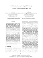
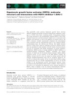
Fig. 1. Processing of pro-MSP by HGFA. (A) Immunoblot analysis
of proteolytic cleavage of a His-tagged human pro-MSP recombi-
nant protein by human HGFA. Pro-MSP at a concentration of 5 n
M
was incubated with 0.5 nM of HGFA in 20 mM Tris buffer (pH 7.6),
150 m
M NaCl and 0.05% Chaps for 8 h at 37 °C. Anti-MSP IgG rec-
ognized the a chain of MSP and the anti-His tag IgG recognized the
poly-His tag at the C-terminus of MSP. The N-terminal amino acid
sequence of a product of approximately 30 kDa is indicated.
(B) Effects of NaCl concentration on the processing of pro-MSP.
Pro-MSP at a concentration of 5 n
M was incubated with various
concentrations of HGFA or factor XIa in 20 m
M Tris buffer (pH 7.6)
and 0.05% Chaps, with 50–150 m
M of NaCl, for 4 h at 37 °C. The
processed products were analyzed by immunoblot. (C) Dose-depen-
dent processing of pro-MSP (5 n
M) by HGFA or factor XIa in Tris
buffer (pH 7.6), 50 m
M NaCl and 0.05% Chaps. The reaction mix-
tures were incubated for 4 h at 37 °C. (D) Time-dependent process-
ing of pro-MSP (5 n
M) by HGFA (0.5 nM) or factor XIa (0.5 nM)in
Tris buffer (pH 7.6), 50 m
M NaCl and 0.05% Chaps.
Activation of pro-MSP by HGFA M. Kawaguchi et al.
3482 FEBS Journal 276 (2009) 3481–3490 ª 2009 The Authors Journal compilation ª 2009 FEBS
In this study, we found that recombinant human
HGFA efficiently converts human pro-MSP to its
active form in vitro. Subsequent experiments using an
HGFA-deficient mouse model [18] revealed that
HGFA is a major serum activator of pro-MSP.
Results
Processing of pro-MSP by HGFA
The effect of recombinant human HGFA was tested
on the processing of recombinant human pro-MSP.
Incubation of pro-MSP with different concentrations
of HGFA at 37 °C led to the processing of pro-MSP
in a dose-dependent manner. Immunoblot analysis
using an anti-MSP IgG revealed a band of approxi-
mately 60 kDa, presumably the a chain of mature
MSP (Fig. 1A). Generation of a band of approxi-
mately 30 kDa, presumably the b chain, was also
detected by an anti-His tag IgG (Fig. 1A). Cleavage
site analysis was performed after separating the
products of HGFA cleavage by SDS–PAGE under
reducing conditions. The N-terminal amino acid
sequence of the 30 kDa product was Val-Val-Gly-Gly-
His. Therefore, this 30 kDa band was in fact the b
chain of mature MSP, and HGFA cleaved pro-MSP at
the normal processing site, Arg483–Val484 (Fig. 1A).
The processing was suppressed at higher concentra-
tions of NaCl, and this tendency was also observed for
factor XIa, a known serum activator of pro-MSP
(Fig. 1B). The concentration of HGFA required to acti-
vate 50% of 5 nm pro-MSP (PC50%) after 4 h at 37 °C
was 0.05 nm, whereas that of factor XIa was 0.5 nm.
Therefore, HGFA was a 10-fold more potent convertase
of pro-MSP than factor XIa (Fig. 1C). Further degra-
dation of mature MSP was not observed by HGFA. We
also examined the time course of pro-MSP processing
by HGFA (Fig. 1D). More than 50% of pro-MSP
(5 nm) was processed within 30 min by 0.5 nm of
HGFA, again showing superior efficiency to factor XIa.
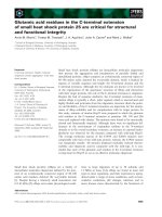
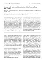
Biological activity of MSP processed by HGFA
The biological activity of MSP after HGFA processing
was determined using macrophage chemotaxis assays.
Pro-MSP could not efficiently induce the chemotactic
migration of macrophages. However, after incubation
of pro-MSP with HGFA, the processed products
showed a significant induction of macrophage migra-
tion, and the activity was comparable with that of
commercially available recombinant mature human
MSP a/b heterodimer (Fig. 2A). Macrophages derived
from HGFA
)/)
mice also responded to the recombi-
nant mature MSP a/b heterodimer (data not shown).
HGFA alone did not detectably induce the chemotac-
tic response. We also examined the effect of HGFA
processing on the culture morphology of mouse perito-
neal macrophages. The MSP processed by HGFA
induced an elongated, migratory morphology of
macrophages within 1 h, showing an effect similar to
+–+ ––
0
20
80
AB
+–+––
–– +––
pro-MSP
HGFA
MSP
Migrated cells/field
40
60
*
*
No treatment + MSP
+ pro-MSP + HGFA-treated pro-MSP
Control
pro-MSP
pro-MSP + HGFA
Fig. 2. Biological activity of MSP processed by HGFA. (A) Results of chemotaxis assays. Murine peritoneal resident macrophages
(1 · 10
5
cells) were placed in the upper well of Chemotaxicells and incubated for 3.5 h at 37 °C. The bottom well contained pro-MSP
(1.25 n
M) with or without HGFA (0.125 nM) pretreatment (2 h) or recombinant active MSP (1.25 nM). Values are the mean number ± stan-
dard deviation of migrated cells per high-power field in triplicate experiments. *P < 0.01 compared with control (pro-MSP only, HGFA only or
no addition) (Mann–Whitney U-test). Representative photographs of migrating cells are also shown. (B) Morphology of macrophages in the
presence of pro-MSP (1.25 n
M), MSP (1.25 nM) or pro-MSP (1.25 nM) pretreated with HGFA (0.125 nM). After 1 h in culture, the cells were
observed by phase-contrast microscopy.
M. Kawaguchi et al. Activation of pro-MSP by HGFA
FEBS Journal 276 (2009) 3481–3490 ª 2009 The Authors Journal compilation ª 2009 FEBS 3483
that of the recombinant mature MSP a/b heterodimer
(Fig. 2B).
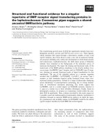
Impaired processing of endogenous pro-MSP
in serum from HGFA
-/-
mice
In order to study further the role of HGFA in the acti-
vation of pro-MSP, we used HGFA
)/)
mice. After
incubation at 37 °C for 2 h, most of the endogenous
MSP proteins in the plasma from both wild-type and
HGFA
)/)
mice were the 90 kDa single-chain pro-
forms (Fig. 3A). In contrast, pro-MSP was apparently
processed in the sera from wild-type mice after a 2 h
incubation at 37 °C (Fig. 3B), indicating the presence
of pro-MSP convertase in the wild-type serum, as pre-
viously observed in bovine serum [7]. However, the
processing of pro-MSP was reduced significantly in the
sera from HGFA
)/)
mice (Fig. 3B). A subsequent time
course study also confirmed the significantly reduced
processing activity of pro-MSP in HGFA-deficient sera
relative to that in wild-type sera (n = 5 for each
group) (Fig. 3C). Although the processing of endo-
genous pro-MSP was apparent within 15 min of incu-
bation and had reached 20% at 30 min in wild-type
serum, there was less than 10% processing even after
120 min of incubation in HGFA-deficient serum
(Fig. 3C). Therefore, the absence of HGFA resulted in
a markedly delayed and reduced processing of pro-
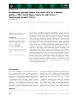
MSP in mouse serum. Indeed, the addition of recombi-
nant HGFA to the sera of HGFA
)/)
mice restored the
pro-MSP processing activity (Fig. 4).
Inhibition of pro-MSP processing activity
in serum by anti-HGFA neutralizing IgG
The efficient pro-MSP activating activity of human
HGFA in vitro and the significantly reduced processing
of endogenous pro-MSP in HGFA-deficient mouse
serum suggest that HGFA is a major serum activator
of pro-MSP in vivo. Therefore, we examined the effect
of a neutralizing antibody raised against HGFA (P1-4)
on the pro-MSP convertase activity of wild-type mouse
serum. The P1-4 antibody suppressed significantly the
processing of pro-MSP in sera obtained from wild-type
mice (Fig. 5). We concluded that HGFA is a major
activator of pro-MSP in mouse serum.
Delayed infiltration of macrophages in HGFA
-/-
mice at a site of tissue injury
To test the physiological role of HGFA-mediated acti-
vation of pro-MSP, we compared the recruitment of
macrophages in injured tissues, in which the activation
of pro-HGFA was anticipated by thrombin and/or
0
10
20
30
40
50
60
70
01530 60 120
pro-MSP
pro-MSP
pro-MSP
α-chain
α-chain
α-chain
1212
1212
120 min
120 min
60 min
Incubation
time
Incubation
time
PlasmaA
B
C
Serum
Wild HGFA
–/–
HGFA
–/–
Wild
% converted pro-MSP
Incubation time (min)
Wild-type serum
HGFA-deficient serum
*
Fig. 3. Impaired processing of endogenous pro-MSP in HGFA-defi-
cient serum. (A) Processing of endogenous pro-MSP in plasma
from wild-type and HGFA
)/)
mice. Plasma was incubated at 37 °C
and the processing of endogenous pro-MSP was analyzed by
immunoblot using anti-MSP antibody. (B) Processing of endo-
genous pro-MSP in sera from wild-type and HGFA
)/)
mice. Serum
was incubated at 37 °C and the processing of endogenous pro-
MSP was analyzed by immunoblot. (C) Time course of pro-MSP
processing in serum. Values are the mean processing rate ± stan-
dard deviation (n = 5). *P < 0.001, Mann–Whitney U-test.
0 0.018 0.18 1.8 18 180
I
ncu
b
at
i
on: 120 m
i
n
HGFA (n
M)
% converted
pro-MSP
pro-MSP
MSP
α
chain
56 85 > 90% > 90 > 90
Fig. 4. Reversion of pro-MSP convertase activity in serum from
HGFA
)/)
mice by recombinant HGFA. HGFA-deficient serum was
incubated with recombinant human HGFA (0–180 n
M) for 2 h, and the
processing of endogenous pro-MSP was analyzed by immunoblot.
Activation of pro-MSP by HGFA M. Kawaguchi et al.
3484 FEBS Journal 276 (2009) 3481–3490 ª 2009 The Authors Journal compilation ª 2009 FEBS
KLKs, between wild-type mice and HGFA
)/)
mice.
We generated a small mechanical wound in the dorsal
skin of mice and examined the infiltration of macro-
phages by measuring the CD68 mRNA level. One day
after injury, the levels of CD68 mRNA in the wounds
of HGFA
)/)
mice were significantly lower than those
of wild-type mice (Fig. 6A). The level of pro-MSP pro-
cessing was also low in HGFA
)/)
wounds (Fig. 6B).
However, at the fifth day of injury, the CD68 mRNA
level was comparable between HGFA
)/)
wounds and
wild-type wounds (Fig. 6A). These results suggest that
serum HGFA is required for the early-phase recruit-
ment of macrophages at the injured tissue, possibly via
its efficient pro-MSP processing activity.
Discussion
Pro-MSP is primarily produced by the liver [6] and cir-
culates in blood with a concentration of 2–5 nm [5]. It is
converted to its mature active form during blood coagu-
lation and local inflammation [5,7]. Wound fluids also
contain pro-MSP convertase activity, and a cellular sur-
face proteinase is also an important convertase [5,8,9].
This activation step of pro-MSP might serve as a critical
regulatory mechanism in MSP-induced physiological
and pathophysiological tissue responses. After proteo-
lytic cleavage, it stimulates resident macrophages via its
specific receptor tyrosine kinase, RON [5,11]. Epithelial
cells and neoplastic cells also frequently express RON
[11,20,21]. The establishment of RON-induced signaling
appears to have an important role in inflammatory pro-
cesses [1,5,22–24], cellular survival and wound healing
[25,26]. It is also important in the progression and meta-
static spread of various types of tumor [11,27,28]. In this
study, we have shown that human HGFA efficiently
activates human pro-MSP in vitro. In mice, serum
HGFA represents the major pro-MSP convertase activ-
ity of the serum. Indeed, the conversion of endogenous
pro-MSP to its mature form was impaired in sera from
HGFA
)/)
mice and the convertase activity in wild-type
sera was significantly attenuated by the addition of anti-
HGFA neutralizing IgG. Moreover, initial infiltration
of macrophages into the site of mechanical skin injury
was delayed in HGFA
)/)
mice. Together with the fact
that matriptase, a cell surface activator of pro-MSP [9],
is also a potent activator of pro-HGF/SF [29], we sug-
gest that pro-MSP might share its activation machiner-
ies with pro-HGF/SF (Fig. 7).
The identification of HGFA as a major serum activa-
tor of pro-MSP may explain why the serum convertase
activity of pro-MSP is different between species.
Although bovine serum [7] and mouse serum showed
significant processing activity for endogenous pro-MSP,
the processing activity of human serum was very weak
and the molecular form of MSP in human serum was
mostly proforms (data not shown), as described previ-
ously [8]. HGFA is resistant to major serum proteinase
inhibitors [12] and is active in mouse serum [18]. How-
ever, it can be inhibited by PCI, a serpin-type protein-
ase inhibitor present in human plasma [14,30].
However, mouse plasma does not contain PCI [30].
Therefore, HGFA-mediated conversion of pro-MSP
may be tightly regulated by PCI in human serum,
whereas HGFA remains active and easily converts pro-
MSP in mouse serum because of the absence of PCI.
HGFA is present in plasma as an inactive zymogen
at a concentration of approximately 40 nm in humans
[14]. During tissue injury, pro-HGFA is converted to
the active heterodimeric form by thrombin [12,15].
Human kallikrein 1-related peptidases, KLK4 and
KLK5, are also candidate activators of pro-HGFA in
the local tissue environment [17]. After conversion,
mature HGFA very efficiently activates pro-HGF/SF
at the site of injury [16], which might have important
roles in survival, repair and regeneration of the injured
tissue [12,19]. The activity of HGFA is tightly regu-
lated by PCI in human serum and also by HGFA
inhibitor type 1 and type 2 on the epithelial cell surface
[12,14,19]. Nonetheless, the activity of HGFA is
detectable in injured human tissues, such as invasive
tumors, accompanying the activation of pro-HGF/SF
[31]. To date, the possible involvement of HGFA in
tissue repair and cancer progression has been discussed
primarily in the context of its presumed capability to
activate pro-HGF/SF and the subsequent MET signal-
ing cascade [11,12,18]. This study indicates that MSP-
induced RON signaling can be initiated by HGFA
activity and may contribute to the role of HGFA in
tissue repair and cancer progression. Furthermore, the
activation of pro-MSP by HGFA prompts the consid-
eration of the possible role of HGFA in inflammation
via modulation of macrophage function.
IgG1 P1-4 IgG1 P1-4 IgG1 P1-4
0 min 30 min 60 min
Incubation time
Antibody
pro-MSP
MSP
α
chain
— — —
35 38 14 52 61 29%
% converted
pro-MSP
Fig. 5. Inhibition of pro-MSP processing activity in serum by anti-
HGFA IgG. Serum from wild-type mice was incubated for the
indicated time periods at 37 °C without or with 400 lgÆmL
)1
of
anti-HGFA neutralizing IgG (P1-4) or nonspecific mouse IgG1. The
processing of endogenous pro-MSP was analyzed by immunoblot.
M. Kawaguchi et al. Activation of pro-MSP by HGFA
FEBS Journal 276 (2009) 3481–3490 ª 2009 The Authors Journal compilation ª 2009 FEBS 3485
Evidence suggests that MSP exerts a dual function,
both stimulatory and inhibitory, on macrophages [5].
Stimulatory functions include its ability to induce mac-
rophage spreading, migration, phagocytosis and the
production of cytokines [1,5,32]. However, MSP inhib-
its lipopolysaccharide (LPS)-induced production of
inflammatory mediators and, consequently, RON-defi-
cient mice show increased inflammatory responses and
susceptibility to LPS-induced septic death [5,22–24].
Therefore, MSP is also required to attenuate an exces-
sive inflammatory response to LPS stimulation, and
thus may have an important regulatory role in septic
Wild-type HGFA KO
1.11 ± 0.22 1.08 ± 0.16
CD68
β
-actin
Wild-type
A
B
HGFA KO
0 day
0.33 ± 0.08
CD68/actin
0.30 ± 0.06
Wild-type HGFA KO
1.25 ± 0.05 0.79 ± 0.15
*
1 day 5 day
Wild KO
Wild
KO
Incubated
Serum
1 day 0 day
pro-MSP
MSP α chain
% converted pro-MSP 44 33 — < 5 18 48 84 < 5 — — —
Fig. 6. Delayed infiltration of macrophages in cutaneous wounds of HGFA
)/)
mice. (A) Infiltration of macrophages in wounded skin tissue
was evaluated by CD68 mRNA level. *P < 0.05, Mann–Whitney U-test (n = 4). (B) Processing level of pro-MSP in injured tissues (1 day after
injury). Skin tissues without injury (0 day) were also examined. For positive control, wild-type serum after incubation for the processing of
endogenous pro-MSP (incubated serum) was also applied. Wild, wild-type mice; KO, HGFA
)/)
mice.
Tissue injury
Activation of
coagulation cascade
pro-thrombin
Thrombin
pro-HGFA
pro-MSP
MSP
pro-HGF/SF
HGF/SF
RON
MET
HGFA
EGF-BP, NGF-γ
Matriptase
KLK4, KLK5
Cell surface
proteinase(s)
Macrophages
Epithelial
cells
Tumour cells
Endothelial
cells
Factor XIa
Factor XIIa
Fig. 7. Hypothetical model for the activation of pro-MSP. There may be diverse pathways for the activation of pro-MSP, and pro-MSP might
share the activation machinery with its homologous protein, pro-HGF/SF. One pathway is mediated by membrane-bound serine proteinases
(cell surface activator), such as matriptase [9]. Matriptase is also a potent activator of pro-HGF/SF [12,30]. The second pathway is mediated
by humoral activators that are generated in injured tissues. The activation of the coagulation cascade by tissue injury eventually results in
the active form of HGFA that efficiently activates both pro-MSP and pro-HGF/SF. Other coagulation proteinases, such as factor XIa and fac-
tor XIIa, may also mediate the activation of pro-MSP [7] and pro-HGF/SF [12]. Wound fluids in the injured tissues contain other pro-MSP acti-
vators, such as epidermal growth factor-binding protein (EGF-BP) and nerve growth factor c (NGF-c) [10]. The effects of EGF-BP and NGF-c
on pro-HGF/SF are unknown.
Activation of pro-MSP by HGFA M. Kawaguchi et al.
3486 FEBS Journal 276 (2009) 3481–3490 ª 2009 The Authors Journal compilation ª 2009 FEBS
inflammation. In the septic condition, intravascular
hypercoagulation occurs, which might result in the
conversion of pro-HGFA to its active form. The
HGFA-mediated activation of pro-MSP may be an
important process in the regulation of macrophage
functions in septic inflammatory responses. To test this
hypothesis, future studies of HGFA
)/)
mice under vari-
ous inflammatory stimuli, including LPS stimulation,
will be required. Moreover, the difference in serum
HGFA activity between human and mouse may
have implications in the different susceptibility to
LPS-induced septic death between these species.
In summary, we have demonstrated for the first time
that HGFA is a potent activator of pro-MSP. Although
the activation of pro-MSP is a redundant system which
can be mediated by various proteinases (Fig. 7) [7–10],
the major pro-MSP convertase in serum is HGFA. As
pro-HGFA is activated in response to tissue injury, we
suggest that HGFA-mediated activation may play an
important role in the regulation of MSP/RON signaling
involved in inflammation, wound healing and cancer
progression. Further experiments of tissue injury and
inflammation using genetically engineered mouse mod-
els of the HGFA and MSP genes are needed to explore
the in vivo significance of HGFA in MSP/RON signal-
ing. However, our study also indicates that caution
should be exercised when interpreting the function of
MSP/RON signaling using a mouse model in vivo,as
HGFA activity would be much higher in mouse serum
than in human serum because of the absence of circulat-
ing PCI in mice [30].
Experimental procedures
Antibodies
Anti-human MSP goat polyclonal IgG and the recombinant
active form of human MSP were purchased from R&D Sys-
tems (Minneapolis, MN, USA). Recombinant human factor
XIa was obtained from Haematologic Technologies, Inc.
(Essex Junction, VT, USA). Anti-mouse MSP goat
polyclonal IgG (T-19) was obtained from Santa Cruz
Biotechnology (Santa Cruz, CA, USA). The preparation of
the recombinant active form of human HGFA and anti-
human HGFA mouse monoclonal neutralizing IgG P1-4,
which is also cross-reactive to mouse HGFA, has been
described previously [15]. Anti-His tag rabbit polyclonal IgG
was purchased from MBL (Nagoya, Japan).
Preparation of recombinant proteins
The preparation of the recombinant active form of HGFA
has been described previously [19]. To obtain a recombinant
pro-MSP protein, the entire coding region of the MSP gene
was subcloned into the pcDNA3.1/myc-HisA expression
plasmid (Invitrogen, Carlsbad, CA, USA) and transfected
into Chinese hamster ovary (CHO) cells using Lipofectamine
2000 reagent (Invitrogen). After transfection, the cells were
cultured in DMEM containing 10% fetal bovine serum and
gradually changed to serum-free medium (CHO-S-SFMII;
Invitrogen) containing 250 lgÆmL
)1
G418 (Sigma-Aldrich,
St Louis, MO, USA). To prevent the cleavage of pro-MSP
by cellular and fetal bovine serum-derived proteases, cells
were cultured in the presence of 50 lm nafamostat mesilate
(Torii Pharmaceutical Co., Tokyo, Japan). G418-resistant
colonies were selected and screened for the expression and
production of pro-MSP. Supernatants were collected from
the serum-free cultures every day and 0.1% Chaps (Sigma-
Aldrich) was added. Recombinant pro-MSP in the condi-
tioned medium was affinity purified with TALON His-Tag
Purification Resins (Clontech Laboratories, Mountain View,
CA, USA) according to the manufacturer’s instructions.
Activation of pro-MSP
Recombinant pro-MSP (final concentration, 5 nm) was
incubated with various concentrations of HGFA or factor
XIa in 20 lL reactions containing 20 mm Tris/HCl,
50–150 mm NaCl and 0.05% Chaps (pH 7.6) for the indi-
cated time periods at 37 °C. The processing of pro-MSP
was determined by immunoblot analysis under reducing
conditions, and the extent of processing was verified using
photoshop software (Adobe Systems, San Jose, CA, USA).
The specific activity for pro-MSP processing was expressed
as the enzyme concentration required for the conversion of
50% of 5 nm pro-MSP to its mature form, and was desig-
nated as the processing concentration 50% (PC50%). To
assess the time course of cleavage by HGFA or factor XIa,
pro-MSP (5 nm) was incubated with 0.5 nm of each
proteinase at 37 °C for various time periods (0–120 min).
Immunoblot analysis
Each sample was mixed with SDS–PAGE sample buffer
and heated for 15 min at 70 °C. SDS–PAGE was per-
formed under reducing conditions using 4–12% gradient
gels. After electrophoresis, samples were transferred to
Immobilon poly(vinylidene difluoride) membranes (Milli-
pore, Bedford, MA, USA). After blocking with 3% BSA in
Tris-buffered saline (TBS) with 0.05% Tween-20 (TBS-T),
the membranes were incubated with primary antibody at
4 °C overnight, followed by washing in TBS-T and incuba-
tion with a horseradish peroxidase-conjugated rabbit anti-
goat IgG (DAKO, Glostrup, Denmark) diluted in TBS-T
with 1% BSA for 1 h at room temperature. The labeled
proteins were visualized with a chemiluminescence reagent
(PerkinElmer Life Science, Boston, MA, USA).
M. Kawaguchi et al. Activation of pro-MSP by HGFA
FEBS Journal 276 (2009) 3481–3490 ª 2009 The Authors Journal compilation ª 2009 FEBS 3487
N-terminal amino acid sequencing of cleaved
pro-MSP
Pro-MSP (final concentration, 416 nm) was incubated with
97 nm HGFA in a 40 lL reaction containing 20 mm Tris/
HCl, 150 mm NaCl and 0.05% Chaps (pH 7.6) at 37 °C for
11 h. The reaction mixture was subjected to SDS–PAGE,
after which the proteins were transferred to an Immobilon
membrane and stained with 0.1% Coomassie Brilliant Blue
in a water–methanol–acetic acid solution (4.5 : 4.5 : 1, v/v).
The cleaved MSP protein band was cut and processed for
N-terminal amino acid sequencing by automated Edman
degradation using the Procise 494 HT Protein Sequencing
System (Applied Biosystems, Foster City, CA, USA).
Preparation of peritoneal macrophages and
bioassays
Murine peritoneal resident macrophages were obtained
from C57BL/6 mice by washing the peritoneal cavity with
3 mL per mouse of serum-free RPMI-1640 medium. Cells
were washed and resuspended in RPMI-1640 medium con-
taining 25 mm Hepes at a concentration of 1 · 10
6
cell-
sÆmL
)1
. The macrophage chemotaxis assay was performed
using a polycarbonate membrane with a pore size of 5 lm
(Chemotaxicells; Kurabo, Osaka, Japan). One hundred
microliters of the cell suspension (i.e. 10
5
macrophages)
were added to the upper wells of the Chemotaxicells. The
bottom wells were filled with RPMI-1640 medium contain-
ing purified pro-MSP treated or not with HGFA at 37 °C
for 2 h. The recombinant active form of human MSP
(R&D Systems) was used as a positive control. After incu-
bation at 37 °C for 3.5 h, the cells on the upper surface of
the membrane were wiped off with a cotton swab and the
membranes were fixed with 3.7% formaldehyde in NaCl/P
i
and stained with hematoxylin. Migration was quantified by
counting the cells on the lower surface in 10 randomly
selected high-power fields (200-fold magnification). To test
the effect of MSP on the morphological changes of macro-
phages, murine peritoneal resident macrophages
(1 · 10
6
cellsÆmL
)1
) were cultured in serum-free RPMI-1640
medium overnight. After incubation, nonadherent cells were
removed and pro-MSP (1.25 nm), pretreated or not with
HGFA, was added to the culture medium. After an addi-
tional incubation at 37 °C for 1 h, morphological changes
of the macrophages were observed by phase-contrast
microscopy.
Analysis of molecular forms of MSP in wild-type
and HGFA-deficient mice
The generation of HGFA knockout (HGFA
)/)
) mice by
the targeting of gene disruption has been reported previ-
ously [18]. Sera and EDTA-treated plasma were obtained
from C57BL/6 wild-type (HGFA
+/+
) and HGFA
)/)
mice,
and diluted 10-fold with phosphate buffer (pH 7.4).
Molecular forms of endogenous MSP in the plasma and
serum were analyzed by immunoblots. To test the effect
of complementation of HGFA activity on serum pro-
MSP convertase activity, the diluted serum from an
HGFA
)/)
mouse was incubated with varying concentra-
tions of recombinant HGFA at 37 °C for 2 h, and ana-
lyzed by immunoblot. For a neutralizing study, the
diluted serum from a C57BL/6 mouse was incubated with
or without 400 lgÆmL
)1
of anti-HGFA neutralizing anti-
body at 37 °C for the indicated time periods. The molec-
ular forms of endogenous MSP were analyzed by
immunoblot.
Skin injury model
Eight-week-old male wild-type and HGFA
)/)
mice were
deeply anesthetized by intraperitoneal administration of
ketamine hydrochloride [100 lgÆ(g body weight)
)1
; Sankyo,
Tokyo, Japan] and xylazine [10 lgÆ(g body weight)
)1
;
Bayer, Tokyo, Japan]. After shaving the dorsal hair and
cleaning with 70% ethanol, two full-thickness excisional
skin wounds (5 mm in diameter) were made. Mice were sac-
rificed at 1 or 5 days after the generation of wounds. The
wounded tissues were excised and used for RT–PCR,
immunoblot analysis for pro-MSP processing and routine
histological analysis with hematoxylin and eosin staining.
For control, normal skin tissues were also biopsied (0 day).
For RT-PCR, total RNA was prepared with TRIzol (Invi-
trogen Japan, Tokyo, Japan) followed by DNase I (Takara
Bio, Shiga, Japan) treatment. Three micrograms of total
RNA were reverse transcribed with a mixture of oligo
(dT)
12)18
(Invitrogen Japan) and random primers (6-mer)
(Takara Bio) using 200 units of ReverTraAceÔ (TOYOBO,
Osaka, Japan), and 1/30 of the resultant cDNA was pro-
cessed for each PCR with 0.1 lm of both forward and
reverse primers and 2.5 units of HotStarÔ Taq DNA poly-
merase (Qiagen, Tokyo, Japan). The following primers were
used: b-actin: forward, 5¢-TGACAGGATGCAGAAGGA
GA; reverse, 5¢-GCTGGAAGGTGGACAGTGAG; CD68:
forward, 5¢-TCTACCTGGACTACATGGCGGTGG; reverse,
5¢-ACATGGCCCGAAGTGTCCCTTGTC. For immuno-
blot, tissues were homogenized on ice in lysis buffer
(CelLyticÔ-MT; Sigma-Aldrich) supplemented with prote-
ase inhibitor cocktail (Sigma-Aldrich). The extracts were
centrifuged at 20 000 g for 20 min at 4 °C, and the result-
ing supernatants were used for immunoblot.
Statistical analysis
Statistical analyses were carried out using spss 15.0 (SPSS
JAPAN Inc., Tokyo, Japan). P values of less than 0.05
were considered to be statistically significant.
Activation of pro-MSP by HGFA M. Kawaguchi et al.
3488 FEBS Journal 276 (2009) 3481–3490 ª 2009 The Authors Journal compilation ª 2009 FEBS
Acknowledgements
This study was supported by a Grant-in-Aid for Scien-
tific Research (B) No. 20390114 from the Ministry of
Education, Science, Sports and Culture, Japan. We
thank Dr Miyuki Daio for assistance and Dr Takeshi
Shimomura for helpful discussions.
References
1 Skeel A, Yoshimura T, Showalter SD, Tanaka S,
Appella E & Leonard EJ (1991) Macrophage
stimulating protein: purification, partial amino
acid sequence, and cellular activity. J Exp Med 173,
1227–1234.
2 Yoshimura T, Yuhki N, Wang MH, Skeel A & Leon-
ard EJ (1993) Cloning, sequencing, and expression of
human macrophage stimulating protein (MSP, MST1)
confirms MSP as a member of the family of kringle
proteins and locates the MSP gene on chromosome 3.
J Biol Chem 268, 15461–15468.
3 Shimamoto A, Kimura T, Matsumoto K & Nakamura
T (1993) Hepatocyte growth factor-like protein is identi-
cal to macrophage stimulating protein. FEBS Lett 333,
61–66.
4 Wang MH, Ronsin C, Gesnel MC, Coupey L, Skeel A,
Leonard EJ & Breathnach R (1994) Identification of
the ron gene product as the receptor for the
human macrophage stimulating protein. Science 266,
117–119.
5 Wang MH, Zhou YQ & Chen YQ (2002) Macrophage-
stimulating protein and RON receptor tyrosine kinase:
potential regulators of macrophage inflammatory activi-
ties. Scand J Immunol 56, 545–553.
6 Bezerra JA, Witte DP, Aronow BJ & Degen SJ (1993)
Hepatocyte-specific expression of the mouse hepatocyte
growth factor-like protein. Hepatology 18, 394–399.
7 Wang MH, Yoshimura T, Skeel A & Leonard EJ
(1994) Proteolytic conversion of single chain precursor
macrophage-stimulating protein to a biologically active
heterodimer by contact enzymes of the coagulation
cascade. J Biol Chem 269, 3436–3440.
8 Wang MH, Skeel A & Leonard EJ (1996) Proteolytic
cleavage and activation of pro-macrophage-stimulating
protein by resident peritoneal macrophage membrane
proteases. J Clin Invest 97, 720–727.
9 Bhatt AS, Welm A, Farady CJ, Va
´
squez M, Wilson K
& Craik CS (2007) Coordinate expression and func-
tional profiling identify an extracellular proteolytic
signaling pathway. Proc Natl Acad Sci USA 104,
5771–5776.
10 Wang MH, Gonias SL, Skeel A, Wolf BB, Yoshimura
T & Leonard EJ (1994) Proteolytic activation of single-
chain precursor macrophage-stimulating protein by
nerve growth factor-gamma and epidermal growth
factor-binding protein, members of the kallikrein
family. J Biol Chem 269, 13806–13810.
11 Benvenuti S & Comoglio PM (2007) The MET receptor
tyrosine kinase in invasion and metastasis. J Cell
Physiol 213, 316–325.
12 Kataoka H, Miyata S, Uchinokura S & Itoh H (2003)
Roles of hepatocyte growth factor (HGF) activator
and HGF activator inhibitor in the pericellular
activation of HGF/scatter factor. Cancer Metastasis
Rev 22, 223–236.
13 Miyazawa K, Shimomura T, Kitamura A, Kondo J,
Morimoto Y & Kitamura N (1993) Molecular cloning
and sequence analysis of the cDNA for a human serine
protease responsible for activation of hepatocyte growth
factor. Structural similarity of the protease precursor
to blood coagulation factor XII. J Biol Chem 268,
10024–10028.
14 Hayashi T, Nishioka J, Nakagawa N, Kamada H,
Gabazza EC, Kobayashi T, Hattori A & Suzuki K
(2007) Proteinb C inhibitor directly and potently
inhibits activated hepatocyte growth factor activator.
J Thromb Haemost 5, 1477–1485.
15 Miyazawa K, Shimomura T & Kitamura N (1996) Acti-
vation of hepatocyte growth factor in the injured tissues
is mediated by hepatocyte growth factor activator.
J Biol Chem 271, 3615–3618.
16 Shimomura T, Kondo J, Ochiai M, Naka D, Miyazawa
K, Morimoto Y & Kitamura N (1993) Activation of
the zymogen of hepatocyte growth factor activator by
thrombin. J Biol Chem 268, 22927–22932.
17 Mukai S, Fukushima T, Naka D, Tanaka H, Osada Y
& Kataoka H (2008) Activation of hepatocyte growth
factor activator zymogen (pro-HGFA) by human kallik-
rein 1-related peptidases. FEBS J 275, 1003–1017.
18 Itoh H, Naganuma S, Takeda N, Miyata S, Uchinok-
ura S, Fukushima T, Uchiyama S, Tanaka H, Nagaike
K, Shimomura T et al. (2004) Regeneration of injured
intestinal mucosa is impaired in hepatocyte growth
factor activator-deficient mice. Gastroenterology 127,
1423–1435.
19 Kataoka H, Shimomura T, Kawaguchi T, Hamasuna
R, Itoh H, Kitamura N, Miyazawa K & Koono M
(2000) Hepatocyte growth factor activator inhibitor
type 1 is a specific cell surface binding protein of hepa-
tocyte growth factor activator (HGFA) and regulates
HGFA activity in the pericellular microenvironment.
J Biol Chem 275, 40453–40462.
20 Okino T, Egami H, Ohmachi H, Takai E, Tamori Y,
Nakagawa A, Nakano S, Sakamoto O, Suda T &
Ogawa M (1991) Immunohistochemical analysis of dis-
tribution of RON receptor tyrosine kinase in human
digestive organs. Dig Dis Sci 46, 424–429.
21 Wang MH, Lee W, Luo YL, Weis MT & Yao HP
(2007) Altered expression of the RON receptor tyrosine
kinase in various epithelial cancers and its contribution
M. Kawaguchi et al. Activation of pro-MSP by HGFA
FEBS Journal 276 (2009) 3481–3490 ª 2009 The Authors Journal compilation ª 2009 FEBS 3489
to tumorigenic phenotypes in thyroid cancer cells.
J Pathol 213, 402–411.
22 Correll PH, Iwama A, Tondat S, Mayrhofer G, Suda T
& Bernstein A (1997) Deregulated inflammatory
response in mice lacking the STK/RON receptor tyro-
sine kinase. Genes Funct 1, 69–83.
23 Muraoka RS, Sun WY, Colbert MC, Waltz SE, Witte
DP, Degen JL & Friezner Degen SJ (1999) The Ron/
STK receptor tyrosine kinase is essential for peri-
implantation development in the mouse. J Clin Invest
103, 1277–1285.
24 Morrison AC, Wilson CB, Ray M & Correll PH (2004)
Macrophage-stimulating protein, the ligand for the stem
cell-derived tyrosine kinase/RON receptor tyrosine
kinase, inhibits IL-12 production by primary peritoneal
macrophages stimulated with IFN-gamma and lipopoly-
saccharide. J Immunol 172, 1825–1832.
25 Danilkovitch-Miagkova A & Leonard EJ (2001) Anti-
apoptotic action of macrophage stimulating protein
(MSP). Apoptosis 6, 183–190.
26 Cantaluppi V, Biancone L, Romanazzi GM, Figliolini
F, Beltramo S, Galimi F, Camboni MG, Deriu E,
Conaldi P, Bottelli A et al. (2008) Macrophage
stimulating protein may promote tubular regeneration
after acute injury. J Am Soc Nephrol 19, 1904–1918.
27 O’Toole JM, Rabenau KE, Burns K, Lu D, Mangalam-
palli V, Balderes P, Covino N, Bassi R, Prewett M,
Gottfredsen KJ et al. (2006) Therapeutic implications
of a human neutralizing antibody to the
macrophage-stimulating protein receptor tyrosine
kinase (RON), a c-MET family member. Cancer Res
66, 9162–9170.
28 Welm AL, Sneddon JB, Taylor C, Nuyten DS, van de
Vijver MJ, Hasegawa BH & Bishop JM (2007) The
macrophage-stimulating protein pathway promotes
metastasis in a mouse model for breast cancer and pre-
dicts poor prognosis in humans. Proc Natl Acad Sci
USA 104, 7570–7575.
29 Lee SL, Dickson RB & Lin CY (2000) Activation of
hepatocyte growth factor and urokinase/plasminogen
activator by matriptase, an epithelial membrane serine
protease. J Biol Chem 275, 36720–36725.
30 Suzuki K (2008) The multi-functional serpin, protein C
inhibitor: beyond thrombosis and hemostasis. J Thromb
Haemost 6, 2017–2026.
31 Kataoka H, Hamasuna R, Itoh H, Kitamura N &
Koono M (2000) Activation of hepatocyte growth
factor/scatter factor in colorectal carcinoma. Cancer
Res 60, 6148–6159.
32 Suzuki Y, Funakoshi H, Machide M, Matsumoto K &
Nakamura T (2008) Regulation of cell migration and
cytokine production by HGF-like protein (HLP)/macro-
phage stimulating protein (MSP) in primary microglia.
Biomed Res 29, 77–84.
Activation of pro-MSP by HGFA M. Kawaguchi et al.
3490 FEBS Journal 276 (2009) 3481–3490 ª 2009 The Authors Journal compilation ª 2009 FEBS