Báo cáo khoa học: Quenched hydrogen ⁄deuterium exchange NMR characterization of amyloid-b peptide aggregates formed in the presence of Cu2+ or Zn2+ ppt
Bạn đang xem bản rút gọn của tài liệu. Xem và tải ngay bản đầy đủ của tài liệu tại đây (488.39 KB, 10 trang )
Quenched hydrogen
⁄
deuterium exchange NMR
characterization of amyloid-b peptide aggregates
formed in the presence of Cu
2+
or Zn
2+
Anders Olofsson
1
, Malin Lindhagen-Persson
1
, Monika Vestling
1
, A. Elisabeth Sauer-Eriksson
2
and
Anders O
¨
hman
2
1 Department of Medical Biochemistry and Biophysics, Umea
˚
University, Sweden
2 Department of Chemistry, Umea
˚
University, Sweden
Introduction
Abnormal protein assemblies in form of amyloid are
linked to more than 25 different syndromes, of which
the neurodegenerative disorder Alzheimer’s disease
(AD) is the most well-known example [1]. The main
Keywords
Alzheimer’s disease; amyloid-b peptide;
Cu
2+
;H⁄ D exchange NMR; Zn
2+
Correspondence
A. O
¨
hman, Department of Chemistry, Umea
˚
University, SE-901 87 Umea
˚
, Sweden
Fax: +46 90 786 5944
Tel: +46 90 786 5919
E-mail:
A. Olofsson, Department of Medical
Biochemistry and Biophysics, Umea
˚
University, SE-901 87 Umea
˚
, Sweden
Fax: +46 90 786 5944
Tel: +46 90 786 5921
E-mail:
(Received 10 March 2009, revised 4 May
2009, accepted 26 May 2009)
doi:10.1111/j.1742-4658.2009.07113.x
Alzheimer’s disease, a neurodegenerative disorder causing synaptic impair-
ment and neuronal cell death, is strongly correlated with aggregation of the
amyloid-b peptide (Ab). Divalent metal ions such as Cu
2+
and Zn
2+
are
known to significantly affect the rate of aggregation and morphology of
Ab assemblies in vitro and are also found at elevated levels within cerebral
plaques in vivo. The present investigation characterized the architecture of
the aggregated forms of Ab(1–40) and Ab(1–42) in the presence or absence
of either Cu
2+
or Zn
2+
using quenched hydrogen ⁄ deuterium exchange
combined with solution NMR spectroscopy. The NMR analyses provide a
quantitative and residue-specific structural characterization of metal-
induced Ab aggregates, showing that both the peptide sequence and the
type of metal ion exert an impact on the final architecture. Common
features among the metal-complexed peptide aggregates are two solvent-
protected regions with an intervening minimum centered at Asn27, and a
solvent-accessible N-terminal region, Asp1–Lys16. Our results suggest that
Ab in complex with either Cu
2+
or Zn
2+
can attain an aggregation-prone
b-strand–turn–b-strand motif, similar to the motif found in fibrils, but
where the metal binding to the N-terminal region guides the peptide into
an assembly distinctly different from the fibril form.
Structured digital abstract
l
MINT-7102414, MINT-7102427: Abeta (uniprotkb:P05067) and Abeta (uniprotkb:P05067)
bind (
MI:0407)byfluorescence technologies (MI:0051)
l
MINT-7103341, MINT-7103348: Abeta (uniprotkb:P05067) and Abeta (uniprotkb:P05067)
bind (
MI:0407)byatomic force microscopy (MI:0872)
l
MINT-7102371, MINT-7102380: Abeta (uniprotkb:P05067) and Abeta (uniprotkb:P05067)
bind (
MI:0407)bycircular dichroism (MI:0016)
l
MINT-7102390, MINT-7102399: Abeta (uniprotkb:P05067) and Abeta (uniprotkb:P05067)
bind (
MI:0407)bybiophysical (MI:0013)
l
MINT-7103367, MINT-7103374: Abeta (uniprotkb:P05067)andAbeta (uniprotkb:P05067) bind
(
MI:0407)bynuclear magnetic resonance (MI:0077)
Abbreviations
AD, Alzheimer’s disease; AFM, atomic force microscopy; Ab, amyloid-b peptide; H ⁄ D, hydrogen ⁄ deuterium; ThT, thioflavin T.
FEBS Journal 276 (2009) 4051–4060 ª 2009 The Authors Journal compilation ª 2009 FEBS 4051
protein component of the plaques found in patients
with AD is the amyloid-b peptide (Ab). This is a
proteolytic excision product derived from the signifi-
cantly larger amyloid precursor protein, representing
an ensemble of peptides of various lengths, each of
which has distinguishing biophysical properties. Frag-
ments of 39–43 residues are all clinically relevant, but
the most abundant are Ab(1–40) and Ab(1–42) [2].
The fold and mode of assembly of Abs are deter-
mined by their primary sequences in combination with
the chemical properties of the solvent. Abs assemble
via several different routes into aggregates ranging in
size from dimers [3] to smaller oligomers [4–7], protofi-
brils [8,9], and fully mature amyloid fibers. Elucidation
of amyloid fiber and aggregate structures, as well as
their path of formation, is crucial to understanding the
pathological process of AD. Although the size, insolu-
bility and noncrystalline behavior of fibers and aggre-
gates make conventional structural studies difficult,
extensive solid-state and solution-state NMR investiga-
tions of both Ab(1–40) and Ab(1–42) fibrils have dem-
onstrated that the peptides stack perpendicularly along
the fibril axis via a parallel in-register assembly of a
b-strand–turn–b-strand motif, forming a characteristic
cross-b structure with a highly solvent-protected core
of two intermolecular b-sheets [10–13]. X-ray micro-
crystallography of short Ab fragments has revealed
very tight packing of side chains within the cross-b
structure [14].
The aggregation properties of Abs are strongly
affected by their affinities for certain metal ions [15–
19]. Elevated concentrations of Cu
2+
,Zn
2+
and Fe
2+
are found within deposited plaques in vivo [20], and
both Cu
2+
and Zn
2+
affect the toxicity of Ab [21–25].
The in vivo effect of metals can be complex [26,27],
and the presence of Cu
2+
,Zn
2+
or Ca
2+
significantly
accelerates aggregation of Ab in vitro [19,28,29]. The
binding of Cu
2+
and Zn
2+
to monomeric Ab has been
extensively studied and occurs via three histidines
(His6, His13, and His14) and a fourth ligand suggested
to be either Tyr10 [30–32], Glu11 [33], or the N-termi-
nus [34,35]. A weaker binding site for zinc, involving
Asp23–Lys28, has also been identified [35]. Ab assem-
blies formed in the presence of metals are not charac-
terized as amyloid, because they lack tinctorial
properties and fibrillar morphology, and they can be
completely dissociated using chelating agents or by a
slight increases in pH [19,28]. Interesting parallels can
be drawn between these assemblies and the diffuse pla-
ques found in AD patients, which also lack fibrillar
morphology and which are largely solubilized by che-
lating agents [36]. Intriguingly, it appears that metal
binding induces a fold that effectively protects against
further propagation into fibrillar form [37] and that
removal of the bound metals causes dissociation of the
peptide aggregates rather than further assembly into
amyloid. Identification of structural differences
between the fibrillar and the metal-bound aggregate
forms is therefore of interest with respect to under-
standing the mode of assembly and pinpointing the
mechanism by which metals guide the path of aggre-
gation away from the fibrillar trail.
The present investigation assessed how the growth
and architecture of Ab(1–40) and Ab(1–42) aggregates
are affected by the presence or absence of Cu
2+
or
Zn
2+
, using turbidity measurements, thioflavin T
(ThT) fluorescence, CD spectroscopy, atomic force
microscopy (AFM), and, in particular, a very powerful
quenched hydrogen ⁄ deuterium (H ⁄ D) exchange NMR
method. Although this NMR method was developed
and utilized for studies of amyloid fibrils [12,38–43], it
is equally applicable to the quantitative and residue-
specific identification of the core structures in metal-
bound Ab aggregates. Significant structural changes
with an overall less protected structure were observed
in the metal-complexed Ab aggregates as compared
with the Ab fibrils. Differences were also identified
between Cu
2+
-bound and Zn
2+
-bound aggregates and
between Ab(1–40) and Ab(1–42) aggregates, indicating
that both the type of bound metal and the peptide
sequence dictate the mode of peptide assembly.
Results and Discussion
Ab aggregation in the presence of Cu
2+
and Zn
2+
Turbidity measurements showed that the aggregation
rates of both Ab(1–40) and Ab(1–42) were significantly
increased in the presence of either Cu
2+
or Zn
2+
(Fig. 1A), in agreement with previous investigations
[19,28,44,45]. Both Ab(1–40) and Ab(1–42) reached a
plateau where aggregation was essentially complete
after 40 min (Fig. 1A). It is noteworthy that the pla-
teau values for both peptides were considerably higher
when Zn
2+
was added, suggesting a different mode of
assembly within the metal-induced aggregates. Addi-
tion of EDTA to our samples efficiently reversed the
aggregation and reduced the intensity to background
levels (Fig. 1A), which parallels previous findings
[19,28]. During the time of the analysis, no detectable
turbidity was observed for either Ab(1–40) or Ab(1–42)
in the absence of divalent metals, i.e. when EDTA was
included in the reaction mixture. SDS ⁄ PAGE and gel
filtration analysis of the EDTA-containing samples
confirmed that Ab was present in its monomeric form
(data not shown).
NMR analysis of metal-complexed Ab aggregates A. Olofsson et al.
4052 FEBS Journal 276 (2009) 4051–4060 ª 2009 The Authors Journal compilation ª 2009 FEBS
The amount of amyloid or amyloid-like structure
within the different Ab assemblies was quantified by
ThT fluorescence. To ensure that trace amounts of
divalent metals did not affect the formation and analy-
sis of the fibril samples, a chelator in a two-fold molar
excess of the peptide was present in all experiments on
fibrillar samples. With Cu
2+
present, approximately
10% of the normal fibrillar ThT signal for both
Ab(1–40) and Ab(1–42) aggregates was detected,
whereas Zn
2+
-induced aggregates resulted in a larger
ThT response, corresponding to approximately 40% of
the fibrillar counterpart (Fig. 1B).
A considerable change between the spectra from
metal-complexed Ab aggregates and from fibrils was
detected using CD spectroscopy (Fig. 1C). The spectra
from the metal-complexed peptides were characteristic
for aggregated samples, and suggest a lower degree of
secondary structure content as compared with the
spectra from the fibril samples. The CD measurements
were carried out at two different protein concentra-
tions (50 and 100 lm) to ensure that the results were
not biased by the inherent light scattering of aggre-
gates (data shown only for 50 lm).
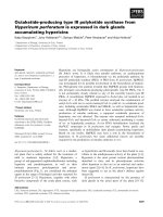
All forms of aggregates were examined using AFM
(Fig. 2). The results were in accordance with previous
investigations in which aggregates formed in the pres-
ence of a molar excess of divalent metal ions failed to
attain the classic morphology of amyloid fibrils [37,46].
The aggregates of Ab(1–40) and Ab(1–42) formed in
the presence of a chelating agent gave rise to the clas-
sic fibrillar morphology, with an average fibril diame-
ter of about 7 nm. It is well known that Ab(1–40) and
Ab(1–42) fibrils consist of bundles of thinner filaments,
each having a cross-section of about 3 nm [47]. This
was most clearly seen within the Ab(1–42) variant
(Fig. 2F), which also had a twinned ultrastructure. For
both Ab(1–40) and Ab(1–42), the presence of divalent
metal ions efficiently inhibited fibril formation and, as
expected, resulted in more amorphous aggregates. It
was not, however, possible to morphologically distin-
guish aggregates formed in the presence of Cu
2+
from
those formed in the presence of Zn
2+
.
Taken together, these results suggest that Ab aggre-
gates formed in the presence of Cu
2+
or Zn
2+
are less
compact than fibrillar complexes and are assembled
from Abs with altered secondary structure. The results
of ThT analysis suggest that Zn
2+
-complexed Ab may
tolerate assembly into a more fibrillar-like fold,
whereas Cu
2+
more efficiently prevents formation of a
fibrillar assembly. Furthermore, all metal-bound Ab
aggregates could be efficiently converted to monomers
through the addition of a chelating agent, suggesting
that their mode of assembly is different from that of
the fibrillar forms.
Quenched H
⁄
D exchange NMR on Ab assemblies
The use of quenched H⁄ D exchange in combination
with NMR spectroscopy is a highly useful technique
with which to efficiently pinpoint the solvent accessibil-
ity of individual amide protons within peptide assem-
blies in a residue-specific and quantitative manner,
thereby providing detailed structural information
[12,38–43]. In this study, aggregates of Ab(1–40) and
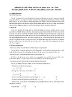
AB C
Fig. 1. Abs were characterized by using (A) turbidity measurements, (B) a ThT assay, and (C) CD spectroscopy. (A) Turbidity measurements
on samples containing 50 l
M Ab(1–40) (solid line) and Ab(1–42) (broken lines) were started immediately after the addition of Cu
2+
[(iii) and
(iv)], Zn
2+
[(i) and (ii)] or EDTA [(v) and (vi)] and continued for 164 min. After 100 min, 400 lM EDTA was added, and the absorbance was
measured for an additional 64 min. (B) ThT analysis of aggregated Ab samples containing 100 l
M Ab(1–40) and Ab(1–42) in the presence of
Cu
2+
(light gray bars), Zn
2+
(open bars), or EDTA (dark gray bars). The respective emissions of the fibrillar forms of Ab(1–40) and Ab(1–42)
were set to 100% intensity. (C) Far-UV CD spectra from samples containing 50 l
M Ab(1–40) (solid lines) or Ab(1–42) (broken lines), either
in the form of aggregates formed in the presence of Cu
2+
[(i) and (iii)] or Zn
2+
[(ii) and (iv)], or as fibrils formed in the presence of EDTA [(v)
and (vi)].
A. Olofsson et al. NMR analysis of metal-complexed Ab aggregates
FEBS Journal 276 (2009) 4051–4060 ª 2009 The Authors Journal compilation ª 2009 FEBS 4053
Ab(1–42) peptides in the presence of Cu
2+
,Zn
2+
or
EDTA were investigated. The chelating agent EDTA
was used to remove trace amounts of divalent metals
that may affect fibril formation. H ⁄ D exchange was
carried out by preincubating samples in D
2
O for 24 h
before analysis. The length of incubation ensures
detection of only those amide protons that are pro-
tected as a result of hydrogen bonding within second-
ary structure elements or because they are deeply
buried in the core of the fibril [12,13].
Samples verified by AFM and ThT analysis to con-
tain the fibrillar forms of Ab(1–40) and Ab(1–42)
(fibrils formed in the presence of EDTA) displayed
amide protection patterns with two bell-shaped pro-
tected regions (Fig. 3C,F). Ab(1–40) fibrils showed
partial to full protection for Glu3–Ser26 and Gly29–
Val40, separated by exposed residues in the turn region
centered at Asn27–Lys28 (Fig. 3C), whereas the pro-
tection pattern of the fibrillar form of Ab(1–42)
included residues Phe4–Arg5, Tyr10–Ser26, and
Gly29–Ala42 (Fig. 3F). The degree of protection in the
N-terminal region of Ab(1–42) is notably lower than
in Ab(1–40). These results are similar to those of our
recently published investigations on Ab(1–40) and
Ab(1–42) fibrils [12,13]; the small differences observed
are probably due to the use of different growth condi-
tions during fibril preparation [150 mm NaCl (pH 7.4)
and 1 mm EDTA versus 50 mm NaCl (pH 7.0) in the
previous investigations]. The protection patterns
observed are in good agreement with current models
extrapolated from solid-state NMR data [10,48], dou-
ble compensatory mutagenesis combined with H ⁄ D
exchange NMR [11], and cysteine scanning mutagene-
sis [49], where the Abs (which form two b-strands with
a turn region involving Val24–Ala30) stack in an in-
register parallel arrangement to form the fibril [10,48]
(Fig. 4). The pattern for Ab(1–40) fibrils also fits well
with our recent report, in which a new general method
to quantitatively determine the exchange rates of
amide protons within fibrils is described and applied to
fibrils formed by Ab(1–40) [43].
The presence of either Cu
2+
or Zn
2+
during aggre-
gate ⁄ fibril growth had a significant impact on the
solvent protection pattern for both Ab(1–40) and
Ab(1–42). When compared to the protection ratios in
the absence of metal (Fig. 3C,F), the overall protection
ratio in the presence of metal was significantly reduced
(Fig. 3A,B,D,E). In the metal-complexed aggregates,
the Asp1–Lys16 region essentially lacks solvent protec-
tion. This is reasonable, as metal binding occurs within
the first 14 residues, inducing a low degree of second-
ary structure [33,50]. However, the remaining parts of
both Ab(1–40) and Ab(1–42) still display two fairly
well-protected bell-shaped regions with an intervening
minimum centered on Asn27. This is strong evidence
that metal-complexed Ab aggregates attain a b-strand-
turn–b-strand structural arrangement similar to the
structural arrangement within mature fibrils (Fig. 4).
The metal-complexed Ab(1–40) aggregates also show
pronounced differences from the fibrillar form in the
C-terminal region as well as in the turn region of the
peptide. Assuming a fibril-like structural arrangement
in which two protofilaments formed from stacked pep-
tides are laterally assembled, the N-terminal metal-
binding region of the peptide will be in close proximity
to both the C-terminal and turn residues, possibly
interfering with these regions. This explains how an
altered conformation in the N-terminal region upon
metal binding can affect sections of the molecule closer
to the C-terminus (see the fibril cross-section in Fig. 4).
Furthermore, differences in solvent protection are
A
B
C
D
E
F
Fig. 2. Tapping mode AFM images of recombinant Ab(1–40) and
Ab(1–42) aggregates. (A–C) Morphology of Ab(1–40) in the pres-
ence of Cu
2+
,Zn
2+
, and EDTA, respectively. (D–F) Morphology of
Ab(1–42) in the presence of Cu
2+
,Zn
2+
, and EDTA, respectively.
Scale bar: 1 lm.
NMR analysis of metal-complexed Ab aggregates A. Olofsson et al.
4054 FEBS Journal 276 (2009) 4051–4060 ª 2009 The Authors Journal compilation ª 2009 FEBS
observed between the Cu
2+
-induced and Zn
2+
-induced
aggregates, reflecting the differences in metal-binding
properties of Ab [26,30,33–35]. Zn
2+
significantly
reduces the overall protection, in particular for residues
close to the turn region (Fig. 3B). For Zn
2+
-induced
Ab(1–40) aggregates, the solvent-protected residues
include Leu17–Glu22 and Ile31–Val36 (Fig. 3B),
whereas in Cu
2+
-induced Ab(1–40) aggregates, Leu17–
Gly25 and Lys28–Val36 are protected (Fig. 3A).
The increased solvent accessibility of the turn region
within Zn
2+
-induced Ab(1–40) aggregates correlates
well with a proposed second weak binding site for
Zn
2+
involving Asp23–Lys28 in the turn region [35].
Comparison of the solvent protection pattern of
Cu
2+
-induced and Zn
2+
-induced Ab(1–42) aggregates
(Fig. 3D,E) with Ab(1–42) fibrils (Fig. 3F) reveals a
reduced overall level of protection and completely
exposed residues in the N-terminal region. The
solvent-protected residues in the Ab(1–42) metal-
complexed aggregates (Leu17–Glu22 ⁄ Asp23 and Ile31–
Ile41) form two characteristic bell-shaped regions,
suggesting a mode of assembly similar to that of the
fibrillar samples. Figure 3D,E demonstrates that there
is no significant difference in solvent exposure in the
turn regions of A b(1–42) aggregates formed in the
presence of either Cu
2+
or Zn
2+
. The C-terminal
region in the Ab(1–42) aggregates is affected by metals
to a lesser extent than the C-terminal region of the
Ab(1–40) aggregates, suggesting they are assembled in
a way that places the C-terminal residues in the aggre-
gate core. This is similar to the arrangement found in
Ab(1–42) fibrils, where the C-terminal residues
strengthen the hydrophobic interactions within the
core of the fibrillar fold via a shift in the protofilament
assembly that positions the C-terminal residues of
Ab(1–42) in a more hydrophobic environment [10,48]
(Fig. 4).
Implications for aggregate
⁄
fibril formation
Although the properties of fibrils and metal-bound
aggregates of Abs differ significantly, the H ⁄ D protec-
tion patterns of metal-bound Ab aggregates still sug-
gest a partly preserved b-strand–turn–b-strand fold
similar to the fibrillar form (Fig. 3). The metal-induced
Ab aggregates have fewer protected residues and a
A
B
C
D
E
F
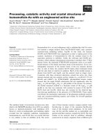
Fig. 3. Solvent protection ratios for the backbone amide protons of Ab(1–40) and Ab(1–42) aggregates. Protection is defined as the ratio of
the observed intensity after a 24 h preincubation period in D
2
O over the intensity in a completely protonated sample (defined as 100%).
(A–C) Solvent protection ratios for Ab(1–40) aggregates in the presence of Cu
2+
,Zn
2+
, and EDTA, respectively. (D–F) Ratios for Ab(1–42)
aggregates in the presence of Cu
2+
,Zn
2+
, and EDTA, respectively. Rings correspond to 0% protection, and crosses represent residues that
exchange too quickly to be detected. Error bars indicate the experimental uncertainty of the measurements.
A. Olofsson et al. NMR analysis of metal-complexed Ab aggregates
FEBS Journal 276 (2009) 4051–4060 ª 2009 The Authors Journal compilation ª 2009 FEBS 4055
lower degree of solvent protection than fibrils, suggest-
ing that formation of b-strands and subsequently
fibrils is hindered, and that fewer intermolecular inter-
actions are needed for assembly. This scenario would
explain the high aggregation rates observed in the tur-
bidity measurements (Fig. 1A). The pronounced lack
of H ⁄ D protection close to the known metal-binding
site within the N-terminal regions of both Ab(1–40)
and Ab(1–42) clearly shows that only the protected
C-terminal regions are involved in the assembly. How-
ever, assembly in the presence of metals is unlikely to
involve the same intermolecular interactions as within
fibrils, as removal of the metals causes dissociation
into monomers instead of the continued assembly into
amyloid fibrils expected from a more fibril-like fold.
One speculative explanation of this behavior is that, in
the presence of metals, the peptides assemble into an
array with b-strand–turn–b-strand motifs (Fig. 4)
stacked in an antiparallel fashion, distinctly different
from the parallel assembly within fibrils. This sugges-
tion is supported by previous observations that peptide
fragments of Ab preferably stack in an antiparallel
fashion along the fibril axis [14,51–53].
In conclusion, our results support the previous notion
that metal binding by the N-terminal residues (Asp1–
Lys16) prevents assembly into a fibrillar structure. On
the basis of our quenched H ⁄ D exchange NMR data,
we propose that both Cu
2+
-induced and Zn
2+
-induced
Ab aggregates assemble via a b-strand–turn–b-strand
motif, resembling the motif found in fibrils.
Experimental procedures
Isotope-enriched chemicals were purchased from Cambridge
Isotope Laboratories (Andover, MA, USA). All peptides,
including uniformly
15
N-labeled Ab(1–40) and Ab(1–42),
were obtained in lyophilized form from Alexotech AB
(Umea
˚
, Sweden) (). Reagents and
buffers were purchased from Sigma-Aldrich (St Louis, MO,
USA), unless otherwise stated.
Sample preparation
Lyophilized Ab(1–40) and Ab(1–42) were briefly dissolved
in 10 mm NaOH, sonicated for 30 s, and centrifuged
(10 min at 12 000 g) to remove residual oligomeric species,
as previously described [54]. This treatment efficiently
monomerizes the peptides and facilitates dilution in 20 mm
Tris (pH 7.4) and 150 mm NaCl (for turbidity measure-
ments) or 10 mm Tris (pH 7.4) and 150 m m NaCl (for ThT
assay and CD spectroscopy) to a peptide concentration of
either 50 or 100 lm, with a two-fold molar excess of CuCl
2
,
ZnCl
2
, or EDTA. Fibrils ⁄ aggregates were formed by incu-
bating the samples for 14 days at 37 °C with agitation.
Turbidity measurements
Turbidity was measured by recording the absorbance
at 405 nm. Measurements on samples containing 50 lm
Ab(1–40) and Ab(1–42) were started immediately after the
addition of Cu
2+
,Zn
2+
, or EDTA, and thereafter recorded
for 164 min. The samples were kept still at room tempera-
ture except from 5 s before each measurement. Each mea-
surement was performed in triplicate. After 100 min,
A
B
C
D
E
F
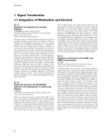
Fig. 4. The determined solvent protection ratios for residues within
Ab(1–40) and Ab(1–42) aggregates are mapped onto corresponding
dimer of two cross-b units taken from cross-sections of the
Ab(1–40) and Ab(1–42) fibril (aggregate) models, respectively. The
color code is varied between the following extremes: navy blue for
complete, and red for no solvent protection. Residues with no pro-
tection ratios available are depicted in gray. Cu
2+
is shown in
brown, and Zn
2+
in bluish gray. (A–C) Protection ratios of Ab(1–40)
in the presence of Cu
2+
,Zn
2+
, and EDTA, respectively. (D–F) Pro-
tection ratios for Ab(1–42) in presence of Cu
2+
,Zn
2+
, and EDTA,
respectively. The image was prepared in
MOLMOL [55].
NMR analysis of metal-complexed Ab aggregates A. Olofsson et al.
4056 FEBS Journal 276 (2009) 4051–4060 ª 2009 The Authors Journal compilation ª 2009 FEBS
400 lm EDTA was added to all wells and the absorbance
was measured for an additional 64 min.
ThT analysis
Samples containing 100 lm aggregated Abs were mixed
with 10 lm ThT and 50 lm phosphate buffer (pH 6.5) in a
10 mm quartz cuvette. Emission intensities were recorded
at 25 °C, using a JASCO spectrofluorometer (Jasco Int.
Co., Ltd., Tokyo, Japan), with excitation and emission
wavelengths of 450 and 482 nm, respectively, and a 3 nm
bandwidth for both emission and excitation. Each measure-
ment was performed in triplicate.
CD spectroscopy
Far-UV CD spectra were collected on samples containing
either 50 or 100 lm Ab, using a JASCO J-810 spectropola-
rimeter (Jasco) at 25 °C. Spectra were recorded between
200 and 250 nm, and averaged over 10 scans with a band-
width of 1 nm, a response time of 1 s, a pitch of 0.5 nm,
and a scan rate of 20 nmÆs
)1
, in a 2 mm quartz cuvette.
AFM
A portion of each Ab aggregate sample was diluted in
water to approximately 1 lm peptide and applied to freshly
cleaved ruby red mica (Goodfellow, Cambridge, UK). The
material was allowed to adsorb for 30 s, and then washed
with distilled water three times and air dried. AFM analysis
was performed using a Nanoscope IIIa multimode atomic
force microscope (Digital Instruments, Santa Barbara,
USA) in tapping mode in air. A silicon probe was oscillated
at abount 280 kHz, and images were collected at an opti-
mized scan rate corresponding to 1 Hz.
Quenched H
⁄
D exchange NMR
Lyophilized
15
N-labelled Ab(1–40) and Ab(1–42) (obtained
from Alexotech AB) () were
monomerized as described above, and this was followed by
addition of 10· buffer (20 mm Tris, pH 7.4, 150 mm NaCl)
to a final peptide concentration of 500 lm. A two-fold
molar excess of either ZnCl
2
, CuCl
2
or EDTA was added
to each sample. Fibril ⁄ aggregate solutions were prepared
(5–8 days of incubation at 37 °C with agitation at
130 r.p.m.) and recovered by centrifugation (2 min at
13 000 g), and each was then split into two; one half was
preincubated in D
2
O for 24 h (by dissolving the pellet 30
times in 20 mm Tris, pD 7.0, 150 mm NaCl and a two-fold
molar excess of ZnCl
2
, CuCl
2
, or EDTA), and one served
as a fully protonated reference sample. At the end of the
incubation period and immediately prior to NMR analysis,
the Ab assemblies were recovered by short centrifugation
steps (13 000 g) and subsequently dissociated into NMR-
detectable monomers (to 2mm monomeric Ab) using an
optimized solution of hexafluoroisopropanol as described in
[12], with the addition of 2 m m diethylenetriamine penta-
acetic acid as chelating agent. NMR data were recorded
and analyzed as previously described [12,13], resulting in
residue-specific solvent protection ratios for the backbone
amide protons within Ab(1–40) and Ab(1–42) aggregates.
Observed ratios were mapped onto models of the various
aggregates. These models are based on our previous fibril
models [12,13], the model of Ab(9–40) by Tycko et al. [10],
the solution structure of Ab(1–16) with and without bound
Zn
2+
[33], and the proposed filament packing arrangement
for Ab(1–42) [10,48]. Modifications and energy minimiza-
tion were performed in molmol [55] and swiss-pdbviewer
[56], respectively.
Acknowledgements
We thank R. Tycko for kindly providing us with the
coordinates of the Ab(9–40) amyloid model. This work
was supported by the Magn. Bergvalls Foundation,
Carl Trygger Foundation, Alzheimerfonden, Socialsty-
relsen, Hja
¨
rnfonden, A
˚
ke Wibergs Foundation, Bern-
hard och Signe Ba
¨
ckstro
¨
ms stiftelse, O. E. och Edla
Johanssons vetenskapliga stiftelse, Go
¨
ran Gustafssons
Foundation, Swedish Research Science Council, Ernst
Schering Foundation, Centre for Biomedical Engineer-
ing at Wrocaw University of Technology, Gun och
Bertil Stohnes stiftelse, Loo och Hans Ostermans stift-
else fo
¨
r geriatrisk forskning and patients’ association
FAMY ⁄ AMYL.
References
1 Chiti F & Dobson CM (2006) Protein misfolding, func-
tional amyloid, and human disease. Annu Rev Biochem
75, 333–366.
2 Iwatsubo T, Odaka A, Suzuki N, Mizusawa H, Nukina
N & Ihara Y (1994) Visualization of A beta 42(43) and
A beta 40 in senile plaques with end-specific A beta
monoclonals: evidence that an initially deposited species
is A beta42(43). Neuron 13, 45–53.
3 Roher AE, Chaney MO, Kuo YM, Webster SD, Stine
WB, Haverkamp LJ, Woods AS, Cotter RJ, Tuohy JM,
Krafft GA et al. (1996) Morphology and toxicity of
Abeta-(1-42) dimer derived from neuritic and vascular
amyloid deposits of Alzheimer’s disease. J Biol Chem
271, 20631–20635.
4 Lambert MP, Barlow AK, Chromy BA, Edwards C,
Freed R, Liosatos M, Morgan TE, Rozovsky I, Trom-
mer B, Viola KL et al. (1998) Diffusible, nonfibrillar
ligands derived from Abeta1–42 are potent central
A. Olofsson et al. NMR analysis of metal-complexed Ab aggregates
FEBS Journal 276 (2009) 4051–4060 ª 2009 The Authors Journal compilation ª 2009 FEBS 4057
nervous system neurotoxins. Proc Natl Acad Sci USA
95, 6448–6453.
5 Lacor PN, Buniel MC, Chang L, Fernandez SJ, Gong
Y, Viola KL, Lambert MP, Velasco PT, Bigio EH,
Finch CE et al. (2004) Synaptic targeting by Alzhei-
mer’s-related amyloid beta oligomers. J Neurosci 24,
10191–10200.
6 Klein WL, Stine WB Jr & Teplow DB (2004) Small
assemblies of unmodified amyloid beta-protein are the
proximate neurotoxin in Alzheimer’s disease. Neurobiol
Aging 25, 569–580.
7 Necula M, Kayed R, Milton S & Glabe CG (2007)
Small molecule inhibitors of aggregation indicate that
amyloid beta oligomerization and fibrillization path-
ways are independent and distinct. J Biol Chem 282,
10311–10324.
8 Walsh DM, Lomakin A, Benedek GB, Condron MM &
Teplow DB (1997) Amyloid beta-protein fibrillogenesis.
Detection of a protofibrillar intermediate. J Biol Chem
272, 22364–22372.
9 Kheterpal I, Lashuel HA, Hartley DM, Walz T,
Lansbury PT Jr & Wetzel R (2003) Abeta protofibrils
possess a stable core structure resistant to hydrogen
exchange. Biochemistry 42, 14092–14098.
10 Petkova AT, Yau WM & Tycko R (2006) Experimental
constraints on quaternary structure in Alzheimer’s
beta-amyloid fibrils. Biochemistry 45, 498–512.
11 Lu
¨
hrs T, Ritter C, Adrian M, Riek-Loher D,
Bohrmann B, Do
¨
beli H, Schubert D & Riek R (2005)
3D structure of Alzheimer’s amyloid-beta(1-42) fibrils.
Proc Natl Acad Sci USA 102, 17342–17347.
12 Olofsson A, Sauer-Eriksson AE & O
¨
hman A (2006)
The solvent protection of alzheimer amyloid-beta-(1-42)
fibrils as determined by solution NMR spectroscopy.
J Biol Chem 281, 477–483.
13 Olofsson A, Lindhagen-Persson M, Sauer-Eriksson AE
&O
¨
hman A (2007) Amide solvent protection
analysis demonstrates that amyloid-beta(1-40) and
amyloid-beta(1-42) form different fibrillar
structures under identical conditions. Biochem J 404,
63–70.
14 Sawaya MR, Sambashivan S, Nelson R, Ivanova MI,
Sievers SA, Apostol MI, Thompson MJ, Balbirnie M,
Wiltzius JJ, McFarlane HT et al. (2007) Atomic
structures of amyloid cross-beta spines reveal varied
steric zippers. Nature 447, 453–457.
15 Bush AI, Pettingell WH, Multhaup G, Paradis M,
Vonsattel JP, Gusella JF, Beyreuther K, Masters CL
& Tanzi RE (1994) Rapid induction of Alzheimer
A beta amyloid formation by zinc. Science 265,
1464–1467.
16 Garzon-Rodriguez W, Yatsimirsky AK & Glabe CG
(1999) Binding of Zn(II), Cu(II), and Fe(II) ions to
Alzheimer’s A beta peptide studied by fluorescence.
Bioorg Med Chem Lett 9, 2243–2248.
17 Tougu V, Karafin A & Palumaa P (2008) Binding of
zinc(II) and copper(II) to the full-length Alzheimer’s
amyloid-beta peptide. J Neurochem 104, 1249–1259.
18 Noy D, Solomonov I, Sinkevich O, Arad T, Kjaer K
& Sagi I (2008) Zinc-amyloid beta interactions on a
millisecond time-scale stabilize non-fibrillar Alzheimer-
related species. J Am Chem Soc 130, 1376–1383.
19 Atwood CS, Moir RD, Huang X, Scarpa RC, Bacarra
NM, Romano DM, Hartshorn MA, Tanzi RE & Bush
AI (1998) Dramatic aggregation of Alzheimer abeta by
Cu(II) is induced by conditions representing physiologi-
cal acidosis. J Biol Chem 273, 12817–12826.
20 Lovell MA, Robertson JD, Teesdale WJ, Campbell JL
& Markesbery WR (1998) Copper, iron and zinc in
Alzheimer’s disease senile plaques. J Neurol Sci 158,
47–52.
21 Huang X, Cuajungco MP, Atwood CS, Hartshorn MA,
Tyndall JD, Hanson GR, Stokes KC, Leopold M,
Multhaup G, Goldstein LE et al. (1999) Cu(II) potenti-
ation of alzheimer abeta neurotoxicity. Correlation with
cell-free hydrogen peroxide production and metal reduc-
tion. J Biol Chem 274, 37111–37116.
22 Yoshiike Y, Tanemura K, Murayama O, Akagi T,
Murayama M, Sato S, Sun X, Tanaka N & Takashima
A (2001) New insights on how metals disrupt amyloid
beta-aggregation and their effects on amyloid-beta
cytotoxicity. J Biol Chem 276, 32293–32299.
23 Garai K, Sahoo B, Kaushalya SK, Desai R & Maiti S
(2007) Zinc lowers amyloid-beta toxicity by selectively
precipitating aggregation intermediates. Biochemistry
46, 10655–10663.
24 Lovell MA, Xie C & Markesbery WR (1999) Protection
against amyloid beta peptide toxicity by zinc. Brain Res
823, 88–95.
25 Huang X, Cuajungco MP, Atwood CS, Moir RD,
Tanzi RE & Bush AI (2000) Alzheimer’s disease, beta-
amyloid protein and zinc. J Nutr 130, 1488S–1492S.
26 Bush AI (2003) The metallobiology of Alzheimer’s
disease. Trends Neurosci 26, 207–214.
27 Phinney AL, Drisaldi B, Schmidt SD, Lugowski S,
Coronado V, Liang Y, Horne P, Yang J, Sekoulidis J,
Coomaraswamy J et al. (2003) In vivo reduction of
amyloid-beta by a mutant copper transporter. Proc Natl
Acad Sci USA 100, 14193–14198.
28 Huang X, Atwood CS, Moir RD, Hartshorn MA,
Vonsattel JP, Tanzi RE & Bush AI (1997) Zinc-induced
Alzheimer’s Abeta1-40 aggregation is mediated
by conformational factors. J Biol Chem 272, 26464–
26470.
29 Isaacs AM, Senn DB, Yuan M, Shine JP & Yankner
BA (2006) Acceleration of amyloid beta-peptide
aggregation by physiological concentrations of calcium.
J Biol Chem 281, 27916–27923.
30 Curtain CC, Ali F, Volitakis I, Cherny RA, Norton
RS, Beyreuther K, Barrow CJ, Masters CL, Bush AI
NMR analysis of metal-complexed Ab aggregates A. Olofsson et al.
4058 FEBS Journal 276 (2009) 4051–4060 ª 2009 The Authors Journal compilation ª 2009 FEBS
& Barnham KJ (2001) Alzheimer’s disease amyloid-beta
binds copper and zinc to generate an allosterically
ordered membrane-penetrating structure containing
superoxide dismutase-like subunits. J Biol Chem 276,
20466–20473.
31 Tickler AK, Smith DG, Ciccotosto GD, Tew DJ,
Curtain CC, Carrington D, Masters CL, Bush AI,
Cherny RA, Cappai R et al. (2005) Methylation of the
imidazole side chains of the Alzheimer disease
amyloid-beta peptide results in abolition of superoxide
dismutase-like structures and inhibition of neurotoxic-
ity. J Biol Chem 280, 13355–13363.
32 Antzutkin ON (2004) Amyloidosis of Alzheimer’s Abeta
peptides: solid-state nuclear magnetic resonance,
electron paramagnetic resonance, transmission electron
microscopy, scanning transmission electron microscopy
and atomic force microscopy studies. Magn Reson
Chem 42, 231–246.
33 Zirah S, Kozin SA, Mazur AK, Blond A, Cheminant
M, Segalas-Milazzo I, Debey P & Rebuffat S (2006)
Structural changes of region 1–16 of the
Alzheimer disease amyloid beta-peptide upon zinc
binding and in vitro aging. J Biol Chem 281, 2151–
2161.
34 Syme CD, Nadal RC, Rigby SE & Viles JH (2004)
Copper binding to the amyloid-beta (Abeta) peptide
associated with Alzheimer’s disease: folding, coordina-
tion geometry, pH dependence, stoichiometry, and
affinity of Abeta-(1-28): insights from a range of
complementary spectroscopic techniques. J Biol Chem
279, 18169–18177.
35 Danielsson J, Pierattelli R, Banci L & Gra
¨
slund A
(2007) High-resolution NMR studies of the zinc-binding
site of the Alzheimer’s amyloid beta-peptide. FEBS J
274, 46–59.
36 Cherny RA, Legg JT, McLean CA, Fairlie DP, Huang
X, Atwood CS, Beyreuther K, Tanzi RE, Masters CL
& Bush AI (1999) Aqueous dissolution of Alzheimer’s
disease Abeta amyloid deposits by biometal depletion.
J Biol Chem 274, 23223–23228.
37 Smith DP, Ciccotosto GD, Tew DJ, Fodero-Tavoletti
MT, Johanssen T, Masters CL, Barnham KJ & Cappai
R (2007) Concentration dependent Cu
2+
induced
aggregation and dityrosine formation of the Alzheimer’s
disease amyloid-beta peptide. Biochemistry 46, 2881–
2891.
38 Alexandrescu AT (2001) An NMR-based quenched
hydrogen exchange investigation of model amyloid
fibrils formed by cold shock protein A. Pac Symp
Biocomput 6, 67–78.
39 Reference withdrawn.
40 Ippel JH, Olofsson A, Schleucher J, Lundgren E &
Wijmenga SS (2002) Probing solvent accessibility of
amyloid fibrils by solution NMR spectroscopy. Proc
Natl Acad Sci USA 99, 8648–8653.
41 Olofsson A, Ippel JH, Wijmenga SS, Lundgren E &
O
¨
hman A (2004) Probing solvent accessibility of trans-
thyretin amyloid by solution NMR spectroscopy. J Biol
Chem 279, 5699–5707.
42 Hoshino M, Katou H, Hagihara Y, Hasegawa K, Naiki
H & Goto Y (2002) Mapping the core of the beta(2)-
microglobulin amyloid fibril by H ⁄ D exchange. Nat
Struct Biol 9, 332–336.
43 Olofsson A, Sauer-Eriksson AE & O
¨
hman A (2009)
Amyloid fibril dynamics revealed by combined hydro-
gen ⁄ deuterium exchange and nuclear magnetic reso-
nance. Anal Biochem 385, 374–376.
44 Huang X, Atwood CS, Moir RD, Hartshorn MA,
Tanzi RE & Bush AI (2004) Trace metal contamination
initiates the apparent auto-aggregation, amyloidosis,
and oligomerization of Alzheimer’s Abeta peptides.
J Biol Inorg Chem 9, 954–960.
45 House E, Collingwood J, Khan A, Korchazkina
O, Berthon G & Exley C (2004) Aluminium, iron, zinc
and copper influence the in vitro formation of amyloid
fibrils of Abeta42 in a manner which may have
consequences for metal chelation therapy in Alzheimer’s
disease. J Alzheimers Dis 6, 291–301.
46 Ha C, Ryu J & Park CB (2007) Metal ions differentially
influence the aggregation and deposition of Alzheimer’s
beta-amyloid on a solid template. Biochemistry 46,
6118–6125.
47 Malinchik SB, Inouye H, Szumowski KE & Kirschner
DA (1998) Structural analysis of Alzheimer’s beta(1–40)
amyloid: protofilament assembly of tubular fibrils. Bio-
phys J 74, 537–545.
48 Sato T, Kienlen-Campard P, Ahmed M, Liu W, Li H,
Elliott JI, Aimoto S, Constantinescu SN, Octave JN &
Smith SO (2006) Inhibitors of amyloid toxicity based
on beta-sheet packing of Abeta40 and Abeta42. Bio-
chemistry 45, 5503–5516.
49 Shivaprasad S & Wetzel R (2006) Scanning cysteine
mutagenesis analysis of Abeta-(1-40) amyloid fibrils.
J Biol Chem 281, 993–1000.
50 Ma QF, Hu J, Wu WH, Liu HD, Du JT, Fu Y, Wu
YW, Lei P, Zhao YF & Li YM (2006) Characterization
of copper binding to the peptide amyloid-beta(1-16)
associated with Alzheimer’s disease. Biopolymers 83,
20–31.
51 Bu Z, Shi Y, Callaway DJ & Tycko R (2007) Molecular
alignment within beta-sheets in Abeta(14-23) fibrils:
solid-state NMR experiments and theoretical predic-
tions. Biophys J 92, 594–602.
52 Klimov DK & Thirumalai D (2003) Dissecting the
assembly of Abeta16-22 amyloid peptides into antipar-
allel beta sheets. Structure 11, 295–307.
53 Lansbury PT Jr, Costa PR, Griffiths JM, Simon EJ,
Auger M, Halverson KJ, Kocisko DA, Hendsch ZS,
Ashburn TT, Spencer RG et al. (1995) Structural model
for the beta-amyloid fibril based on interstrand align-
A. Olofsson et al. NMR analysis of metal-complexed Ab aggregates
FEBS Journal 276 (2009) 4051–4060 ª 2009 The Authors Journal compilation ª 2009 FEBS 4059
ment of an antiparallel-sheet comprising a C-terminal
peptide. Nat Struct Biol 2, 990–998.
54 Fezoui Y, Hartley DM, Harper JD, Khurana R,
Walsh DM, Condron MM, Selkoe DJ, Lansbury PT
Jr, Fink AL & Teplow DB (2000) An improved
method of preparing the amyloid beta-protein for
fibrillogenesis and neurotoxicity experiments. Amyloid
7, 166–178.
55 Koradi R, Billeter M & Wu
¨
thrich K (1996) MOLMOL:
a program for display and analysis of macromolecular
structures. J Mol Graph 14, 51–55, 29–32.
56 Guex N & Peitsch MC (1997) SWISS-MODEL and the
Swiss-PdbViewer: an environment for comparative
protein modeling. Electrophoresis 18, 2714–2723.
NMR analysis of metal-complexed Ab aggregates A. Olofsson et al.
4060 FEBS Journal 276 (2009) 4051–4060 ª 2009 The Authors Journal compilation ª 2009 FEBS