Báo cáo khoa học: Mutagenic probes of the role of Ser209 on the cavity shaping loop of human monoamine oxidase A docx
Bạn đang xem bản rút gọn của tài liệu. Xem và tải ngay bản đầy đủ của tài liệu tại đây (385.74 KB, 13 trang )
Mutagenic probes of the role of Ser209 on the cavity
shaping loop of human monoamine oxidase A
Jin Wang
1
, Johnny Harris
1,
*, Darrell D. Mousseau
2
and Dale E. Edmondson
1
1 Departments of Biochemistry and Chemistry, Emory University, Atlanta, GA, USA
2 Cell Signaling Laboratory, Department of Psychiatry, University of Saskatchewan, Saskatoon, Canada
Introduction
Monoamine oxidase (MAO; EC 1.4.3.4) A (MAO A)
serves an important role in the degradation of seroto-
nin and has been the object of intense experimental
interest because this enzyme has been implicated in a
range of human conditions, from aggressive trait disor-
ders [1–3] to cardiovascular disease [4–6]. Although a
considerable amount of structural and functional infor-
mation is available [7,8] regarding this membrane-
bound mitochondrial flavoenzyme, very little is known
about any possible processes that could regulate its
function. The involvement of MAO A in pro-apoptotic
signaling pathways is suggested by a variety of studies
demonstrating that staurosporine (a kinase inhibitor)
induces MAO A-sensitive apoptosis [9]. Ou et al. [10]
have shown that MAO A and a protein (R1) that
inhibits the MAO A promoter are downstream of the
Keywords
cavity-shaping loop; membrane; monoamine
oxidase A; mutagenesis; phosphomimic
Correspondence
D. E. Edmondson, Department of
Biochemistry, Emory University, Atlanta,
GA 30322, USA
Fax: +1 404 727 2738
Tel: +1 404 727 5972
E-mail:
*Present address
Departments of Biochemistry and Molecular
Biology, University of Florida, Gainesville,
FL, USA
(Received 6 May 2009, revised 17 June
2009, accepted 19 June 2009)
doi:10.1111/j.1742-4658.2009.07162.x
The available literature implicating human monoamine oxidase A (MAO A)
in apoptotic processes reports levels of MAO A protein that do not corre-
late with activity, suggesting that unknown mechanisms may be involved in
the regulation of catalytic function. Bioinformatic analysis suggests Ser209
as a possible phosphorylation site that may be relevant to catalytic function
because it is adjacent to a six-residue loop termed the ‘cavity shaping loop’
from structural data. To probe the functional role of this site, MAO A
Ser209Ala and Ser209Glu mutants were created and investigated. In its
membrane-bound form, the MAO A Ser209Glu phosphorylation mimic
exhibits catalytic and inhibitor binding properties similar to those of wild-
type MAO A. Solubilization in detergent solution and purification of the
Ser209Glu mutant results in considerable decreases in these functional
parameters. By contrast, the MAO A Ser209Ala mutant exhibits similar
catalytic properties to those of wild-type enzyme when purified. Compared
to purified wild-type and Ser209Ala MAO A proteins, the Ser209Glu
MAO A mutant shows significant differences in covalent flavin fluorescence
yield, CD spectra and thermal stability. These structural differences in the
purified MAO A Ser209Glu mutant are not exhibited in quantitative struc-
ture–activity relationship patterns using a series of para-substituted benzyl-
amine analogs similar to the wild-type enzyme. These data suggest that
Ser209 in MAO A does not appear to be the putative phosphorylation site
for regulation of MAO A activity and demonstrate that the membrane
environment plays a significant role in stabilizing the structure of MAO A
and its mutant forms.
Abbreviations
MAO A, monoamine oxidase A; QSAR, quantitative structure–activity relationship.
FEBS Journal 276 (2009) 4569–4581 ª 2009 The Authors Journal compilation ª 2009 FEBS 4569
functions of p38 mitogen-activated protein kinase, sup-
porting their involvement in an apoptotic signaling
pathway. MAO A catalysis appears to be an important
factor in the induction of apoptosis because treatment
of cells with clorgyline (a specific MAO A inhibitor)
appears to have a protective role. Data from several
studies [9,11,12] reveal that the level of MAO A
expression does not correlate well with MAO A cata-
lytic activity levels. These observations suggest that the
investigation of any regulatory post-translational mod-
ification of MAO A that might influence its catalytic
activity would be a worthwhile endeavor.
Protein phosphorylation is a well-known mechanism
for the regulation of the functional activity [13,14] of
enzymes and several observations provide the rationale
for the experiments conducted in the present study.
The sequence of MAO A was subjected to netphos
[15] (a bioinformatic neural network method) to pre-
dict potential phosphorylation sites. The results shown
in Fig. S1 suggest that eight Ser sites are predicted to
be available for phosphorylation, of which Ser81 and
Ser209 exhibit the highest prediction ranking scores
(0.994 and 0.990, respectively). Of these two sites,
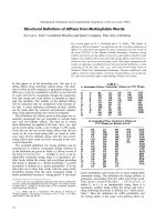
Ser209 is of interest because the crystal structures of
human MAO A [16,17] show differing conformations
of a six-residue loop that is termed the ‘cavity shaping
loop’. One conformer is more extended and the other
is in a more coiled structure, similar to that of MAO
B (Fig. 1). Ser209 is situated adjacent to the ‘cavity
shaping loop’ and its proximity from the carboxyl of
Glu216 would result in electrostatic repulsion if Ser209
were to be phosphorylated. This ‘cavity shaping loop’
may serve to alter the shape of the catalytic site of
MAO A, which would result in alterations in MAO A
catalytic function and serve a regulatory function.
Therefore, Ser209 could be a target for phosphoryla-
tion. We chose to investigate the functional conse-
quences of Ser209 phosphorylation in human MAO A.
To date, there are no published data demonstrating
the in vivo phosphorylation of MAO A. To investigate
potential influences of Ser209 phosphorylation on
MAO A catalytic function, we report studies on two
mutant proteins in which Ser209 is substituted with
either a Glu residue, thereby generating a ‘phosphory-
lation mimic’ [18–20], or an alanine residue, which pre-
cludes any phosphorylation on this residue. The
structural and functional consequences of these muta-
tions are determined and compared with wild-type
enzyme. The results obtained demonstrate a remarkable
stabilizing influence in the mitochondrial outer mem-
brane environment on the Ser209Glu MAO A and sug-
gest that the phosphorylation of Ser209 likely does not
occur as a primary mode of enzyme regulation in vivo.
Results
Kinetic properties of human wild-type
MAO A and MAO A Ser209Glu mutant in
membrane-bound form
Preliminary studies showed that the Ser209Glu mutant,
but not the Ser209Ala mutant, of MAO A was unsta-
ble to purification unless measurements were per-
formed on freshly purified enzyme and the preparation
was kept on ice. Therefore, initial comparative studies
of this mutant with wild-type enzyme were performed
in membrane preparations. Previous studies of Tyr444
mutants of MAO A showed their membrane-bound
forms to be stable, whereas the purified forms readily
inactivate [21]. To determine active site concentrations
so that k
cat
values could be calculated, we conducted
titration of membrane particles of wild-type MAO A
and MAO A Ser209Glu mutant with clorgyline.
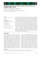
As shown in Fig. 2, the MAO concentrations in
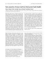
Fig. 1. The different conformations of the cavity-shaping loop in two
human MAO A crystal structures. The two crystal structures by De
Colibus (in green) and by Son (in cyan) are superimposed. For quality
of viewing specific residues, the superimposed structures are
displayed in 60% translucent mode. The flavin cofactor is shown in
yellow. The cavity shaping loops in De Colibus’ and Son’s structure
are shown in red and black, respectively. Ser209 and Glu216 are
indicated in stick mode. The figure was drawn using
PYMOL (Delano
Scientific, San Carlo, CA, USA; ).
Ser209 and the structure of human MAO A J. Wang et al.
4570 FEBS Journal 276 (2009) 4569–4581 ª 2009 The Authors Journal compilation ª 2009 FEBS
membrane particles of wild-type and the Ser209Glu
mutant are 11.5 lm and 6.5 lm, respectively. It should
be noted that the differences of MAO concentrations
(i.e. wild-type and the mutant enzymes) in membrane
particles result from differences in the total protein
concentrations in these experiments. Both wild-type
MAO A and the MAO A Ser209Glu mutant in mem-
brane preparations exhibit similar specific activities.
Another interesting phenomenon that we observed is
that membrane particles of the MAO A Ser209Glu
mutant show a 10-fold lower activity in potassium
phosphate buffer containing 0.5% reduced Triton
X-100 than in potassium phosphate buffer in which the
detergent was omitted, whereas wild-type MAO A in
membrane-bound form exhibits similar activities in the
presence and absence of 0.5% reduced Triton X-100.
Using four different substrates, a comparison of the
MAO A Ser209Glu mutant in membrane-bound form
(Table 1) with wild-type MAO A shows similar turn-
over numbers (k
cat
) and catalytic efficiencies (k
cat
⁄ K
m
).
Similar binding affinities of MAO A specific reversible
inhibitors are observed for both the MAO A Ser209-
Glu mutant as well as wild-type MAO A. These cata-
lytic and binding data demonstrate that, in their
membrane bound forms, substitution of Ser209 with a
negatively-charged Glu residue does not alter the cata-
lytic and structural properties of the active site of the
protein. However, as demonstrated below, solubiliza-
tion and purification of the mutant enzyme in deter-
gent solution results in considerable changes in these
parameters.
UV-visible spectral properties of human
MAO A Ser209 mutants
The purified human MAO A Ser209Ala and Ser209-
Glu mutants show the expected absorption spectral
properties for covalent flavin cofactors (Fig. S2, solid
lines). Addition of the acetylenic inhibitor clorgyline
results in the conversion of the oxidized flavin cofac-
tors to their respective N(5) flavocyanine adducts [22],
which exhibit a characteristic absorption maximum at
415 nm with an e = 23 400 m
)1
Æcm
)1
(Fig. S2, dashed
lines). These data demonstrate that the freshly purified
mutant enzymes exhibit > 90% functionality and that
A
B
Fig. 2. Determination of MAO A active site concentrations in mem-
brane particles by titration with clorgyline. (A) Wild-type MAO A. (B)
MAO A Ser209Glu mutant.
Table 1. Steady-state kinetic properties of membrane-bound wild-type MAO A and the MAO A Ser209Glu mutant.
Wild-type MAO A MAO A Ser209Glu
Substrate k
cat
(min
)1
) K
m
(mM) k
cat
⁄ K
m
(min
)1
ÆmM
)1
) k
cat
(min
)1
) K
m
(mM) k
cat
⁄ K
m
(min
)1
ÆmM
)1
)
Benzylamine 2.44 ± 0.03 1.67 ± 0.12 1.46 ± 0.11 2.05 ± 0.02 2.83 ± 0.11 0.72 ± 0.03
Kynuramine 93.33 ± 0.79 0.14 ± 0.01 666.64 ± 19.86 77.50 ± 0.62 0.093 ± 0.003 836.81 ± 7.23
Phenylethylamine 48.57 ± 1.06 0.47 ± 0.04 103.34 ± 9.70 64.05 ± 1.43 0.85 ± 0.07 75.35 ± 6.25
Serotonin 145.77 ± 1.80 0.094 ± 0.004 1542.59 ± 72.10 153.57 ± 1.80 0.069 ± 0.002 2221.75 ± 79.58
Competitive inhibitor K
i
(lM) K
i
(lM)
Harmane 0.14 ± 0.03 0.18 ± 0.02
Pirlindole mesylate 0.25 ± 0.04 0.29 ± 0.06
Tetrindole mesylate 2.43 ± 0.15 2.76 ± 0.11
J. Wang et al. Ser209 and the structure of human MAO A
FEBS Journal 276 (2009) 4569–4581 ª 2009 The Authors Journal compilation ª 2009 FEBS 4571
they react stoichiometrically with irreversible inhibitors
in a manner similar to that observed with wild-type
MAO A.
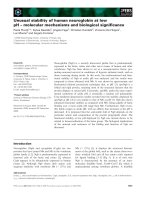
Thermal stability of human MAO A Ser209
mutants
Because the purified Ser209Glu mutant exhibits low-
ered stability relative to the wild-type and the Ser209-
Ala enzymes, their respective thermal stabilities were
compared to establish conditions that would facilitate
further comparisons. At five different temperatures (0,
10, 15, 25 and 30 °C), the purified MAO A Ser209Ala
mutant exhibits stability that is comparable to wild-
type MAO A. (Fig. 3A). At 25 °C, the purified MAO
A Ser209Ala mutant lost approximately 40% activity
within 120 min, whereas, at 30 °C, approximately 50%
of MAO A Ser209Ala mutant activity is lost. By con-
trast, the purified MAO A Ser209Glu mutant is only
thermally stable at 0 °C (Fig. 3B). After incubation for
120 min at 10 and 15 °C, this mutant retains 70% and
55% activity, respectively. Increasing the incubation
temperature to 25 and 30 °C results in greater losses in
activity (approximately 40% of and 25% of activity
remaining, respectively). These data demonstrate that
substituting Ser209 with Glu markedly reduces the
stability of human MAO A.
Comparison of the kinetic properties of
detergent-solubilized forms of human wild-type
MAO A and the MAO A Ser209 mutants
Although no major functional effect of placing a nega-
tive charge at position 209 in MAO A is observed in
membrane-bound forms of the enzyme, large differ-
ences are observed on comparing the purified forms in
detergent solution. Comparisons of the steady-state
kinetic parameters for the oxidation of benzylamine,
kynuramine, phenylethylamine and serotonin for the
human wild-type MAO A, Ser209Ala MAO A mutant
and Ser209Glu MAO A mutant are shown in Table 2.
For the MAO A Ser209Ala mutant, only modest
changes in catalytic efficiencies are observed (approxi-
mately 1.5–3.7-fold lower than wild-type MAO A). By
contrast, the k
cat
values of the MAO A Ser209Glu
mutant for these substrates are more than 10-fold
lower and the respective K
m
values are more than
10-fold higher than those exhibited by the wild-type
enzyme. Therefore, the relative catalytic efficiencies
(k
cat
⁄ K
m
values) for these substrates tested with the
Ser209Glu mutant are 0.5–1% of those determined for
the wild-type MAO A.
A similar pattern is observed with several MAO
competitive inhibitors. The MAO A Ser209Ala mutant
exhibits similar K
i
values (i.e. one- to two-fold differ-
ence) to those of wild-type MAO A (Table 3). Large
changes in inhibition affinities were observed on com-
parison of the wild-type MAO A and MAO A Ser209-
Glu mutant (Table 3). d-Amphetamine and isatin,
which are nonselective reversible MAO inhibitors, inhi-
bit the human MAO A Ser209Glu mutant with much
lower affinities (160-fold and 20-fold, respectively)
compared to the wild-type enzyme (Table 3), and
phentermine binds to the Ser209Glu mutant with a K
i
of 6682 lm, which is 13-fold lower than that found for
wild-type MAO A. The MAO A specific reversible
inhibitors, harmane, pirlindole and tetrindole are also
bound to the Ser209Glu mutant much more weakly
than the values observed with either wild-type or the
Ser209Ala MAO A mutant. These results demonstrate
that, in purified preparations of MAO A, placing a
negative charge at position 209 has a major influence
A
B
Fig. 3. Comparison of thermal stabilities of the purified human
MAO A Ser209Ala mutant (A) and Ser209Glu mutant (B). Loss of
catalytic activities versus incubation time at 0, 10, 15, 25 and 30 °C
are shown [enzyme buffer: 50 m
M potassium phosphate, 20%
(v ⁄ v) glycerol and 0.8% (w ⁄ v) b-octylglucopyranoside, pH 7.5].
Ser209 and the structure of human MAO A J. Wang et al.
4572 FEBS Journal 276 (2009) 4569–4581 ª 2009 The Authors Journal compilation ª 2009 FEBS
on the properties of the substrate binding site of MAO
A, suggesting that structural alterations are occurring
in the conformation of the cavity shaping loop
(Fig. 1).
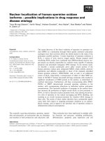
Flavin fluorescence and CD spectral properties of
human wild-type MAO A and the MAO A Ser209
mutant proteins
To investigate whether any differential structural alter-
ations occur in the catalytic site of MAO A as a conse-
quence of these mutations, the spectral properties of
the active site covalent flavin coenzyme was compared
for wild-type MAO A and the two Ser209 mutant
enzymes. As shown in Fig. 4A, both human wild-type
MAO A (i.e. solid line) and MAO A Ser209Ala
mutant (i.e. dashed line) exhibit similar fluorescence
intensities and emission maxima. However, for the
MAO A Ser209Glu mutant (the dotted line), a marked
decrease in fluorescence intensity and a blue-shift
(maximum emission at 510 nm) are observed. The fluo-
rescence intensity of the covalent flavin is known to be
influenced by solvent dielectric [23] and by other envi-
ronmental influences [24–26]. If the observed fluores-
cence spectral properties reflect their differential
Table 2. Comparison of steady-state kinetic properties of the purified wild-type human MAO A and purified human MAO A Ser209Ala and
Ser209Glu mutants.
Benzylamine Kynuramine Phenylethylamine Serotonin
Human MAO A k
cat
(min
)1
) 2.5 ± 0.1
a
125.4 ± 8.5
b
53.8 ± 1.0
c
175.1 ± 2.1
c
K
m
(mM) 1.04 ± 0.15
a
0.13 ± 0.01
b
1.48 ± 0.08
c
0.30 ± 0.05
c
k
cat
⁄ K
m
(min
)1
ÆmM
)1
) 2.4 ± 0.4
a
964.6 ± 98.9
b
36.4 ± 2.1
c
583.7 ± 97.5
c
Human MAO A Ser209Ala k
cat
(min
)1
) 1.56 ± 0.03 39.69 ± 0.86 18.84 ± 0.10 161.9 ± 4.0
K
m
(mM) 0.91 ± 0.09 0.15 ± 0.01 0.78 ± 0.02 0.41 ± 0.04
k
cat
⁄ K
m
(min
)1
ÆmM
)1
) 1.72 ± 0.17 262.8 ± 18.3 24.2 ± 0.6 396.2 ± 35.4
Human MAO A Ser209Glu k
cat
(min
)1
) 0.226 ± 0.002 1.48 ± 0.04 3.37 ± 0.07 25.38 ± 0.44
K
m
(mM) 9.73 ± 0.28 0.32 ± 0.02 19.11 ± 1.11 3.54 ± 0.20
k
cat
⁄ K
m
(min
)1
ÆmM
)1
) 0.023 ± 0.001 4.57 ± 0.37 0.18 ± 0.01 7.17 ± 0.43
a
Values from Miller et al.[27].
b
Values from Nandigama et al. [41].
c
Values from Li et al. [35].
Table 3. Comparison of competitive inhibition constants [K
i
(lM)]
for purified wild-type human MAO A and human MAO A Ser209Ala
and Ser209Glu mutants.
Human
MAO A
Human
MAO A
Ser209Ala
Human
MAO A
Ser209Glu
D-Amphetamine 3.69 ± 0.45 4.72 ± 0.63 608.83 ± 31.61
Isatin 15
a
24.5 ± 5.6 314.67 ± 2.13
Phentermine 498 ± 60
b
944 ± 25 6682 ± 245
Harmane 0.58 ± 0.02 1.37 ± 0.04 15.74 ± 0.93
Pirlindole mesylate 0.92 ± 0.04 0.88 ± 0.18 21.52 ± 1.36
Tetrindole mesylate 5.27 ± 0.24 4.11 ± 0.67 16.13 ± 0.57
a
Value from Hubalek et al. [42].
b
Value from Nandigama et al.
[43].
A
B
Fig. 4. Fluorescence spectra of human wild-type MAO A (—),
MAO A Ser209Ala mutant (- - -) and MAO A Ser209Glu mutant (ÆÆÆ)
before (A) and after (B) guanidine chloride denaturation. All spectral
data were acquired in 50 m
M potassium phosphate containing 20%
glycerol and 0.8% (w ⁄ v) b-octylglucopyranoside, pH 7.5. The con-
centrations of all samples were normalized to 20 l
M.
J. Wang et al. Ser209 and the structure of human MAO A
FEBS Journal 276 (2009) 4569–4581 ª 2009 The Authors Journal compilation ª 2009 FEBS 4573
environments, denaturation of the proteins should
result in samples exhibiting identical spectral proper-
ties. Unfolding of the proteins by incubation with gua-
nidine chloride resulted in all three enzyme samples
exhibiting essentially identical fluorescence emission
intensities and maxima (Fig. 4B). Thus, the covalent
flavin cofactors in all denatured proteins are present in
identical levels and are now in identical environments.
The fluorescence intensities of all denatured proteins
are higher than that shown in Fig. 4A, demonstrating
that the quantum yields of fluorescence are higher in
their respective denatured forms than in their native
forms. Therefore, the fluorescence spectral differences
observed in the native forms of the proteins reflect
structural alterations to the active site on incorporating
the mutations.
To further investigate the environment of flavin
cofactor in the active site of MAO A, CD spectros-
copy was used to monitor the alterations in the ellip-
ticity of the bound flavin chromophore in visible
region (300–550 nm). Because the flavin ring is opti-
cally inactive, any alterations in CD spectral properties
reflect alterations of the asymmetric protein environ-
ment about the flavin binding site. The CD spectra
presented in Fig. 5 show that the oxidized forms of the
flavin in either human wild-type MAO A (the solid
line) or in the MAO A Ser209Ala mutant (the dashed
line) exhibit quite similar dichroic spectra: two positive
bands at 380 and 460 nm, respectively. The CD spec-
trum of the MAO A Ser209Glu mutant shows that the
band at 460 nm exhibits a negative signal (the dotted
line). Because, in the UV-visible absorption spectrum
of the MAO A Ser209Glu mutant (Fig. S2B, the solid
line), the purified enzyme showed characteristic
absorption of oxidized flavin at 456 nm, which does
not differ from wild-type enzyme, the negative absorp-
tion at 460 nm in the CD spectrum does not result
from the introduction of other chromophoric forms of
the flavin (i.e. semiquinone or hydroquinone redox
forms) or other components exhibiting absorption in
this spectral region. These results are in agreement
with the observed different fluorescence spectrum of
the MAO A Ser209Glu mutant, indicating a structural
change in the active site that affects the interaction of
the isoalloxazine ring of the FAD cofactor with its
surrounding environment.
Structure
⁄
activity studies of human MAO A
Ser209 mutants as a probe of active site
structure
The above spectroscopic and catalytic studies of the
MAO A Ser209Glu mutant enzyme suggest consider-
able alterations of the catalytic site affected by this
mutation in the solubilized form of the enzyme. One
way to provide further information on the nature of
these alterations is to probe the behavior of the mutant
enzyme with para-substituted benzylamine substrate
analogs. Previous studies conducted in our laboratory
have shown that wild-type MAO A catalyzes the oxi-
dation of these analogs. Large deuterium kinetic iso-
tope effects are observed, demonstrating that C-H
bond cleavage is rate limiting in catalysis. A Hammett
plot of log k
cat
versus the electronic parameter of the
para-substituent exhibits a q value of +1.89 (± 0.43),
demonstrating a H
+
abstraction mechanism for C-H
bond cleavage. In addition, log K
d
for substrate analog
binding correlates with the van der Waals volume of
the para-substituent (where a higher affinity is
observed with an increase in substituent volume) [27].
These quantitative structure–activity relationship
(QSAR) approaches were applied to the Ser209 mutant
forms of MAO A as a sensitive probe of active site
structures. The steady-state kinetic parameters for cat-
alyzed oxidation of seven para -substituted benzylamine
analogs by the MAO A Ser209Ala and Ser209Glu
mutants were determined and their respective values of
k
cat
and K
m
are shown in Table 4. The turnover num-
bers [k
cat
(H)] of the MAO A Ser209Ala and Ser209Glu
mutants determined for each substrate show a marked
dependence on the nature of the para-substituent. The
k
cat
and K
m
values determined for the MAO A Ser209-
Ala mutant for these analogs are quite similar to those
previously published for wild-type MAO A [27]. By
Fig. 5. Visible CD spectra of the oxidized human wild-type MAO A
(—), MAO A Ser209Ala mutant (- ) and MAO A Ser209Glu mutant
(ÆÆÆ). All spectral data were acquired in 50 m
M potassium phosphate
containing 20% (v ⁄ v) glycerol and 0.8% (w ⁄ v) b-octylglucopyrano-
side, pH 7.5.
Ser209 and the structure of human MAO A J. Wang et al.
4574 FEBS Journal 276 (2009) 4569–4581 ª 2009 The Authors Journal compilation ª 2009 FEBS
contrast, significant decreases in k
cat
values and
increases in K
m
values for the MAO A Ser209Glu
mutant enzyme are observed (Table 4). These data
demonstrate that substitution of Ser209 with Glu dra-
matically reduces the catalytic efficiency of human
MAO A, as shown above for the catalytic activity data
of the solubilized mutant enzyme with other amine
substrates (Table 2).
To determine whether these mutations altered the
relative rates of C-H bond cleavage, the oxidation of
the a,a [
2
H]-benzylamine analogs was determined
(Table 4).
D
k
cat
values in the range 5–13 (Table 4) are
observed for each mutant enzyme, demonstrating that
the C-H bond cleavage step (in the reductive half-reac-
tion) remains rate-limiting in catalysis [27]. Kinetic iso-
tope effects on k
cat
⁄ K
m
[
D
(k
cat
⁄ K
m
)] values are in the
range 6–12 for both mutants. Analysis of these kinetic
data provides the basis for a comparison of QSAR
substituent effects both on the mechanism of C-H
bond cleavage and substrate analog binding parame-
ters to the mutant enzymes.
Linear regression analysis of the rate of steady-state
turnover of the MAO A Ser209Ala and Ser209Glu
mutants with the electronic substituent parameter (r)
was performed using the data set obtained for seven
benzylamine substrate analogs (Table 4). The correla-
tions of log k
cat
with r are shown in Fig. 6. For both
mutant enzymes, a linear correlation of rate with the
electron withdrawing ability of the para-substituent is
observed. The correlations for the two mutant enzymes
are:
MAO A Ser209Ala
log k
cat
([
1
H]) = 2.30 (± 0.41)r + 0.61 (± 0.11)
log k
cat
([
2
H]) = 2.31 (± 0.46)r – 0.40 (± 0.12)
MAO A Ser209Glu
log k
cat
([
1
H]) = 1.58 (± 0.29)r – 0.36 (± 0.08)
log k
cat
([
2
H]) = 1.39 (± 0.34)r – 1.19 (± 0.09)
A lower q value is observed with the Ser209Glu
mutant enzyme than with either wild-type MAO A or
the Ser209Ala mutant, but, given the error in the esti-
mation of this value, it can be concluded that no
major effects on the mechanism of C-H bond cleavage
result from these mutations. The higher q value
observed for the Ser209Ala mutant enzyme is also
within the range of experimental uncertainty of the
wild-type enzyme. No significant correlations of log
k
cat
with other QSAR parameters (hydrophobicity or
steric effects) are observed with either mutant enzyme
and the correlations with the electronic parameter are
not improved in two-component correlations.
With the knowledge of deuterium kinetic isotope
effect data for both mutant enzymes, the apparent sub-
strate dissociation constants that represent all pre-iso-
topically sensitive steps could be calculated by the
method of Klinman and Matthews [28]. Because MAO
A binds only the deprotonated form of the amine
Table 4. Comparison of steady-state kinetic constants for human MAO A Ser209Ala and Ser209Glu mutants catalyzed oxidation of para-
substituted benzylamine analogs.
Para-substituent
Human MAO A Ser209Ala Human MAO A Ser209Glu
k
cat
(H) K
m
(H)
D
(k
cat
)
D
(V ⁄ K)
k
cat
(H)
(min
)1
)
K
m
(H)
(l
M)
D
(k
cat
)
D
(V ⁄ K)(min
)1
)(lM)
H 1.56 ± 0.03 905 ± 86 11.6 ± 0.2 12.2 ± 0.7 0.226 ± 0.002 9734 ± 283 7.1 ± 0.2 6.2 ± 0.7
CF
3
64.00 ± 0.43 948 ± 27 7.0 ± 0.2 9.3 ± 0.9 3.36 ± 0.10 6840 ± 607 7.7 ± 0.2 9.3 ± 0.9
Br 24.15 ± 0.56 278 ± 29 12.7 ± 0.3 10.8 ± 0.5 1.23 ± 0.02 3529 ± 186 8.0 ± 0.1 6.1 ± 0.4
Cl 18.49 ± 0.61 341 ± 51 13.7 ± 0.5 11.8 ± 2.0 0.703 ± 0.008 1893 ± 112 9.0 ± 0.2 9.1 ± 0.6
F 3.38 ± 0.05 675 ± 35 10.7 ± 0.2 8.4 ± 0.8 1.05 ± 0.37 14441 ± 1356 6.5 ± 2.3 10.5 ± 3.9
Me 3.22 ± 0.04 181 ± 15 8.1 ± 0.1 8.8 ± 0.9 0.249 ± 0.003 2586 ± 109 6.7 ± 0.2 8.9 ± 0.5
MeO 0.99 ± 0.02 249 ± 42 9.5 ± 0.2 7.9 ± 1.4 0.179 ± 0.010 3273 ± 451 5.4 ± 0.3 5.7 ± 0.9
Fig. 6. Hammett plots of k
cat
values of human MAO A Ser209Ala
mutant (—,
) and MAO A Ser209Glu mutant (- - -, s) for the oxida-
tion of para-substituted benzylamine analogs (r). F
1,6
values for the
human MAO A Ser209Ala and Ser209Glu mutants are 35 and 28,
respectively. Purified enzyme preparations were used and the k
cat
values were measured at air saturation.
J. Wang et al. Ser209 and the structure of human MAO A
FEBS Journal 276 (2009) 4569–4581 ª 2009 The Authors Journal compilation ª 2009 FEBS 4575
substrates [29], the dissociation constant K
d
values are
corrected according to McEwen [30]. Correlations of
these calculated binding data with QSAR parameters
and comparison with the available data on wild-type
MAO A provide insights into any environmental
changes in the active sites of the mutant enzymes.
QSAR analysis of para-substituted benzylamine analog
binding to the two mutant enzymes was performed
using the data shown in Table 4. Linear correlations
of para-substituted benzylamine analog binding affini-
ties to the MAO A Ser209Ala and Ser209Glu mutants
are observed only with the van der Waals volume (V
w
)
of each substituent (Fig. 7). The values of V
w
are
scaled by a factor of 0.1 to make their magnitudes sim-
ilar to the other substituent parameters. The QSAR
binding correlations for the MAO A Ser209 mutants
are described by the relationships:
MAO A Ser209Ala
log K
d
= )0.58 (± 0.27) (0.1 · V
w
)
) 4.58 (± 0.33)
MAO A Ser209Glu
log K
d
= )0.62 (± 0.24) (0.1 · V
w
)
) 3.46 (± 0.29)
By comparison, wild-type MAO A exhibits the
following relationship [27]:
log K
d
= )0.45 (± 0.05)(0.1 · V
w
) ) 4.8 (± 0.1)
Therefore, within the range of experimental uncer-
tainty, essentially parallel correlations of log K
d
with
the V
w
of the para-substituent are observed for wild-
type and the Ser209 mutant forms of MAO A. These
data suggest similar structures of the substrate binding
sites for both mutant and wild-type enzymes. Substitu-
tion of Ser209 with Ala has only minor effects on ben-
zylamine binding affinity, whereas the Glu substitution
decreases the apparent affinity by approximately 10-
fold. Therefore, the observed conformational alteration
in the active site in the Glu mutant enzyme decreases
the binding affinities of both substrates and reversible
inhibitors. Paradoxically, the QSAR properties of
wild-type enzyme appear to be maintained. The molec-
ular basis for these observations remains to be deter-
mined in future investigations.
Discussion
Ser209 as a site for the putative regulation of
MAO A activity by phosphorylation
Other than studies of regulation of MAO A activity by
gene promoter activation ⁄ deactivation, there are no
reports of any regulatory mechanism. Yet there are
numerous studies documenting levels of MAO A
expression that do not correlate with the levels of cata-
lytic activity observed. One example relating to a
human condition is the study of placental tissues from
pre-eclampsic patients where low levels of MAO A
activity are observed (relative to placental tissues from
normal patients), whereas MAO A levels, as detected
immunochemically or by mRNA analysis, appear to
be normal [31]. Other studies outlined in the Introduc-
tion to the present study document low correlations of
MAO A catalytic activity with levels of enzyme expres-
sion. To date, no definitive evidence exists for phos-
phorylated forms of MAO A in a biological system
and its putative influence on catalytic activity. The
present study attempts to address this question via the
generation of a ‘phosphomimic’ form of MAO A by
the Glu substitution of a Ser residue, identified
through bioinformatics analysis and structural analy-
sis, as a reasonable candidate for phosphorylation.
The evidence presented here demonstrates the pre-
dicted effects on structure and catalytic properties for
the purified solubilized form of the enzyme. This, how-
ever, is not reflected in the membrane-bound form.
The structure and activity of MAO A has been
known for some time to be much more stable in its
membrane environment compared to a detergent-con-
taining aqueous solution. The replacement of Ser209
with Ala has little effect on either the structure or
activity of MAO A, whereas its replacement with Glu
has a considerable effect on its non-membrane bound
Fig. 7. Correlations of calculated K
d
values for the binding of
para-substituted benzylamine analogs to human MAO A Ser209Ala
mutant (—,
) and MAO A Ser209Glu mutant (- - -, s) with the van
der Waals volume (V
w
) of the para-substituent. F
1,5
values for the
human MAO A Ser209Ala and Ser209Glu mutants are 4.6 and 6.5,
respectively. All binding constants are corrected for the concen-
tration of deprotonated amine in the assays.
Ser209 and the structure of human MAO A J. Wang et al.
4576 FEBS Journal 276 (2009) 4569–4581 ª 2009 The Authors Journal compilation ª 2009 FEBS
form. Interestingly, both mutants of MAO A appear
to fold properly on expression and to incorporate
covalently bound FAD cofactors. Previous data
obtained in our laboratory have demonstrated that the
apo-(deflavinated) (Cys406Ala MAO A) mutant form
is capable of proper folding and incorporation in the
mitochondrial outer membrane in Saccharomyces cere-
visiae cells and that activity can be reconstituted by the
addition of FAD [32]. Therefore, we predict that the
apoform of wild-type and the Ser209 mutant forms of
MAO A are also incorporated into the mitochondrial
outer membrane prior to covalent flavin incorporation
(although this was not determined in the present
study). Structural studies of MAO A [17] demonstrate
that it is held to the mitochondrial outer membrane
via a single trans-membrane C-terminal a-helix. Other
protein–membrane interactions are also likely to occur,
which currently are not well-defined. The results
obtained in the present study demonstrate that such
membrane–protein interactions are important for the
stable conformation of the six-residue ‘cavity shaping
loop’. This loop does not appear to be in direct con-
tact with the membrane (Fig. 1) and therefore long-
range interactions are probably involved, as suggested
in a recent theoretical study on rat MAO A [33].
Indeed, placing a negative charge at a residue pre-
dicted to be electrostatically repulsed by a nearby Glu
residue does not appear to influence the structure in
the membrane-bond form, but certainly does in the
detergent solubilized form. Presumably, the membrane
could be acting as a ‘pseudo-scaffold’ for MAO that
restricts its conformation and charge effects in the
membrane, or neutralize this unstable electrostatic
interaction, whereas placement of the mutant enzyme
in a micelle of a neutral detergent does not.
The major conclusion of the present study is that a
putative phosphorylation of Ser209 in MAO A does
not appear to be a viable post-translational mechanism
for the regulation of enzyme activity, at least not in its
membrane-bound form. At this point, it is difficult to
state with any certainty whether such a modification
would serve a purpose, such as in the case of the non-
mitochondrial MAO A observed in pre-eclampic tissue
[31], because no phosphorylated form of MAO has
been found in vivo. No dramatic effects are observed
on the membrane-bound form of the enzyme either via
catalytic turnover or sensitivity to active site-directed
inhibitors. If the investigation was limiled to the deter-
gent soluble, purified form of the enzyme, a quite dif-
ferent conclusion would be reached. This conclusion
also assumes that mammalian tissue mitochondrial
outer membranes have properties similar to those
exhibited by the Pichia mitochondrial outer mem-
branes. This is probably an incorrect assumption. In
addition, our knowledge of the different and similar
properties of mitochondrial outer membranes from
different tissues in the same mammalian organism is
inadequate to allow any definitive conclusions to be
made. Therefore, whether MAO A is phosphorylated
in vivo and, if this is the case, the identification of the
site that is targeted for phosphorylation as well as its
influence on catalytic activity, all remain to be deter-
mined in future studies. The results obtained in the
present study emphasize the usefulness of studies inves-
tigating both membrane-bound as well as purified,
detergent solutions of mutant forms of MAO A (or of
MAO B), and this caveat should also be extended to
other membrane-associated enzymes ⁄ receptors.
Experimental procedures
Reagents
The QuikChange XL Site-Directed Mutagenesis Kit was
obtained from Stratagene (La Jolla, CA, USA). The plas-
mid (pPIC3.5K), strain (KM71) and Amplex Red reagent
were obtained from Invitrogen Corp (Carlsbad, CA, USA).
b-Octylglucopyranoside was from Anatrace Inc. (Maumee,
OH, USA). Reduced Triton X-100 was from Fluka (Buchs,
Switzerland). Potassium phosphate, glycerol, phenylmethyl-
sulfonyl fluoride, triethylamine, isatin, benzylamine, kynur-
amine, b-phenylethylamine, serotonin, d-amphetamine,
phentermine, horseradish peroxidase and guanidine chloride
were purchased from Sigma–Aldrich (St Louis, MO, USA).
Dithiothreitol was from US Biological (Swampscott, MA,
USA). Harmane, pirlindole mesylate and tetrindole mesy-
late were purchased from Tocris Bioscience (Ellisville, MO,
USA). DEAE SepharoseÔ Fast Flow resin was obtained
from Amersham Biosciences (Upsala, Sweden). All benzyl-
amine analogs were synthesized as described previously
[34].
Expression and purification of human MAO A
Ser209Ala and Ser209Glu mutants
Recombinant human liver MAO A Ser209Ala and Ser209-
Glu mutants were generated using the Stratagene Quik-
ChangeÒ XL Site-Directed Mutagenesis Kit. The desired
sequence alterations were confirmed by DNA sequence
analysis. The mutant enzymes were expressed in Pichia pas-
toris (strain KM71) using methods described previously
[35]. The process for purification of the MAO A Ser209Ala
mutant is identical to that for the wild-type enzyme [35].
However, purification of the MAO A Ser209Glu mutant
required some modifications. Briefly, the DEAE Sepha-
roseÔ Fast Flow anion exchange column was pre-equili-
brated with 10 mm potassium phosphate containing 20%
J. Wang et al. Ser209 and the structure of human MAO A
FEBS Journal 276 (2009) 4569–4581 ª 2009 The Authors Journal compilation ª 2009 FEBS 4577
(v ⁄ v) glycerol and 0.5% (w ⁄ v) Triton X-100 (pH 7.2). Dur-
ing the Triton extraction step, homogenized pellets were
suspended in 10 mm potassium phosphate (pH 7.2).
d-Amphetamine, a reversible MAO inhibitor, was added in
the elution step to stabilize enzyme activity. Purified
enzyme was stored in 50 mm potassium phosphate (pH 7.5)
containing 20% (v ⁄ v) glycerol, 0.8% (w ⁄ v) b-octylglucopyr-
anoside, 1 mm phenylmethylsulfonyl fluoride and 30 lm
dithiothreitol. The purified mutant enzymes exhibit homo-
geneous bands on SDS ⁄ PAGE and migrated with an
apparent molecular mass of 60 kDa. Both mutants contain
covalently bound flavin cofactors, as detected by Western
blot analysis using antisera specific for the covalent flavins
[36].
Preparation of membrane particles of human
wild-type MAO A and MAO A Ser209Glu mutant
Yeast cells from 0.5 L of culture were suspended in 0.5 L
of breakage buffer with an equal volume of silica-zirconia
beads (0.5 mm in diameter) and then disrupted in Biospec
Beadbeater (Bartlesville, OK, USA) with six cycles of beat-
ing for 2 min and chilling on ice for 5 min. After removal
of glass beads by filtration through a layer of Miracloth
(Calbiochem, San Diego, CA, USA), the cell lysate (sepa-
rated from unbroken cells and large cell debris by centrifu-
gation at 1500 g for 10 min at 4 °C) was centrifuged at
100 000 g for 30 min at 4 °C to isolate the membrane frac-
tion. The pellets were suspended in 0.1 m triethylamine (pH
7.2). Protein concentration was determined using the Biuret
method [37].
To determine the stoichiometry of catalytic sites of MAO
A in membrane-bound preparations, suspensions of mem-
brane preparations of the recombinant enzymes were incu-
bated overnight at 4 °C with various molar ratios of
clorgyline and the levels of catalytic activity remaining were
determined. Linear extrapolation of the activity versus
moles clorgyline results in plots that allow the determina-
tion of active site concentrations of MAO A and mutant
forms.
Spectroscopic experiments
All UV-visible absorption spectral studies of human MAO
A Ser209 mutants ( 10 lm)in50mm potassium phos-
phate (pH 7.5) containing 20% (v ⁄ v) glycerol and 0.8%
(w ⁄ v) b-octylglucopyranoside were carried out on a Cary
50 UV-visible spectrophotometer (Varian Inc., Palo Alto,
CA, USA).
Steady-state fluorescence measurements of both the wild-
type MAO A and MAO A Ser209 mutants were conducted
on an AMINCO-Bowman Series 2 luminescence spectrome-
ter (American Intrument Company, Silver Spring, MD,
USA) equipped with a 150 W Xenon lamp. The flavin fluo-
rescence signal was excited at 450 nm and emission
recorded in the range 480–600 nm. All protein samples were
in 50 mm potassium phosphate (pH 7.5) containing 20%
(v ⁄ v) glycerol and 0.8% (w ⁄ v) b-octylglucopyranoside.
Denaturation of the wild-type MAO A and MAO A Ser
mutants was achieved by dilution of the stock protein solu-
tion with guanidine chloride in protein buffer, leading to
final denaturant concentrations of 4 m.
CD spectral measurements were performed at 0 °C using
an Aviv model 62DS spectrophotometer (Aviv Biomedical
Inc., Lakewood, NJ, USA). A quartz cell with pathlength
of 1 cm was used in the 500–300 nm region at a scan rate
of 5 nmÆs
)1
at a bandwidth of 1.5 nm with a 1 s dwell-time.
All samples were in 50 mm potassium phosphate (pH 7.5)
containing 20% (v ⁄ v) glycerol and 0.8% (w ⁄ v) b-octylg-
lucopyranoside, and were analyzed with concentrations in
the range 20–35 lm. A total of five repetitive scans were
averaged, and the spectra smoothed using an adjacent-point
averaging function.
Thermal stability of human MAO A Ser209
mutants
Human MAO A Ser209Ala mutant and MAO A Ser209-
Glu mutant in 50 mm potassium phosphate (pH 7.5)
containing 20% (v ⁄ v) glycerol and 0.8% (w ⁄ v)
b-octylglucopyranoside were incubated at five different tem-
peratures: 0, 10, 15, 25 and 30 °C. The loss of enzyme
activity was determined over a 2-h period. For the MAO A
Ser209Ala mutant, 5 lL aliquots were removed every
10 min for the determination of catalytic activity using
kynuramine as substrate. The rate of 1 mm kynuramine oxi-
dation in 50 mm potassium phosphate with 0.5% reduced
Triton X-100 (pH 7.5) was monitored at 316 nm (product
4-hydroxyquinone absorbance, e =12000m
)1
Æcm
)1
) [38]
over time using a Perkin Elmer Lambda 2 spectrophotome-
ter (Perkin Elmer, Waltham, MA, USA). One unit activity
of MAO A was defined as the amount of enzyme that is
able to catalyze the formation of 1 molÆmin
)1
of 4-hydroxy-
quinone. Because the enzymatic activity of the MAO A
Ser209Glu mutant was much lower than wild-type MAO
A, the oxidation rate of kynuramine by the purified Ser209-
Glu mutant was too low to accurately monitor product
formation. Amplex Red–peroxidase coupled assays, which
increase the detection sensitivity by approximately five-fold,
were used to monitor the loss of enzyme activity of the
MAO A Ser209Glu mutant. Briefly, 20 lL aliquots of the
MAO A Ser209Ala mutant were removed from the incuba-
tion buffer every 10 min and applied to an Amplex Red–
peroxidase coupled assay.
Steady-state enzymatic activity assays
All steady-state enzymatic activity assays of the purified
human MAO A Ser209 mutants were performed in 50 mm
potassium phosphate assay buffer (pH 7.5) with 0.5%
Ser209 and the structure of human MAO A J. Wang et al.
4578 FEBS Journal 276 (2009) 4569–4581 ª 2009 The Authors Journal compilation ª 2009 FEBS
reduced Triton X-100 and the conversion of substrates was
spectrophotometrically determined using a Perkin Elmer
Lambda 2 UV-visible spectrophotometer at 25 °C. The
concentrations of the purified MAO A mutants were deter-
mined by the absorbance of flavin cofactor (e
456 nm
=
11 800 m
)1
Æcm
)1
). d-Amphetamine was removed by gel fil-
tration (Sephadex G-25) before performing steady-state
kinetic studies. All assays were performed in air-saturated
solutions (oxygen concentration 240 lm).
Competitive inhibition experiments with the purified
MAO A Ser209Ala mutant were performed using kynur-
amine as substrate, whereas those for the purified MAO A
Ser209Glu mutant were performed using p-CF
3
-benzyl-
amine as substrate (Amplex Red–peroxidase coupled
assay).
The kinetics studies (i.e. kinetic property and competitive
inhibition) of membrane particles of wild-type MAO A and
MAO A Ser209Glu mutant were performed in 50 mm
potassium phosphate assay buffer (pH 7.5). The concentra-
tion of MAO A protein in the membrane particles was
determined by titration with the irreversible inhibitor,
clorgyline, as described above.
All steady-state kinetic measurements of para-substituted
benzylamine analog oxidation with the purified MAO A
Ser209 mutants were performed in 50 mm potassium phos-
phate (pH 7.5) containing 0.5% (w ⁄ v) reduced Triton
X-100 at 25 °C. The steady-state rate of benzylamine
analog oxidation to the corresponding benzaldehyde was
measured spectrophotometrically. Monitoring wavelength
and molar absorption extinction coefficients for each alde-
hyde were reported by Walker and Edmondson [34]. It is
noted that, because the oxidation rates of p-F-BA,
p-Me-BA, p-MeO-BA as well as a,a-[
2
H]benzylamine
analogs are too low to accurately monitor the formation of
the corresponding aldehyde, Amplex Red–peroxidase cou-
pled assays were again used to achieve a higher sensitivity.
In addition, as noted above, the MAO A Ser209Glu
mutant exhibits very low enzymatic activity, and Amplex
Red–peroxidase coupled assays were performed to obtain
all steady-state kinetic data of the MAO A Ser209Glu
mutant.
Data analysis
All steady-state kinetic data were fit either by Michaelis–
Menten equation (hyperbolic equation) or by a Linewe-
aver–Burk plot (linear fit) using the program origin 7.0
pro (MicroCal, Inc., Northampton, MA, USA) to calculate
turnover number (k
cat
) and Michaelis constant (K
m
). Inhibi-
tion constant values (K
i
) were calculated by analyzing the
apparent K
m
of substrates at various concentrations of
inhibitor. Values of substituent parameters r and V
w
were
obtained from Hansch et al. [39] and Bondi [40], respec-
tively. Binding data for the benzylamine analogs were
determined from steady-state deuterium kinetic isotope
effect data, as described by Klinman and Matthews [28].
Multivariate linear regression analysis of rate and binding
data as a function of substituent parameters was performed
using the software package statview (Abacus Concepts,
Berkeley, CA, USA).
Acknowledgements
The authors thank Ms Milagros Aldeco for providing
the purified human MAO A preparations used in the
present study. This work was supported by National
Institute of Health Grant GM-29433 (DEE). J.H. par-
ticipated in this work as a summer undergraduate
research student under the ‘SURE’ program at Emory
University, with support from the National Science
Foundation as part of a Career Award to Dr. G. Fanucci
(Department of Chemistry, University of Florida).
References
1 Cases O, Seif I, Grimsby J, Gaspar P, Chen K, Pournin
S, Muller U, Aguet M, Babinet C, Shih JC et al. (1995)
Aggressive behavior and altered amounts of brain sero-
tonin and norepinephrine in mice lacking MAOA.
Science 268, 1763–1766.
2 Brunner HG, Nelen MR, Vanzandvoort P, Abeling
NGGM, Vangennip AH, Wolters EC, Kuiper MA,
Ropers HH & Vanoost BA (1993) X-linked borderline
mental retardation with prominent behavioral distur-
bance: phenotype, genetic localization, and evidence for
disturbed monoamine metabolism. Am J Human Genet
52, 1032–1039.
3 McDermott R, Tingley D, Cowden J, Frazzetto G &
Johnson DDP (2009) Monoamine oxidase A gene
(MAOA) predicts behavioral aggression following
provocation. Proc Natl Acad Sci USA 106, 2118–
2123.
4 Lairez O, Calise D, Bianchi P, Ordener C, Spreux-Var-
oquaux O, Guilbeau-Frugier C, Escourrou G, Seif I,
Roncalli J, Pizzinat N et al. (2009) Genetic deletion of
MAO-A promotes serotonin-dependent ventricular
hypertrophy by pressure overload. J Mol Cell Cardiol
46, 587–595.
5 Maurel A, Hernandez C, Kunduzova O, Bompart G,
Cambon C, Parini A & Frances B (2003) Age-depen-
dent increase in hydrogen peroxide production by car-
diac monoamine oxidase A in rats. Am J Physiol Heart
Circ Physiol 284, H1460–H1467.
6 Bianchi P, Kunduzova O, Masini E, Cambon C, Bani
D, Raimondi L, Seguelas MH, Nistri S, Colucci W,
Leducq N et al. (2005) Oxidative stress by monoamine
oxidase mediates receptor-independent cardiomyocyte
apoptosis by serotonin and postischemic myocardial
injury. Circulation 112, 3297–3305.
J. Wang et al. Ser209 and the structure of human MAO A
FEBS Journal 276 (2009) 4569–4581 ª 2009 The Authors Journal compilation ª 2009 FEBS 4579
7 Edmondson DE, Binda C, Wang J, Upadhyay AK &
Mattevi A (2009) Molecular and mechanistic properties
of the membrane-bound mitochondrial monoamine ox-
idases. Biochemistry 48, 4220–4230.
8 Edmondson DE, Binda C & Mattevi A (2007) Struc-
tural insights into the mechanism of amine oxidation by
monoamine oxidases A and B. Arch Biochem Biophys
464, 269–276.
9 Fitzgerald JC, Ufer C, De Girolamo LA, Kuhn H &
Billett EE (2007) Monoamine oxidase-A modulates
apoptotic cell death induced by staurosporine in human
neuroblastoma cells. J Neurochem 103, 2189–2199.
10 Ou XM, Chen K & Shih JC (2006) Monoamine oxidase
A and repressor R1 are involved in apoptotic signaling
pathway. Proc Natl Acad Sci USA 103, 10923–10928.
11 Fowler JS, Alia-Klein N, Kriplani A, Logan J, Williams
B, Zhu W, Craig IW, Telang F, Goldstein R, Volkow
ND et al. (2007) Evidence that brain MAO A activity
does not correspond to MAO A genotype in healthy
male subjects. Biol Psychiatry 62, 355–358.
12 Sivasubramaniam SD, Finch CC, Rodriguez MJ, Mahy
N & Billett EE (2003) A comparative study of the
expression of monoamine oxidase-A and -B mRNA and
protein in non-CNS human tissues. Cell Tissue Res 313,
291–300.
13 Stock JB, Ninfa AJ & Stock AM (1989) Protein-Phos-
phorylation and Regulation of Adaptive Responses in
Bacteria. Microbiol Rev 53, 450–490.
14 Barford D, Das AK & Egloff MP (1998) The structure
and mechanism of protein phosphatases: insights into
catalysis and regulation. Annu Rev Biophys Biomol
Struct 27, 133–164.
15 Blom N, Gammeltoft S & Brunak S (1999) Sequence
and structure-based prediction of eukaryotic protein
phosphorylation sites. J Mol Biol 294, 1351–1362.
16 De Colibus L, Li M, Binda C, Lustig A, Edmondson
DE & Mattevi A (2005) Three-dimensional structure of
human monoamine oxidase A (MAO A): relation to the
structures of rat MAO A and human MAO B. Proc
Natl Acad Sci USA 102, 12684–12689.
17 Son SY, Ma A, Kondou Y, Yoshimura M, Yamashita
E & Tsukihara T (2008) Structure of human mono-
amine oxidase A at 2.2-angstrom resolution: the control
of opening the entry for substrates ⁄ inhibitors. Proc Natl
Acad Sci USA 105, 5739–5744.
18 Egelhoff TT, Lee RJ & Spudich JA (1993) Dictyosteli-
um myosin heavy chain phosphorylation sites regulate
myosin filament assembly and localization in vivo. Cell
75, 363–371.
19 Huang WD & Erikson RL (1994) Constitutive activa-
tion of Mek1 by mutation of serine phosphorylation
sites. Proc Natl Acad Sci USA 91, 8960–8963.
20 Finley NL & Rosevear PR (2004) Introduction of nega-
tive charge mimicking protein kinase C phosphorylation
of cardiac troponin I. Effects on cardiac troponin C. J
Biol Chem 279, 54833–54840.
21 Li M, Binda C, Mattevi A & Edmondson DE (2006)
Functional role of the ‘aromatic cage’ in human mono-
amine oxidase B: structures and catalytic properties of
Tyr435 mutant proteins. Biochemistry 45, 4775–4784.
22 Maycock AL, Abeles RH, Salach JI & Singer TP (1976)
Structure of covalent adduct formed by the interaction
of 3-dimethylamino-1-propyne and the flavine of mito-
chondrial amine oxidase. Biochemistry 15, 114–125.
23 Kotaki A & Yagi K (1970) Fluorescence properties of
flavins in various solvents. J Biochem (Tokyo) 68, 509–
516.
24 Kimura S, Nishida H & Iyanagi T (2001) Effects of fla-
vin-binding motif amino acid mutations in the NADH-
cytochrome b5 reductase catalytic domain on protein
stability and catalysis. J Biochem (Tokyo) 130, 481–
490.
25 Brunner K, Tortschanoff A, Hemmens B, Andrew PJ,
Mayer B & Kungl AJ (1998) Sensitivity of flavin fluo-
rescence dynamics in neuronal nitric oxide synthase to
cofactor-induced conformational changes and dimeriza-
tion. Biochemistry 37, 17545–17553.
26 Dekok A & Visser AJWG (1987) Flavin binding-site
differences between lipoamide dehydrogenase and gluta-
thione-reductase as revealed by static and time-resolved
flavin fluorescence. FEBS Lett 218, 135–138.
27 Miller JR & Edmondson DE (1999) Structure–activity
relationships in the oxidation of para-substituted ben-
zylamine analogues by recombinant human liver mono-
amine oxidase A. Biochemistry 38, 13670–13683.
28 Klinman JP & Matthews RG (1985) Calculation of sub-
strate dissociation-constants from steady-state isotope
effects in enzyme-catalyzed reactions. J Am Chem Soc
107, 1058–1060.
29 McEwen CM, Sasaki G & Lenz WR (1968) Human
liver mitochondrial monoamine oxidase. I. Kinetic stud-
ies of model interactions. J Biol Chem 243, 5217–5225.
30 McEwen CM, Sasaki G & Jones DC (1969) Human
liver mitochondrial monoamine oxidase. 2. Determi-
nants of substrate and inhibitor specificities. Biochemis-
try 8, 3952–3962.
31 Gujrati VR, Shanker K, Vrat S, Chandravati MS &
Parmar SS (1996) Novel appearance of placental
nuclear monoamine oxidase: biochemical and histo-
chemical evidence for hyperserotonomic state in
preeclampsia-eclampsia. Am J Obstet Gynecol 175,
1543–1550.
32 Nandigama RK & Edmondson DE (2000) Influence of
FAD structure on its binding and activity with the
C406A mutant of recombinant human liver monoamine
oxidase A. J Biol Chem 275, 20527–20532.
33 Apostolov R, Yonezawa Y, Standley DM, Kikugawa
G, Takano Y & Nakamura H (2009) Membrane attach-
Ser209 and the structure of human MAO A J. Wang et al.
4580 FEBS Journal 276 (2009) 4569–4581 ª 2009 The Authors Journal compilation ª 2009 FEBS
ment facilitates ligand access to the active site in mono-
amine oxidase A. Biochemistry 48.
34 Walker MC & Edmondson DE (1994) Structure–activ-
ity relationships in the oxidation of benzylamine ana-
logues by bovine liver mitochondrial monoamine
oxidase B. Biochemistry 33, 7088–7098.
35 Li M, Hubalek F, Newton-Vinson P & Edmondson DE
(2002) High-level expression of human liver monoamine
oxidase A in Pichia pastoris: comparison with the
enzyme expressed in Saccharomyces cerevisiae. Protein
Expr Purif 24, 152–162.
36 Barber MJ, Eichler DC, Solomonson LP & Ackrell BA
(1987) Anti-flavin antibodies. Biochem J 242, 89–95.
37 Oser BL (1965). In Hawk’s Physiological Chemistry. pp.
179–181. McGraw-Hill, New York, NY.
38 Weyler W & Salach JI (1985) Purification and proper-
ties of mitochondrial monoamine oxidase type A from
human placenta. J Biol Chem 260, 13199–13207.
39 Hansch C, Leo A & Hoekman D (1995). In Exploring
QSAR: hydrophobic, electronic, and steric constants.
American Chemical Society, Washington, DC.
40 Bondi A (1964) Van der waals volumes and radii.
J Phys Chem 68, 441–451.
41 Nandigama RK, Miller JR & Edmondson DE (2001)
Loss of serotonin oxidation as a component of the
altered substrate specificity in the Y444F mutant of
recombinant human liver MAO A. Biochemistry 40,
14839–14846.
42 Hubalek F, Binda C, Khalil A, Li M, Mattevi A,
Castagnoli N & Edmondson DE (2005) Demonstration
of isoleucine 199 as a structural determinant for the selec-
tive inhibition of human monoamine oxidase B by specific
reversible inhibitors. J Biol Chem 280 , 15761–15766.
43 Nandigama RK, Newton-Vinson P & Edmondson DE
(2002) Phentermine inhibition of recombinant human
liver monoamine oxidases A and B. Biochem Pharmacol
63, 865–869.
Supporting information
The following supplementary material is available:
Fig. S1. Potential Ser phosphorylation sites in human
MAO A predicted using netphos 2.0.
Fig. S2. UV-visible spectral changes of the purified
MAO A Ser209Ala mutant (A) and the Ser209Glu
mutant (B) on irreversible inhibition by clorgyline.
This supplementary material can be found in the
online article.
Please note: As a service to our authors and readers,
this journal provides supporting information supplied
by the authors. Such materials are peer-reviewed and
may be re-organized for online delivery, but are not
copy-edited or typeset. Technical support issues arising
from supporting information (other than missing files)
should be addressed to the authors.
J. Wang et al. Ser209 and the structure of human MAO A
FEBS Journal 276 (2009) 4569–4581 ª 2009 The Authors Journal compilation ª 2009 FEBS 4581