Báo cáo khoa học: Advanced glycation end products and lipopolysaccharide synergistically stimulate proinflammatory cytokine⁄chemokine production in endothelial cells via activation of both mitogen-activated protein kinases and nuclear factor-jB pdf
Bạn đang xem bản rút gọn của tài liệu. Xem và tải ngay bản đầy đủ của tài liệu tại đây (339.45 KB, 9 trang )
Advanced glycation end products and lipopolysaccharide
synergistically stimulate proinflammatory
cytokine
⁄
chemokine production in endothelial cells
via activation of both mitogen-activated protein kinases
and nuclear factor-jB
Jinghua Liu
1
, Shanchao Zhao
1
, Jing Tang
1
, Zhijie Li
1
, Tianyu Zhong
1
, Yawei Liu
1
, Dengyu Chen
1
,
Mingzhe Zhao
1
, Yusheng Li
1
, Xiaowei Gong
1
, Peng Deng
1
, Jiang H. Wang
2
and Yong Jiang
1
1 Key Laboratory of Functional Proteomics of Guangdong Province, Department of Pathophysiology, Southern Medical University,
Guangzhou, China
2 Department of Surgery, University College Cork, Cork University Hospital, Cork, Ireland
Introduction
As a barrier between circulating blood and extra-
vascular tissues, endothelial cells are persistently exposed
to diverse circulatory mediators, including endogenous
proinflammatory mediators and exogenous pathogens
Keywords
advanced glycation end products;
cytokines ⁄ chemokines; lipopolysaccharide;
MAP kinases; NF-jB
Correspondence
J. H. Wang or Y. Jiang, Department of
Surgery, University College Cork, Cork
University Hospital, Cork, Ireland; Key
Laboratory of Functional Proteomics of
Guangdong Province, Department of
Pathophysiology, Southern Medical
University, Guangzhou 510515, China
Fax: +353 21 4901240; +86 20 87277521
Tel: +353 21 4901275; +86 20 61648231
E-mail: ; yjiang@fimmu.com
(Received 17 April 2009, revised 17 June
2009, accepted 22 June 2009)
doi:10.1111/j.1742-4658.2009.07165.x
It has been well documented that both endogenous inflammatory mediator
advanced glycation end products (AGEs) and exogenous inflammatory
inducer lipopolysaccharide play key roles in the initiation and development
of inflammatory diseases. However, the combined inflammation-stimulatory
effect of AGEs and lipopolysaccharide on endothelial cells, and, further-
more, the underlying signal transduction pathways involved, have not been
fully elucidated. We found that in vitro co-stimulation with AGE-human
serum albumin (HSA) and lipopolysaccharide exhibits a synergistic effect
on proinflammatory cytokine ⁄ chemokine interleukin-6, interleukin-8 and
monochemoattractant protein-1 production in human umbilical vein endo-
thelial cells. Similar to lipopolysaccharide, AGE-HSA stimulation induced
mitogen-activated protein kinase phosphorylation and nuclear factor-jB
nuclear translocation in human umbilical vein endothelial cells, which was
further enhanced by a combination of the two stimulants. Pharmacological
inhibitions of each individual signaling pathway, including p38, extracellu-
lar signal-regulated kinase 1 ⁄ 2, Jun N-terminal kinase and nuclear factor-
jB, revealed that activation of all of these four pathways is necessary for
the effective induction of interleukin-6, interleukin-8 and monochemoattr-
actant protein-1 by both AGE-HSA and lipopolysaccharide. These results
suggest that AGEs and lipopolysaccharide cooperatively induce proinflam-
matory cytokine ⁄ chemokine production by activating mitogen-activated
protein kinases and nuclear factor-jB in endothelial cells, thus amplifying
the inflammatory response and resulting in tissue damage.
Abbreviations
AGE, advanced glycation end product; ERK, extracellular signal-regulated kinase; GM-CSF, granulocyte-macrophage colony-stimulating factor;
HSA, human serum albumin; HUVEC, human umbilical vein endothelial cell; IFN, interferon; IL, interleukin; IP, interferon-inducible protein;
JNK, Jun N-terminal kinase; LPS, lipopolysaccharide; MAP, mitogen-activated protein; MCP, monochemoattractant protein; NF, nuclear
factor; PDTC, pyrrolidine dithiocarbamate.
4598 FEBS Journal 276 (2009) 4598–4606 ª 2009 The Authors Journal compilation ª 2009 FEBS
and their toxic components [1,2]. Studies have confirmed
that endothelial cells are the major target for these
inflammatory initiators, which participate in the devel-
opment of diseases by promoting proinflammatory cyto-
kine ⁄ chemokine release, adhesion molecule expression
and reactive oxygen species production [2,3].
Advanced glycation end products (AGEs) are a het-
erogeneous group of molecules formed from the non-
enzymatic glycation and oxidation reaction between
reducing sugars and free amino groups of proteins, lip-
ids and nucleic acids [4]. AGEs normally form at a
constant but slow rate in the body, and accumulate
markedly by aging and diabetes as a result of the
increased availability of glucose [4,5]. A large body of
evidence suggests that AGEs are important pathogenic
mediators for the development of various diseases,
such as diabetic complications, Alzheimer’s disease,
atherosclerosis and dialysis-related amyloidosis [6–8].
Experimental studies have shown that interaction of
AGEs with the receptor for advanced glycation end
products activates monocytes and endothelial cells
through intracellular signal transduction pathways to
induce the expression of cytokines, adhesion molecules
and tissue factors [9]. It has been demonstrated that
endothelial cells play a critical role in the development
of many diseases that involve elevation of AGEs. For
example, AGEs are found in the retinal vessels of dia-
betic subjects, and their levels correlate with the sever-
ity of the subject’s retinopathy [10]. Upon the action
of AGEs, endothelial cells upregulate adhesion mole-
cule expression and increase proinflammatory cyto-
kine ⁄ chemokine release, resulting in firm adhesion and
recruitment of leucocytes to the loci of inflammation
[7,9].
Lipopolysaccharide (LPS), a major outer membrane
component of Gram-negative bacteria, signals through
Toll-like receptor 4 and is a potent inducer of systemic
inflammatory response by stimulating monocytes ⁄
macrophages to produce proinflammatory cyto-
kines ⁄ chemokines [11–13]. Previous studies have
revealed that endothelial cells are also a major target
for LPS; with the help of soluble CD14 in the circula-
tion, LPS directly activates endothelial cells by inter-
acting with Toll-like receptor 4, thus ultimately
causing endothelial cell dysfunction and damage to the
barrier function of blood vessels [11,14].
Clinical studies have shown that patients with high
levels of circulating AGEs, such as diabetics and the
elderly, are prone to complicating bacterial infections
as a result of their depressed immune function [15,16].
In clinical practice, antibiotics and blood dialysis are
intensively applied to these patients to control bacterial
infection or to remove the metabolic toxic products
from the body. However, both therapeutic approaches
might elevate LPS in the circulation because bacteria
killed by antibiotics release endotoxin ⁄ LPS from the
outer membrane and dialysis procedures increase the
risk of bacterial infection and ⁄ or blood contamination
with LPS [17,18].
It remains unclear whether AGEs and LPS act syner-
gistically to amplify the inflammatory response in endo-
thelial cells. In the present study, we show that
stimulation of human endothelial cells with a combina-
tion of AGEs and LPS demonstrates a synergistic effect
on proinflammatory cytokine ⁄ chemokine production,
which requires both mitogen-activated protein (MAP)
kinase and nuclear factor-jB (NF-jB) activation.
Results
AGE-human serum albumin (HSA) and LPS
stimulate a time- and dose-dependent
cytokine
⁄
chemokine production
To determine the expression profiles of cyto-
kines ⁄ chemokines in human umbilical vein endothelial
cells (HUVECs) upon stimulation with AGE-HSA and
LPS, fourteen cytokines ⁄ chemokines, including tumor
necrosis factor-a, interleukin (IL)-1b, IL-2, IL-4, IL-6,
IL-8, IL-10, IL-12, interferon (IFN)-c, granulocyte-
macrophage colony-stimulating factor (GM-CSF),
monochemoattractant (MCP)-1, macrophage inflam-
matory protein-1, interferon-inducible protein (IP)-10
and RANTES (regulated upon activation, normal T
cell expressed and secreted) were analyzed using a
LiquiChip work station (Qiagen, Chatsworth, CA,
USA). Incubation of HUVECs with AGE-HSA or LPS
resulted in a significantly increased production of IL-6,
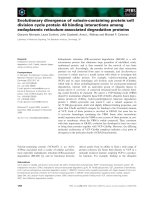
IL-8 and MCP-1 in both a time-dependent (Fig. 1A)
and dose-dependent (Fig. 1B,C) fashion. Notably,
AGE-HSA and LPS induced a very similar pattern of
cytokine ⁄ chemokine expression, although LPS stimula-
tion caused a much higher cytokine ⁄ chemokine
response than did AGE-HSA (Fig. 1). LPS, but not
AGE-HSA, also significantly induced IFN-c, IP-10
and GM-CSF expression in HUVECs (data not
shown). However, neither LPS, nor AGE-HSA stimu-
lated HUVECs to produce tumor necrosis factor-a,
IL-1b, IL-2, IL-4, IL-10 and IL-12 (data not shown).
AGE-HSA and LPS have a synergistic effect on
the induction of cytokines
⁄
chemokines
To determine whether there is any combined effect of
AGE-HSA and LPS on the induction of cytokines ⁄
chemokines, we stimulated HUVECs with AGE-HSA
J. Liu et al. Glycation end products and lipopolysaccharide in cytokine production
FEBS Journal 276 (2009) 4598–4606 ª 2009 The Authors Journal compilation ª 2009 FEBS 4599
at 25 lg ÆmL
)1
, LPS at 10 ngÆmL
)1
, or their combina-
tion, for up to 24 h. Compared to each single stimulus,
co-stimulation with both stimuli led to a much stron-
ger response on IL-6, IL-8 and MCP-1 production
(Fig. 2A). For example, on stimulation with either
AGE-HSA alone or LPS alone, IL-6 increased
from 12.1 ± 2.1 pgÆmL
)1
of basal level to 48.3 ±
13.2 pgÆmL
)1
and 343.3 ± 93.1 pgÆmL
)1
, respectively;
upon co-stimulation with both stimuli, IL-6 increased
to 1783.3 ± 131.8 pgÆmL
)1
, which is much higher than
the addition of the IL-6 levels induced by each single
stimulus (P < 0.01) (Fig. 2A). Consistently, the induc-
tion of IL-8 and MCP-1 also showed synergistic effects
after stimulation with both stimuli (P < 0.01), but this
was not as strong as that of IL-6 (Fig. 2A). However,
we failed to demonstrate any synergistic effects on
IFN-c, IP-10 and GM-CSF production after AGE-
HSA and LPS co-stimulation (data not shown),
suggesting that the synergistic induction of cytokines ⁄
chemokines by AGE-HSA and LPS in HUVECs is
highly selective.
We further performed RT-PCR and found that
either AGE-HSA alone or LPS alone increased the
mRNA transcripts of IL-6, IL-8 and MCP-1 compared
to their low levels in naive cells (Fig. 2B). Convinc-
ingly, co-stimulation with AGE-HSA and LPS led to a
further increase in gene transcription of these cyto-
kines ⁄ chemokines (P < 0.05) (Fig. 2B).
Activation of both MAP kinases and NF-jBis
required for the AGE-HSA and LPS amplified
cytokine ⁄ chemokine response
To elucidate the signal mechanisms involved in the
AGE-HSA and LPS amplified cytokine ⁄ chemokine
response, we first compared the signal transduction
pathways in HUVECs induced by LPS with those
induced by AGE-HSA. As expected, incubation of
HUVECs with LPS led to activation of both MAP
kinases (Fig. 3A) and NF-jB (Fig. 3B). Similar to
LPS, AGE-HSA stimulation also induced phosphory-
lation of p38, extracellular signal-regulated kinase
pg·mL
–1
LPS (ng·mL
–1
)
0
1 10 100 1000
0
200
400
600
800
1000
*
*
*
*
0
200
400
600
800
IL-6
pg·mL
–1
A
B
C
AGE-HSA
LPS
Incubation time (h)
04
612
24
0
500
1000
1500
2000
2500
3000
*
*
*
*
LPS (ng·mL
–1
)
0
1 10 100 1000
0
800
1600
2400
3200
4000
*
*
*
*
LPS (ng·mL
–1
)
0
1 10 100 1000
0
400
800
1200
1600
2000
IL-8
Incubation time (h)
04 612
24
AGE-HSA
LPS
0
Incubation time (h)
04612
24
600
1200
1800
2400
3000
MCP-1
AGE-HSA
LPS
pg·mL
–1
0 50 12.5 25 50 100
HSA
AGE-HSA
(µg·mL
–1
)
25
50
75
1
00
125
0
*
*
*
*
0
200
400
600
800
1000
*
*
*
*
0 50 12.5 25 50 100
HSA
AGE-HSA
0
500
1000
1500
2000
2500
*
*
*
*
0 50 12.5 25 50 100
HSA
AGE-HSA
(µg·mL
–1
)
(µg·mL
–1
)
Fig. 1. (A) Time- and (B, C) dose-dependent cytokine ⁄ chemokine release induced by AGE-HSA or LPS. HUVECs were incubated with various
doses of AGE-HSA or LPS for up to 24 h. Cytokines ⁄ chemokines, including IL-6, IL-8 and MCP-1, in the culture supernatants were mea-
sured. Data are expressed as the mean ± SD of four independent experiments. *P < 0.05 versus (B) unstimulated cells or (C) HSA.
Glycation end products and lipopolysaccharide in cytokine production J. Liu et al.
4600 FEBS Journal 276 (2009) 4598–4606 ª 2009 The Authors Journal compilation ª 2009 FEBS
(ERK) 1 ⁄ 2, Jun N-terminal kinase (JNK) (Fig. 3A)
and nuclear translocation of NF-jB (Fig. 3B),
although the activated levels of MAP kinases and NF-
jB induced by AGE-HSA were less than those induced
by LPS. Furthermore, co-stimulation with AGE-HSA
and LPS resulted in further increases in phosphory-
lated MAP kinases (Fig. 3A) and NF-jB nuclear
translocation (Fig. 3B), which is consistent with the
observed synergistic induction of cytokines ⁄ chemokines
by AGE-HSA and LPS.
After demonstrating that both AGE-HSA and LPS
activate similar signal transduction pathways in
HUVECs, we next aimed to clarify which of these
pathways is involved in the AGE-HSA and LPS ampli-
fied cytokine ⁄ chemokine response using specific phar-
macological inhibitors to block each individual
signaling pathway. Blockage of each individual path-
way [i.e. p38 with SB203580, ERK1 ⁄ 2 with PD98059,
JNK with JNK inhibitor II, and NF-jB with pyrroli-
dine dithiocarbamate (PDTC)] significantly attenuated
AGE-HSA and LPS co-stimulation-induced IL-6
(Fig. 4A), IL-8 (Fig. 4B) and MCP-1 (Fig. 4C) produc-
tion, indicating that the activation of all of these four
pathways is necessary for the effective induction of
cytokines ⁄ chemokines in HUVECs by AGE-HSA and
LPS. Notably, blocking p38 with SB203580 led to a
more profound inhibition in IL-6, IL-8 and MCP-1
production compared to blockade of either ERK1 ⁄ 2,
JNK or NF-jB, indicating a predominant role of the
p38 pathway in the AGE-HSA and LPS amplified
inflammatory response. Furthermore, blockade of both
MAP kinases and NF-jB with a combination of inhib-
itors completely abolished AGE-HSA and LPS co-
stimulated IL-6, IL-8 and MCP-1 production (Fig. 4),
suggesting that these two pathways act co-operatively.
Discussion
Previous studies have demonstrated that AGEs are
produced from oxidative modification by myeloperoxi-
IL-6 (pg·mL
–1
)
IL-8 (pg·mL
–1
)
0
200
400
600
800
1000
Control
AGE-HSA
LPS
AGE-HS
A
+ LPS
*
Control
LPS
AGE-HSA
+ LPS
800
0
400
1200
1600
2000
*
MCP-1 (pg·mL
–1
)
0
500
1000
1500
2000
2500
Control
AGE-HS
A
LPS
AGE-HS
A
+ LPS
*
AGE-HSA
GAPDH
IL-6
AGE-HSA
+ LPS
Control
AGE-HSA
LPS
10
0
2
4
6
8
Induction fold
*
*
*
AGE-HSA
+ LPS
Control
AGE-HSA
LPS
IL-8
GAPDH
Induction fold
0
2
4
6
8
10
*
*
*
AGE-HSA
+ LPS
Control
AGE-HSA
LPS
MCP-1
GAPDH
Induction fold
0
2
4
6
8
10
*
*
*
A
B
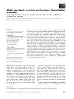
Fig. 2. Synergistic induction of cytokines ⁄ chemokines by AGE-HSA and LPS co-stimulation. HUVECs were incubated with AGE-HSA
(25 lgÆmL
)1
), LPS (10 ngÆmL
)1
), or their combination, for 2 and 24 h. (A) IL-6, IL-8 and MCP-1 release from HUVECs 24 h after stimulation
was measured and (B) gene expression of IL-6, IL-8 and MCP-1 in HUVECs 2 h after stimulation was assessed by RT-PCR. Data are
expressed as the mean ± SD of four (A) or three (B) independent experiments. *P < 0.05 versus AGE-HSA alone or LPS alone.
J. Liu et al. Glycation end products and lipopolysaccharide in cytokine production
FEBS Journal 276 (2009) 4598–4606 ª 2009 The Authors Journal compilation ª 2009 FEBS 4601
dase released from the activated leukocytes in the
lesion regions of inflammatory diseases [9,19]. When
bacterial infection occurs in patients with elevated
AGEs, such as diabetics or the elderly, and those
receiving long-term dialysis, the endothelial cells are
exposed concomitantly to a variety of proinflammatory
mediators, especially AGEs and LPS. It has been dem-
onstrated that both AGEs and LPS play key roles in
the development of inflammatory diseases by stimulat-
ing cytokine ⁄ chemokine production from leukocytes
and endothelial cells [1,8,9]. However, the combined
inflammation-stimulatory effect of AGEs and LPS
on endothelial cells, and, furthermore, the underlying
signal mechanisms involved, have not been fully under-
stood.
In the present study, we found that both AGE-HSA
and LPS, in a very similar patter, induced a time- and
dose-dependent proinflammatory cytokine ⁄ chemokine
production in HUVECs, although a much higher cyto-
kine ⁄ chemokine response was induced by LPS com-
pared to that induced by AGE-HSA. We further
demonstrated that AGE-HSA and LPS co-stimulation
Control
AGE-HSA
+ LPS
AGE-HSA
LPS
ERK1/2
P-ERK1
0
2
4
6
8
10
*
*
*
Control
AGE-HSA
+ LPS
AGE-HSA
LPS
JNK
P-JNK
12
0
2
4
6
8
10
*
*
*
Control
AGE-HSA
+ LPS
AGE-HSA
LPS
p38
P-p38
Induction fold
A
B
0
2
4
6
8
10
*
*
*
Control
AGE-HSA
AGE-HSA
+LPS
LPS
Relative nuclear fluorescence intensity
Control
AGE-HSA
AGE-HSA
+ LPS
LPS
2
4
0
6
8
10
12
*
*
*
Fig. 3. AGE-HSA and LPS stimulate MAP kinase phosphorylation and NF-jB nuclear translocation. HUVECs were incubated with AGE-HSA
(25 lgÆmL
)1
), LPS (10 ngÆmL
)1
), or their combination, for 30 min. (A). Total (upper panel) and phosphorylated (lower panel) p38, ERK1 ⁄ 2 and
JNK were detected by western blot analysis. (B) The nuclear translocation of NF-jB was detected by immunofluorescent staining with a
primary antibody against NF-jB p65 and Alexa Fluor 488-conjugated secondary antibody. Data are expressed as the mean ± SD and the
results shown represent one experiment from a total of four independent experiments. *P < 0.05 versus unstimulated cells.
Glycation end products and lipopolysaccharide in cytokine production J. Liu et al.
4602 FEBS Journal 276 (2009) 4598–4606 ª 2009 The Authors Journal compilation ª 2009 FEBS
exhibited a synergistic effect on the induction of IL-6,
IL-8 and MCP-1, but not other cytokines ⁄ chemokines,
at both gene and protein levels. This is important for
understanding the mechanisms underlying diseases
involving AGEs that are complicated with bacterial
infection. As proinflammatory mediators, these cyto-
kines ⁄ chemokines play an important role in inflamma-
tory processes. For example, IL-6 has been shown to
promote smooth muscle cell proliferation and increase
the permeability of endothelium, thus exaggerating the
inflammatory response [20]; the chemoattractants IL-8
and MCP-1 induce leukocyte–endothelial cell adhesion
and promote the migration and infiltration of leuko-
cytes to the inflammatory loci, which is a key step in
inflammation [21,22]. Therefore, the cooperation of
AGE-HSA and LPS on the induction of cyto-
kines ⁄ chemokines in endothelial cells observed in the
present study may amplify the inflammatory reactions
by enhancing the production of IL-6, IL-8 and MCP-
1, consequently leading to intensive inflammation and
aggravation of the disease.
It is well established that proinflammatory media-
tors initiate a cytokine⁄ chemokine storm by activating
the intracellular signaling pathways, including MAP
kinases and NF-jB [12,23,24]. Previous studies have
demonstrated that LPS and AGEs activate MAP
kinases in various cells, including monocytes ⁄ macro-
phages and endothelial cells [12,24,25]. In agreement
with previous work, we also observed increased MAP
kinase phosphorylation and NF-jB nuclear transloca-
tion in HUVECs after either AGE-HSA or LPS stim-
ulation, indicating that both AGE-HSA and LPS
activate similar intracellular signaling pathways.
Moreover, we demonstrated that AGE-HSA and LPS
co-stimulation led to a further activation of both
MAP kinases and NF-jB, which may explain the
observed much greater production of cyto-
kines ⁄ chemokines induced by a combination of the
two stimulants. We further used specific pharmacolog-
ical inhibitors to block each individual intracellular
signaling pathway, in an attempt to determine which
of these pathways is responsible for the observed
up-regulation of cytokine ⁄ cchemokine expression by
AGE-HSA and LPS co-stimulation. Blockage of
either MAP kinases or NF-jB resulted in a significant
reduction in AGE-HSA and LPS co-stimulation-
induced IL-6, IL-8 and MCP-1 release from
HUVECs. Of note, blocking p38 alone achieved the
maximal attenuation in proinflammatory cyto-
kine ⁄ chemokine production, indicating that the p38
pathway acts predominately in the AGE-HSA and
LPS amplified inflammatory response and, thus, may
serve as a main therapeutic target for AGE-related
inflammatory diseases. Furthermore, blockade of both
MAP kinases and NF-jB completely abolished AGE-
HSA and LPS co-stimulated IL-6, IL-8 and MCP-1
production, suggesting that activation of these two
pathways is required for the effective induction of
these cytokines ⁄ chemokines.
A
*
0
500
1000
1500
2000
2500
*
*
*
*
IL-6 (pg·mL
–1
)
AGE-HSA + LPS
SB 203580
PD 98059
JNK inhibitor II
PDTC
−
−
−
−
−
+
−
−
−
−
+
+
−
−
−
+
−
+
−
−
+
−
−
+
−
+
−
−
−
+
+
+
+
+
+
B
0
AGE-HSA + LPS
SB 203580
PD 98059
JNK inhibitor II
PDTC
−
−
−
−
−
+
−
−
−
−
+
+
−
−
−
+
−
+
−
−
+
−
−
+
−
+
−
−
−
+
+
+
+
+
+
200
400
600
800
1000
1200
*
*
*
*
*
IL-8 (pg·mL
–1
)
C
*
*
*
0
500
1000
1500
2000
2500
*
*
MCP-1 (pg·mL
–1
)
AGE-HSA + LPS
SB 203580
PD 98059
JNK inhibitor II
PDTC
−
−
−
−
−
+
−
−
−
−
+
+
−
−
−
+
−
+
−
−
+
−
−
+
−
+
−
−
−
+
+
+
+
+
+
Fig. 4. Inhibition of either MAP kinases or NF-jB attenuates AGE-
HSA and LPS-stimulated cytokine ⁄ chemokine production. HUVECs
were pretreated with SB253580 (20 l
M), PD98059 (20 lM), JNK
inhibitor II (50 n
M), PDTC (50 lM), or their combination, for 1 h and
then co-stimulated with AGE-HSA (25 lgÆmL
)1
) and LPS
(10 ngÆmL
)1
) for 24 h. (A) IL-6, (B) IL-8 and (C) MCP-1 in the culture
supernatants were measured. Data are expressed as the
mean ± SD from four independent experiments. *P < 0.05 versus
AGE-HSA + LPS co-stimulated cells.
J. Liu et al. Glycation end products and lipopolysaccharide in cytokine production
FEBS Journal 276 (2009) 4598–4606 ª 2009 The Authors Journal compilation ª 2009 FEBS 4603
In summary, we demonstrate that AGE-HSA and
LPS synergistically stimulate IL-6, IL-8 and MCP-1
production in endothelial cells, thus contributing to
the development of AGE-related inflammatory dis-
eases. Furthermore, activation of both MAP kinases
and NF-jB is required for the AGE-HSA and LPS
co-stimulation amplified inflammatory response, impli-
cating that the blockade of these signal pathways, in
particular the p38 pathway, may provide a novel
approach for the treatment of AGE-related diseases,
such as diabetes, complicated with bacterial infection.
Experimental procedures
Preparation of AGE-modified proteins
AGE-modified proteins were prepared as previously
described [6]. Briefly, 1.75 gÆL
)1
purified HSA (Sigma-
Aldrich, St Louis, MO, USA) was incubated with 0.1 m
d-glucose at 37 °C for 8 weeks, and then dialyzed against
NaCl ⁄ P
i
to remove the unbound glucose. HSA incubated
without glucose was used as the control. The content of
AGEs in the AGE-HSA preparation was 85.24 UÆmg
)1
protein, whereas that of the control was less than
0.9 UÆmg
)1
protein, indicating that glycation modified
protein was successfully obtained. Endotoxin levels in all
preparations were measured with an E-toxate kit (Sigma-
Aldrich) and found to be below the detection limit
(< 0.1 EUÆmL
)1
).
Isolation and culture of endothelial cells
Primary HUVECs were isolated from normal human
umbilical cord veins and cultured as described previously
[26]. Briefly, the separated cells were suspended in
RPMI 1640 with 20% fetal bovine serum (HyClone, Logan,
UT, USA) and plated in tissue culture dishes. The next
day, non-attached cells were removed and the medium was
replaced with complete RPMI 1640 supplemented with
20% fetal bovine serum, 100 ngÆmL
)1
endothelial cell
growth factor (Clontech, Mountain View, CA, USA) and
antibiotics. Endothelial cells were identified by the charac-
teristic monolayer cobblestone appearance and positive
staining for von Willebrand factor.
AGE-HSA and LPS stimulation and protein kinase
inhibition
HUVECs were seeded on 96- or six-well culture plates for
cytokine ⁄ chemokine detection, RT-PCR or western blot
analysis. Cells were incubated with different concentrations
of AGE-HSA and LPS (Escherichia coli 0111:B4) (Sigma-
Aldrich) for up to 24 h. Recombinant human CD14 (final
concentration: 0.1 lgÆmL
)1
) (R&D Systems, Minneapolis,
MN, USA) was used in each experiment to enhance the
LPS-mediated cell response because of a deficiency of
CD14 expression on the endothelial cells [14].
To observe the effect of MAP kinase inhibitor or NF-jB
inhibitor on AGE-HSA and LPS-stimulated cell activation,
HUVECs were pretreated with PD98059 (20 lm), JNK
inhibitor II (50 nm), SB203580 (20 lm) (all from Merck,
Darmstadt, Germany), an NF-jB inhibitor PDTC (50 lm)
(Sigma-Aldrich), or their combination, 1 h before AGE-
HSA and LPS stimulation. The concentration of each
inhibitor used was based on the dose–response experiment
(data not shown), with the maximal inhibitory effect. For
each experiment, cell viability was always more than 90%,
as determined by exclusion of trypan blue dye. Cell-free
supernatants were collected and stored at )80 °C until
analysis.
Cytokine
⁄
chemokine measurement
Cytokines ⁄ chemokines in the culture supernatants were
analyzed simultaneously using a LiquiChip work station,
which employs a bead-based xMAP (flexible multi-analyte
profiling) technology [27], according to the manufacturer’s
instructions.
RT-PCR
Total RNA was extracted using a single-step method of
RNA isolation by acid guanidinium thiocyanate–phenol–
chloroform extraction [20]. PCR amplification was per-
formed on the resulting cDNAs with specific primers for
human IL-6 (forward, 5¢-CAGGAGCCCAGCTATGA
ACT-3¢; reverse, 5¢-TAAGTTCTGTGCCCAGTGGA-3¢),
IL-8 (forward, 5¢-AGGGTTGCC AGATGCAATAC-3¢;
reverse, 5¢-ACACAGCTGGCAATGACAAG-3¢), MCP-1
(forward, 5¢-GTGAGGAGCCACCAACATTT-3¢; reverse,
5¢-GGGGGATCCCAAGTACTGTT-3¢) and GAPDH (for-
ward, 5¢-CCCATCACCATCTTCCAGGA-3¢; reverse, 5¢-
TGCTTCACCACCTTCTTGAT-3¢)at94°C for 30 s,
56 °C for 15 s and 72 °C for 2 min, for 30 cycles. The
expected lengths of the fragments for IL-6, IL-8, MCP-1
and GAPDH were 730, 870, 733 and 520 bp, respectively.
Western blot analysis
Cells were lysed with the ice-cold lysis buffer (Cell signaling
Technology, Danvers, MA, USA) to extract cytoplamic
proteins. Equal amounts of protein extracts were subjected
to 12% SDS ⁄ PAGE and blotted onto a poly(vinylidene
difluoride) membrane. The membrane was blocked and
probed overnight at 4 °C with antibodies against total or
phosphorylated p38, ERK1 ⁄ 2 or JNK (Cell Signaling Tech-
nology), followed by incubation with horseradish peroxi-
dase-conjugated secondary antibodies for 1 h at room
Glycation end products and lipopolysaccharide in cytokine production J. Liu et al.
4604 FEBS Journal 276 (2009) 4598–4606 ª 2009 The Authors Journal compilation ª 2009 FEBS
temperature. Blots were developed using an ECL detection
system (Amersham Biosciences, Little Chalfont, UK). Each
image was captured and the intensity of each band was
analyzed with a Kodak image workstation (Eastman
Kodak, Rochester, NY, USA).
Immunofluorescent staining
HUVECs were incubated with AGE-HSA (25 lgÆmL
)1
)
and ⁄ or LPS (10 ngÆmL
)1
) in the presence or absence of an
NF-jB inhibitor PDTC (50 lm). To detect the nuclear
translocation of NF-jB, immunofluorescent staining was
performed by staining the cells with a primary antibody
against NF-jB p65 (Abcam, Cambridge, UK) and Alexa
Fluor 488-conjugated secondary antibody (Molecular
Probes, Eugene, OR, USA). The nuclear fluorescent inten-
sity was analyzed by Axio Vision (Carl Zeiss, Oberkochen,
Germany).
Statistical analysis
All data are expressed as the mean ± SD and analyzed
with the Statistical Package for Social Sciences (SPSS Inc.,
Chicago, IL, USA). The statistical significance of differ-
ences was determined using one-way analysis of variance
and t-test. P < 0.05 was considered statistically significant.
Acknowledgements
This work was supported by the National Key Basic
Research Program of China (973 Program) (No.
2002CB513005), the Program for Changjiang Scholars
and Innovative Research Team in University (No.
IRT0731), the General Program of the National Natu-
ral Science Foundation of China (No. 30670828, No.
30572151 and No. 30670829), the Joint Program of
NSFC and GPG (No. U0632004), PSTPGP (No.
A1090202) and PSTPGC (No. 2007J1-C0301).
References
1 Bannerman DD & Goldblum SE (1999) Direct effects
of endotoxin on the endothelium: barrier function and
injury. Lab Invest 79, 1181–1199.
2 Cines DB, Pollak ES, Buck CA, Loscalzo J, Zimmer-
man GA, McEver RP, Pober JS, Wick TM, Konkle
BA, Schwartz BS et al. (1998) Endothelial cells in physi-
ology and in the pathophysiology of vascular disorders.
Blood 91, 3527–3561.
3 Brownlee M (1995) Advanced protein glycosylation in
diabetes and aging. Annu Rev Med 46, 223–234.
4 Bucala R & Cerami A (1992) Advanced glycosylation:
chemistry, biology, and implications for diabetes and
aging. Adv Pharmacol 23, 1–34.
5 Ulrich P & Cerami A (2001) Protein glycation, diabetes,
and aging. Recent Prog Horm Res 56, 1–21.
6 Hou FF, Chertow GM, Kay J, Boyce J, Lazarus JM,
Braatz JA & Owen WFJr (1997) Interaction between
beta 2-microglobulin and advanced glycation end prod-
ucts in the development of dialysis related-amyloidosis.
Kidney Int 51, 1514–1519.
7 Yan SF, Ramasamy R, Naka Y & Schmidt AM (2003)
Glycation, inflammation, and RAGE: a scaffold for the
macrovascular complications of diabetes and beyond.
Circ Res 93, 1159–1169.
8 Sun M, Yokoyama M, Ishiwata T & Asano G (1998)
Deposition of advanced glycation end products (AGE)
and expression of the receptor for AGE in cardiovascu-
lar tissue of the diabetic rat. Int J Exp Pathol 79, 207–
222.
9 Basta G, Lazzerini G, Massaro M, Simoncini T, Tanga-
nelli P, Fu C, Kislinger T, Stern DM, Schmidt AM &
De CR (2002) Advanced glycation end products acti-
vate endothelium through signal-transduction receptor
RAGE: a mechanism for amplification of inflammatory
responses. Circulation 105, 816–822.
10 Miura J, Yamagishi S, Uchigata Y, Takeuchi M,
Yamamoto H, Makita Z & Iwamoto Y (2003) Serum
levels of non-carboxymethyllysine advanced glycation
endproducts are correlated to severity of microvascular
complications in patients with Type 1 diabetes. J Diabe-
tes Complications 17, 16–21.
11 Leon CG, Tory R, Jia J, Sivak O & Wasan KM (2008)
Discovery and development of toll-like receptor 4
(TLR4) antagonists: a new paradigm for treating sepsis
and other diseases. Pharm Res 25, 1751–1761.
12 Hippenstiel S, Soeth S, Kellas B, Fuhrmann O, Seybold
J, Krull M, Eichel-Streiber C, Goebeler M, Ludwig S &
Suttorp N (2000) Rho proteins and the p38-MAPK
pathway are important mediators for LPS-induced
interleukin-8 expression in human endothelial cells.
Blood 95, 3044–3051.
13 Chow JC, Young DW, Golenbock DT, Christ WJ &
Gusovsky F (1999) Toll-like receptor-4 mediates lipo-
polysaccharide-induced signal transduction. J Biol Chem
274, 10689–10692.
14 Arditi M, Zhou J, Dorio R, Rong GW, Goyert SM &
Kim KS (1993) Endotoxin-mediated endothelial cell
injury and activation: role of soluble CD14. Infect
Immun 61, 3149–3156.
15 Meyer KC (2001) The role of immunity in susceptibility
to respiratory infection in the aging lung. Respir Physiol
128, 23–31.
16 Shah BR & Hux JE (2003) Quantifying the risk of
infectious diseases for people with diabetes. Diabetes
Care 26, 510–513.
17 Dofferhoff AS, Nijland JH, de Vries-Hospers HG,
Mulder PO, Weits J & Bom VJ (1991) Effects of different
types and combinations of antimicrobial agents on
J. Liu et al. Glycation end products and lipopolysaccharide in cytokine production
FEBS Journal 276 (2009) 4598–4606 ª 2009 The Authors Journal compilation ª 2009 FEBS 4605
endotoxin release from gram-negative bacteria: an
in-vitro and in-vivo study. Scand J Infect Dis 23, 745–754.
18 Szeto CC, Kwan BC, Chow KM, Lai KB, Chung KY,
Leung CB & Li PK (2008) Endotoxemia is related to
systemic inflammation and atherosclerosis in peritoneal
dialysis patients. Clin J Am Soc Nephrol 3, 431–436.
19 Anderson MM, Hazen SL, Hsu FF & Heinecke JW
(1997) Human neutrophils employ the myeloperoxidase-
hydrogen peroxide-chloride system to convert hydroxy-
amino acids into glycolaldehyde, 2-hydroxypropanal,
and acrolein. A mechanism for the generation of highly
reactive alpha-hydroxy and alpha,beta-unsaturated alde-
hydes by phagocytes at sites of inflammation. J Clin
Invest 99, 424–432.
20 Remick DG, Bolgos G, Copeland S & Siddiqui J (2005)
Role of interleukin-6 in mortality from and physiologic
response to sepsis. Infect Immun 73, 2751–2757.
21 Tedgui A & Mallat Z (2006) Cytokines in atherosclero-
sis: pathogenic and regulatory pathways. Physiol Rev
86, 515–581.
22 Gerszten RE, Garcia-Zepeda EA, Lim YC, Yoshida M,
Ding HA, Gimbrone MAJr, Luster AD, Luscinskas
FW & Rosenzweig A (1999) MCP-1 and IL-8 trigger
firm adhesion of monocytes to vascular endothelium
under flow conditions. Nature 398, 8–723.
23 Denk A, Goebeler M, Schmid S, Berberich I, Ritz O,
Lindemann D, Ludwig S & Wirth T (2001) Activation
of NF-kappa B via the Ikappa B kinase complex is
both essential and sufficient for proinflammatory gene
expression in primary endothelial cells. J Biol Chem
276, 28451–28458.
24 Yeh CH, Sturgis L, Haidacher J, Zhang XN, Sherwood
SJ, Bjercke RJ, Juhasz O, Crow MT, Tilton RG &
Denner L (2001) Requirement for p38 and p44 ⁄ p42
mitogen-activated protein kinases in RAGE-mediated
nuclear factor-kappaB transcriptional activation and
cytokine secretion. Diabetes 50, 1495–1504.
25 Berbaum K, Shanmugam K, Stuchbury G, Wiede F,
Korner H & Munch G (2008) Induction of novel cyto-
kines and chemokines by advanced glycation endprod-
ucts determined with a cytometric bead array. Cytokine
41, 198–203.
26 Marin V, Kaplanski G, Gres S, Farnarier C &
Bongrand P (2001) Endothelial cell culture: protocol to
obtain and cultivate human umbilical endothelial cells.
J Immunol Methods 254, 183–190.
27 Jiang Y, Xu J, Zhou C, Wu Z, Zhong S, Liu J, Luo W,
Chen T, Qin Q & Deng P (2005) Characterization of
cytokine ⁄ chemokine profiles of severe acute respiratory
syndrome. Am J Respir Crit Care Med 171, 850–857.
Glycation end products and lipopolysaccharide in cytokine production J. Liu et al.
4606 FEBS Journal 276 (2009) 4598–4606 ª 2009 The Authors Journal compilation ª 2009 FEBS