Báo cáo khoa học: High molecular weight kininogen binds to laminin – characterization and kinetic analysis pot
Bạn đang xem bản rút gọn của tài liệu. Xem và tải ngay bản đầy đủ của tài liệu tại đây (353.82 KB, 11 trang )
High molecular weight kininogen binds to
laminin – characterization and kinetic analysis
Inger Schousboe and Birthe Nystrøm
Department of Biomedical Sciences, The Panum Institute, University of Copenhagen, Denmark
Keywords
extracellular matrix; high molecular
weight kininogen; kininostatin; laminin;
Zn2+-independent
Correspondence
I. Schousboe, Department of Biomedical
Sciences, The Panum Institute, University of
Copenhagen, Blegdamsvej 3C, DK-2200
Copenhagen, Denmark
Fax: +45 3536 7980
Tel: +45 3532 7800
E-mail:
(Received 11 March 2009, revised 8 July
2009, accepted 16 July 2009)
doi:10.1111/j.1742-4658.2009.07218.x
High molecular weight kininogen (HK) is an abundant plasma protein that
plays a central role for the function of the kallikrein ⁄ kinin ⁄ kininogen system. Thus, cleavage of HK by kallikrein liberates bradykinin, which stimulates vascular repair and a two-chain protein, activated HK (HKa), which
induces apoptosis in proliferating endothelial cells. The localization of these
events remains obscure, although the basement membrane may be of
importance. Analyzing the interaction between HK and HKa and selected
basement membrane proteins, we observed that they bound to the major
noncollageneous proteins laminin, but not to vitronectin or fibronectin
coated on microtiter plates. The binding to laminin was Zn2+ independent.
However, at low but not at high concentrations of albumin, Zn2+
increased the affinity for the binding by abolishing an inhibitory effect of
Ca2+. Recombinant human kininostatin encompassing the amino acid
sequence, Arg439-Ser532 but not the endothelial cell binding peptide
sequence (His479-His498; HKH20) within kininostatin inhibited the binding of HKa to laminin. This established that the amino acid sequence
Arg439-Lys478 in domain 5 of HK is of importance for its binding to laminin. Extensive proteolytic cleavage of HK and HKa with kallikrein abolished the binding to laminin, releasing a 12 kDa anti-kininostatin reacting
peptide. On the basis of these results, we propose that the binding of HK
to laminin is a primary event, which secures proper localization of the
cleavage products for subsequent interaction with the endothelium to
promote inflammatory and pro- and anti-angiogenic activities.
Structured digital abstract
l
MINT-7218019: Laminin alpha 5 (uniprotkb:Q61001), Laminin beta 1 (uniprotkb:P02469),
Laminin gamma 1 (uniprotkb:P02468), Laminin alpha 5 (uniprotkb:Q61001), Laminin beta 2
(uniprotkb:Q61292) and Laminin gamma 1 (uniprotkb:P02468) physically interact (MI:0915)
with HK (uniprotkb:P01042) by solid phase assay (MI:0892)
l
MINT-7219326: Laminin alpha 1 (uniprotkb:P19137), Laminin beta 1 (uniprotkb:P02469) and
Laminin gamma 1 (uniprotkb:P02468) physically interact (MI:0915) with HK (uniprotkb:
P01042) by solid phase assay (MI:0892)
Introduction
The extracellular matrix (ECM) controls a variety of
cellular functions by interacting with a vast array of
macromolecules, exhibiting a wide range of activities
[1]. Recent in vitro investigations have indicated that
Abbreviations
ECM, extracellular matrix; FN, fibronectin; HK, high molecular weight kininogen; HKa, activated HK; HRP, horseradish peroxidase; HUVEC,
human umbilical vein endothelial cells; LM, laminin; uPAR, urokinase plasminogen activator receptor; VN, vitronectin.
5228
FEBS Journal 276 (2009) 5228–5238 ª 2009 The Authors Journal compilation ª 2009 FEBS
I. Schousboe and B. Nystrøm
this array of macromolecules includes the surface binding proteins in the contact activation system of the
blood coagulation system. This system consists of
factor XII, high molecular weight kininogen (HK),
prekallikrein and factor XI, but only factor XII and
HK interact directly with the ECM. However, in the
plasma, prekallikrein and factor XI form complexes
with HK, enabling the assembly of the entire contact
activation system on the vascular wall [2–5]. After
being assembled, the system functions locally dependent on the demand for activation. Thus, as a result of
factor XI activation by activated factor XII, the rate
of fibrin formation becomes enhanced [6], whereas
activation of prekallikrein by activated factor XII
enhances the proteolytic cleavage of HK by plasma
kallikrein [7]. However, the localization of the activation remains to be revealed, although factor XII
recently was shown to bind to fibronectin (FN) [8],
and several endothelial cell membrane proteins have
been suggested as receptors for HK, including the
globular C1q receptor [9,10] and cytokeratin-1 [10–12].
The binding of HK to these receptors is strictly dependent upon the free Zn2+ concentration. Cleavage of
HK releases a short-lived strong inflammatory nonapeptide, bradykinin, leaving behind a two-chain activated HK (HKa), which Zn2+-dependently binds to
the urokinase plasminogen activator receptor (uPAR)
[13]. This binding is considered to inhibit angiogenesis
[14].
The process of angiogenesis is a complex event
requiring signals from both plasma and the extracellular basement membrane, and adhesive interactions of
endothelial cells with the underlying basement membrane are instrumental in regulating the development
and maintenance of the vascular wall. The basement
membrane contains a network of collagen and laminin
(LM) [15]. Formation of new vessels involves the
migration and proliferation of cells. To assist the cells
in their migration, the extravascular matrix provides
an environment consisting of hyaluronic acid, vitronectin (VN) and FN [16]. LM plays an important role in
cell adhesion to the basement membrane by interacting
in the endothelium with a series of integrins, including
a3b1, a6b1, avb1, avb3 and avb5 [16–18]. The latter
three of these integrins are activated and engaged by
VN [19] when VN interacts with uPAR [20–22]. The
induction of apoptosis in proliferating endothelial cells
by HKa [23] has been suggested to be the result of the
binding of HKa not only to uPAR, but also to VN
[24–26], preventing the interaction between VN and
uPAR. However, the apoptotic effect of HKa is apparently regulated by several ECM proteins [11,24]. Thus,
Guo et al. [24] observed that the adhesion of endothe-
HK binding to laminin
lial cells cultured on VN, but not that of cells cultured
on FN, was inhibited by HKa, whereas Sun and McCrae [14] demonstrated that HKa induced apoptosis of
endothelial cells cultured on not only VN, but also on
FN and LM. Whether this inhibition is the result of
the binding of HKa to the ECM proteins remains to
be revealed.
LMs are a family of glycoprotein heterotrimers composed of an a, b and c chain. To date, five a, four b
and three c LM chains have been identified that can
combine to form 15 different isoforms [15,27]. The
prototype of LMs is LM 1. The LM 1 isoform is characterized by the presence of one LM a1 chain, which
combines with one LM b1 chain and one LM c1
chain. LMs expressed in endothelial cells are characterized by the presence of LM a4 and a5 chains, which
combine with LM b1 and c1 chains to form LM 8 and
LM 10, respectively. In LM 11, which is also present
in the endothelium, one a5 chain combines with b2
and c1 chains [15].
In the present study, using a solid phase binding
assay, we analyzed the binding of HK and HKa to
LM, FN and VN, and showed that both HK and
HKa bind with high affinity to LM. The LMs used in
the study comprised LM 1 and LM 10 ⁄ 11, the latter of
which are characteristic of the endothelium.
Results
HK and HKa binding to proteins of the
extracellular membrane
To analyze the ability of HK to bind to selected proteins present in the extracellular membrane, freshly
drawn citrate anti-coagulated plasma was incubated on
LM, VN or FN coated on microtiter plates. The
amount of HK absorbed from the plasma by these
matrix proteins was subsequently analyzed by immunoreactions using a monoclonal antibody to the heavy
chain of HK (mAb 2B5) as the primary antibody. This
demonstrated that HK apparently could be extracted
from plasma by binding to all three matrix proteins.
However, only the amount extracted by LM was
higher than the amount extracted by the noncoated
surface (Fig. 1A). Analyzing the binding of purified
HK and HKa to LM, VN and FN showed that, overall, a larger amount of HKa than of HK bound
when incubated at the same concentration. However,
the amount of HKa bound to VN was identical to the
amount bound to the noncoated plate, whereas the
amount bound to LM and FN was lower. By contrast,
relative to the amount bound to the noncoated plate,
more HK bound to LM and VN than to FN (Fig. 1B).
FEBS Journal 276 (2009) 5228–5238 ª 2009 The Authors Journal compilation ª 2009 FEBS
5229
HK binding to laminin
A
I. Schousboe and B. Nystrøm
problem of nonspecific binding. This was achieved by
increasing the concentration of the LM and VN coated
on the microtiter plate. As indicated above, HK did
not bind to the noncoated plate. With increasing LM
concentrations up to 5 lgỈmL)1, increasing amounts of
HK bound to LM. By contrast, the amount of HKa
that bound to the noncoated plate decreased as the
concentration of LM increased to 5 lgỈmL)1. A constant amount of HK and HKa bound to LM at concentrations higher than 5 mgỈmL)1 (Fig. 2A). The
binding of HKa to LM was inhibited by the presence
of soluble LM, but not by the presence of soluble VN
or FN (data not shown).
Zn2+ has been shown to play a determining role for
the interaction of HK and HKa with all previously
identified receptors and ligands. The presence of Zn2+
increased the amount of HK as well as HKa bound to
the noncoated plate but, analogous to the binding in
Plasma
VN
Ligand coated on the microtiter plate
FN
LM
None
0
0.2
0.4
0.6
0.8
1
1.2
HK/HKa bound (absorbance units)
B
Purified HK and HKa
VN
FN
LM
0
0.2
0.4
0.6
0.8
1
1.2
1.4
1.6
HK/HKa bound (absorbance units)
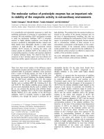
Fig. 1. Extraction of HK from plasma (A) and of HK and HKa (B)
from solutions of purified proteins by immobilized LM, VN and FN.
(A) Blood was drawn by the syringe method using one volume of
3.8% (w ⁄ v) sodium citrate to nine volumes of blood and centrifuged at 1180 g for 5 min. Aliquots of 100 lL of the supernatant
(citrate anti-coagulated plasma) were then added to wells coated
overnight with LM (10 lgỈmL)1), VN (5 lgỈmL)1) and FN
(10 lgỈmL)1), respectively, and blocked with Locke’s buffer containing 1% (w ⁄ v) BSA. (B) Alternatively, the wells were incubated with
purified solutions of 20 nM HK (hatched columns) or 20 nM HKa
(grey columns) in Locke’s buffer containing 0.35% (w ⁄ v) BSA. After
1 h of incubation, the content in the wells was removed and the
plates were washed and incubated with primary and secondary
antibodies, as described in the Experimental procedures. Binding to
the surface coated with buffer alone was used as a measure
of nonspecific binding. The results are the mean ± SD (n = 3) as
indicated by vertical bars.
Attempts to reduce the binding to the noncoated plate
by increasing the BSA concentration from the standard
concentration of 3.5 mgỈmL)1 to as much as
75 mgỈmL)1 in the block buffer gradually decreased
the amount of HKa bound nonspecifically, but had no
influence on the amount of HKa bound to LM, VN
and FN relative to the amount bound to the noncoated surface (results not shown). Because a considerable
amount of HKa bound to the noncoated microtiter
plate, we next analyzed whether the concentration of
LM and VN was sufficiently high to saturate the surface of the microtiter plate, thereby preventing the
5230
4.0
HK
HK + Zn
HKa
HKa + Zn
3.5
3.0
2.5
2.0
1.5
1.0
0.5
0.0
0
2
4
6
8
10
12
LM concentration (µg·mL–1)
B
HKa bound (absorbance units)
None
HKa bound (absorbance units)
A
4.5
4
HK
HK + Zn
HKa
HKa + Zn
3.5
3
2.5
2
1.5
1
0.5
0
0
5
10
15
VN concentration (µg·mL–1)
20
25
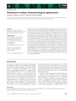
Fig. 2. Binding of HK and HKa to wells coated with increasing concentrations of LM (A) and VN (B). Wells were coated overnight with
increasing concentrations of LM (A) and VN (B) in coating buffer as
indicated and subsequently blocked with Locke’s buffer containing
1% (w ⁄ v) BSA. Next, the wells were incubated for 1 h with 20 nM
HK or 20 nM HKa in Locke’s buffer containing 0.35% (w ⁄ v) BSA in
the presence (open symbols) or absence of 50 lM Zn2+ (closed
symbols). The amount of HK (squares) and HKa (circles) bound
after extensive washing was determined by incubation with mAb
2B5. The results are the mean ± SD (n = 3) as indicated by vertical
bars when extending beyond the symbols.
FEBS Journal 276 (2009) 5228–5238 ª 2009 The Authors Journal compilation ª 2009 FEBS
I. Schousboe and B. Nystrøm
HK binding to laminin
the absence of Zn2+, the binding to LM decreased
with increasing concentrations of LM and became
constant at concentrations higher than 5 lgỈmL)1
(Fig. 2A). Therefore, a standard concentration of
10 lgỈmL)1 LM was used throughout the study.
A similar analysis, performed with VN as the immobilized ligand, showed no binding of HK at any concentration of VN in the absence of Zn2+ (Fig. 2B).
The high amount of HKa that bound to the noncoated
plate (Fig. 1B), particularly in the presence of Zn2+,
decreased exponentially with increasing VN concentrations over the whole VN concentration range
(0–20 lgỈmL)1) (Fig. 2B). This indicated that any
concentration of VN lower than 20 lgỈmL)1 was
insufficient to saturate completely the surface in the
microtiter plate. Thus, the binding of HK and HKa to
VN shown in Fig. 1B was most likely nonspecific.
The effect of Zn2+ on the binding of HK and HKa
to LM
Further investigations of the effect of Zn2+ on the
binding of HKa to LM showed that the amount of
bound HKa increased with increasing Zn2+ concentrations up to 15–20 lm and then decreased (Fig. 3).
Because the enhancement was more pronounced at
200
3.000
150
100
50
0
0
20
40
60
80
100
120
Concentration of Zn2+ (µM)
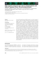
Fig. 3. Binding of HKa to LM as a function of the concentration of
Zn2+. A microtiter plate was coated overnight with LM (10 lgỈmL)1)
and blocked in Locke’s buffer containing 1% (w ⁄ v) BSA. Next, it
was incubated with either 10 nM HKa (open squares) or 30 nM HKa
(open circles) diluted in Locke’s buffer containing 0.35% (w ⁄ v) BSA
and supplemented with increasing concentrations of Zn2+. The
amount of HKa bound to the LM after incubation for 1 h was determined using mAb 2B5, as described in the Experimental procedures. Using the amount of HKa bound to LM in the absence of
Zn2+ as the reference (100%), the enhancement of the binding at
the varying Zn2+ concentrations is shown as a percentage. The
results are the mean ± SD (n = 3) as indicated by vertical bars
when extending beyond the symbols.
Hka bound (absorbance units)
Relative amount of Hka bound (%)
250
lower than at higher HKa concentrations, this indicated that the effect of Zn2+ might be caused by a
change in the affinity for the binding of HKa to LM.
Titration of LM with increasing concentrations of
HKa in the presence and absence of 20 lm Zn2+
showed that the presence of Zn2+ enhanced the affinity of the binding, but apparently had no or only little
effect on the maximum amount of bound HKa
(Fig. 4). Moreover, in the presence of Zn2+, the
amount of HKa bound at high HKa concentrations
was constant, indicating that HKa did not bind nonspecifically to LM.
The LM isoform used in the above measurements
was the prototype, LM 1. To determine whether HK
and HKa would bind also to LM 10 ⁄ 11, the dissociation constants, KD, for the binding were determined in
a series of identical experiments using LM 1 as well as
LM 10 ⁄ 11. This revealed that, in the absence of Zn2+,
HK and HKa bound with the same affinity, regardless
of whether LM 1 or LM 10 ⁄ 11 had been coated on
the microtiter plate (Table 1). The presence of Zn2+
affected more the affinity of the binding of HKa than
HK. Thus, a five- to seven-fold increase was observed
in the affinity for the binding of HKa to both LM 1
and LM 10 ⁄ 11, whereas only a three-fold increase was
observed for the binding of HK to LM 1 (Table 1).
The presence of Zn2+ had no or only a minimal effect
on the maximal amount bound in each individual
experiment (data not shown).
2.500
2.000
1.500
1.000
0.500
0.000
0.0
20.0
40.0
60.0
80.0
100.0 120.0 140.0 160.0
Concentration of HKa (nM)
Fig. 4. Concentration dependent binding of HKa to LM in the presence or absence of Zn2+. A microtiter plate was coated overnight
with LM (10 lgỈmL)1) and blocked in Locke’s buffer containing 1%
(w ⁄ v) BSA, as described in the legend to Fig. 2. Next, it was incubated with increasing concentrations of HKa diluted in Locke’s buffer containing 0.35% (w ⁄ v) BSA in the presence (open circles) or
absence (closed circles) of 20 lM Zn2+. The amount of HKa bound
to the LM after incubation for 1 h was subsequently determined
using mAb 2B5, as described in the Experimental procedures. The
results are the mean ± SD (n = 3) as indicated by vertical bars
when extending beyond the symbols.
FEBS Journal 276 (2009) 5228–5238 ª 2009 The Authors Journal compilation ª 2009 FEBS
5231
HK binding to laminin
I. Schousboe and B. Nystrøm
Table 1. The effect of Zn2+ on KD for the binding of HK and HKa
to LM 1 and LM 10 ⁄ 11. The concentration of Zn2+ was 20 lM.
Data are the mean ± SD (n).
KD (nM)
LM 1
Hka
None
+Zn2+
HK
None
+Zn2+
LM 10 ⁄ 11
35.8 ± 9.8 (5)
7.1 ± 3.0 (5)
34.3 ± 0.9 (3)
4.6 ± 1.7 (4)
27.0 ± 4.6 (6)
8.7 ± 1.6 (4)
22.2 ± 6.1 (4)
16.6 ± 5.7 (3)
The nonhyperbolic shape of the Zn2+-dependency
curve (Fig. 3) indicated further that the effect of Zn2+
might be explained by the presence of other divalent
cations in the incubation mixture. To verify this, the
standard constituents, Ca2+ and Mg2+ in the incubation buffer (Locke’s buffer) were excluded. This
enhanced the amount of HKa bound to LM in the
absence, but not in the presence, of Zn2+ and indicated that the effect of Zn2+ was to abolish an inhibitory effect of Mg2+ or Ca2+ or both. Excluding one
of these ions at a time showed that the inhibition was
mainly the result of the presence of Ca2+. The amount
of HKa bound to LM in the presence of Zn2+ was
not or only slightly affected by the depletion of Ca2+
and Mg2+, as long as the BSA concentration was low
(3.5 mgỈmL)1). At a higher BSA concentration, the
presence of 20 lm Zn2+ was unable to abolish the
inhibitory effect of Mg2+ and Ca2+ (Table 2).
Identification of the binding region in HKa
The amino acid sequence Arg439-Ser532 in domain 5
of HK (Fig. 5) [28,29], and particularly the His479His498 sequence, have been mapped as the endothelial
cell-, ECM- and surface-binding site [11,13,30]. To
define the binding site within HK for the binding to
LM, we determined whether a recombinant human
kininostatin peptide (rhkininostatin; Arg439-Ser532) and
a synthetic dodeca-peptide (His479-His498; HKH20)
competitively inhibited the binding of HKa to LM.
We also tested whether a synthetic sequence of amino
acids copying the Ser372-Arg419 sequence N-terminally to kininostatin affected the binding. This
sequence is cleaved off secondary to bradykinin. Only
rhkininostatin inhibited the binding and, both in the
presence and absence of Zn2+, a 50% inhibition was
observed at a 500 molar excess of rhkininostatin compared to the concentration of HKa (Fig. 6). However,
the presence of HKH20, even at a 5000 molar excess
5232
Table 2. The effect of Ca2+, Mg2+ and Zn2+ on the binding of HKa
to LM. The complete binding buffer (Locke’s buffer) contained
physiological concentrations of Ca2+ (2.3 mM) and Mg2+ (1 mM).
The experiment was performed as described using the buffer composition of Locke’s buffer, but lacking the divalent cations indicated. Some of the experiments were performed at a 10-fold
higher BSA concentration, as indicated. The results are representative of one of three experiments performed in triplicate and are
shown as the mean ± SD. A statistical analysis was performed
using one-way analysis of variance followed by the Bonferoni
post-hoc test.
Locke’s buffer
HKa bound in
the absence
of Zn2+
[BSA]
(absorbance
(mgỈmL)1) units)
Complete (control)
3.5
Lacking 2.3 mM Ca2+ 3.5
and 1 mM Mg2+
Lacking 1 mM Mg2+
3.5
Lacking 2.3 mM Ca2+ 3.5
Complete (control)
35.0
Lacking 2.3 mM Ca2+ 35.0
and 1 mM Mg2+
HKa bound in
the presence
of Zn2+ (20 lM)
(absorbance
units)
0.806 ± 0.014 2.133 ± 0.005
1.580 ± 0.044* 1.929 ± 0.032**
0.963
1.538
0.835
1.470
Statistically significant difference
*P < 0.001; **P < 0.005.
±
±
±
±
0.017
0.027*
0.036
0.018*
from
1.795
2.190
0.879
1.675
respective
±
±
±
±
0.058**
0.042
0.024
0.072*
control:
of HKa, had no effect. This indicates that the
N-terminal region of kininostatin encompassing the
Arg439- Lys478 sequence might be of importance
for the binding of HKa to LM. Further investigations
reveled that the binding to LM was abolished if HK
and HKa had been pre-incubated at increasing lengths
of time up to 60 min with kallikrein at a 1 : 1 molar
concentration ratio (Fig. 7A). This was not the result
of a time-dependent binding of kallikrein to HK and
HKa because no inhibition was observed when HK
and HKa were incubated with kallikrein for the same
length of time in the presence of protease inhibitors (0
time data point). The use of western blotting at
reduced conditions to follow the progression of the
kallikrein catalyzed cleavage of HK revealed the generation of a 55 kDa heavy chain fragment as visualized
by the mAb 2B5 antibody. Complete cleavage was seen
only at equimolar concentrations of HK and kallikrein
(Fig. 7B). Visualization of the cleavage products using
anti-rhkininostatin IgG revealed that the 45 kDa light
chain generated after 60 min of incubation of HK with
a 1 : 10 molar concentration of kallikrein became partially cleaved, generating a 12 kDa anti-rhkininostatin
reacting peptide, when HK was incubated for 60 min
with an equimolar concentration of kallikrein
(Fig. 7C).
FEBS Journal 276 (2009) 5228–5238 ª 2009 The Authors Journal compilation ª 2009 FEBS
I. Schousboe and B. Nystrøm
HK binding to laminin
Domain 5
LM binding sequence
Fig. 5. Functionally identified fragments of
domain 5. Numbering of the N- and C- terminal amino acids of indicated fragments
and the generally accepted cleavage sites of
kallikrein are based on the previously
reported sequence [28].
Bradykinin
Kininostatin (1 µM)
None
1
N499 – S 532
rhKininostatin
Plus zink
Minus zink
0.5
H479 – H498
Surface binding sequence
S372-R419 (10 µM)
0
R 439 – K478
Kallikrein
HKH20 (50 µM)
Kininostatin (5 µM)
S372 –R419 K420 – Q438
1.5
2
HKa bound (absorbance units)
Fig. 6. Specificity of HKa binding to LM in the presence or absence
of Zn2+. A LM coated microtiter plate (Fig. 2) was incubated with
HKa diluted in Locke’s buffer containing 0.35% (w ⁄ v) BSA in the
presence (closed columns) or absence (open columns) of 20 lM
Zn2+. In the experiments indicated, binding was measured in the
presence of rhkininostatin (1 and 5 lM), the amino terminal
sequence of the light chain (Ser372-Arg419; 10 lM) and the surface
binding peptide, HKH20 (50 lM). The amount of HKa bound to LM
after incubation for 1 h in the presence or absence of the effectors
was determined using mAb 2B5, as described in the Experimental
procedures. The antibodies did not react with any of the effectors.
The results are the mean ± SD (n = 3) as indicated by vertical bars.
Binding of HK to ECM
Although HK has been shown to bind Zn2+-dependently to the ECM generated during the growth of
human umbilical vein endothelial cells (HUVEC) and
ECV304 cells of a carcinoma cell line [5], the target for
the binding was not identified. Because LMs are being
deposited in vivo in the basement membrane by proliferating cells, it was next determined whether LM was
present in ECM generated during growth of HUVEC.
Immunostaining of ECM with anti-LM IgG verified
the presence of LM in this matrix (data not shown).
Analysing the binding of HK to ECM, the influence
from binding to the cell-free surface in the culture dish
had to be taken into account because a confluent layer
of cells covers only the bottom of the cell culture dish.
Thus, the cell-free surface would be expected to
account for approximately two-thirds of the surface in
a 96-well cell culture plate incubated with 100 lL per
well, and hence be accessible for nonspecific binding.
Because a considerably higher amount of HKa than
HK bound nonspecifically to the noncoated surface
(Figs 1 and 2), and this could not be prevented by
increasing the concentration of BSA, only the binding
of HK was used in this part of the study. However, at
conditions in which cell-free areas were blocked with a
0.2% (w ⁄ v) gelatin or a 0.35% (w ⁄ v) BSA, no HK
bound to ECM in the absence of Zn2+ and, in its
presence, a higher amount of HK bound to the cellfree surface than to ECM (Fig. 8). Increasing the BSA
concentration increased the optimal concentration of
Zn2+ for the binding of HK without blocking the nonspecific binding (data not shown). This excludes the
possibility of measuring the binding of HK to ECM
and suggests that HK might bind to one of the compounds in the cell culture medium that was absorbed
on the surface of the well when the cells were growing.
Discussion
There are numerous investigations showing that
HK ⁄ HKa binds Zn2+-dependently to different receptors on the surface of endothelial cells, and only a few
analyzing the possibility of the interaction of the kininogens with the proteins in the basal membrane, which
is of equal importance when explaining the in vivo
effect of HKa. Therefore, the binding of HK and HKa
to selected noncollageneous proteins could be important for the function of the kallikrein ⁄ kinin ⁄ kininogen
system on the vascular wall. During the present study,
it was shown that both HK and HKa bind to LM,
which is the most abundant noncollageneous protein
in the basal membrane [15]. The binding was inhibited
by rhkininostatin, but not by the surface binding
peptide sequence (His479-His498; HKH20) within
kininostatin, which has been identified as the sequence
that is responsible for the Zn2+-dependent binding of
HK ⁄ HKa to endothelial cell [11]. Equimolar concentrations of HK ⁄ HKa and kallikrein cleaved off an
anti-rhkininostatin reacting peptide from HKa, abol-
FEBS Journal 276 (2009) 5228–5238 ª 2009 The Authors Journal compilation ª 2009 FEBS
5233
HK binding to laminin
I. Schousboe and B. Nystrøm
HK/HKa bound (absorbance units)
A
2.5
2
HK: PK (10 : 1)
HK: PK (1 : 1)
HKa: PK (10 : 1)
HKa: PK (1 : 1)
1.5
1
0.5
0
C
0
10
20
30
40
50
Incubation period (min)
B b/0.5 b/30 b/60
60
HK b/60 c/60
70
c/0.5 c/30 c/60
120 kDa
120 kDa
45 kDa
55 kDa
12 kDa
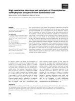
Fig. 7. Incubation with kallikrein abolished the binding of HKa and HK to LM (A) by cleaving the light chain of HKa (B, C). In Locke’s buffer
containing 0.35% (w ⁄ v) BSA, one volume of HK and HKa, respectively, at a concentration of 60 nM was incubated for varying lengths of
time with one volume of either 6 nM (10 : 1) or 60 nM (1 : 1) kallikrein. (A) At the times indicated, cleavage was stopped by adding one volume of a protease inhibitor cocktail consisting of 40 lgỈmL)1 each of leupeptin, aprotenin, benzamidin and soy bean trypsin inhibitor, giving
a final concentration of 20 nM of HK and HKa, respectively. The binding of the proteolysed HK (squares) and HKa (circles) to LM coated on a
microtiter plate at a concentration of 10 lgỈmL)1 (Fig. 2) are shown after incubation at high (closed symbols) and low (open symbols) concentrations of kallikrein. Western blots of incubation mixtures of HK and kallikrein followed the proteolysis. (B) Using the mAb 2B5 towards
the heavy chain showed that, at a low kallikrein concentration, a higher amount of HK was cleaved after 60 min (b ⁄ 60) than after 30 min
(b ⁄ 30) of incubation, whereas, at equimolar concentrations of HK and kallikrein, HK was completely cleaved after 30 min (c ⁄ 30) incubation
and a considerable amount was already cleaved after 0.5 min (c ⁄ 0.5). (C) Using the anti-rhkininostatin IgG specific for the light chain, it was
found that 60 min of incubation of equimolar concentrations of HK with kallikrein (c ⁄ 60) apparently cleaved the light chain generating a
12 kDa anti-rhkininostatin recognizable peptide.
ishing the binding of HK ⁄ HKa to LM. This, when
combined with the inhibition of the binding by rhkininostatin, indicates that the binding of HK ⁄ HKa to
LM is mediated via the N-terminal region in kininostatin encompassing the amino acid sequence
Arg439-Lys478 (Fig. 5).
Zn2+ has been assumed to have a decisive role with
respect to the binding and function of HK and HKa.
In the present study, we show that Zn2+ is not
required for the binding of either HK or HKa to LM.
However, it enhanced the affinity of the binding of
both HK and HKa to LM, although the total amount
bound was not affected. However, this was without
significance because the binding affinity, even in the
absence of Zn2+, was approximately 15-fold higher
than the plasma concentration of HK, demonstrating
5234
no obvious need for the presence of Zn2+. Furthermore, the effect of Zn2+ on the binding to LM was
only evident at low concentrations of BSA, at which it
abolished the inhibitory effect of Ca2+. At a higher
BSA concentration, the effect of Ca2+ remained,
whereas the effect of Zn2+ was eliminated by reducing
its free concentration by binding to BSA. Because the
total plasma concentration of Zn2+ is 25 lm [31], the
free Zn2+ concentration in vivo may never be sufficiently high to influence either the function of HKa or
the binding of HK and HKa to LM. Therefore, the
present study, showing that HK and HKa bind Zn2+independently to LM, must be physiologically relevant.
Experimentally, there was a considerable difference
in the minimal amount of matrix protein required to
cover the microtiter plate. The concentration of LM
FEBS Journal 276 (2009) 5228–5238 ª 2009 The Authors Journal compilation ª 2009 FEBS
I. Schousboe and B. Nystrøm
HK binding to laminin
HK bound (absorbance units)
1.2
1
0.8
0.6
0.4
0.2
0
0
50
100
150
200
Concentrarion of Zn2+ (µM)
Fig. 8. Binding of HK to ECM and cell-free wells. Cell-free wells,
which had been exposed to growth medium, exactly as for the cultures of HUVEC were, similarly to a confluent layer of the cells,
rinsed with NaCl ⁄ Pi and incubated with EDTA. Next, the wells
(ECM and cell-free) were blocked for 30 min with Locke’s buffer
containing 0.2% (w ⁄ v) or 0.35% (w ⁄ v) BSA (blocking solutions) and
subsequently incubated for 1 h with 20 nM HK diluted in the
respective blocking solutions containing varying concentrations of
Zn2+. The amount of HK bound was measured by immunoreactions, as described in the Experimental procedures. A higher
amount of HK bound Zn2+-dependently to cell-free wells (open
symbols) than to ECM (closed symbols), regardless of whether the
surface had been blocked with gelatin (circles) or BSA (triangles).
The results are shown as the mean ± SD (n = 3) when extending
beyond the symbols.
was 5 lgỈmL)1, whereas more than 20 lgỈmL)1 was
required for VN, and almost no HKa bound to VN at
this concentration, even in the presence of Zn2+. A
previous study reported that HKa binds Zn2+-dependently to VN coated at a concentration of 5 lgỈmL)1
[26]. At present, this discrepancy cannot be explained
because the use of a considerably higher BSA concentration for blocking did not prevent Zn2+-dependent
nonspecific binding. However, the nonspecific binding,
particularly that of HKa, in the presence of Zn2+ may
explain the controversial results reported on the effect
of matrix proteins on regulation of the apoptotic effect
of HKa on HUVEC grown in culture. HKa induced
Zn2+-dependent apoptosis of the cells, regardless of
the cells being cultured on 2 lgỈmL)1 VN or LM
[14,24,26,32], although not on 10 lgỈmL)1 FN
[24,26,32].
It was not possible to measure binding of HK to
LM in ECM generated during the growth of HUVEC.
This may likewise be the result of too low a concentration of the LM deposited during the 2–4 days of cell
growth. The addition of Zn2+ enhanced the nonspecific binding and indicated that the previously shown
Zn2+-dependent binding of HK to ECM [5] might
have been nonspecific. The strongest evidence for this
was that a lower amount of HK bound Zn2+-depen-
dently to ECM than to the cell-free wells, implying
that ECM blocked the binding of HK to the cell-free
area of the well. Furthermore, the Zn2+ optimum for
the binding of HK to the surface of the microtiter
plate was exactly the same as those reported for the
binding of HK to ECM and HUVEC [4,5,33,34],
regardless of whether the binding assay was performed
in buffer supplemented with gelatin or albumin.
Accordingly, and because the correction for nonspecific binding in these previous studies was performed
by the subtraction of binding in the absence of Zn2+,
a series of investigations investigating the significance
of Zn2+ for the interaction between HK and endothelial cells in cell culture systems [4,5,13,23,34,35] needs
to be revisited.
In vivo, LM might be a matrix protein of significance with respect to the function of HKa on the
activity of the endothelial cells in as much as LM plays
an important role in cell adhesion to the basement
membrane [16–18]. Furthermore, both HK and HKa
bind to LM, suggesting that this may be a prerequisite
for the kallikrein ⁄ kinin ⁄ kininogen system to be assembled and activated to enable the local liberation of the
short-lived bradykinin and its participation in inflammatory and pro- and anti-angiogenic reactions
[25,36,37]. Thus, the binding of HK to LM in the
basement membrane may be followed by activation of
prekallikrein bound in a 1 : 1 molecular complex to
HK. The activation of prekallikrein is accomplished
either by factor XIIa, heat shock protein 90 [38] or a
prolylcarboxypeptidase [39]. The observation that
kallikrein cleaves off a 12 kDa anti-rhkininostatin
reacting peptide shows that kallikrein, secondary to
bradykinin, releases kininostatin from HK, as previously anticipated [36]. This fits neatly with the observation that the HKa signaling of an apoptotic effect is
promoted as a result of extensive cleavage by kallikrein
[37,40].
Experimental procedures
Materials
One-chain HK, two-chain HKa and kallikrein delivered
lyophilized from Enzyme Research Laboratories Ltd.
(Swansea, UK) were dissolved, aliquoted and stored in siliconized test tubes at –80 °C. All dilutions of HK, HKa and
kallikrein were likewise performed in siliconized test tubes
and excess dilutions were discarded. VN, FN, LM from
Engelbreth-Holm-Swarm murine sarcoma basement membrane (LM 1) and LM from placenta (LM 10 ⁄ 11) were
obtained from Sigma Chemicals (St Louis, MO, USA). The
surface binding peptide sequence within the light chain of
FEBS Journal 276 (2009) 5228–5238 ª 2009 The Authors Journal compilation ª 2009 FEBS
5235
HK binding to laminin
I. Schousboe and B. Nystrøm
HK, His479-Lys-His-Gly-His-Gly-His-Gly-Lys-His-Lys-AsnLys-Gly-Lys-Lys-Asn-Gly-Lys-His498 (HKH20), was a
kind gift from Dr Alvin Schmaier (Case Western Reserve
University, Cleveland, OH, USA). The sequence of amino
acids copying the Ser372-Arg419 region [28] was synthe˚
sized at Novo Nordisk (Maløv, Denmark). Recombinant
human kininostatin (rhkininostatin) and goat anti-(human
rhkininostatin) were obtained from R&D Systems (Abingdon, UK). The monoclonal antibody to the heavy chain of
human HK (mAb 2B5) was obtained from Abcam (Cambridge, UK). Horseradish peroxidase (HRP) conjugated
rabbit anti-(mouse IgG) (P-0260), HRP-conjugated rabbit
anti-(goat IgG) (P-0449) and ortophenylenediamine were
obtained from Dako (Glostrup, Denmark). SuperSignalÒ
West Femto Maximum Sensitivity Substrate was from
Pierce Biotechnology, Inc. (Rockford, IL, USA). Essentially
fatty acid free BSA (A7030) was obtained from Sigma
Chemicals. All other reagents were of the purest grade
commercially available.
Solid phase binding assay
Maxisorp microtiter plates (high binding capacity; Nunc,
Roskilde, Denmark) were coated overnight at 4 °C with
150 lL of 5 lgỈmL)1 VN or 10 lgỈmL)1 of either FN, LM
1 or LM 10 ⁄ 11 in coating buffer (0.1 m potassium phosphate, pH 7.4), or at the concentrations indicated. After
blocking for 30 min with 200 lL of 1% (w ⁄ v) BSA in
Locke’s buffer (154 mm NaCl, 5.6 mm KCl, 3.6 mm NaHCO3, 2.3 mm CaCl2, 1 mm MgCl2, 5.6 mm glucose, 5 mm
Hepes, pH 7.4), 100 lL of HK or HKa (20 nm or as indicated) diluted in Locke’s buffer containing 0.35% (w ⁄ v)
BSA was added. After incubation for 1 h at room temperature, the wells were washed three times with Locke’s buffer,
one time with ice-cold methanol and five times with wash
buffer [50 mm Tris, 0.15 mm NaCl, pH 8.0, containing
0.05% (v ⁄ v) Tween 20]. This was followed by 1 h of incubation with mAb 2B5 diluted 5000-fold in wash buffer containing 1% (w ⁄ v) dry skim milk. After a further washing
cycle with wash buffer (five times), the plate was incubated
for 1 h with HRP-conjugated rabbit anti-(mouse IgG)
diluted 5000-fold in wash buffer. Finally, the plates were
incubated for 10–30 min with ortophenylenediamine,
dissolved in water according to the manufacturer’s instructions. The peroxidase reaction was stopped by a two-fold
dilution with 0.5 m H2SO4 and the relative amount of HK
bound to the wells determined as absorbance units (A) at
490 nm. All experiments were performed in triplicates and
repeated at least twice. To obtain estimates of affinity constants, the data were analyzed according to the isotherm:
A = Amax · [B] ⁄ (KD + [B])
where [B] is the molar concentration of the analyt, which is
either HK or HKa; A is the absorbance of the oxidized
HRP substrate, which is assumed to be proportional to the
5236
amount of bound analyt; and Amax represents the absorbance at saturating concentrations of the analyt.
Western blotting
Proteolytic cleavage of HK was visualized by western
blotting of aliquots of HK incubated with kallikrein in
Locke’s buffer. Reduced samples were separated on
4–12% SDS-PAGE simultaneously with a standard sample
of a mixture of Mr markers. After blotting to a poly
(vinylidene difluoride) membrane according to standard
procedures, the membrane was incubated for 10 min in
Tween-BSA block-buffer [50 mm Tris, 0.15 mm NaCl, pH
8.0, containing 0.1% (v ⁄ v) Tween 20 and 0.1% (w ⁄ v)
BSA], and subsequently overnight with the primary antibody; either mAb 2B5 or goat anti-(human rhkininostatin), diluted 5000-fold in 1% (w ⁄ v) dry skim milk in
the Tween-BSA block-buffer. The positions of the light
and the heavy chains of HK were visualized by incubation
with HRP conjugated rabbit anti-(mouse IgG) (P-0260)
and HRP-conjugated rabbit anti-(goat IgG) (P-0449),
respectively, followed by SuperSignalÒ West Femto
Maximum Sensitivity Substrate as recommended by the
manufacturer. The results were monitored using a chemiluminator.
Endothelial cell culture
HUVEC (Clonetics, Cambrex Bio Science, Verviers,
Belgium) were prepared as previously described [41].
Briefly, the cells were grown to confluence under standard
conditions, cryopreserved and sub-cultured in half of a
96-well microtiter plate (Nunc) The microtiter plate was
not pre-coated. The other half of the plate (without cells)
was used as a control. The wells in this half were incubated
with growth medium in absence of cells, and the medium
was changed in accordance with the exact same schedule as
for the exchange of medium in the wells with cells.
HK binding to ECM
At confluence, all wells including the cell-free wells were
washed with NaCl ⁄ Pi (50 mm phosphate, 0.1 m NaCl, pH
7.5), and incubated with 5 mm EDTA in NaCl ⁄ Pi for
30 min at room temperature, as previously described [41].
This treatment detached the cells from the surface of the
wells as visualized microscopically and as analyzed by the
absence of anti-actin binding components [ELISA using
mouse-anti actin IgG (M-0635); Dako]. The wells were next
washed twice with Locke’s buffer and incubated for minimum of 30 min at room temperature with 200 lL per well
of 1% (w ⁄ v) BSA or otherwise as noted. LM deposited in
the matrix during growth of the cells was detected using
rabbit anti-LM (Z-0097; Dako). Binding of HK was determined as described above.
FEBS Journal 276 (2009) 5228–5238 ª 2009 The Authors Journal compilation ª 2009 FEBS
I. Schousboe and B. Nystrøm
HK binding to laminin
Statistical analysis
The results are shown as the mean ± SD and statistically
significant differences were estimated using analysis of
variance and the Bonferoni post-hoc test. P < 0.05 was
considered statistically significant.
11
12
Acknowledgements
The present study was supported by grant 2005-1-192
and 2007-01-0355 from the Carlsberg Foundation.
13
References
1 Aumailley M & Gayraud B (1998) Structure and biological activity of the extracellular matrix. J Mol Med
76, 253–265.
2 Berrettini M, Schleef RR, Heeb MJ, Hopmeier P &
Griffin JH (1992) Assembly and expression of an
intrinsic factor IX activator complex on the surface of
cultured human endothelial cells. J Biol Chem 267,
19833–19839.
3 Colman RW & Schmaier AH (1997) Contact system: a
vascular biology modulator with anticoagulant, profibrinolytic, antiadhesive, and proinflammatory attributes. Blood 90, 3819–3843.
4 Motta G, Rojkjaer R, Hasan AA, Cines DB &
Schmaier AH (1998) High molecular weight kininogen
regulates prekallikrein assembly and activation on endothelial cells: a novel mechanism for contact activation.
Blood 91, 516–528.
5 Motta G, Shariat-Madar Z, Mahdi F, Sampaio CA &
Schmaier AH (2001) Assembly of high molecular weight
kininogen and activation of prekallikrein on cell matrix.
Thromb Haemost 86, 840–847.
6 Renne T, Nieswandt B & Gailani D (2006) The intrinsic
pathway of coagulation is essential for thrombus stability in mice. Blood Cells Mol Dis 36, 148–151.
7 Iwaki T & Castellino FJ (2006) Plasma level of bradykinin are suppressed in factor XII-deficient mice. Thromb
Haemost 95, 1003–1010.
8 Schousboe I, Nystrøm BT & Hansen GH (2008) Differential binding of factor XII and activated factor XII to
soluble and immobilized fibronectin. Localization of the
Hep-1 ⁄ Fib-1 binding site for activated factor XII. FEBS
J 275, 5161–5172.
9 Joseph K, Ghebrehiwet B, Peerschke EI, Reid KB &
Kaplan AP (1996) Identification of the zinc-dependent
endothelial cell binding protein for high molecular
weight kininogen and factor XII: identity with the
receptor that binds to the globular ‘heads’ of C1q
(gC1q-R). Proc Natl Acad Sci USA 93, 8552–8557.
10 Joseph K, Ghebrehiwet B & Kaplan AP (1999) Cytokeratin 1 and gC1qR mediate high molecular weight
14
15
16
17
18
19
20
21
22
23
24
kininogen binding to endothelial cells. Clin Immunol 92,
246–255.
Shariat-Madar Z, Mahdi F & Schmaier AH (1999)
Mapping binding domains of kininogens on endothelial
cell cytokeratin 1. J Biol Chem 274, 7137–7145.
Hasan AA, Zisman T & Schmaier AH (1998) Identification of cytokeratin 1 as a binding protein and presentation receptor for kininogens on endothelial cells. Proc
Natl Acad Sci USA 95, 3615–3620.
Mahdi F, Shariat-Madar Z, Kuo A, Carinato M, Cines
DB & Schmaier AH (2004) Mapping the interaction
between high molecular weight kininogen and the urokinase plasminogen activator receptor. J Biol Chem 279,
16621–16628.
Sun D & McCrae KR (2006) Endothelial-cell apoptosis
induced by cleaved high-molecular-weight kininogen
(HKa) is matrix dependent and requires the generation
of reactive oxygen species. Blood 107, 4714–4720.
Hallmann R, Horn N, Selg M, Wendler O, Pausch F &
Sorokin LM (2005) Expression and function of laminins
in the embryonic and mature vasculature. Physiol Rev
85, 979–1000.
Grant DS & Kleinman HK (1997) Regulation of capillary formation by laminin and other components of the
extracellular matrix. EXS 79, 317–333.
Belkin AM & Stepp MM (2000) Integrins as receptors
for laminins. Microsc Res Tech 51, 280–301.
Kikkawa Y, Sanzen N & Sekiguchi K (1998) Isolation
and characterization of laminin 10 ⁄ 11 secreated by
human lung carcinoma cells. Laminin 10 ⁄ 11 mediates
cell adhesion through integrins alpha3beta1. J Biol
Chem 273, 15854–15859.
Madsen CD & Sidenius N (2008) The interaction
between urokinase receptor and vitronectin in cell
adhesion and signalling. Eur J Cell Biol 87, 617–629.
Wei Y, Waltz DA, Rao N, Drummond RJ, Rosenberg
S & Chapman HA (1994) Identification of the urokinase receptor as an adhesion receptor for vitronectin.
J Biol Chem 269, 32380–32388.
Kugler MC, Wei Y & Chapman HA (2003) Urokinase
receptor and integrin interactions. Curr Pharm Des 9,
1565–1574.
Wei Y, Lukashev M, Simon DI, Bodary SC, Rosenberg
S, Doyle MV & Chapman HA (1996) Regulation of integrin function by the urokinase receptor. Science 273,
1551–1555.
Zhang JC, Claffey K, Sakthivel R, Darzynkiewicz Z,
Shaw DE, Leal J, Wang YC, Lu FM & McCrae KR
(2000) Two-chain high molecular weight kininogen
induces endothelial cell apoptosis and inhibits angiogenesis: partial activity within domain 5. FASEB J 14,
2589–2600.
Guo Y-L, Wang S, Cao DJ & Colman RW (2003)
Apoptotic effect of cleaved high molecular weight kinin-
FEBS Journal 276 (2009) 5228–5238 ª 2009 The Authors Journal compilation ª 2009 FEBS
5237
HK binding to laminin
25
26
27
28
29
30
31
32
I. Schousboe and B. Nystrøm
ogen is regulated by extracellular matrix proteins. J Cell
Biochem 89, 622–632.
Guo Y-L & Colman RW (2005) Two faces of high
molecular weight kininogen (HK) in angiogenesis:
bradykinin turns it on and cleaved HK (HKa) turns it
off. J Thromb Haemost 3, 670–676.
Chavakis T, Kanse SM, Lupu F, Hammes HP, Mulleră
Esterl W, Pixley RA, Colman RW & Preissner KT
(2000) Different mechanisms define the antiadhesive
function of high molecular weight kininogen in integrinand urokinase receptor-dependent interactions. Blood
96, 514–522.
Tzu J & Marinkovich MP (2008) Bridging structure
with function: structural, regulatory, and developmental role of laminins. Int J Biochem Cell Biol 40, 199–
214.
DeLa Cadena RA, Wachtfogel YT & Colman RW
(1993) Contact activation pathway: Inflammation and
coagulation. In Hemostastis and Thrombosis: Basic
Principles and Clinical Practice, 3rd Edn (Colman RW,
Hirsh J, Marder VJ & Slazman EW Eds), pp. 219–240.
JB Lippinscott, Philadelphia, PA.
Guo YL, Wang S & Colman RW (2001) Kininostatin,
an angiogenic inhibitor, inhibits proliferation and
induces apoptosis of human endothelial cells. Arterioscler Thromb Vasc Biol 21, 1427–1433.
Hasan AA, Cines DB, Herwald H, Schmaier AH &
Muller-Esterl W (1995) Mapping the cell binding site
on high molecular weight kininogen domain 5. J Biol
Chem 270, 19256–19261.
Oster O, Dahm M, Oelert H & Prellwitz W (1989) Concentration of some trace elements (Se, Zn, Cu, Fe, Mg,
K) in blood and heart tissue of patients with coronary
heart disease. Clin Chem 35, 851–856.
Cao DJ, Guo YL & Colman RW (2004) Urokinasetype plasminogen activator receptor is involved in mediating the apoptotic effect of cleaved high molecular
5238
33
34
35
36
37
38
39
40
41
weight kininogen in human endothelial cells. Circ Res
94, 1227–1234.
van Iwaarden F, de Groot PG & Bouma BN (1988)
The binding of high molecular weight kininogen to
cultured human endothelial cells. J Biol Chem 263,
4698–4703.
Rojkjaer R, Hasan AA, Motta G, Schousboe I &
Schmaier AH (1998) Factor XII does not initiate
prekallikrein activation on endothelial cells. Thromb
Haemost 80, 74–81.
Zhao Y, Qiu Q, Mahdi F, Shariat-Madar Z, Rojkjaer
R & Schmaier AH (2001) Assembly and activation of
HK-PK complex on endothelial cells results in bradykinin liberation and NO formation. Am J Physiol Heart
Circ Physiol 280, H1821–H1829.
Guo YL, Wang S & Colman RW (2002) Kininostatin
as an antiangiogenic inhibitor: what we know and what
we do not know. Int Immunopharmacol 2, 1931–1940.
Colman RW, Jameson BA, Lin Y, Johnson D & Mousa
SA (2000) Domain 5 of high molecular weight kininogen (kininostatin) down-regulates endothelial cell proliferation and migration and inhibits angiogenesis. Blood
95, 543–550.
Joseph K, Tholanikunnel BG & Kaplan AP (2009)
Heat shock protein 90 catalyzes activation of the prekallikrein-kininogen complex in the absence of factor
XII. Proc Natl Acad Sci USA 99, 896–900.
Shariat-Madar Z, Mahdi F & Schmaier AH (2002)
Identification and characterization of prolylcarboxypeptidase as an endothelial cell prekallikrein activator.
J Biol Chem 277, 17962–17969.
Colman RW (2006) Regulation of angiogenesis by
the kallikrein-kinin system. Curr Pharm Des 12, 2599–
2607.
Schousboe I (2006) Endothelial cells express a matrix
protein which binds activated factor XII in a zink-independent manner. Thromb Haemost 95, 312–319.
FEBS Journal 276 (2009) 5228–5238 ª 2009 The Authors Journal compilation ª 2009 FEBS