Báo cáo khoa học: A single intersubunit salt bridge affects oligomerization and catalytic activity in a bacterial quinone reductase pptx
Bạn đang xem bản rút gọn của tài liệu. Xem và tải ngay bản đầy đủ của tài liệu tại đây (812.21 KB, 12 trang )
A single intersubunit salt bridge affects oligomerization
and catalytic activity in a bacterial quinone reductase
Alexandra Binter
1
, Nicole Staunig
2
, Ilian Jelesarov
3
, Karl Lohner
4
, Bruce A. Palfey
5
, Sigrid Deller
1
,
Karl Gruber
2
and Peter Macheroux
1
1 Institute of Biochemistry, Graz University of Technology, Austria
2 Institute of Molecular Biosciences, University of Graz, Austria
3 Institute of Biochemistry, University of Zu
¨
rich, Switzerland
4 Institute of Biophysics and Nanosystems Research, Austrian Academy of Sciences, Graz, Austria
5 Department of Biological Chemistry, University of Michigan, Ann Arbor, MI, USA
Keywords
NADPH:FMN oxidoreductase;
oligomerization; quinone reductase; salt
bridge; thermostability
Correspondence
K. Gruber, Institute of Molecular
Biosciences, University of Graz,
Humboldtstrasse 50 ⁄ III, A-8010 Graz,
Austria
Fax: +43 316 380 9897
Tel: +43 316 380 5483
E-mail:
P. Macheroux, Institute of Biochemistry,
Graz University of Technology, Petersgasse
12 ⁄ II, A-8010 Graz, Austria
Fax: +43 316 873 6952
Tel: +43 316 873 6450
E-mail:
(Received 25 May 2009, revised 14 July
2009, accepted 17 July 2009)
doi:10.1111/j.1742-4658.2009.07222.x
YhdA, a thermostable NADPH:FMN oxidoreductase from Bacillus subtil-
is, reduces quinones via a ping-pong bi-bi mechanism with a pronounced
preference for NADPH. The enzyme occurs as a stable tetramer in solu-
tion. The two extended dimer surfaces are packed against each other by a
90° rotation of one dimer with respect to the other. This assembly is stabi-
lized by the formation of four salt bridges between K109 and D137 of the
neighbouring protomers. To investigate the importance of the ion pair con-
tacts, the K109L and D137L single replacement variants, as well as the
K109L ⁄ D137L and K109D ⁄ D137K double replacement variants, were gen-
erated, expressed, purified, crystallized and biochemically characterized.
The K109L and D137L variants form dimers instead of tetramers, whereas
the K109L ⁄ D137L and K109D ⁄ D137K variants appear to exist in a
dimer–tetramer equilibrium in solution. The crystal structures of the
K109L and D137L variants confirm the dimeric state, with the
K109L ⁄ D137L and K109D ⁄ D137K variants adopting a tetrameric assem-
bly. Interestingly, all protein variants show a drastically reduced quinone
reductase activity in steady-state kinetics. Detailed analysis of the two half
reactions revealed that the oxidative half reaction is not affected, whereas
reduction of the bound FMN cofactor by NADPH is virtually abolished.
Inspection of the crystal structures indicates that the side chain of K109
plays a dual role by forming a salt bridge to D137, as well as stabilizing a
glycine-rich loop in the vicinity of the FMN cofactor. In all protein vari-
ants, this glycine-rich loop exhibits a much higher mobility, compared to
the wild-type. This appears to be incompatible with NADPH binding and
thus leads to abrogation of flavin reduction.
Structured digital abstract
l
MINT-7229866, MINT-7229874, MINT-7229885, MINT-7229894, MINT-7229905: YhdA
(uniprotkb:
O07529) and YhdA (uniprotkb:O07529) bind (MI:0407)byblue native page
(
MI:0276)
l
MINT-7229854: YhdA (uniprotkb:O07529) and YhdA (uniprotkb:O07529) bind (MI:0407)by
x-ray crystallography (
MI:0114)
Abbreviations
DSC, differential scanning calorimetry; DLS, dynamic light scattering; Ni-NTA, nickel-nitrilotriacetic acid agarose.
FEBS Journal 276 (2009) 5263–5274 ª 2009 The Authors Journal compilation ª 2009 FEBS 5263
Introduction
The search for enzymes with catalytic properties of
potential application in biocatalysis and biotechnology
has led to the discovery of bacterial enzymes known as
azoreductases. These enzymes are found in several
diverse bacterial species catalysing the reductive cleav-
age of azo dyes containing one or more azo-bonds (R
1
-
N=N-R
2
) to their corresponding amines [1–7]. Aro-
matic azo dyes are artificial chemicals with potentially
harmful properties resulting in health and environmen-
tal concerns. Recently, we described a FMN-containing
flavoenzyme from Bacillus subtilis, termed YhdA, capa-
ble of cleaving azo dyes such as Cibachron Marine
(Ciba, Basel, Switzerland) at the expense of NADPH
[8]. YhdA shares sequence similarity with a family of fla-
vin- (FMN or FAD) dependent quinone reductases such
as mammalian NQO1 and yeast Lot6p [9]. Moreover,
YhdA and these eukaryotic quinone reductases possess
a similar protein topology, a so-called flavodoxin fold
consisting of five a-helices sandwiching a five-stranded
parallel b-sheet in the centre [10,11]. Because of these
similarities, we were interested in analyzing whether
YhdA accepts quinones as substrates. In the present
study, it is demonstrated that the enzyme reduces a vari-
ety of quinones by a ping-pong bi-bi kinetic mechanism
with a clear preference for NADPH as reducing agent.
As could be expected, the turnover rates for quinones
are much higher than those obtained for artificial azo-
compounds, in line with the assumption that quinones,
unlike azo dyes, are cognate enzyme substrates.
Bacterial and eukaryotic quinone reductases possess
a similar protein topology, but diverge with respect to
their oligomeric structure. Although eukaryotic proteins
form dimers, YhdA and Azo1 (from Staphylococ-
cus aureus) [4] form tetramers [8,9]. In the case of
YhdA, the tetramer is formed by two dimers, which
interact through an extended concave surface. The
interface between dimers is stabilized by four salt
bridges formed by the side chains of residues K109 and
D137 of structural neighbours (Scheme 1). This higher
oligomerization state was considered to be responsible
for the increased thermal stability of YhdA (T
m
=
87 °C) compared to the dimeric yeast homolog Lot6p
(T
m
= 60.2 °C) [12]. To test this hypothesis and to
obtain more insight into the importance of these salt
bridges for tetramer assembly, we created four YhdA
protein variants: K109L, D137L, K109L ⁄ D137L and
K109D ⁄ D137K. Characterization of the variants
showed that single replacement of K109 or D137
disrupts the tetramer, whereas the two double replace-
ment protein variants appear to have conserved some
tendency to form tetramers. Interestingly, all of the
variants have a melting point similar to the wild-type
protein, suggesting that the high thermostability is an
intrinsic property of the dimer. Surprisingly, the protein
variants showed dramatically reduced enzymatic activ-
ity, which is a result of the breakdown of the reductive
half reaction, indicating that structural changes impede
docking of NADPH to the active site. Crystallization
and concomitant X-ray crystallographic determination
of variant structures revealed that increased mobility of
a highly conserved glycine-rich loop in the vicinity of
the isoalloxazine ring system might be responsible for
the loss of enzyme activity.
Results
YhdA has recently been classified as an NADPH:FMN
oxidoreductase with the ability to reductively cleave azo
dyes [8]. Lot6p, the YhdA homolog in yeast, was shown
to reduce quinones to their hydroquinone state [12];
hence, we tested YhdA for its quinone reductase activ-
ity. Steady-state measurements using several quinone
substrates as electron acceptors resulted in much higher
turnover rates than those reported for the reduction of
azo dyes (data not shown). For a more detailed charac-
terization, 2-hydroxy-p-naphthoquinone was chosen
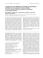
as a representative substrate. Figure 1 shows the double
reciprocal plot of initial velocity measurements in
the presence of NADPH as the electron donor and
2-hydroxy-p-naphthoquinone as electron acceptor. The
family of parallel lines obtained from data analysis
indicates a ping-pong bi-bi mechanism, where both
substrates consecutively bind to the catalytic site (i.e.
the electron donor NADPH binds first, then dissociates
K109
D137
D137
D137
D137
K109
K109
K109
Scheme 1. Schematic representation of the four salt-bridges in the
YhdA tetramer. One dimer is formed by the green and magenta
chains; the second by the pink and blue chains.
Effect of oligomerization in a quinone reductase A. Binter et al.
5264 FEBS Journal 276 (2009) 5263–5274 ª 2009 The Authors Journal compilation ª 2009 FEBS
to vacate the active site for binding of the electron
accepting quinone substrate). The same mechanism was
proposed for the homologous yeast enzyme Lot6p and
other quinone reductases [12].
Using [S-
2
H]-NADPH and [R-
2
H]-NADPH as
reducing agents and subsequent analysis of NADP
+
by
1
H-NMR spectroscopy, it was revealed that the
pro-S hydride of NADPH is preferentially transferred
to FMN.
Heterologous expression of the four generated pro-
tein variants was performed in the same way as that
described for wild-type protein, resulting in similar
amounts of soluble protein. All hexahistidine-tagged
protein variants were purified by nickel-nitrilotriacetic
acid agarose (Ni-NTA) chromatography according to
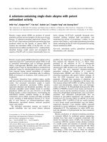
the protocol established for wild-type YhdA. YhdA
possesses a noncovalently bound FMN cofactor, which
is also present in the protein variants, but showed
some minor changes in their UV ⁄ visible absorbance
spectra (Fig. 2). The extinction coefficients of enzyme-
bound FMN were determined using an extinction coef-
ficient of e
450
= 12 400 m
)1
Æcm
)1
for free FMN and
are summarized in Table 1.
The native molecular mass was estimated by mole-
cular sieve chromatography. Each protein variant
eluted as a single species; however, all protein variants
exhibited larger elution volumes indicating a lower
apparent mass (Table 2). These data suggest that the
two single protein variants and the K109L ⁄ D137L
double protein variant form dimers in solution. On the
other hand, the K109D ⁄ D137K variant showed a
native molecular mass of 61 kDa, suggesting that the
protein may exist in a dimer–tetramer equilibrium.
This result was qualitatively confirmed by dynamic
light scattering (DLS) experiments (Table 2), demon-
strating that the single replacement variants form
dimers rather than tetramers. On the other hand, both
double replacement protein variants show a tendency
to form a tetramer similar to wild-type protein.
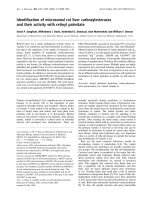
To further characterize the oligomerization of the
protein variants, native PAGE was employed. As
shown in Fig. 3A, both single protein variants have a
higher mobility compared to wild-type protein; this
can be interpreted in terms of the formation of dimers
Fig. 1. Double-reciprocal plot of initial rate measurements in
steady-state experiments as a function of NADPH at 2 (m), 5 (
•
),
10 (j) and 30 (. ) l
M of 2-hydroxy-p-naphthoquinone (from top to
bottom).
Fig. 2. UV ⁄ visible absorbance spectra of wild-type YhdA and the
four protein variants.
Table 1. Extinction coefficients of wild-type YhdA and the four
protein variants at 450 nm.
Protein e
450
(M
–1
Æcm
–1
) k
max
(nm)
Wild-type 11 690 451
D137L 10 660 452
K109L 11 070 450
K109L ⁄ D137L 10 760 452
K109D ⁄ D137K 10 720 453
Table 2. Native molecular mass estimation by molecular sieve
chromatography and DLS and apparent unfolding temperatures T
m
in °C as determined by CD spectroscopy and DSC. The void
volume of the column was determined to V
o
= 44.04 mL.
Protein
V
E
(mL)
Molecular
mass (kDa)
DLS
a
(kDa)
T
m
(CD)
T
m
(DSC)
Wild-type 56.69 72.7 85 87
b
93
D137L 66.29 33.5 53 84 95
K109L 64.61 44.0 68 88 95
K109L ⁄ D137L 64.21 45.1 72 89 95
K109D ⁄ D137K 59.45 61.0 88 89 > 95
a
The values given are the average of two independent measure-
ments.
b
Value taken from [8].
A. Binter et al. Effect of oligomerization in a quinone reductase
FEBS Journal 276 (2009) 5263–5274 ª 2009 The Authors Journal compilation ª 2009 FEBS 5265
rather than tetramers. The different isoelectric points
resulting from the aspartate to leucine (pI = 6.92) and
the lysine to leucine (pI = 6.09) replacements, respec-
tively, account for the mobility shift between the two
single protein variants. The two double protein vari-
ants give rise to bands positioned between the K109L
and D137L variant. Considering that the two double
protein variants have an intermediate isoelectric point
of 6.43 (i.e. the same as wild-type protein), this result
also suggests that both of these protein variants occur
as dimers. However, at higher protein concentra-
tions (Fig. 3B; and barely visible in Fig. 3A), the
K109L ⁄ D137L protein variant exhibits an additional
band at lower mobility, indicating that this protein
variant may form tetramers under these conditions.
Interestingly, the ‘inverse’ K109D ⁄ D137K variant pro-
duces only a single band at high mobility (no change
between Fig. 3A, B) in contrast to the dimer–tetramer
equilibrium suggested by molecular sieve chromatogra-
phy. Obviously, both double replacement variants have
some tendency to form tetramers, albeit much weaker
than the wild-type protein.
Next, we characterized the protein variants with
respect to their quinone reductase activity. Initial rate
measurements show that all protein variants retain less
than 1% of wild-type activity, using molecular oxygen
as well as various quinones as final electron acceptors
(Table 3). Stopped-flow measurements were performed
to determine whether the reductive or the oxidative
half reaction is impaired in the protein variants. The
reductive half reaction of wild-type, the D137L and
the K109L ⁄ D137L protein variant was investigated in
more detail. With both protein variants, the rate of
reduction of the FMN cofactor was very small,
amounting to 0.6% and 3% of the wild-type rate for
the D137L and K109L ⁄ D137L protein variants,
respectively. The rate of reduction for the other two
protein variants was much smaller and could not be
determined accurately in the stopped-flow instrument.
On the other hand, the oxidative half reaction using
2-hydroxy-p-naphthoquinone as a substrate was not
affected in any of the protein variants, yielding compa-
rable rates for wild-type and all protein variants
(Table 3). Hence, it can be concluded that the loss of
enzymatic activity in the four protein variants observed
in steady-state measurements is a result of the collapse
of the reduction step (i.e. the transfer of electrons from
NADPH to the flavin cofactor).
YhdA has been described as an enzyme exhibiting
high thermostability with a melting temperature of
86.5 °C, as determined by monitoring thermal unfold-
A
B
Fig. 3. Native PAGE. From left to right, WT = YhdA wild-type,
D137L variant, K109L variant; DM = K109L ⁄ D137L variant, IM
K109D ⁄ D137K variant. Protein solution (6.5 lL) at (A) 15 l
M and (B)
60 l
M, respectively, was applied onto each lane.
Table 3. Steady-state and rapid reaction parameters for wild-type YhdA and protein variants. Turnover measurements were carried out with
NADPH and oxygen as substrates. The rate of reduction and oxidation was measured with NADPH and 2-hydroxy-p-naphthoquinone
(2OHpNQ), respectively. ND, not determined (rates were very small compared to wild-type YhdA).
Protein Turnover
(s
)1
)
K
M
(NADPH)
(m
M)
Reduction
k
red
(s
)1
)
K
D
(NADPH)
(m
M)
Oxidation
k
ox
(s
)1
)
K
D
(2OHpNQ)
(m
M)
Wild-type 57.9 ± 6.7 0.52 ± 0.09 100 ± 3 0.54 ± 0.05 90 ± 9.7 0.27 ± 0.05
D137L 0.28 ± 0.02 0.65 ± 0.07
a
0.45
b
– 115 ± 7.1 0.21 ± 0.02
K109L 0.02 ± 0.004 0.42 ± 0.14
a
ND – 96 ± 28.8 0.31 ± 0.018
K109L ⁄ D137L 0.55 ± 0.12 1.37 ± 0.35
a
3.07 ± 0.3 2.29 ± 0.42 103 ± 10.9 0.24 ± 0.044
K109D ⁄ D137K 0.16 ± 0.04 0.69 ± 0.18
a
ND – 124 ± 6.6 0.12 ± 0.014
a
Accurate determination of K
M
values was hampered by low activity of the variants.
b
Rate of reduction increased linearly to k
red
= 0.45 s
)1
at 4 mM NADPH.
Effect of oligomerization in a quinone reductase A. Binter et al.
5266 FEBS Journal 276 (2009) 5263–5274 ª 2009 The Authors Journal compilation ª 2009 FEBS
ing of the protein by CD spectroscopy [8]. The high
thermostability of the tetrameric YhdA in comparison
to its dimeric yeast homologue Lot6p (T
m
=60°C)
gave rise to the assumption that the tetrameric state
stabilizes the protein toward thermal unfolding [13].
Both proteins possess a common structural topology,
the so-called flavodoxin-like fold. They exhibit the
same dimer architecture, forming a large, slightly con-
cave surface, characterized by four a-helices spanning
its entire width [11]. In the case of Lot6p, several
charged residues in the central part of this surface
appear to interfere with the tetrameric assembly. These
residues are replaced by hydrophobic or uncharged
residues in YhdA, which allows tetramer formation by
rotating the two dimers against each other by 90° and
packing of the two dimers. To test the hypothesis that
tetramerization is responsible for increased thermosta-
bility, the apparent melting temperatures of the protein
variants were determined by CD spectrometry and
differential scanning calorimetry (DSC). Surprisingly,
no significant changes in the thermostability of the
protein variants were observed (Table 2). Thus, it can
be concluded that the higher oligomerization state of
YhdA compared to Lot6p is not the governing
factor for achieving higher thermostability because
all four predominantly dimeric protein variants
show unfolding temperatures similar to the tetrameric
wild-type protein.
All four YhdA variants were crystallized. Three
structures (of the K109L, the D137L and
K109D ⁄ D137K variant) were determined and refined
to varying crystallographic resolution (Table 4). The
crystals obtained for the K109L ⁄ D137L variant were
isomorphous to the hexagonal crystal form of wild-
type YhdA [Protein Data Bank (PDB) entry: 1NNI]
but diffracted to lower resolution; therefore, this struc-
ture was not refined further.
The YhdA protomer belongs to the SCOP family
[14] of ‘NADPH-dependent FMN reductases’. The
closest structural neighbours, according to a SSM
analysis [15], are a NAD(P)H-dependent FMN reduc-
tase from Pseudomonas aeruginosa (PDB entry: 1x77)
[16], ArsH from Sinorhizobium meliloti (2q62) [17], a
NADH-dependent FMN reductase from the EDTA-
degrading bacterium bnc1 (2vzf, 2vzh, 2vzj) [18] and
ArsH from Shigella flexneri (2fzv) [19]. The rmsd are
in the range 1.6–2.0 A
˚
for 150–160 aligned C-a atoms.
The oligomeric states of the different variants in the
crystalline state were analyzed using the msd-pisa
server [20] taking into account all the interactions of
protein chains within the asymmetric unit as well as
with symmetry equivalent molecules. The crystal
Table 4. Data collection and refinement statistics. Values in parentheses are for highest-resolution shell.
K109L D137L K109D ⁄ D137K
Data collection
Space group P22
1
2
1
P1 P2
1
Cell dimensions
a, b, c (A
˚
) 47.54, 66.24, 220.75 51.49, 56.08, 64.18 68.63, 170.13, 93.29
a, b, c (°) 90.0, 90.0, 90.0 84.1, 77.0, 74.5 90.0, 92.3, 90.0
Resolution (A
˚
) 29.0–3.20 (3.31–3.20) 20.0–2.40 (2.44–2.40) 30.0–2.11 (2.16–2.11)
R
sym
0.161 (0.597) 0.057 (0.274) 0.096 (0.460)
I ⁄ rI 10.4 (2.9) 12.8 (3.2) 15.8 (2.0)
Completeness (%) 99.5 (99.5) 95.0 (88.8) 96.7 (52.5)
Redundancy 4.9 (4.5) 2.0 (1.9) 3.6 (2.0)
Refinement
Resolution (A
˚
) 29.0–3.20 19.9–2.50 29.3–2.11
Number of reflections 12 155 24 878 119 393
R
work
⁄ R
free
0.212 ⁄ 0.262 0.199 ⁄ 0.251 0.185 ⁄ 0.221
Number of atoms
Protein 5124 5175 15 379
Cofactor 124 124 372
Water – 319 977
B-factors
Protein 58.0 36.5 36.1
Cofactor 53.6 28.3 29.4
Water – 39.3 40.7
rmsd
Bond lengths (A
˚
) 0.003 0.002 0.002
Bond angles (°) 0.6 0.6 0.6
A. Binter et al. Effect of oligomerization in a quinone reductase
FEBS Journal 276 (2009) 5263–5274 ª 2009 The Authors Journal compilation ª 2009 FEBS 5267
structure of wild-type YhdA contains only one protein
chain in the asymmetric unit, but a tetramer is formed
by two crystallographic diads (space group P6
2
22),
which is predicted to be stable also in solution. This
prediction was confirmed experimentally by molecular
sieve chromatography and native PAGE (Fig. 3 and
Table 2). This tetramer exhibits 222 symmetry and can
be considered as a dimer of dimers (Fig. 4) with a sig-
nificantly larger interaction surface within the dimers
( 1100 A
˚
2
buried surface area per chain) than
between them ( 660 A
˚
2
). Four individual salt bridges
involving K109 and D137 are formed across the
dimer–dimer interface (Scheme 1). Lys109 also forms
hydrogen bonds to three carbonyl groups in the gly-
cine rich loop (G
106
-GG-K
109
-GG
111
) of the neigh-
bouring subunit, thereby stabilizing this loop, which is
in close proximity of the N(1)-C(2=O) locus of the fla-
vin (Fig. 5). Based on the observed isomorphicity of
crystals obtained for the K109L ⁄ D137L variant, the
same oligomeric state can safely be assumed to be
present, although the salt bridge between Lys109 and
Asp137 cannot be formed in this case. Most of the
closest structural neighbours mentioned above also
form tetramers in the crystal, and the mode of oligo-
merization is the same as in YhdA. The only exception
is the NAD(P)H-dependent FMN reductase from
P. aeruginosa, which forms a dimer again equivalent to
YhdA. In this context, it is noteworthy that K109 and
D137 are only conserved among putative oxidoreduc-
tases in the genera Bacillus and are not found in any
of the other structurally related proteins. This clearly
indicates that tetramer formation is not solely depen-
dent on the presence of the salt bridges formed
between these residues.
The asymmetric unit of crystals of the K109D ⁄
D137K variant contains 12 protein chains forming
three tetramers, which are each very similar to the
wild-type tetramer. rmsd were in the range 0.3–0.4 A
˚
after superposition of 664–669 C-a atoms (> 90% of
the total number of C-a atoms in the tetramers).
Although, in principle, the ‘inverse’ amino acid
exchange should allow the formation of an inter-dimer
salt bridge, this interaction is not observed in the crys-
tal structure. In addition, the stabilizing interactions of
the lysine with the carbonyl groups in the glycine rich
loop in the neighbouring protomer are not formed
(Fig. 5). Accordingly, pisa analysis predicts a lower
stability for this tetramer (calculated DG
diss
= 4.5–
6.2 kcalÆmol
)1
compared to 9.8 kcalÆmol
)1
for native
YhdA) which thus could more easily dissociate into
dimers in solution.
The remaining two variants (K109L and D137L)
show different oligomeric states in each case, with four
protomers in the asymmetric unit, which form two
dimers identical to the dimers found in the other
YhdA structures. In the crystal, these two dimers also
form tetramers, which, according to the pisa analysis,
should only be marginally stable in solution (DG
diss
of
)0.1 and 1.4 kcalÆmol
)1
). These tetramer arrangements
are very similar in the two structures (rmsd of 0.6 A
˚
for 626 superimposed C-a atoms). Compared to the
wild-type tetramer, the two dimers interact differently
with each other. Although, in wild-type YhdA (as well
as in the studied double mutant proteins), the two
dimers are aligned almost perpendicular to each other,
they are essentially parallel in the single mutant
proteins (Fig. 6).
The isolated protomers of the YhdA variant struc-
tures show only small structural changes compared
to the wild-type (rmsd in the range 0.3–0.6 A
˚
for
166–168 superimposed C-a atoms). The largest
changes are observed in the region around residue
109, which is in the centre of the above mentioned
glycine-rich loop region (Fig. 7) This loop also
becomes more flexible upon amino acid exchange,
which is clearly indicated by the lesser quality of the
electron density and the significantly higher B-factors
in this region (Fig. 8).
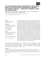
Fig. 4. Crystal structure of wild-type YhdA from Bacillus subtilis
(PDB entry: 1NNI). The oligomeric state can be described as a
dimer of dimers. One dimer is formed by the green and magenta
chains; the second by the pink and blue chains. The FMN cofactors
are shown as spheres. The figure was prepared using the software
PYMOL ( />Effect of oligomerization in a quinone reductase A. Binter et al.
5268 FEBS Journal 276 (2009) 5263–5274 ª 2009 The Authors Journal compilation ª 2009 FEBS
Discussion
Quinone reductases are present in many different
organisms in the eubacterial, fungal, plant and animal
kingdom. YhdA, previously described as an azoreduc-
tase [8], clearly possesses quinone reductase activity.
Considering the similarity of YhdA both in sequence
and structure with confirmed quinone reductases such
as mammalian NQO1 and yeast Lot6p, this finding is
not unexpected. On the other hand, YhdA differs with
regard to its quaternary structure. Although quinone
reductases of eukaryotic origin form dimers, YhdA
assembles into a tetramer, made up by a dimer of
dimers (Scheme 1 and Fig. 4). The reasons for adopt-
ing higher quaternary protein structures are still elusive
and appear to be case-dependent. Comparisons of the
quaternary structure of proteins from thermophilic
organisms with their mesophilic counterparts have
indicated that higher oligomeric structures provide
increased thermal stability required for adaptation to
elevated temperatures [13,21,22]. Although B. subtilis is
not a thermophilic organism, YhdA possesses a
surprisingly high thermal stability, with an apparent
melting temperature of T
m
= 86.5 °C. By contrast to
YhdA, its ortholog from Saccharomyces cerevisiae has
a much lower apparent melting temperature
(T
m
= 60.2 °C), as could be expected as a result of its
lower quaternary structure. Inspection of the tetra-
meric structure of YhdA revealed that the main con-
tacts between the two dimers are set up by four
reciprocal salt bridges between the side chains of K109
and D137 (Scheme 1). Therefore, we hypothesized that
these interactions are responsible for tetramer stabiliza-
tion and this in turn will lead to increased thermosta-
bility. Based on this hypothesis, we generated two
variants with either K109 or D137 replaced by leucine
and a third variant with both residues exchanged to
leucine. In a fourth variant, we swapped the interact-
ing residues in an attempt to restore the salt bridge
and thus redesign an intact dimer–dimer interaction.
The role of the salt bridges for tetramer assembly was
confirmed by our experimental results because the two
single replacement variants were exclusively found as
dimers both in solution and in the crystal. The two
double variants predominantly exist as dimers in solu-
tion, although both showed some tendency to form tet-
ramers in solution (Fig. 3 and Table 2). In the crystal,
both of them were clearly present as tetramers showing
packing similar to the wild-type protein (Fig. 6). Inter-
estingly, inverting the position of the interaction
partners in the K109D ⁄ D137K protein variant does
not rebuild the salt bridge, as is clearly seen in the
crystal structure (Fig. 5B). Instead, K137 forms a
hydrogen bond to the backbone C=O of G108 and
not to the carboxyl group of D109.
However, none of the protein variants exhibited
decreased thermostability (Table 2), clearly contradict-
ing our initial hypothesis that tetramer assembly is
responsible for higher thermostability. Obviously, ther-
mostability in this case is not a function of quaternary
structure but an intrinsic property of YhdA protomers
and ⁄ or the dimer.
Surprisingly, quinone reductase activity was severely
compromised in all variants because of a lack of
reduction of the FMN cofactor by NADPH. This was
clearly unexpected because all variants appear to have
A
B
Fig. 5. Close-up view of a portion of the dimer–dimer interface.
One subunit is shown in light blue; the other in magenta. The FMN
cofactor is shown in yellow; hydrogen bonding interactions are indi-
cated using dashed green lines. (A) In wild-type YhdA, a salt bridge
between Lys109 and Asp137 is formed across this interface. (B) In
the K109D ⁄ D137K variant, the number of interactions is greatly
reduced and the salt bridge can no longer be formed. The figure
was prepared using the software
PYMOL ( />A. Binter et al. Effect of oligomerization in a quinone reductase
FEBS Journal 276 (2009) 5263–5274 ª 2009 The Authors Journal compilation ª 2009 FEBS 5269
similar active sites as judged by their UV ⁄ visible absor-
bance spectrum. Moreover, initially, the determined
structures of the variants showed no conspicuous dif-
ferences that would have predicted altered enzymatic
properties. Closer inspection, however, revealed that a
glycine-rich loop in the vicinity of the isoalloxazine
ring system with Gly106 directly interacting with
C(2=O) of the pyrimidine moiety shows substantially
altered mobility (Fig. 8). In wild-type YhdA, this loop
is stabilized by K109 of a neighbouring subunit
(Scheme 1 and Fig. 5); in the variants, this interaction
is lost either as a result of replacement of the lysine
residue or, in the case of the D137L variant, by abro-
gated tetramer formation. It appears that the higher
mobility of the glycine-rich loop is incompatible with
binding of NADPH and ⁄ or delivery of a hydride to
the flavin cofacor and hence K109 plays a dual role by
supporting tetramer assembly through its interaction
with D137 and stabilization of the glycine-rich loop
necessary to enable flavin reduction by NADPH.
Unfortunately, attempts to obtain a crystal structure
with NADPH or NADP
+
bound to the active site
have so far proved unsuccessful (for a model, see
Fig. S1).
Fig. 7. Stereo representation of the superposition of all YhdA protomers found in the crystal structures of the wild-type (PDB entry: 1nni)
and the different variants. In total, 21 structures are shown in different colours. The two sites of amino acid exchanges are indicated. The
protomer structures are very similar to each other (for details, see text) and differ only in some loop regions, especially around residues 109
and 137. The figure was prepared using the software
PYMOL ( />Fig. 6. Schematic representation of the
observed oligomeric states and dimer–dimer
interactions in different YhdA variants. In
each case, one two-fold symmetric dimer
subunit is shown in its surface representa-
tion, whereas the other is shown in a car-
toon representation. Two views are
presented, which are rotated by 90° around
the x-axis. Wild-type YhdA, as well as the
double replacement variants, form stable
tetramers with the two dimers rotated by
approximately 60° relative to each other
(left). In the single replacement variants
(right), the two dimers are oriented parallel
to each other. The latter interaction is only
present in the crystal. The figure was pre-
pared using the software
PYMOL (http://
www.pymol.org/).
Effect of oligomerization in a quinone reductase A. Binter et al.
5270 FEBS Journal 276 (2009) 5263–5274 ª 2009 The Authors Journal compilation ª 2009 FEBS
The findings obtained in the present study suggest
that tetramer assembly of YhdA is not responsible for
the unusual thermostability; however, the quaternary
structure appears to be required for catalytic activity.
Although wild-type enzyme clearly exists as a tetramer
in solution, it is conceivable that extreme environmen-
tal conditions (e.g. high temperature) may cause disso-
ciation of the tetramer into dimers and hence result in
the deactivation of quinone reductase activity. Because
YhdA dimers exhibit the same thermal stability as tet-
ramers, this mode of regulation is reversible (i.e. tetra-
mers can reform once conditions favouring tetramer
assembly are restored). At this point, it is not clear
whether regulation of quinone reductase activity
through reversible dimer–tetramer equilibrium is rele-
vant for the bacterium and to which end it serves in
adaptation to environmental challenges.
Experimental procedures
Reagents
The Ni-NTA was obtained from Qiagen (Hilden, Germany).
All chemicals were of the highest grade commercially avail-
able and obtained from Sigma-Aldrich (St Louis, MO,
USA), Fluka (Buchs, Switzerland), Merck (Darmstadt, Ger-
many), or Carl Roth GmbH (Karlsruhe, Germany).
Cloning, recombinant expression and purification
The cloning of yhdA from B. subtilis into the pET21a vector,
the recombinant expression of YhdA using the host expres-
sion strain Escherichia coli BL21 (DE3) and the protein puri-
fication procedure have been described previously [8]. The
Sephadex Desalting Column PD-10 from GE Healthcare
(Amersham, UK) was used for buffer exchange.
Site-directed mutagenesis
Site-directed mutagenesis was carried out as specified in the
QuikChange
Ò
XL Site-Directed Mutagenesis Kit from
Stratagene (Cedar Creek, TX, USA). The pETyhdA plasmid
described previously [8] served as a template, performing the
PCR-based mutagenesis. To obtain the two single muteins
YhdA K109L and YhdA D137L, as well as the two double
muteins YhdA K109L ⁄ D137L and YhdA K109D ⁄ D137K,
the following primers and their complementary counterparts
were used: K109L: 5¢-GGG CGG CGG A
CTT GGC GGC
ATC AAT G-3¢ (sense), D137L: 5¢-GCA GCT GGT GCT
T
CT TCC GGT GCA TAT TG-3¢ (sense), K109D: 5¢-GGG
CGG CGG A
GA TGG CGG CAT CAA TG-3¢ (sense),
D137K: 5¢-GCA GCT GGT GCT T
AA ACC GGT GCA
TAT TG-3¢ (sense) (where the underlined nucleotides repre-
sent the mutated codon). After the mutagenesis protocol,
the sequences of the transformation constructs were verified
by sequencing analysis. The generation of the double
mutations was achieved using pETyhdA(K109L) and
pETyhdA(D137L), respectively, as templates and the relat-
ing primer pairs for the PCR. The mutated plasmids were
purified according to the Plasmid DNA Purification Kit
from Macherey-Nagel (Du
¨
ren, Germany) and transformed
into the host expression strain E. coli BL21 (DE3). Recom-
binant expression of the protein variants and the purifica-
tion procedure by Ni-NTA chromatography were
performed as described for the wild-type enzyme [8].
AB
DC
Fig. 8. ‘B-factor putty’ representation of
structures of different YhdA variants (A,
wild-type; B, D137L; C, K109L; D,
K109D ⁄ D137K) focusing in the glycine-rich
loop around residue 109. Orange to red col-
ours and a wider tube indicate regions with
higher B-factors, whereas shades of blue
and a narrower tube indicate regions with
lower B-factors. The FMN cofactor is shown
as a stick representation. The figure was
prepared using the software
PYMOL (http://
www.pymol.org/).
A. Binter et al. Effect of oligomerization in a quinone reductase
FEBS Journal 276 (2009) 5263–5274 ª 2009 The Authors Journal compilation ª 2009 FEBS 5271
Molecular mass determination
For the native molecular mass determination of the vari-
ants, molecular sieve chromatography was used [8]. The
results obtained from the molecular sieve chromatography
were verified by native PAGE, in accordance with the
standard procedures for SDS-PAGE, using 12.5% separat-
ing gels and 5% stacking gels. The gels and the running
buffer, respectively lacked SDS or dithiothreitol to main-
tain the native state of proteins. Native PAGE was per-
formed for 4 h at 90 V and 4 °C. In addition, DLS of
wild-type YhdA and the four protein variants was carried
out with a DynaProÔ (Wyatt Technology, Santa Barbara,
CA, USA).
Spectrophotometric methods
To determine the extinction coefficient of enzyme-bound
FMN, 0.2% SDS was used to release the cofactor.
UV ⁄ visible absorbance spectra were recorded before and
after denaturation of the enzyme with a photometer
(model specord 205) from Analytik Jena AS (Jena, Ger-
many). All measurements were performed in 100 mm
Tris ⁄ HCl (pH 7.5) using 1 cm quartz cuvettes, unless
stated otherwise.
Steady-state kinetics
To determine the reaction mechanism, steady-state turnover
of wild-type YhdA was measured by monitoring the oxida-
tion of NADPH spectrophotometrically in the presence of
2-hydroxy-p-naphthoquinone. Steady-state turnover of the
protein variants was determined by monitoring the oxida-
tion of NADPH in the presence of molecular dioxygen as
substrate. Initial velocities were measured by monitoring
the decrease in A
340
. All reactions were carried out in
100 mm Tris-HCl (pH 7.5) at 37 °C. The reaction mixture
contained 4 lm enzyme, 10 lm FMN, and NADPH in the
concentration range 25–275 lm. The enzyme activity was
calculated by using a molar absorption coefficient of
6220 m
)1
Æcm
)1
for NADPH.
Determination of the stereospecificity of YhdA
YhdA was exchanged into appropriate buffer (30 mm
Tris-HCl, pH 8.0, in D
2
O) using Econo-Pac 10DG desalting
columns (Bio-Rad, Hercules, CA, USA). A solution (1 mL)
containing the buffer mentioned above, 10 lm YhdA, and
3 mg of either [4R-
2
H]-NADPH or [4S-
2
H]-NADPH was left
to react for 2 h at 37 °C. Enzyme was removed using
size-exclusion chromatography, the remaining solution was
lyophilized, and the product analyzed by
1
H-NMR [23]. All
listed signals are given relative to tetramethylsilane as an
internal standard.
Stopped-flow kinetics
Stopped-flow measurements were carried out with a Hi-Tech
(SF-61DX2) stopped-flow device (TgK Scientific Limited,
Bradford-on-Avon, UK) positioned in a glove box from
Belle Technology (Weymouth, UK) at 25 °C. Two reactant
solutions were joined in single mixing mode, using a 0.5 mL
stopping syringe. FMN oxidation and reduction were
measured respectively, by monitoring changes in A
453
with a
KinetaScanT diode array detector (MG-6560) (TgK Scien-
tific Limited). Initial rates were calculated by fitting the
curves with Specfit 32 (Spectrum Software Associates,
Chapel Hill, NC, USA) using a function of two exponentials.
To perform the reductive half reaction, 40 lm enzyme
and NADPH at a concentration in the range 0.5–8.0 mm in
100 mm Tris-HCl (pH 7.5) were mixed by the stopped-flow
device. The decrease in A
453
was monitored spectrophoto-
metrically.
To determine rate constants for the oxidative half reac-
tion, 40 lm enzyme in 100 mm Tris-HCl (pH 8.4) was first
reduced chemically by titration of 14 mm sodium dithionite.
After mixing with 2-hydroxy-p-naphthoquinone at concen-
trations in the range 25–500 lm, the reoxidation of the
FMN cofactor was monitored by measuring A
453
. For
preparation of the quinone solution, a 10 mm stock solu-
tion of 2-hydroxy-p-naphthoquinone in ethanol was diluted
with 100 mm Tris-HCl (pH 8.4) to the final concentrations.
All samples were prepared by flushing with nitrogen
followed by incubation in the glove box environment.
Thermal unfolding experiments
Thermal unfolding of the muteins was monitored in 0.1 cm
cuvettes using a Jasco J-500 spectropolarimeter (Jasco Inc.,
Easton, MD, USA) at 225 nm. The cuvette was placed in a
thermostated cell holder. The temperature was raised con-
tinuously from 5 to 95 ° C at a heating rate of 1 °CÆmin
)1
.
The enzyme concentration was 50 lm, in 100 mm Tris-HCl
(pH 7.5). DSC was performed with a VP-DSC, MicroCal
calorimeter (MicroCal Inc., Northampton, MA, USA).
After scanning a buffer–buffer baseline of 100 mm Tris-
HCl (pH 7.5), 600 lL samples containing 1–3 mgÆmL
)1
protein were scanned at a heating rate of 1 °CÆmin
)1
at a
temperature in the range 5–110 °C.
X-ray crystal structures
The YhdA variants were crystallized at room temperature
using the batch crystallization method with drops of 1 lL
of protein solution (c = 10–18 mgÆmL
)1
) plus 1 lL of res-
ervoir solution. Diffraction quality crystals were obtained
under the conditions: 0.1 m Hepes (pH 7.5), 0.2 m
(NH
4
)
2
SO
4
, 25% w ⁄ v PEG 3350 (K109L variant); 0.1 m
Bis-Tris (pH 6.5), 20% w ⁄ v PEG MME 5000 (D137L
Effect of oligomerization in a quinone reductase A. Binter et al.
5272 FEBS Journal 276 (2009) 5263–5274 ª 2009 The Authors Journal compilation ª 2009 FEBS
variant); 0.1 m Tris-HCl (pH 8.5), 2.0 m (NH
4
)
2
SO
4
(K109D ⁄ D137K variant); 0.1 m Hepes (pH 7.0), 0.2 m
(NH
4
)
2
SO
4
, 0.5% w ⁄ v PEG 8000 (K109L ⁄ D137L variant).
For cryoprotection, the crystals were transferred to
corresponding solutions containing 25% glycerol before
flash-cooling in liquid nitrogen.
Diffraction datasets were collected at beamlines X13
(k = 0.8148 A
˚
) and X11 (k = 0.8010 A
˚
) at the DESY/
EMBL (Hamburg, Germany). In all cases, data reduction
involved the hkl software package [24], as well as software
from the ccp4 suite [25].
The structures were solved by molecular replacement
with the software phaser [26] using the structure of wild-
type YhdA (PDB entry: 1NNI) as a search model and were
further refined using the software phenix [27]. Model build-
ing and fitting steps involved the graphics software coot
[28] using r
A
-weighted 2F
o
–F
c
and F
o
–F
c
electron density
maps [29]. R
free
-values [30] were computed from 5% ran-
domly chosen reflections that were not used throughout the
refinement. In the higher resolution structures, water mole-
cules were placed automatically into difference electron
density maps and were retained or rejected based on geo-
metric criteria, as well as on their refined B-factors. NCS
restraints were applied during all refinement steps. Details
of the data collection, processing and structure refinement
are summarized in Table 4. Molprobity Ramachandran
plots [31] showed no outliers in the higher resolution struc-
tures (D137L and K109D ⁄ D137K) and 0.6% outliers in the
lower resolution structure (K109L). The coordinates and
structure factors have been deposited with the PDB under
accession numbers 3GFQ (K109L), 3GFR (D137L) and
3GFS (K109D ⁄ D137K).
Acknowledgements
We appreciate the support of staff scientists at the
synchrotron beamlines at DESY ⁄ EMBL (Hamburg,
Germany) during the diffraction data collection.
Support provided by the Austrian Science Fund (FWF)
through Doktoratskolleg ‘Molecular Enzymology’
(W901-B05) to K.G. and P.M. is gratefully acknowl-
edged.
References
1 Bin Y, Jiti Z, Jing W, Cuihong D, Hongman H,
Zhiyong S & Yongming B (2004) Expression and
characteristics of the gene encoding azoreductase from
Rhodobacter sphaeroides AS1.1737. FEMS Microbiol
Letters 236, 129–136.
2 Blu
¨
mel S, Knackmuss H-J & Stolz A (2002) Molecular
cloning and characterization of the gene coding for the
aerobic azoreductase from Xenophilus azovorans
KF46F. Appl Environ Microbiol 68, 3948–3955.
3 Blu
¨
mel S & Stolz A (2003) Cloning and characterization
of the gene coding for the aerobic azoreductase from
Pigmentiphaga kullae K24. Appl Microbiol Biotechnol
62, 186–190.
4 Chen H, Hopper SL & Cerniglia CE (2005) Biochemical
and molecular characterization of an azoreductase from
Staphylococcus aureus, a tetrameric NADPH-dependent
flavoprotein. Microbiology 151, 1433–1441.
5 Chen H, Wang R-F & Cerniglia CE (2004) Molecular
cloning, overexpression, purification, and characteriza-
tion of an aerobic FMN-dependent azoreductase from
Enterococcus faecalis. Protein Expr Purif 34, 302–310.
6 Nakanishi M, Yatome C, Ishida N & Kitade Y (2001)
Putative ACP phosphodiesterase gene (acpD) encodes
an azoreductase. J Biol Chem 276, 46394–46399.
7 Suzuki Y, Yoda T, Ruhul A & Sugiura W (2001)
Molecular cloning and characterisation of the gene
coding for azoreductase from Bacillus sp. OY1-2
isolated from soil. J Biol Chem 276, 9059–9065.
8 Deller S, Sollner S, Trenker-El-Toukhy R, Jelesarov I,
Gubitz GM & Macheroux P (2006) Characterization of
a thermostable NADPH:FMN oxidoreductase from the
mesophilic bacterium Bacillus subtilis. Biochemistry 45,
7083–7091.
9 Deller S, Macheroux P & Sollner S (2008) Flavin-
dependent quinone reductases. Cell Mol Life Sci 65,
141–160.
10 Li R, Bianchet MA, Talalay P & Amzel LM (1995) The
three-dimensional structure of NAD(P)H:quinone
reductase, a flavoprotein involved in cancer chemopro-
tection and chemotherapy: mechanism of the two-elec-
tron reduction. Proc Natl Acad Sci USA 92, 8846–8850.
11 Liger D, Graille M, Zhou C-Z, Leulliot N, Quevillon-
Cheruel S, Blondeau K, Janin J & van Tilbeurgh H
(2004) Crystal structure and functional characterization
of yeast YLR011wp, an enzyme with NAD(P)H-FMN
and ferric iron reductase activities. J Biol Chem 279,
34890–34897.
12 Sollner S, Nebauer R, Ehammer H, Prem A, Deller S,
Palfey BA, Daum G & Macheroux P (2007) Lot6p from
Saccharomyces cerevisiae is a FMN-dependent reductase
with a potential role in quinone detoxification. FEBS J
274, 1328–1339.
13 Dams T, Auerbach G, Bader G, Jacob U, Ploom T,
Huber R & Jaenicke R (2000) The crystal structure of
dihydrofolate reductase from Thermotoga maritima:
molecular features of thermostability. J Mol Biol 297,
659–672.
14 Murzin AG, Brenner SE, Hubbard T & Chothia C
(1995) SCOP: a structural classification of proteins
database for the investigation of sequences and struc-
tures.
J Mol Biol 247, 536–540.
15 Krissinel E & Henrick K (2004) Secondary-structure
matching (SSM), a new tool for fast protein structure
A. Binter et al. Effect of oligomerization in a quinone reductase
FEBS Journal 276 (2009) 5263–5274 ª 2009 The Authors Journal compilation ª 2009 FEBS 5273
alignment in three dimensions. Acta Crystallogr 60,
2256–2268.
16 Agarwal R, Bonanno JB, Burley SK & Swaminathan S
(2006) Structure determination of an FMN reductase
from Pseudomonas aeruginosa PA01 using sulfur anom-
alous signal. Acta Crystallogr 62, 383–391.
17 Ye J, Yang HC, Rosen BP & Bhattacharjee H (2007)
Crystal structure of the flavoprotein ArsH from Sino-
rhizobium meliloti. FEBS Lett 581, 3996–4000.
18 Nissen MS, Youn B, Knowles BD, Ballinger JW, Jun
SY, Belchik SM, Xun L & Kang C (2008) Crystal struc-
tures of NADH:FMN oxidoreductase (EmoB) at differ-
ent stages of catalysis. J Biol Chem 283, 28710–28720.
19 Vorontsov II, Minasov G, Brunzelle JS, Shuvalova L,
Kiryukhina O, Collart FR & Anderson WF (2007)
Crystal structure of an apo form of Shigella flexneri
ArsH protein with an NADPH-dependent FMN reduc-
tase activity. Protein Sci 16, 2483–2490.
20 Krissinel E & Henrick K (2007) Inference of macromo-
lecular assemblies from crystalline state. J Mol Biol 372,
774–797.
21 Thoma R, Hennig M, Sterner R & Kirschner K (2000)
Structure and function of mutationally generated mono-
mers of dimeric phosphoribosylanthranilate isomerase
from Thermotoga maritima. Structure 8, 265–276.
22 Walden H, Bell GS, Russell RJM, Siebers B, Hensel R
& Taylor GL (2001) Tiny TIM: a small, tetrameric,
hyperthermostable triosephosphate isomerase. J Mol
Biol 306, 745–757.
23 Ottolina G, Riva S, Carrea G, Danieli B & Buckmann
AF (1989) Enzymatic synthesis of [4R-2H]NAD(P)H
and [4S-2H]NAD(P)H and determination of the stereo-
specificity of 7 alpha- and 12 alpha hydroxysteroid
dehydrogenase. Biochim Biophys Acta 998, 173–178.
24 Otwinowski Z & Minor W (1997) Processing of x-ray
diffraction data collected in oscillation mode. Methods
Enzymol 276, 307–326.
25 CCP4 (1994) The CCP4 suite – programs for protein
crystallography. Acta Crystallogr 50, 760–763.
26 McCoy AJ, Grosse-Kunstleve RW, Adams PD,
Winn MD, Storoni LC & Read RJ (2007) Phaser
crystallographic software. J Appl Crystallogr 40,
658–674.
27 Adams PD, Grosse-Kunstleve RW, Hung LW, Ioerger
TR, McCoy AJ, Moriarty NW, Read RJ, Sacchettini
JC, Sauter NK & Terwilliger TC (2002) PHENIX:
building new software for automated crystallographic
structure determination. Acta Crystallogr 58, 1948–
1954.
28 Emsley P & Cowtan K (2004) Coot: Model-building
tools for molecular graphics. Acta Crystallogr D Biol
Crystallogr 60, 2126–2132.
29 Read RJ (1986) Improved Fourier coefficients for maps
using phases from partial structures with errors. Acta
Crystallogr 42, 140–149.
30 Kleywegt GJ & Brunger AT (1996) Checking your
imagination – applications of the free R-value. Structure
4, 897–904.
31 Lovell SC, Davis IW, Arendall WB III, de Bakker PI,
Word JM, Prisant MG, Richardson JS & Richardson
DC (2003) Structure validation by Calpha geometry:
phi,psi and Cbeta deviation. Proteins 50, 437–450.
Supporting information
The following supplementary material is available:
Fig. S1. Structure of the docked YhdA-NADP(H)
complex.
This supplementary material can be found in the
online article.
Please note: As a service to our authors and readers,
this journal provides supporting information supplied
by the authors. Such materials are peer-reviewed and
may be re-organized for online delivery, but are not
copy-edited or typeset. Technical support issues arising
from supporting information (other than missing files)
should be addressed to the authors.
Effect of oligomerization in a quinone reductase A. Binter et al.
5274 FEBS Journal 276 (2009) 5263–5274 ª 2009 The Authors Journal compilation ª 2009 FEBS