Báo cáo khoa học: De novo synthesis, uptake and proteolytic processing of lipocalin-type prostaglandin D synthase, b-trace, in the kidneys pptx
Bạn đang xem bản rút gọn của tài liệu. Xem và tải ngay bản đầy đủ của tài liệu tại đây (1.26 MB, 13 trang )
De novo synthesis, uptake and proteolytic processing of
lipocalin-type prostaglandin D synthase, b-trace, in the
kidneys
Nanae Nagata1, Ko Fujimori1,2, Issey Okazaki1, Hiroshi Oda3, Naomi Eguchi1, Yoshio Uehara4 and
Yoshihiro Urade1
1
2
3
4
Department of Molecular Behavioral Biology, Osaka Bioscience Institute, Japan
Laboratory of Biodefense and Regulation, Osaka University of Pharmaceutical Sciences, Japan
Central Research Institute, Maruha Nichiro Holdings, Inc., Ibaraki, Japan
Health Service Center, The University of Tokyo, Japan
Keywords
kidney; monoclonal antibody; renal disease;
urine; b-trace
Correspondence
Y. Urade, Department of Molecular
Behavioral Biology, Osaka Bioscience
Institute, 6-2-4 Furuedai, Suita, Osaka
565-0874, Japan
Fax: +81 6 6872 2841
Tel: +81 6 6872 4851
E-mail:
(Received 12 August 2009, revised 29
September 2009, accepted 6 October 2009)
doi:10.1111/j.1742-4658.2009.07426.x
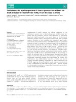
Lipocalin-type prostaglandin D synthase (L-PGDS) is a multifunctional
protein that produces prostaglandin D2 and binds and transports various
lipophilic substances after secretion into various body fluids as b-trace.
L-PGDS has been proposed to be a useful diagnostic marker for renal
injury associated with diabetes or hypertension, because the urinary and
plasma concentrations are increased in patients with these diseases. However, it remains unclear whether urinary L-PGDS is synthesized de novo in
the kidney or taken up from the blood circulation. In crude extracts of
monkey kidney and human urine, we found L-PGDS with its original N-terminal sequence starting from Ala23 after the signal sequence, and also its
N-terminal-truncated products starting from Gln31 and Phe34. In situ
hybridization and immunohistochemical staining with monoclonal antibody
5C11, which recognized the amino-terminal Ala23–Val28 loop of L-PGDS,
revealed that both the mRNA and the intact form of L-PGDS were localized in the cells of Henle’s loop and the glomeruli of the kidney, indicating
that L-PGDS is synthesized de novo in these tissues. However, truncated
forms of L-PGDS were found in the lysosomes of tubular cells, as visualized
by immunostaining with 10A5, another monoclonal antibody, which recognized the three-turn a-helix between Arg156 and Thr173. These results
suggest that L-PGDS is taken up by tubular cells and actively degraded
within their lysosomes to produce the N-terminal-truncated form.
Structured digital abstract
l
MINT-7266187: L-PGDS (uniprotkb:P41222) and Cathepsin D (uniprotkb:Q4R4P0) colocalize (MI:0403) by fluorescence microscopy (MI:0416)
l
MINT-7266176: L-PGDS (uniprotkb:P41222) and Cathepsin B (uniprotkb:Q4R5M2) colocalize (MI:0403) by fluorescence microscopy (MI:0416)
Introduction
Lipocalin-type prostaglandin D synthase (L-PGDS;
EC 5.3.99.2) catalyzes the isomerization of prostaglandin H2 (PGH2), a common precursor of various pro-
stanoids, to produce PGD2, an endogenous regulator
of sleep and pain [1–5]. L-PGDS was originally purified from rat brain [6] and found to be a monomeric
Abbreviations
CSF, cerebrospinal fluid; DIG, digoxigenin; GST, glutathione S-transferase; KO, knockout; L-PGDS, lipocalin-type prostaglandin D synthase;
MAb, monoclonal antibody; PG, prostaglandin; SPR, surface plasmon resonance.
7146
FEBS Journal 276 (2009) 7146–7158 ª 2009 The Authors Journal compilation ª 2009 FEBS
N. Nagata et al.
protein with a molecular mass of approximately
26 000 [1], and was later demonstrated to be N-glycosylated at two positions, Asn51 and Asn78 [7].
L-PGDS is known to be identical to b-trace [8,9], the
major protein in human cerebrospinal fluid (CSF) [10].
Moreover, L-PGDS is a member of the lipocalin gene
family, as judged from its amino acid homology with
several conserved motifs in this family [11]. L-PGDS
binds various lipophilic substances, such as retinoids,
thyroid hormones [12], bilirubin, biliverdin [13], gangliosides [14] and amyloid b-peptide [15], with high
affinities (Kd = 0.02–2 lm), similar to those of other
proteins in the lipocalin gene family. L-PGDS is dominantly expressed in the brain, heart and male genital
organs [1], is secreted into various body fluids, such as
CSF, plasma, seminal plasma and urine [5], and functions as both a PGD2-producing enzyme and an extracellular transporter of various lipophilic substances.
Previously, we have generated several types of mouse
monoclonal antibody (MAb) against human L-PGDS
and developed a sandwich ELISA with MAb-1B7 and
MAb-7F5 [16]. The L-PGDS level in various human
body fluids, measured by the ELISA system, revealed
that the urinary excretion of L-PGDS is increased in the
early stage of diabetes [16] and declines with the control
of the blood glucose level by hospitalization [17,18].
Moreover, urinary L-PGDS excretion is increased in
patients with hypertension with latent renal injury [19].
Thus, L-PGDS is thought to be a useful diagnostic marker for these diseases [1,20]. Urinary L-PGDS is believed
to reflect the change in glomerular permeability because
of its small molecular mass and anionic property. However, the origin of urinary L-PGDS remains unclear.
Furthermore, only 10% of L-PGDS administered intravenously in canines is recovered in the urine [21], suggesting that the permeability of L-PGDS may be low
and ⁄ or urinary L-PGDS is reabsorbed or metabolized
after filtration through the glomerular membrane.
In this study, we examined the localization of
L-PGDS in monkey kidney by in situ hybridization
and immunohistochemical analysis with two novel
MAbs against L-PGDS, and found that L-PGDS was
synthesized de novo in monkey kidney, and that
N-terminal-truncated forms of L-PGDS were present
in monkey kidney and human urine.
Results
Production and characterization of novel
anti-human L-PGDS MAbs
We purified b-trace from human CSF or the human
D1)22Cys65,167Ala L-PGDS from Escherichia coli trans-
Lipocalin-type prostaglandin D synthase in kidney
formants. In SDS-PAGE, b-trace showed a broad band
at position Mr = 25 000–27 000 as a result of its glycosylation (Fig. 1A, lane 1), whereas D1)22Cys65,167Ala
L-PGDS showed a sharp band at position Mr = 19 000
because of a lack of glycosylation (Fig. 1A, lane 2). We
generated two novel anti-human L-PGDS MAbs, i.e.
5C11 and 10A5, by immunizing L-PGDS knockout
(KO) mice with b-trace and rats with D1)22Cys65,167Ala
L-PGDS, respectively. Western blot analysis showed
that both MAb-5C11 (Fig. 1A, lanes 3 and 4) and
MAb-10A5 (Fig. 1A, lanes 5 and 6) recognized b-trace
and D1)22Cys65,167Ala L-PGDS, similar to the case of
polyclonal antibody against human L-PGDS (Fig. 1A,
lanes 9 and 10). By contrast, previously generated MAb1B7 (Fig. 1A, lanes 7 and 8) bound to D1)22Cys65,167Ala
L-PGDS efficiently but to b-trace only slightly. These
results indicate that novel MAb-5C11 and MAb-10A5
recognize both highly glycosylated b-trace and non-glycosylated recombinant L-PGDS with affinities greater
than those of the previously obtained MAb-1B7 [16].
Epitopes recognized by MAb-5C11 and MAb-10A5
were determined by western blot analysis using glutathione S-transferase (GST)-fusion proteins containing
different lengths of human L-PGDS (Fig. 1B). Each
GST-fusion protein was purified, separated by SDSPAGE and stained with silver (Fig. 1C, top panel).
MAb-5C11 bound only to the Ala23–Gln190 protein,
whereas MAb-10A5 bound to all constructs, except for
that carrying the Glu174–Gln190 region, indicating
that epitopes for MAb-5C11 and MAb-10A5 were
located in Ala23–Val28 and Arg156–Thr173, respectively. In the tertiary structure of human L-PGDS
(Fig. 1D, E) modeled from the crystallographic (PDB
codes: 2CZT and 2CZU [22]) and NMR (PDB code:
2E4J [23]) structures of mouse L-PGDS, the epitopes
for MAb-5C11 and MAb-10A5 were localized to the
amino-terminal loop and the three-turn a-helical
region of human L-PGDS, respectively. The previously generated MAb-1B7 [16] bound to the EF-loop
on the side opposite the sites for MAb-5C11 and
MAb-10A5.
The immunoglobulin subclass and light chain type
were determined to be IgG1 (j) for MAb-5C11 and
IgG2a (j) for MAb-10A5 (Table 1). Surface plasmon
resonance (SPR) analysis of the antigen–antibody
interaction demonstrated that immobilized MAb-5C11
and MAb-10A5 bound to the soluble form of b-trace
with Kd values of 91 and 2900 nm, respectively, and to
the recombinant D1)22Cys65,167Ala L-PGDS with
values of 167 and 57 nm, respectively. The binding
affinities of MAb-5C11 and MAb-10A5 were increased
about 50–100-fold for the immobilized b-trace to 1.1
and 4.5 nm, respectively, and for the recombinant
FEBS Journal 276 (2009) 7146–7158 ª 2009 The Authors Journal compilation ª 2009 FEBS
7147
Lipocalin-type prostaglandin D synthase in kidney
N. Nagata et al.
A
MAb
SYPRO
orange
kDa
5C11
10A5
1B7
PAb
26
19
1
B
1
2
*
Ala23
3
5
6
7
C
8
9
10
1 2 3 4 5 6 7 8 9 10 11
160 190
Gln190
*
Ser29
3
Silver staining
Gln35
Ser52
4
Asp74
5
6
Glu90
MAb-5C11
Tyr107
7
Val121
8
Gly140
9
Arg156
10
11
D
4
(Amino acids)
80
120
40
1
2
Glu174
MAb-10A5
E
2
Asn51
1B7
C
D
C
H B
Asn51
1B7
Asn78
G
F
E
A
1
3
10A5
10A5
5C11
N
5C11
Fig. 1. Detection of human CSF b-trace and recombinant L-PGDS by MAbs against human L-PGDS and characterization of their antigenic
epitopes. (A) b-trace (lanes 1, 3, 5, 7 and 9) and recombinant D1)22Cys65,167Ala human L-PGDS (lanes 2, 4, 6, 8 and 10) were separated by
SDS-PAGE and stained with SYPRO Orange (lanes 1 and 2), followed by blotting onto a polyvinylidine difluoride membrane. The blots were
then reacted with human L-PGDS MAb-5C11 (lanes 3 and 4), MAb-10A5 (lanes 5 and 6), MAb-1B7 (lanes 7 and 8) or polyclonal antibody
(PAb, lanes 9 and 10) for western blot analysis. Molecular size markers are shown on the left. (B) Schematic representation of GST-fusion
proteins containing a series of amino-terminus-truncated human L-PGDS. The amino-terminal amino acid residue of each mutant is indicated
on the left. The signal sequence of the amino-terminal 22 amino acid residues of human L-PGDS was removed in the parent mutant,
D1)22Cys65,167Ala L-PGDS (line 1). Asterisks indicate the two N-glycosylation sites. (C) The purified GST-fusion proteins containing the series
of amino-terminus-truncated L-PGDS were separated by SDS-PAGE, followed by silver staining (top panel) or used for western blot analysis
with MAb-5C11 (middle panel) or MAb-10A5 (bottom panel). Lanes 1–11 correspond to the mutants 1–11 shown in (B). (D) Positions of the
antigenic epitopes for MAbs on the ribbon model of human L-PGDS, which is composed of nine b-strands (strand A, Gly40–Ala49; strand B,
Cys65–Ala72; strand C, Gly76–Arg85; strand D, Gln88–Pro98; strand E, Ser104–Arg108; strand F, Tyr116–Thr123; strand G, Val128–Gly135;
strand H, Phe143–Ser150; strand I, Ile177–Phe179) and a three-turn a-helix (Ala157–Ala169). The epitopes of MAb-5C11, MAb-10A5 and
MAb-1B7 are shown in blue, orange and dark gray, respectively. Two N-glycosylation sites (Asn51 and Asn78) are shown in green and
pink, respectively. (E) Positions of the antigenic epitopes on the surface model of L-PGDS drawn using PYMOL software (DeLano Scientific
LLC, Palo Alto, CA, USA). The epitopes for MAb-5C11, MAb-10A5 and MAb-1B7 are shown in blue, orange and dark gray, respectively. The
N-glycosylation sites (Asn51) is shown in green.
D1)22Cys65,167Ala L-PGDS to 0.52 and 0.16 nm,
respectively. Thus, MAb-5C11 bound to both b-trace
and the recombinant D1)22Cys65,167Ala L-PGDS with
almost the same affinity, in either the soluble or immo7148
bilized state. The binding affinity of MAb-5C11 for
immobilized b-trace was approximately fivefold higher
than the affinities of MAb-1B7 and MAb-10A5. In this
model, the N-terminal and C-terminal regions of
FEBS Journal 276 (2009) 7146–7158 ª 2009 The Authors Journal compilation ª 2009 FEBS
N. Nagata et al.
Lipocalin-type prostaglandin D synthase in kidney
Table 1. Characterization of the three anti-human L-PGDS MAbs used in this study.
Kd (nM)
D1)22Cys65,167,Ala L-PGDS
b-trace protein
Antigen
Animal
Clone
Subclass
Soluble form
Immobilized
Soluble form
Immobilized
b-trace protein
Recombinant L-PGDS
Recombinant L-PGDS
L-PGDS KO mouse
Rat
Wild-type mouse
5C11
10A5
1B7
IgG 1 (j)
IgG 2a (j)
IgG 1 (j)
91
2900
33
1.1
4.5
5.9
167
57
50
0.52
0.16
0.96
L-PGDS are exposed on the surface of the molecule.
However, in the soluble form, the affinities of MAb10A5 for native L-PGDS were lower than those for
the recombinant protein, suggesting that the C-terminal region may be partially covered by a sugar chain
in native L-PGDS, because the N-glycosylation site
(Asn51) is near the C-terminal region in the surface
model (Fig. 1E).
We used these MAbs against human L-PGDS for
immunochemical and immunohistochemical analyses
of monkey kidney, because the homology between
human and monkey L-PGDS is very high (only five
amino acid substitutions of a total of 190 amino acid
residues, giving 97.4% amino acid identity; GenBank
M61900 for human and DDBJ Accession No.
AB032480 for monkey; Fig. 2A). The comparison of
the amino acid sequences revealed that the sequences
of the antigenic regions recognized by MAb-5C11,
MAb-1B7 and MAb-10A5 were also highly conserved
in both species. The sequence of the antigenic epitope
of MAb-5C11 (Ala23–Val28) was the same in both
species; that of MAb-1B7 contained only one substitution (R108 in human and G108 in monkey) of 14
amino acid residues; and that of MAb-10A5 (Arg156–
Thr173) also contained only one substitution (T164 in
human and S164 in monkey) of 18 amino acid residues. As a result of the highly conserved sequences
between both species, all anti-human L-PGDS MAbs
bound to monkey L-PGDS.
Western blot analysis of monkey kidney samples, partially purified by immunoaffinity columns conjugated
with MAb-1B7, MAb-5C11 or MAb-10A5, revealed
that all of these MAbs showed a broad single immunoreactive band at the same position (Mr = 25 000–
27 000) as that of purified human L-PGDS ⁄ b-trace from
human CSF (Fig. 2B). These results indicate that these
MAbs selectively recognized L-PGDS and did not bind
other proteins in monkey kidney.
Localization of L-PGDS in monkey kidney
The localization of L-PGDS in monkey kidney was
then determined by immunohistochemical staining with
MAb-5C11, MAb-10A5 and MAb-1B7 and by in situ
hybridization with antisense RNA for L-PGDS
mRNA (Fig. 3). The staining profile by in situ hybridization of mRNA for L-PGDS was similar to that
obtained with MAb-5C11 (Fig. 3A, B). By contrast,
the staining profile with MAb-10A5 was similar to that
with MAb-1B7 (Fig. 3C, D).
In the cortex and outer medulla, the L-PGDS
mRNA and L-PGDS protein detected with MAb-5C11
were localized in the distal tubules, including Henle’s
loop (Fig. 3E, F). MAb-10A5 showed generally much
weaker staining in the tubules than that obtained with
MAb-1B7 (Fig. 3G, H).
In the glomeruli, the mRNA and L-PGDS protein
detected with MAb-5C11 were localized in the epithelial
cells of Bowman’s capsule and in occasional podocytes
(arrowhead and arrow, respectively, in Fig. 3I, J).
Podocytes were identified by morphological criteria as
cells located adjacent to the outer aspect of the glomerular basement membrane. By contrast, MAb-10A5
showed scarce, and MAb-1B7 weak, immunoreactivity
in the glomeruli (Fig. 3K, L).
In higher magnification analysis, positive staining in
the tubules was observed in perinuclear regions by
in situ hybridization and by immunostaining with
MAb-5C11 (Fig. 3M, N). Although punctuate structures were seen by immunostaining with MAb-10A5
(Fig. 3O), diffuse cytoplasmic staining was observed
with MAb-1B7 (Fig. 3P). No positive signals were
observed when sense RNA probe, mouse IgG or rat
IgG was applied (Fig. 3Q–T).
Confocal laser scanning microscopy revealed that
MAb-5C11-positive fluorescence was colocalized with
L-PGDS mRNA in Henle’s loop (Fig. 4A) and glomeruli (not shown), suggesting that L-PGDS was synthesized de novo in these regions of the kidney. However,
MAb-10A5-positive fluorescence overlapped that of
cathepsin B, a lysosomal marker, in the tubules
(Fig. 4B). In higher magnification analysis, a minor
immunoreactivity of cathepsin B was not colocalized
with MAb-10A5 immunoreactivity, but the majority of
these two immunoreactivities was colocalized to the
same organella (Fig. 4C). Furthermore, MAb-10A5-
FEBS Journal 276 (2009) 7146–7158 ª 2009 The Authors Journal compilation ª 2009 FEBS
7149
Lipocalin-type prostaglandin D synthase in kidney
N. Nagata et al.
Fig. 2. Alignment of the amino acid sequences of monkey and human L-PGDS. (A) Amino acid sequence of human L-PGDS was compared
with that of monkey L-PGDS. Grey tinted boxes indicate substituted amino acid residues. Open boxes indicate the epitopes of MAb-5C11,
MAb-1B7 and MAb-10A5. Conserved residues (*) are indicated below the sequences. (B) Crude extracts of monkey kidney (1 lg protein,
lane 1) and purified human CSF L-PGDS ⁄ b-trace (0.1 lg protein, lane 2) were applied for SDS-PAGE and stained with SYPRO Orange.
The purified human CSF L-PGDS ⁄ b-trace (0.1 lg protein, lane 3) and the partially purified samples from the crude extracts of monkey
kidney (2 mg protein, lanes 4–6), obtained by immunoaffinity chromatography with MAb-1B7, MAb-5C11 or MAb-10A5, were separated by
SDS-PAGE and analyzed by western blot analysis with each MAb (lanes 4, 5 and 6, respectively). Positions of molecular size markers are
shown on the left.
positive fluorescence overlapped that of cathepsin D,
another lysosomal marker (Fig. 4D), indicating that
the MAb-10A5-positive punctate fluorescence was
distributed to lysosomes. These results, taken together,
suggest that L-PGDS is re-absorbed from the urine
into the tubules and proteolytically degraded within
lysosomes in the tubule cells.
Identification of L-PGDS of various sizes in
monkey kidney and human urine
We applied crude extracts of monkey kidney to a
MAb-1B7-conjugated immunoaffinity column and
obtained purified L-PGDS. Because of its N-glycosylation at positions Asn51 and Asn78 [7], purified
L-PGDS showed a broad band of two different sizes,
7150
Mr = 19 000–22 000 and Mr = 25 000–27 000, the
latter of which corresponded to the intact form of
L-PGDS ⁄ b-trace with N-glycosylation. Western blot
analysis showed that MAb-1B7 and MAb-10A5 were
reactive with both sizes of L-PGDS, whereas MAb5C11 selectively reacted with the intact form of
L-PGDS with Mr = 25 000–27 000 (Fig. 5A). After
glycopeptidase F treatment, the two forms of monkey
L-PGDS migrated to positions of Mr = 18 000 and
Mr = 19 000, the latter of which corresponds to the
non-glycosylated form of L-PGDS [7], and both of
which were recognized by MAb-1B7 and MAb-10A5.
By contrast, MAb-5C11 bound to non-, mono- and
di-glycosylated forms of L-PGDS with Mr = 19 000,
Mr = 22 000 and Mr = 27 000, respectively, but not
to the small band at Mr = 18 000 (Fig. 5B).
FEBS Journal 276 (2009) 7146–7158 ª 2009 The Authors Journal compilation ª 2009 FEBS
N. Nagata et al.
Lipocalin-type prostaglandin D synthase in kidney
5C11
ISH
B
A
1B7
10A5
C
D
1 mm
F
E
G
H
50 m
J
I
*
M
*
N
L
K
*
O
20 m 5 m
P
20 m
Q
R
S
50 m
*
5 m
T
20 m
5 m
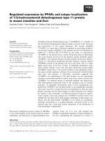
Fig. 3. Localization of L-PGDS-immunoreactive protein and mRNA in monkey kidney. Sections of monkey kidney were used for in situ
hybridization (ISH) and immunoperoxidase staining with MAb-5C11, MAb-10A5 and MAb-1B7. (A–D) Low-magnification views. At high
magnification, L-PGDS signals were detected in the tubules in the cortex and outer medulla (E–H). The signals were also detected in the
glomeruli (I–L). Asterisks, arrowheads and arrows indicate Bowman’s space, Bowman’s capsule and cytoplasm of podocytes, respectively.
(M–P) High-magnification micrographs of L-PGDS signals in tubule cells. (Q–S) Staining profile by in situ hybridization with sense RNA
probe (Q) and immunostaining with mouse IgG (R) or rat IgG (S and T). Scale bars: (A–D) 1 mm; (E–H, Q–S) 50 lm; (I–P, T) 20 lm;
insets, 5 lm.
FEBS Journal 276 (2009) 7146–7158 ª 2009 The Authors Journal compilation ª 2009 FEBS
7151
Lipocalin-type prostaglandin D synthase in kidney
A
Nomarski
*
N. Nagata et al.
DAPI
5C11
*
ISH
*
*
Merge
*
10 µm
B
Nomarski
*
DAPI
10A5
CathepsinB
*
*
*
Merge
*
10 µm
C
Nomarski
DAPI
10A5
CathepsinB
Merge
10 µm
D
Nomarski
DAPI
10A5
CathepsinD
Merge
10 µm
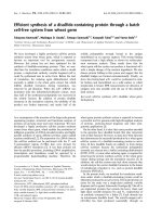
Fig. 4. Confocal laser scanning micrographs of monkey kidney. (A) In situ hybridization (ISH) combined with immunohistochemistry. Antisense cRNA probe for L-PGDS (DIG, red) was used in combination with anti-L-PGDS MAb-5C11 (green). Cells in Henle’s loop positive for
MAb-5C11 fluorescence were also positive for L-PGDS mRNA. (B) Double immunofluorescence for L-PGDS with MAb-10A5 (green) and lysosomal marker cathepsin B (red). (C, D) High-magnification micrographs of double immunofluorescence for L-PGDS with MAb-10A5 (green)
and two lysosomal markers (red) cathepsin B (C) and cathepsin D (D). Asterisks indicate lumen of a tubule. Scale bar, 10 lm for (A)–(D).
N-terminal amino acid sequence analysis revealed
that L-PGDS of the intact form in monkey kidney
started from Ala23, which is the same as the aminoterminal end of human b-trace (GenBank: M61900),
and that the smaller sized proteins of Mr = 18 000
began from Gln31 and Phe34, which were 8 and 11
amino acids shorter, respectively, than the intact
protein (Fig. 5C). These results indicate that the kidney
contained both intact L-PGDS and the N-terminaltruncated form. MAb-5C11 recognized solely the intact
form of L-PGDS, whereas MAb-10A5 detected both
forms. Furthermore, immunohistochemical staining
with MAb-5C11 indicated that the intact forms of
L-PGDS were localized to cells of Henle’s loop and the
glomeruli of the kidney. By contrast, immunostaining
with MAb-10A5 indicated that the truncated forms of
L-PGDS were taken up by tubular cells and degraded
within their lysosomes.
We then analyzed human and monkey urine by western blotting with MAb-5C11 and MAb-10A5 before
7152
and after incubation with glycopeptidase F. Without
glycopeptidase F treatment, both MAbs showed a
single immunoreactive band corresponding to the
intact form of L-PGDS (Mr = 27 000, Fig. 5D). By
contrast, after treatment of the urine samples with
glycopeptidase F, MAb-10A5, but not MAb-5C11,
detected the presence of low-molecular-mass forms
of L-PGDS in human urine (filled arrow). These
results suggest that the N-terminal-truncated form of
L-PGDS is also excreted in human urine.
Discussion
L-PGDS (Mr = 27 000) is much smaller than serum
albumin (Mr = 66 000), although these proteins share
similar chemical properties to secretory proteins, such
as having anionic charges at pH 7.4. Renal filtration is
considered to be a major clearance pathway for
low-molecular-mass proteins (Mr < 30 000 [24]). Thus,
L-PGDS passes through the glomerular capillary walls
FEBS Journal 276 (2009) 7146–7158 ª 2009 The Authors Journal compilation ª 2009 FEBS
N. Nagata et al.
Lipocalin-type prostaglandin D synthase in kidney
Monkey kidney L-PGDS
A Glycopeptidase F (–)
MAb
kDa
1B7
Intact form
5C11
10A5
27 kDa
28
L-PGDS
Truncated form 22
19.3
16.2
B Glycopeptidase F(+)
Fig. 5. Identification of novel forms of
L-PGDS in monkey kidney and human urine.
(A) L-PGDS was purified from the homogenates of monkey kidney by MAb-1B7-conjugated affinity chromatography. The purified
L-PGDS proteins were separated by SDSPAGE and analyzed by western blot analysis
with MAb-1B7, MAb-5C11 and MAb-10A5.
Positions of molecular size markers are
shown on the right. (B) Purified L-PGDS was
treated with glycopeptidase F and used for
western blot analysis with MAb-1B7,
MAb-5C11 and MAb-10A5. Truncated forms
of L-PGDS were detected by MAb-1B7 and
MAb-10A5, but not by MAb-5C11. Positions
of molecular size marker proteins are shown
on the right. (C) N-terminal amino acid
sequences of the three different forms of
L-PGDS. (D) Human and monkey urine were
treated or not with glycopeptidase F (GPF),
and used for western blot analysis with
MAb-5C11 and MAb-10A5. N-terminal-truncated forms of L-PGDS were detected with
MAb-10A5 (filled arrow). Molecular size
markers are shown on the left.
MAb
1B7
5C11
kDa
10A5
28
27 kDa
22
Intact form
L–PGDS
19.3
19
Truncated form 18
16.2
Human L–PGDS
C
30
1
10
20
40
50
MATHHTLWMGLVLLGLLGGLQAAPEAQVSVQPNFQPDKFLGRWFSAGLAS
Intact form APEAQVSVQPNF
23
QPNFQPDKFLGR
31 FQPDKFLGRWFS
Truncated form
34
{
D
Human urine
5C11
kDa
GPF –
+
10A5
–
+
28
–
+
28
19.3
19.3
Intact form
Truncated form
16.2
16.2
of the kidney more easily than does serum albumin.
Previously, we established a sandwich ELISA system
using anti-human L-PGDS MAb-1B7 and MAb-7F5
to estimate the L-PGDS level in urine [16]. Urinary
L-PGDS reflects even slight changes in glomerular
permeability because of its low molecular mass and
anionic property. Thus, urinary L-PGDS is a useful
diagnostic marker for renal diseases [1,20]. However,
our recent study [21] demonstrated that only 10% of
the intravenously administered L-PGDS in canines was
recovered in urine, suggesting that the urinary
L-PGDS concentration is not determined by leakage
through the glomeruli only, and that a large part of
the urinary L-PGDS filtered through the membranes is
reabsorbed or metabolized. To clarify the precise localization and metabolism of L-PGDS in the kidney, we
attempted to generate novel MAbs against the epitopes
distinct from those of previously obtained MAbs.
However, as the orthology of the amino acid sequence
Monkey urine
10A5
5C11
kDa
+
GPF –
Intact form
of L-PGDS is high (70.4%) between human and
mouse [25], the generation of MAbs with a variety of
epitopes is difficult in wild-type mice. Therefore, in this
study, we used rats and L-PGDS KO mice for immunization and obtained two novel anti-human L-PGDS
MAbs recognizing distinct epitopes: Ala23–Val28 for
MAb-5C11 and Arg156–Thr173 for MAb-10A5
(Fig. 1); both antigenic epitopes were distinct from
that for the previously generated MAb-1B7, which is
Tyr107–Val120 [16]. These novel MAbs recognized
efficiently both native and recombinant human
L-PGDS proteins in western blot (Fig. 1) and SPR
analyses (Table 1), and detected immunohistochemically the L-PGDS immunoreactive protein in kidney
tissue with high sensitivity and specificity (Figs 3
and 4). Therefore, these novel MAbs are useful for
further diagnostic analysis to clarify the localization of
L-PGDS in tissues and various body fluids, such as
CSF, plasma and urine.
FEBS Journal 276 (2009) 7146–7158 ª 2009 The Authors Journal compilation ª 2009 FEBS
7153
Lipocalin-type prostaglandin D synthase in kidney
N. Nagata et al.
In this study, we demonstrated that monkey kidney
contained at least three different forms of L-PGDS,
i.e. an intact form (Mr = 19 000 after removing N-glycosylated groups) and two truncated forms lacking 8
or 11 N-terminal amino acid residues (Mr = 18 000
for the non-glycosylated form; Fig. 5B, C). Moreover,
the L-PGDS immunoreactivity of the C-terminal
epitope of MAb-10A5 was detected in lysosomes in the
tubules of monkey kidney (Fig. 4), suggesting that
truncated L-PGDS was proteolytically degraded in the
kidney. We also found that the truncated forms of
L-PGDS were excreted in human urine (Fig. 5D).
These data, taken together, suggest that a major part
of urinary L-PGDS is processed by proteolytic degradation after leakage through the glomeruli. As the
L-PGDS level is altered in clinical samples, such as the
serum, CSF and urine of patients with hypertension
[19], arteriosclerosis [26], hemorrhage [27] and diabetes
[17,18,28–30], L-PGDS has been proposed to be a useful diagnostic marker for these diseases [1,20].
However, the previous ELISA system with anti-human
L-PGDS MAb-1B7 and MAb-7F5 [16] did not detect
N-terminal-truncated L-PGDS. Therefore, a new
ELISA system with MAb-10A5, recognizing the C-terminal a-helical region of L-PGDS, will be useful for
further clinical analysis.
Although the localization of L-PGDS in the kidney
remains controversial [19,28,29], we have clearly
demonstrated by immunohistochemistry with MAb5C11 and by in situ hybridization that the L-PGDS
protein is synthesized de novo in Henle’s loop, Bowman’s capsule and podocytes of the glomeruli in the
kidney (Fig. 3). The cellular distribution of L-PGDS in
the kidney is in good agreement with the results of a
previous report indicating that a substantial amount of
PGH2, a substrate of L-PGDS, is released from rat
glomeruli and glomerular mesangial cells [31].
Although the physiological role of L-PGDS in the kidney has not yet been clarified, Shirahase et al. [32] have
demonstrated previously that pretreatment of rat mesenteric artery with PGD2 attenuates organ damage
induced by endotoxin shock with lipopolysaccharide.
In addition, PGD2 and its metabolites reduce the
expression of mRNA for inducible nitric oxide synthase
stimulated by interleukin-1b [33]. NO generation
induced by the overexpression of inducible nitric oxide
synthase seems to play a pathogenic role in diabetic
nephropathy [34]. The inhibition of cytokine-mediated
NO production following an increase in PGD2 ⁄
L-PGDS in the kidney possibly attenuates the progression of kidney damage. In some renal diseases, the
increase in the biosynthesis of L-PGDS in the kidney
may be a type of adaptation mechanism against kidney
7154
injury. An L-PGDS inhibitor, AT-56, found recently
[35], is useful for clarifying the pathophysiological significance of L-PGDS in Henle’s loop and the glomeruli
in the kidney.
Materials and methods
Animals
Sprague–Dawley rats were purchased from Shizuoka Laboratory Animal Center (Hamamatsu, Japan). L-PGDS KO
mice were generated as described previously [36]. Mice and
rats were maintained under specific pathogen-free conditions in isolated cages with a 12 h light ⁄ 12 h dark photoperiod in a humidity- and temperature-controlled room
(55% at 24 °C). Water and food were available ad libitum.
The protocols used for all animal experiments in this study
were approved by the Animal Research Committee of
Osaka Bioscience Institute.
Purification of recombinant D1)22Cys65,167Ala
L-PGDS and b-trace
We cloned the coding region of human L-PGDS without
the signal sequence at the amino-terminal portion (amino
acid residues 1–22, defined translation initiation codon Met
as 1) [37] into the pGEX-2T vector (GE Healthcare, Amersham, Buckinghamshire, UK) to produce a fusion protein
with GST. The Cys residues at positions 65 and 167 were
substituted for Ala residues using a QuikChange Site-Directed Mutagenesis Kit (Stratagene, La Jolla, CA, USA) to
minimize misfolding of the recombinant protein as a result
of incorrect S–S bridging in E. coli [37]. The plasmid was
designated as GST-D1)22Cys65,167Ala and used to transform
E. coli BL21 (DE3). The recombinant GST-fusion protein
was produced by the addition of isopropyl-b-d-thiogalactopyranoside (final concentration, 0.6 mm). The cells
were further cultured for 6 h at 37 °C, and then harvested
and disrupted by sonication. The resultant cell lysates
were incubated with glutathione-Sepharose 4B resin (GE
Healthcare), followed by digestion of the purified GSTfusion
protein
with
thrombin.
The
D1)22
65,167
Cys
Ala L-PGDS was further purified by gel filtration chromatography with HiLoad Superdex 75 (GE
Healthcare).
b-Trace was purified by MAb-1B7-conjugated immunoaffinity column chromatography [38], followed by gel filtration chromatography with a HiLoad Superdex 75 column
(GE Healthcare), from the culture medium of Chinese hamster ovary cells stably expressing human L-PGDS [39], or
from human CSF provided by Dr M. Mase (Department of
Neurosurgery, Nagoya City University Hospital, Nagoya,
Japan). Proteins were analyzed by SDS-PAGE and stained
with SYPRO Orange (Invitrogen, Carlsbad, CA, USA).
FEBS Journal 276 (2009) 7146–7158 ª 2009 The Authors Journal compilation ª 2009 FEBS
N. Nagata et al.
Protein concentrations were measured using a BCA Protein
Assay Kit (Pierce Biotechnology, Rockford, IL, USA).
Preparation of MAbs for human L-PGDS
Sprague–Dawley rats were subcutaneously immunized with
the recombinant D1)22Cys65,167Ala human L-PGDS protein
expressed in E. coli. Alternatively, purified b-trace from the
culture medium of Chinese hamster ovary cells was used to
immunize L-PGDS KO mice. Splenocytes from the immunized rats or mice were fused with myeloma cells (P3U1)
under standard protocols [40]. Positive hybridomas were
cloned by the limiting dilution method. For MAb-10A5
obtained in rats, ascites was produced in male BALB ⁄
cA-nu mice by the intraperitoneal injection of hybridoma
cells. For MAb-5C11 generated in L-PGDS KO mice, IgG
antibody was purified from the serum-free medium of
hybridoma cultures. The immunoglobulin isotype of MAbs
was determined using a Rat Monoclonal Antibody Isotyping Kit (Serotec, Oxford, UK) or IsoStrip Mouse Monoclonal Antibody Isotyping Kit (Roche Diagnostics, Mannheim,
Germany), according to the manufacturer’s instructions.
SPR analysis
The kinetics of binding of MAbs to human L-PGDS were
determined by SPR analysis using a Biacore 2000 system
(GE Healthcare). D1)22Cys65,167Ala L-PGDS, b-trace or
MAbs for human L-PGDS were coupled to a CM5 sensor
chip (GE Healthcare) by the amine-coupling method,
according to the manufacturer’s protocol. The Kd values
were calculated from the sensorgrams using biaevaluation
3.1 software (GE Healthcare).
Western blot analysis
Protein samples were dissolved in 62.5 mm Tris ⁄ Cl (pH 6.8)
containing 2% (w ⁄ v) SDS, 15% (v ⁄ v) glycerol and 5%
(v ⁄ v) b-mercaptoethanol, and electrophoresed in 10–20%
(w ⁄ v) polyacrylamide gels, followed by silver staining with
Silver Stain Reagent (Daiichi Pure Chemicals, Tokyo,
Japan) or blotted onto a polyvinylidine difluoride membrane (Immobilon P; Millipore, Bedford, MA, USA). The
blots were incubated with human L-PGDS MAbs. After
washing, the blots were incubated with anti-mouse IgG
conjugated with horseradish peroxidase. Immunoreactive
signals were detected using the ECL Western Blotting
Detection System (GE Healthcare).
Mapping of antigenic epitopes recognized by
MAbs
The coding region of human L-PGDS was sequentially
truncated from the 5¢-terminus of the coding strand by
Lipocalin-type prostaglandin D synthase in kidney
PCR. Each amplified fragment was cloned downstream of
GST in the pGEX-2T vector. Expression, purification and
analyses of recombinant proteins were carried out as
described above.
N-terminal amino acid sequence analysis of
L-PGDS
Monkey (Macaca fascicularis) tissues were provided by Drs
Y. Eguchi and R. Torii (Shiga University of Medical
Science, Otsu, Japan). L-PGDS was purified by MAb-1B7conjugated immunoaffinity column chromatography from
monkey kidney [38]. The affinity-purified L-PGDS protein
was separated by SDS-PAGE and transferred onto a polyvinylidine difluoride membrane. The blots were stained with
Coomassie brilliant blue. The protein bands were excised
and utilized for sequencing analysis by Edman degradation
employing the HP G1005A Protein Sequencing System
(Hewlett-Packard, Palo Alto, CA, USA). The blots were
also used for western blot analysis as described above.
Immunohistochemical analysis
Monkey kidney was fixed in Bouin’s fixative and embedded
in paraffin. The paraffin sections (thickness, 5 lm) were
mounted on glass slides, deparaffinized in xylene and rehydrated in ethanol with increasing concentrations of water.
The rehydrated sections were pretreated with 0.3% (v ⁄ v)
H2O2 in methanol for 30 min at room temperature, and
then incubated with 0.3% (w ⁄ v) pepsin (Sigma, St. Louis,
MO, USA) in 0.01 N HCl for 5 min at room temperature.
Next, the sections were incubated for 1 h at room temperature with 10% (v ⁄ v) normal goat serum, 0.1% (v ⁄ v) Triton
X-100 and 0.1% (w ⁄ v) sodium azide in NaCl ⁄ Pi, for 16 h
at 4 °C with anti-human L-PGDS MAbs in NaCl ⁄ Pi containing 1% (v ⁄ v) normal goat serum and 0.1% (v ⁄ v) Triton
X-100, and for 1 h at room temperature with biotinylated
antibody against mouse or rat IgG (Vector Laboratories,
Burlingame, CA, USA). The immunoreactive signals were
visualized as the avidin-biotinylated enzyme complex
(Vectastain Elite ABC Kit; Vector Laboratories) after incubation in 50 mm Tris ⁄ Cl (pH 7.6) containing 0.001% (v ⁄ v)
H2O2 and 0.02% (w ⁄ v) 3,3¢-diaminobenzidine tetrahydrochloride. The sections were then counterstained with hematoxylin and observed under an ECLIPSE E600 microscope
(Nikon, Tokyo, Japan).
For double staining with rabbit polyclonal anti-cathepsin
D IgG (Assay Designs, Ann Arbor, MI, USA) and MAb10A5, after the primary antibodies had been applied, the
sections were sequentially incubated with Alexa Fluor 594conjugated antibody against rabbit IgG (Invitrogen) and
biotinylated antibody against rat IgG (5 lgỈmL)1; Jackson
ImmunoResearch, West Grove, PA, USA) followed by
Alexa Fluor 488-conjugated streptavidin (5 lgặmL)1; Invi-
FEBS Journal 276 (2009) 71467158 ê 2009 The Authors Journal compilation ª 2009 FEBS
7155
Lipocalin-type prostaglandin D synthase in kidney
N. Nagata et al.
trogen). The sections were observed under an Axiovert
100M microscope connected to a Zeiss laser-scanning
microscope 510META (Carl Zeiss, Jena, Germany).
In situ hybridization
In situ hybridization was performed using a RiboMap Kit
and Discovery automatic staining module (Roche Diagnostics). The digoxigenin (DIG)-labeled RNA probe for human
L-PGDS was prepared as follows. The coding region of
human L-PGDS was subcloned into pCR-Script Amp
SK(+) vector (Stratagene). DIG-labeled cRNA probes were
synthesized using T7 (antisense) or SP6 (sense) RNA polymerase according to the manufacturer’s manual (Roche
Diagnostics), and further purified by a Chroma Spin Column (BD Biosciences, San Jose, CA, USA). The efficiency
of DIG incorporation was determined by dot blot analysis.
The kidney sections were hybridized with L-PGDS RNA
probe in Ribohybe hybridization solution (Roche Diagnostics) at 70 °C for 3 h, and then incubated with horseradish
peroxidase-conjugated anti-DIG IgG (DakoCytomation,
Glostrup, Denmark) for 30 min, followed by 2,4-dinitrophenyl-conjugated tyramide for signal amplification. Tyramide
was then activated with H2O2 for 20 min. After heating at
85 °C for 10 min, the sections were sequentially incubated
with anti-2,4-dinitrophenyl IgG for 30 min, avidin for 1 h,
biotin for 1 h (Endogenous Biotin Blocking Kit; Roche
Diagnostics) [41], biotin-conjugated anti-rabbit IgG for
10 min and streptavidin-conjugated alkaline phosphatase for
16 min (AmpMap with TSA; Roche Diagnostics). The chromogen reaction of alkaline phosphatase was performed with
Nitro Blue tetrazolium chloride ⁄ 5-bromo-4-chloro-3¢-indolylphosphatase p-toluidine salt (BlueMap Kit; Roche Diagnostics). The sections were counterstained with Nuclear Fast
Red (ISH Red Counterstain; Roche Diagnostics).
For in situ hybridization combined with immunohistochemistry, after hybridization with the L-PGDS cRNA
probe at 70 °C for 3 h, the sections were incubated with
anti-DIG IgG conjugated with alkaline phosphatase
(1 : 1000, Roche Diagnostics) and MAb-5C11 overnight at
4 °C. The sections were then sequentially incubated for 1 h
each with avidin, biotin (Vector Laboratories), biotinylated anti-mouse IgG (5 lgỈmL)1; Jackson ImmunoResearch) and Alexa Fluor 488-conjugated streptavidin
(5 lgỈmL)1; Invitrogen). The alkaline phosphatase activity
was detected using a 2-hydroxy-3-naphthoic acid-2¢-phenylanilide phosphate fluorescence detection set (Roche Diagnostics), according to the manufacturer’s instructions.
Detection of L-PGDS in human and monkey urine
Human urine samples were obtained from the Health
Service Center of The University of Tokyo and human
plasma samples were provided by Dr Y. Eguchi (Shiga
University of Medical Science, Otsu, Japan). Urine or
7156
plasma samples were pretreated with 0.5% (w ⁄ v) SDS and
0.1 m b-mercaptoethanol at 100 °C for 3 min. Each sample
was incubated with glycopeptidase F (Takara Bio, Kyoto,
Japan) at 37 °C for 24 h in 0.1 m Tris ⁄ Cl (pH 8.6) with 1%
(v ⁄ v) Nonidet P-40. The digested samples were then separated by SDS-PAGE, followed by staining with SYPRO
Ruby (Invitrogen) or western blotting as described above.
Statement of clinical studies
This study was performed in accordance with the guidelines
for human studies in the respective hospitals and clinics,
and was approved by the respective authoritative boards of
the hospitals and departments. Informed consent was
obtained in written form from all subjects on entry to the
study.
Acknowledgements
We acknowledge Dr Mitsuhito Mase (Nagoya City
University Medical School, Nagoya, Japan) for supplying human CSF, Drs Yutaka Eguchi and Ryuzo Torii
(Shiga University of Medical Science, Otsu, Japan) for
providing monkey kidney and supplying human
plasma, and Mr Kosuke Seiki (Maruha Nichiro Holdings, Inc., Ibaraki, Japan) for supplying human urine.
We also thank Megumi Yamaguchi, Megumi Yamada
and Taeko Nishimoto for secretarial assistance. This
work was supported in part by Osaka City.
References
1 Urade Y & Hayaishi O (2000) Prostaglandin D
synthase: structure and function. Vitamins Horm 58,
89–120.
2 Urade Y & Hayaishi O (1999) Prostaglandin D2 and
sleep regulation. Biochim Biophys Acta 1436, 606–615.
3 Matsuoka T, Hirata M, Tanaka H, Takahashi Y,
Murata T, Kabashima K, Sugimoto Y, Kobayashi T,
Ushikubi F, Aze Y et al. (2000) Prostaglandin D2 as a
mediator of allergic asthma. Science 287, 2013–2017.
4 Hayaishi O & Urade Y (2002) Prostaglandin D2 in
sleep–wake regulation: recent progress and perspectives.
Neuroscientist 8, 12–15.
5 Urade Y, Eguchi N & Hayaishi O (2006) Lipocalin-type
prostaglandin D synthase as an enzymic lipocalin. In
Lipocalins (Akerstrom B, Borregaard N, Flower DR &
Salier JP eds), pp 99–109. Landes Bioscience, Georgetown, TX.
6 Urade Y, Fujimoto N & Hayaishi O (1985) Purification
and characterization of rat brain prostaglandin D
synthetase. J Biol Chem 260, 12410–12415.
7 Urade Y, Nagata A, Suzuki Y, Fujii Y & Hayaishi O
(1989) Primary structure of rat brain prostaglandin D
FEBS Journal 276 (2009) 7146–7158 ª 2009 The Authors Journal compilation ª 2009 FEBS
N. Nagata et al.
8
9
10
11
12
13
14
15
16
17
18
19
synthetase deduced from cDNA sequence. J Biol Chem
264, 1041–1045.
Hoffmann A, Conradt HS, Gross G, Nimtz M, Lottspeich F & Wurster U (1993) Purification and chemical
characterization of beta-trace protein from human
cerebrospinal fluid: its identification as prostaglandin D
synthase. J Neurochem 61, 451–456.
Watanabe K, Urade Y, Mader M, Murphy C &
Hayaishi O (1994) Identification of beta-trace as
prostaglandin D synthase. Biochem Biophys Res
Commun 203, 1110–1116.
Clausen J (1961) Proteins in normal cerebrospinal fluid
not found in serum. Proc Soc Exp Biol Med 107,
170–172.
Toh H, Kubodera H, Nakajima N, Sekiya T, Eguchi N
& Tanaka T (1996) Glutation-independent prostaglandin D synthase as a lead molecule for designing new
functional protein. Protein Eng 9, 1067–1082.
Tanaka T, Urade Y, Kimura H, Eguchi N, Nishikawa
A & Hayaishi O (1997) Lipocalin-type prostaglandin D
synthase (b-trace) is a newly recognized type of retinoid
transporter. J Biol Chem 272, 15789–15795.
Beuckmann CT, Aoyagi M, Okazaki I, Hiroike T, Toh
H & Hayaishi O (1999) Binding of biliverdin, bilirubin,
and thyroid hormones to lipocalin-type prostaglandin D
synthase. Biochemistry 28, 8006–8013.
Mohri I, Taniike M, Okazaki I, Kagitani-Shimono K,
Aritake K, Kanekiyo T, Yagi T, Takikita S, Kim HS,
Urade Y et al. (2006) Lipocalin-type prostaglandin D
synthase is up-regulated in oligodendrocytes in
lysosomal storage diseases and binds gangliosides.
J Neurochem 97, 641–651.
Kanekiyo T, Ban T, Aritake K, Huang ZL, Qu WM,
Okazaki I, Mohri I, Murayama S, Ozono K, Taniike M
et al. (2007) Lipocalin-type prostaglandin D synthase ⁄ beta-trace is a major amyloid beta-chaperone in
human cerebrospinal fluid. Proc Natl Acad Sci USA
104, 6412–6417.
Oda H, Shiina Y, Seiki K, Sato N, Eguchi N & Urade
Y (2002) Development and evaluation of a practical
ELISA for human urinary lipocalin-type prostaglandin
D synthase. Clin Chem 48, 1445–1453.
Hirawa N, Uehara Y, Ikeda T, Gomi T, Hamano K,
Totsuka Y, Yamakado M, Takagi M, Eguchi N, Oda
H et al. (2001) Urinary prostaglandin D synthase (betatrace) excretion increases in the early stage of diabetes
mellitus. Nephron 87, 321–327.
Hamano K, Totsuka Y, Ajima M, Gomi T, Ikeda T,
Hirawa N, Eguchi Y, Yamakado M, Takagi M, Nakajima H et al. (2002) Blood sugar control reverses the
increase in urinary excretion of prostaglandin D
synthase in diabetic patients. Nephron 92, 77–85.
Hirawa N, Uehara Y, Yamakado M, Toya Y, Gomi T,
Ikeda T, Eguchi Y, Takagi M, Oda H, Seiki K et al.
Lipocalin-type prostaglandin D synthase in kidney
20
21
22
23
24
25
26
27
28
29
(2002) Lipocalin-type prostaglandin D synthase in
essential hypertension. Hypertension 39, 449–454.
Urade Y & Hayaishi O (2000) Biochemical, structural,
genetic, physiological, and pathophysiological features
of lipocalin-type prostaglandin D synthase. Biochim
Biophys Acta 1482, 259–271.
Li W, Mase M, Inui T, Shimoda M, Isomura K, Oda H,
Yamada K & Urade Y (2008) Pharmacokinetics of
recombinant human lipocalin-type prostaglandin D
synthase ⁄ beta-trace in canine. Neurosci Res 61, 289–293.
Kumasaka T, Aritake K, Ago H, Irikura D,
Tsurumura T, Yamamoto M, Miyano M, Urade Y &
Hayaishi O (2009) Structural basis of the catalytic
mechanism operating in open–closed conformers of
lipocalin-type prostaglandin D synthase. J Biol Chem
284, 22344–22352.
Shimamoto S, Yoshida T, Inui T, Gohda K, Kobayashi
Y, Fujimori K, Tsurumura T, Aritake K, Urade Y &
Ohkubo T (2007) NMR solution structure of lipocalintype prostaglandin D synthase: evidence for partial
overlapping of catalytic pocket and retinoic acidbinding pocket within the central cavity. J Biol Chem
282, 31373–31379.
Maack T, Johnson V, Kau ST, Figueiredo J & Sigulem
D (1979) Renal filtration, transport, and metabolism of
low-molecular-weight proteins: a review. Kidney Int 16,
251–270.
Fujimori K, Inui T, Uodome N, Kadoyama K, Aritake
K & Urade Y (2006) Zebrafish and chicken lipocalintype prostaglandin D synthase homologues: conservation of mammalian gene structure and binding ability
for lipophilic molecules, and difference in expression
profile and enzyme activity. Gene 375, 14–25.
Eguchi Y, Eguchi N, Oda H, Seiki K, Kijima Y,
Matsu-ura Y, Urade Y & Hayaishi O (1997) Expression
of lipocalin-type prostaglandin D synthase (beta-trace)
in human heart and its accumulation in the coronary
circulation of angina patients. Proc Natl Acad Sci USA
94, 14689–14694.
Mase M, Yamada K, Iwata A, Matsumoto T, Seiki
K, Oda H & Urade Y (1999) Acute and transient
increase of lipocalin-type prostaglandin D synthase
(beta-trace) level in cerebrospinal fluid of patients with
aneurysmal subarachnoid hemorrhage. Neurosci Lett
270, 188–190.
Tsuchida T, Eguchi N, Eguchi Y, Numabe A, Nakajima H, Oda H, Seiki K, Hakamada-Taguchi R, Urade
Y & Uehara Y (2004) Lipocalin-type prostaglandin D
synthase in urine in adriamycin-induced nephropathy of
mice. Nephron Physiol 96, 42–51.
Ogawa M, Hirawa N, Tsuchida T, Eguchi N, Kawabata Y, Numabe A, Negoro H, Hakamada-Taguchi R,
Seiki K, Umemura S et al. (2006) Urinary excretions of
lipocalin-type prostaglandin D2 synthase predict the
FEBS Journal 276 (2009) 7146–7158 ª 2009 The Authors Journal compilation ª 2009 FEBS
7157
Lipocalin-type prostaglandin D synthase in kidney
30
31
32
33
34
35
36
N. Nagata et al.
development of proteinuria and renal injury in OLETF
rats. Nephrol Dial Transplant 21, 924–934.
Yoshikawa R, Wada J, Seiki K, Matsuoka T,
Miyamoto S, Takahashi K, Ota S, Taniai K, Hida K,
Yamakado M et al. (2007) Urinary PGDS levels are
associated with vascular injury in type 2 diabetes
patients. Diabetes Res Clin Pract 76, 358–367.
Soler M, Camacho M, Sola R & Vila L (2001)
Mesangial cells release untransformed prostaglandin
H2 as a major prostanoid. Kidney Int 59, 1283–1289.
Shirahase H, Kanda M, Nakamura S, Tarumi T,
Uehara Y & Ichikawa A (2000) Inhibitory effects
of PGD2, PGJ2 and 15-deoxy-delta12,14-PGJ2 on
iNOS induction in rat mesenteric artery. Life Sci 66,
2173–2182.
Nagoshi H, Uehara Y, Kanai F, Maeda S, Ogura T,
Goto A, Toyo-oka T, Esumi H, Shimizu T & Omata M
(1998) Prostaglandin D2 inhibits inducible nitric oxide
synthase expression in rat vascular smooth muscle cells.
Circ Res 82, 204–209.
Craven PA, DeRubertis FR & Melhem M (1997) Nitric
oxide in diabetic nephropathy. Kidney Int Suppl 60,
S46–53.
Irikura D, Aritake K, Nagata N, Maruyama T, Shimamoto S & Urade Y (2009) Biochemical, functional,
and pharmacological characterization of AT-56, an
orally active and selective inhibitor of lipocalin-type
prostaglandin D synthase. J Biol Chem 284, 7623–7630.
Eguchi N, Minami T, Shirafuji N, Kanaoka Y, Tanaka
T, Nagata A, Yoshida N, Urade Y, Ito S & Hayaishi O
7158
37
38
39
40
41
(1999) Lack of tactile pain (allodynia) in lipocalin-type
prostaglandin D synthase-deficient mice. Proc Natl
Acad Sci USA 96, 726–730.
Urade Y, Tanaka T, Eguchi N, Kikuchi M, Kimura H,
Toh H & Hayaishi O (1995) Structural and functional
significance of cysteine residues of glutathione-independent prostaglandin D synthase. Identification
of Cys65 as an essential thiol. J Biol Chem 270,
1422–1428.
Mase M, Yamada K, Shimazu N, Seiki K, Oda H,
Nakau H, Inui T, Li W, Eguchi N & Urade Y (2003)
Lipocalin-type prostaglandin D synthase (beta-trace) in
cerebrospinal fluid: a useful marker for the diagnosis of
normal pressure hydrocephalus. Neurosci Res 47,
455–459.
Manya H, Sato Y, Eguchi N, Seiki K, Oda H,
Nakajima H, Urade Y & Endo T (2000) Comparative
study of the asparagine-linked sugar chains of human
lipocalin-type prostaglandin D synthase purified from
urine and amniotic fluid, and recombinantly expressed
in Chinese hamster ovary cells. J Biochem (Tokyo)
127, 1001–1011.
Harlow E & Lane D (1988) A Laboratory Manual,
Antibodies. Cold Spring Harbor Laboratory Press, Cold
Spring Harbor, NY.
Bussolati G, Gugliotta P, Volante M, Pace M & Papotti
M (1997) Retrieved endogenous biotin: a novel marker
and a potential pitfall in diagnostic immunohistochemistry. Histopathology 31, 400–407.
FEBS Journal 276 (2009) 7146–7158 ª 2009 The Authors Journal compilation ª 2009 FEBS