Báo cáo khoa học: Synergistic co-operation of signal transducer and activator of transcription 5B with activator protein 1 in angiotensin II-induced angiotensinogen gene activation in vascular smooth muscle cells potx
Bạn đang xem bản rút gọn của tài liệu. Xem và tải ngay bản đầy đủ của tài liệu tại đây (330.03 KB, 9 trang )
Synergistic co-operation of signal transducer and
activator of transcription 5B with activator protein 1
in angiotensin II-induced angiotensinogen gene activation
in vascular smooth muscle cells
Mei Han, Ai-Ying Li, Fang Meng, Li-Hua Dong, Bin Zheng, Hai-Juan Hu, Lei Nie and Jin-Kun Wen
Department of Biochemistry and Molecular Biology, Hebei Medical University, Shijiazhuang, China
Angiotensin II (Ang II), an extensively characterized
peptide produced by successive proteolytic cleavage
reactions of its prohormone, angiotensinogen (AGT),
is an important contributor to the regulation of vol-
ume homeostasis and blood pressure in humans and to
the initiation of pathophysiological events that lead to
hypertension and cardiovascular disorders [1,2].
In human genetic studies, a clear linkage has been
established between the AGT gene and hypertension
[3]. Several lines of evidence have indicated that small
variations in AGT concentration result in substantial
changes in the circulating Ang II levels [4]. At the
cellular level, Ang II-mediated signaling is achieved
through its binding to the cell-surface AT1 receptor,
which causes activation of Janus kinase 2 (JAK2) [5,6]
and then activates signal transducer and activator of
transcription (STAT) molecules in cardiac myocytes
and in rat aortic (vascular) smooth muscle cells
(VSMCs) [5,7–9], resulting in the positive feedback of
AGT transcription [5]. The AGT gene itself is the
target for the activated STAT protein in cardiac myo-
cytes through the AGT promoter region [5]. However,
the interaction of STAT5B with the AGT gene
promoter was observed in liver and cardiac myocytes
[8,10], but not in the smooth muscle cell line.
The molecular basis for activation of the AGT gene
is only partially understood. The analysis of biological
information presumes that the 500-bp region of the rat
Keywords
activator protein-1; angiotensinogen; gene
regulation; signal transducer and activator of
transcription-5; vascular smooth muscle
cells
Correspondence
J K. Wen, Department of Biochemistry and
Molecular Biology, No. 361, Zhongshan East
Road, Shijiazhuang 050017, China
Fax: +86 311 8626 6180
Tel: +86 311 8626 5563
E-mail:
(Received 29 October 2008, revised 29
December 2008, accepted 12 January 2009)
doi:10.1111/j.1742-4658.2009.06902.x
The binding sequences for signal transducer and activator of transcription
(STAT) and activator protein 1 have been found in the promoter region of
the angiotensinogen gene. We examined whether the elements for activator
protein 1 and STAT5B function in angiotensinogen gene activation induced
by angiotensin II in vascular smooth muscle cells. Stimulation with angio-
tensin II increased the level of angiotensinogen mRNA by 2.1-fold in
vascular smooth muscle cells. The increased level of angiotensinogen
mRNA occurred with concurrent elevations in the levels of STAT5B and
c-Jun phosphorylation after stimulation with angiotensin II. Likewise,
angiotensin II resulted in similar enhancements of the DNA-binding activ-
ity of STAT5B and c-Jun in angiotensin II-induced angiotensinogen expres-
sion. Notably, the STAT5B–DNA complex interacted with the c-Jun–DNA
complex by forming a stable quaternary complex in angiotensin II-induced
angiotensinogen expression. Our findings support a model in which
co-operative interaction of STAT5B and activator protein 1 bound to the
the promoter region provides maximal activation of angiotensinogen
expression by angiotensin II in vascular smooth muscle cells.
Abbreviations
AGT, angiotensinogen; Ang II, angiotensin II; AP-1, activator protein 1; ChIP, chromatin immunoprecipitation; CoIP, cross-
coimmunoprecipitation; EMSA, electrophoretic mobility shift assay; GAPDH, glyceraldehyde-3-phosphate dehydrogenase; JAK, Janus kinase;
STAT, signal transducer and activator of transcription; VSMCs, vascular smooth muscle cells.
1720 FEBS Journal 276 (2009) 1720–1728 ª 2009 The Authors Journal compilation ª 2009 FEBS
AGT gene promoter contains clusters of regulatory
elements, perfectly or partially matched to consensus
sequences, including binding sequences for STAT5B
and activator protein 1 (AP-1). Previous studies indi-
cate that transcription activation by STATs requires
activated AP-1 [11–13]. AP-1 is a complex composed
of the Fos and Jun proteins [14–16]. In general, Fos
and Jun family proteins function as dimeric transcrip-
tion factors that bind to AP-1 regulatory elements in
the promoter and enhancer regions of the target
[14,16]. However, the role of AP-1 in AGT gene tran-
scription activation is unknown. It has been demon-
strated that STAT proteins co-operate to bind to
target DNA, not only with other STAT family mem-
bers [17–19], but also with other proteins and tran-
scription factors [11,13,20,21]. Recently, the physical
association between STAT and c-Jun on the a
2
-macro-
globulin promoter element has been shown to yield
maximal enhancer function [13].
Based on these pieces of knowledge, we hypothe-
sized that co-operative interaction between c-Jun and
STAT5B may be important in transcription activation
of the AGT gene induced by Ang II. To understand
whether elements for AP-1 and STAT5B function in
AGT gene activation induced by Ang II, we tested the
effect of co-operative interaction between c-Jun and
STAT5B on the AGT promoter activity and AGT
mRNA expression in VSMCs. We showed that
Ang II-induced AGT expression in VSMCs involves
co-operation between AP-1 and STAT5B. We also
demonstrated that there exists a physical interaction
between AP-1 and STAT5B during AGT expression
induced by Ang II.
Results
Ang II increases AGT gene expression with
concurrent increases in the phosphorylation
of STAT5B and c-Jun in VSMCs
It has been demonstrated that Ang II stimulates
AGT expression in hepatocytes [22] and in cardiac
muscle [5]. The present study showed that Ang II
increased the AGT mRNA level in VSMCs. Follow-
ing treatment of VSMCs with Ang II for different
periods of time, AGT mRNA was detected using
RT-PCR. As shown in Fig. 1A, the level of AGT
mRNA peaked 3 h after stimulation with Ang II,
showing an increase of 5.2-fold, and decreased there-
after. STAT5B is necessary for expression of the
AGT gene [10], and Ang II activates JAK-STAT
and AP-1 [23]. To determine the relationship
between the activation of STAT5B and c-Jun, and
the expression of the AGT gene in VSMCs stimu-
lated with Ang II, the effect of Ang II on the phos-
phorylation of STAT5 and c-Jun was measured.
Ang II stimulated the phosphorylation of STAT5B
and c-Jun, with levels of phosphorylated STAT5B
and c-Jun significantly increasing 1 h, and peaking
3 h, after stimulation with Ang II, whereas total
c-Jun and STAT5B were not changed after treatment
with Ang II for different periods of time (Fig. 1B).
However, the phosphorylation of STAT5B induced
by Ang II was dramatically inhibited by pretreating
VSMCs with AG490 (a specific inhibitor of the
JAK-STAT pathway) for 16 h [24], indicating that
the activation of STAT5B and c-Jun may be
involved in Ang II-induced AGT mRNA expression
(Fig. 1C).
DNA-binding activity of STAT5B and c-Jun
increases in Ang II-induced AGT expression
To find out whether the increase in STAT5B and
c-Jun phosphorylation induced by Ang II affects the
binding of STAT5B and c-Jun to their cis-elements,
the activity of STAT5B binding and of c-Jun binding
to DNA was detected, respectively, by electrophoretic
mobility shift assays (EMSAs) using radiolabeled oli-
gonucleotides containing either a STAT5B-binding site
or an AP-1-binding site in the rat AGT gene promoter.
As shown in Fig. 2A,B, DNA–protein complexes were
formed when these two probes were incubated with
nuclear extracts from VSMCs treated with Ang II for
0.5, 1, and 3 h, and the DNA-binding activity of
STAT5B and of AP-1 increased in a time-dependent
manner. The specificities of two DNA–protein com-
plexes were demonstrated by their disappearance upon
the addition of a 100-fold molar excess of unlabeled
probe. Further to confirm whether STAT5B and AP-1
are involved in the shifted complexes, supershift assays
were performed by adding antibodies against STAT5B
or c-Jun. Figure 2A,B showed new supershifted bands,
indicating that the complexes contained STAT5B or
c-Jun. Finally, to verify whether Ang II can stimulate
recruitment of STAT5B and c-Jun to the AGT pro-
moter in vivo, chromatin immunoprecipitation (ChIP)
assays were performed using antibodies to STAT5B
and to c-Jun, respectively. As shown in Fig. 2C, DNA
fragments containing the STAT5B- and AP-1-binding
sites could be detected in the immunoprecipitates
pulled by anti-c-Jun or anti-STAT5B IgGs. Increased
binding of AP-1 or STAT5B to the AGT promoter
was observed in VSMCs treated with Ang II for 3 h.
However, AG490 decreased the recruitment of
STAT5B to the AGT promoter region with the
M. Han et al. STAT5B and AP-1 interaction in AGT gene activation
FEBS Journal 276 (2009) 1720–1728 ª 2009 The Authors Journal compilation ª 2009 FEBS 1721
inhibition of STAT5B phosphorylation (Fig. 1C and
Fig. 2C), suggesting that STAT5B phosphorylation is
necessary for its binding to the AGT promoter.
Co-operation of STAT5B with AP-1 activates the
AGT promoter
To determine whether the binding of STAT5B and
AP-1 with the AGT promoter is essential to AGT
expression induced by Ang II, 293A cells were cotrans-
fected with the pGL3-AGT-Luc reporter plasmid, which
contains both AP-1- and STAT5B-binding sequences in
the AGT promoter from )545 to 39 bp (Fig. 3A), and
pcDNA3.1-STAT5B and ⁄ or pcDNA3.1-c-Jun expres-
sion plasmids. Overexpression of STAT5B or c-Jun
alone modestly increased the reporter activity following
stimulation with Ang II (Fig. 3B). On the other hand,
the cotransfection of STAT5B with c-Jun expression
vectors significantly increased the AGT reporter activity
by 6.8-fold over that seen with the reporter alone. These
results indicate that STAT5B and c-Jun synergistically
activate AGT gene transcription.
STAT5B and AP-1 form a stable complex in the
AGT promoter in Ang II-induced AGT expression
To establish whether there is a direct interaction
between STAT5B and c-Jun in the expression of AGT
induced by Ang II stimulation, DNA–protein inter-
actions were investigated by cross-supershift assays. As
seen in EMSAs, DNA–protein complexes formed by
0
2
4
6
8
10
0 h 1 h 3 h 6 h
Relative level
p-c-Jun
pSTAT5B
Ang II
0136 h
IP: c-Jun/ IB: p-Ser
IP:c-Jun/ IB: c-Jun
IP:STAT5B/IB: PY99
*
*
*
*
*
*
Ang II
A
B
C
03 61224 h
AGT
GAPDH
*
*
*
*
Relative mRNA level
0
2
4
6
8
0 h 3 h 6 h 12 h 24 h
IP: STAT5B/IB: PY99
IP:STAT5B/IB: STAT5B
Ang II – 3366 h
AG490 –– + +–
– 3366 h
––+ +–
*
0
2
4
6
8
10
Relative pSTAT5B level
Ang II
AG490
*
IP:STAT5B/IB: STAT5B
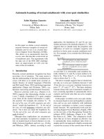
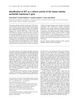
Fig. 1. Ang II induces AGT gene expression with concurrent
increases in the phosphorylation of c-Jun and STAT5B in VSMCs.
(A) VSMCs were treated with Ang II (10
)7
M) for 0, 3, 6, 12 and
24 h. Total RNA was isolated from VSMCs and subjected to
RT-PCR analysis using specific primers of the AGT gene. GAPDH
was used as an internal control. Bar graphs show the relative level
of AGT mRNA for four independent experiments. *P < 0.05, com-
pared with 0 h (n = 3). (B) VSMCs were treated with Ang II
(10
)7
M) for the indicated periods of time. Cell extracts were
immunoprecipitated with antibodies to c-Jun or to STAT5B and
immunoblotted with anti-phospho-Ser IgG or anti-PY99 IgG by
western blot analysis. Bar graphs show the relative level of phos-
phorylated c-Jun or phosphorylated STAT5B for four independent
experiments. *P < 0.05, compared with 0 h (n = 3). (C) VSMCs
were pretreated with or without AG490 (10
)5
M) for 16 h before
stimulation with Ang II (10
)7
M) for 3 and 6 h. Cell extracts were
immunoprecipitated with anti-STAT5B IgG and analyzed by western
blotting using anti-PY99 and anti-STAT5B IgGs, respectively. Bar
graphs show the relative level of phosphorylated STAT5B for four
independent experiments. *P < 0.05, compared with treatment
without AG490 in Ang II-treated cells for 3 and 6 h, respectively
(n = 3).
STAT5B and AP-1 interaction in AGT gene activation M. Han et al.
1722 FEBS Journal 276 (2009) 1720–1728 ª 2009 The Authors Journal compilation ª 2009 FEBS
A
Ang II
Nuclear extract
STAT5B probe
Cold STAT5B probe
Anti-STAT5B IgG
Anti-c-Jun IgG
Rabbit IgG
–
–
0.5 1 3 3
–
33 3 3 3 h
–+++++ –+++++
++++++ ++++++
–––––+ –+––––
–––––– –––––+
–––––– ––––+–
–––––– –––+––
Supershift
Shift
Free probe
Ang II
Nuclear extracts
AP-1 probe
Cold AP-1 probe
Anti-c-Jun IgG
Anti-STAT5B IgG
Rabbit IgG
–
–
0.5 1 3 3 3
–
33 3 h
–++++++–+++
+++++++++++
–––––
+
++
––
––
–––––––––
+–
–––––––––
–+
–––––––
–+– –
Supershift
Shift
Free probe
B
C
Ang II
AG490
–3 3 h
–– +
IP: STAT5B
No antibody
Input
Ang II 0 0.5 1 3 6 12 h
IP: c-Jun
No antibody
Input
STAT5B binding
sequence
AP-1 binding
sequence
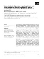
Fig. 2. Ang II increases the DNA-binding
activity of AP-1 and STAT5B. (A and B)
VSMCs were treated with Ang II (10
)7
M)
for 0.5, 1 and 3 h. Nuclear extracts were
analyzed by EMSA using oligonucleotide
probes containing the AP-1-binding site (A)
and the STAT5B-binding site (B) in the AGT
gene promoter. Protein–DNA complexes
were separated by nondenaturing PAGE and
then visualized by autoradiography. Super-
shift assays were performed by adding anti-
bodies against c-Jun or STAT5B. Rabbit IgG
was used as negative control. The data
shown represent the best of three indepen-
dent experiments. (C) VSMCs pretreated
with or without AG490 were treated with
Ang II (10
)7
M) for the indicated periods of
time. Chromatin fragments were immuno-
precipitated by anti-c-Jun and anti-STAT5B
IgG and the AGT promoter region containing
the AP-1 ()644 to )381 bp) or the STAT5B
()200 to )60 bp) binding sequence was
amplified by PCR, respectively. The data
shown represent the best of three indepen-
dent experiments.
M. Han et al. STAT5B and AP-1 interaction in AGT gene activation
FEBS Journal 276 (2009) 1720–1728 ª 2009 The Authors Journal compilation ª 2009 FEBS 1723
nuclear protein with the AP-1 probe were supershifted
by antibody to STAT5B. Similarly, the STAT5B probe–
protein complexes were supershifted by antibody to
c-Jun (Fig. 2A,B). These findings indicate that AP-1
interacts with STAT5B in the AGT expression stimu-
lated by Ang II. The interaction between STAT5B and
AP-1 was also tested by cross-coimmunoprecipitation
(CoIP) of the nuclear extracts. As shown in Fig. 4A
c-Jun protein was detected in the pellets immunoprecipi-
tated with antibody to STAT5B, suggesting that
STAT5B interacts with AP-1. Treatment of VSMCs
with Ang II for 1 and 3 h resulted in an increase in the
interaction of STAT5B with c-Jun. The interaction of
STAT5B with c-Jun induced by Ang II was significantly
decreased by pretreating VSMCs with AG490, suggest-
ing that STAT5B phosphorylation is required for the
interaction of STAT5B with AP-1. To verify this further
in vivo, ChIP was performed by using antibodies to
c-Jun or to STAT5B. STAT5B protein was found
in protein eluates from anti-c-Jun IgG-precipitated
chromatin, whereas the eluates from anti-STAT5B
IgG-precipitated chromatin contained c-Jun protein
(Fig. 4B). Furthermore, ChIP assays showed that the
STAT5B-binding sequence could be amplified by PCR
in the immunoprecipitates formed with anti-c-Jun IgG,
and the AP-1-binding sequence was similarly produced
from the STAT5B–chromatin complexes immunopre-
cipitated by anti-STAT5B IgG (Fig. 4C), indicating that
STAT5B physically interacts with c-Jun by forming a
stable complex with the AGT promoter in Ang II-
induced AGT expression.
Discussion
In this report, we demonstrated, for the first time, that,
in addition to STAT5, AP-1 is an important transcrip-
tion factor which maintains the transcription of AGT
mRNA in VSMCs, and that the activation of AP-1
participates in transcription activation of the AGT gene
to modulate the autocrine Ang II loop in the local
renin-angiotensin system. Jun and Fos family proteins
usually function as dimeric transcription factors that
bind to AP-1 regulatory elements in the promoter of
numerous genes. Jun proteins can form stable homo-
dimers or heterdimers with Fos proteins. Recent study
has indicated that Ang II activates AP-1 to regulate sev-
eral inflammatory genes in VSMCs [23]. We showed
that Ang II could activate AP-1 through enhancement
of the phosphorylation and association to DNA of Jun
proteins in the induction of the AGT gene by Ang II in
VSMCs. ChIP assays confirmed that Ang II increased
the recruitment of AP-1 to the AGT gene promoter.
Overexpression of c-Jun increased AGT-Luc reporter
activity in A293 cells. These findings indicate that AP-1
activation is involved in regulatory mechanisms of
Ang II-induced AGT gene expression in VSMCs.
Ang II is known to activate the JAK-STAT pathway
in several cells [9], STAT1, STAT2 and STAT3 in
VSMCs [9,23,25,26] and STAT5 in cardiac myocytes
[8], whereas the activity of STAT5 is unknown in Ang
II-induced VSMCs under the same conditions
[9,25,27]. However, we demonstrated that Ang II
enhances the phosphorylation of STAT5B and its
association with DNA, and consequently the transacti-
vation transcription of the AGT gene in VSMCs.
Super-EMSA and ChIP confirmed that Ang II could
increase the binding activity of STAT5B to the cis-
element and the recruitment of STAT5B to the
promoter of the AGT gene in vitro and in vivo [28–32].
It was previously demonstrated that the activation
of STAT5B in the liver, and of STAT3 and STAT5A
in the heart, participates in transcription activation of
the AGT gene to modulate the autocrine Ang II loop,
and that Ang II-mediated activation of JAK2 triggers
a pattern of tissue-specific phosphorylation of the
pGL3-AGT-Luc
pcDNA3.1-STAT5B
pcDNA3.1-c-Jun
pcDNA3.1
++++
–+–+
––++
+–––
**
Relative luciferase activity
0
10
20
30
40
50
60
70
80
90
*
A
B
STAT5B
c-Jun
AP-1AP-1 STAT5
tataaa
–419~–412 –282~–277 –172~–163 TATA
+1
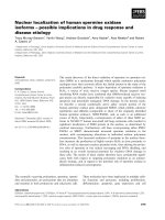
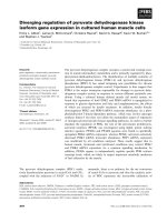
Fig. 3. Co-operation of STAT5B with AP-1 activates the AGT pro-
moter. (A) Schematic representation of the AP-1-binding site and
the STAT5B-binding site in the AGT promoter region. (B) 293A cells
were co-transfected with the pGL3-AGT-Luc reporter and with an
expression vector for c-Jun, STAT5B or c-Jun + STAT5B, respec-
tively. Cell lysates were subjected to luciferase activity assays and
western blotting using anti-STAT5B and anti-c-Jun IgG, respectively.
Bar graphs are expressed as the relative luciferase activity.
*P < 0.05 versus pcDNA3.1-transfected cells (n = 3).
STAT5B and AP-1 interaction in AGT gene activation M. Han et al.
1724 FEBS Journal 276 (2009) 1720–1728 ª 2009 The Authors Journal compilation ª 2009 FEBS
STAT protein in different tissues [10]. Ang II causes
activation of JAK via its interaction with the AT1
receptor and then activates STAT1, STAT2, STAT3,
STAT5A, STAT5B and STAT6 in heart tissues under
different experimental conditions [6,8]. In the present
study, we provided evidence indicating that stimulation
with Ang II results in JAK2 activation, which then
triggers STAT5B phosphorylation and maintains the
transcription of AGT mRNA in VSMCs. Treating
VSMCs with AG490 (10
)5
m), a potent and selective
inhibitor of JAK2 phosphorylation [24], inhibited
STAT5B activation and interaction with DNA, and
consequently caused a decrease in the transcription of
AGT mRNA. There is an elevated level of AGT
mRNA that correlated well with the enhanced
STAT5B phosphorylation. These observations there-
fore lend support to the notion that the activation of
STAT5B and the expression of the AGT gene in
VSMCs are causally linked. STAT5B may be an
important upstream component in the Ang II feedback
circuit that regulates the transcription of AGT mRNA
in VSMCs.
In the context that STAT5B and c-Jun are present
in the binding complexes with the AGT gene pro-
moter, we investigated whether there is any cross-talk
between STAT5B and c-Jun in the induction of expres-
sion of AGT mRNA by Ang II, using super-EMSA,
ChIP, CoIP and western blot analysis. Co-operative
DNA binding of proteins usually involves regions in
close proximity, which functionally represent a com-
posite regulatory element [13,14]. In this study, the
450 bp region encompassing the one STAT5B site and
the two AP-1 sites of the AGT gene promoter may
serve as composite binding elements. These closely
located sites support that the c-Jun interaction with
STAT5B in binding complexes on DNA elements is
important for maximal gene activation. Experimental
support of this is provided by the increased AGT pro-
moter ⁄ luciferase reporter activity with co-transfection
of c-Jun and STAT5B expression vectors. Definitive
evidence of physical association between c-Jun and
STAT5B is provided by the results of the CoIP and
super-EMSA of nuclear extracts, and by the results of
the ChIP assay of immunoprecipitated chromatin from
IP: STAT5B/IB: c-Jun
IP: STAT5B/IB: STAT5B
Ang II
AG490
–131 3 h
–––+ +
A
Ang II 3 h
IP
Input STAT5B c-Jun No Ig
IB: STAT5B
IB: c-Jun
IP: STAT5B
Con Ang II
B
STAT5B binding
sequence
Input
c-Jun IgG
STAT5B IgG
No Ig
AP-1 binding
sequence
C
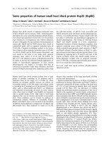
Fig. 4. STAT5B and AP-1 form a stable complex with the AGT promoter in Ang II-induced AGT expression. (A) VSMCs were treated with
Ang II (10
)7
M) for the indicated periods of time after pretreatment with or without AG490 (10
)5
M) for 16 h. Nuclear extracts were immuno-
precipitated with anti-STAT5B IgG and analyzed by western blotting using anti-c-Jun and anti-STAT5B IgG, respectively. (B) VSMCs were
treated with Ang II (10
)7
M) for 3 h. The protein eluates from chromatin precipitated with anti-c-Jun IgG or anti-STAT5B IgG were analyzed
by western blot using anti-STAT5B and anti-c-Jun IgG, respectively. (C) The chromatin fragments immunoprecipitated with anti-c-Jun and
anti-STAT5B IgG were used as templates to amplify the AGT promoter regions containing the AP-1 and STAT5B binding sequences, respec-
tively.
M. Han et al. STAT5B and AP-1 interaction in AGT gene activation
FEBS Journal 276 (2009) 1720–1728 ª 2009 The Authors Journal compilation ª 2009 FEBS 1725
VSMCs using antibodies to c-Jun and STAT5B. Previ-
ous studies have demonstrated that administration of
Ang II stimulates the interaction between p300 and
STAT5B in the liver, and with STAT3 and STAT5A
in the heart, under similar conditions [10]. Taken
together, these observations emphasize the differences
in binding of STAT proteins to the same target
sequence in response to stimulus. The previous reports
and our data suggest a role of JAK2 phosphorylation
in tissue-specific mobilization of STATs, as Ang II-
mediated stimulation of physical interaction between
STATs and c-Jun or the other transcription factors
was effectively reduced by AG490 [10]. Yet, the selec-
tive activation of STATs might require a tissue-specific
factor(s), the identity of which is hitherto unknown.
Previously, it has been shown that the cis-elements
in the AGT promoter, well characterized for their func-
tions in vitro, were dispensable in vivo [33]. We there-
fore complemented our in vitro data with the studies
on protein–protein and protein–DNA interactions and
demonstrated that the requirement for the interaction
of AP-1 with STATs for the AGT promoter activity is
the same as in vitro transient transfection assays in
293A cells. Our study demonstrated that, in addition
to STAT5B, AP-1 is also involved in the signal trans-
duction triggered by Ang II, and pointed to the under-
lying complexity in the regulation of the Ang II
autocrine loop. We speculate that AP-1 and STAT5B
bind to their elements in the AGT gene promoter,
respectively, and meanwhile interact with each other in
Ang II-stimulated VSMCs. Taken together, the inter-
action of STAT5B and c-Jun bound to the promoter
provides maximal activation of AGT expression by
Ang II in VSMCs.
Experimental procedures
Cell culture
Rat VSMCs were isolated and subcultured as described
previously [34]. Cells used in the experiments were from
passages 3–5. VSMCs were allowed to attach to the plate
wall and were then serum-deprived for 24 h in DMEM
(Gibco, Grand Island, NY, USA) containing 0.1% BSA.
Cells were then stimulated with Ang II (10
)7
m; Sigma,
St Louis, MO, USA) dissolved in serum-free DMEM con-
taining 0.1% BSA, with or without pretreatment with AG490
(10
)5
m, Sigma,) for 16 h before the addition of Ang II.
RNA isolation and RT-PCR
Total RNA was isolated from cells using TRizol reagent
according to the manufacturer’s instructions (Invitrogen,
Carlsbad, CA, USA). Reverse transcription was performed
using the Superscript First Stand Synthesis System for
RT-PCR (Invitrogen). The cDNA was then used as a tem-
plate for PCR using specific primers for AGT (forward,
5¢-ACCTTTG AGCCT GTGCCCAT -3¢; reverse, 5¢-GCT ACA
CCTCTTGCCTCAC T-3¢) and glyceraldehyde-3-phosphate
dehydrogenase (GAPDH) (forward, 5¢-CAGGGTGTGATG
GTGGG-3¢; reverse, 5¢-GGAA GAGGA TGCGG CAG-3 ¢).
The amplified RT-PCR products were separated on a 2%
agarose gel containing ethidium bromide and the band
intensities were quantified using NIH image j software.
Nuclear protein extraction
The cells were scraped into cold NaCl ⁄ P
i
and centrifuged
(14 000 g,4°C, 10 min). After the supernatant was dis-
carded, nuclear extracts were prepared by lysing the cells in
ice-cold buffer containing 10 mm Hepes-KOH (pH 7.9),
10 mm KCl, 1 mm Na
3
VO
4
and 0.5% Nonidet P-40 on ice
for 15 min, and then centrifuged at 1500 g to obtain cellu-
lar nuclei. The nuclei were washed in lysis buffer without
Nonidet P-40 and centrifuged again at 1500 g for 5 min.
The supernatant was removed and the pellet was resus-
pended in nuclear resuspension buffer (20 mm Hepes-KOH,
pH 7.9, 400 mm NaCl, 1 mm EDTA, 0.1 mm EGTA, 1 mm
phenylmethanesulfonyl fluoride, 1 mm Na
3
VO
4
,1mm dith-
iothreitol), vigorously vortexed for 10 s and then centri-
fuged at 1500 g for 5 min. The supernatant was separated
into aliquots for use in western blotting and EMSAs.
Immunoprecipitation (CoIP) and western blot
analysis
Equal amounts of proteins were incubated overnight at
4 °C with 1 lg of antibodies to STAT5B and to c-Jun
(Santa Cruz Biotechnologies, Santa Cruz, CA, USA).
Immune complexes were precipitated with 20 lLof
protein A–Sepharose beads (Santa Cruz), extensively
washed, separated by electrophoresis on an 8% SDS-poly-
acrylamide gel and then electrophoretically transferred to
poly(vinylidene difluoride) membranes (Millipore Co.,
Billerica, MA, USA). The membrane was incubated with
anti-phospho-Ser, anti-PY99, anti-STAT5B or anti-c-Jun
IgGs (1 : 1000; Santa Cruz), followed by a secondary
anti-rabbit IgG (1 : 20 000; Santa Cruz), using the chemi-
luminescence protocol (Santa Cruz).
EMSA
The sequences of double-strand oligonucleotide fragments
containing the STAT5-binding site ()172 to )163 bp) or
the AP-1-binding site ()427 to )402 bp) in the rat AGT
gene promoter were 5¢-CTAGGG
TTCCTGGAAGG
GACCC-3¢, and 5¢-AGAGCCGC
TGATGACTTATGAGA
STAT5B and AP-1 interaction in AGT gene activation M. Han et al.
1726 FEBS Journal 276 (2009) 1720–1728 ª 2009 The Authors Journal compilation ª 2009 FEBS
GGT-3¢, respectively. Nuclear extracts (10 lg) were incu-
bated for 30 min with oligonucleotide probes end-labeled
with [
32
P]dATP[cS] and then loaded onto a 6% nondenatu-
rating polyacrylamide gel, as described previously [21,35].
When specified, the reaction proceeded in the presence of
2 lg of anti-c-Jun or anti-STAT5B IgGs.
Chromatin immunoprecipitation assay
VSMCs were treated with Ang II (10
)7
m) for the time peri-
ods indicated and were then fixed with 1% formaldehyde.
ChIP assays were performed using 2 lL of anti-c-Jun or
anti-STAT5B IgGs, as described previously [35]. An aliquot
of the cell lysates was used to isolate total input DNA. PCR
amplification of the immunoprecipitated DNA was per-
formed using primers specific for the AP-1-binding site or
the STAT5B-binding site in the AGT gene promoter. The
sequences of the PCR primers were as follows: AP-1-binding
site ()644 to )381 bp), 5¢-ACTCAAGGGCGGTGCTCT
GA-3¢ and 5¢-TGGCAGATGAGCTTCAGGCA-3¢; and
STAT5B-binding site ()200 to )60 bp), 5¢-TGCCTGA
AGCTCATCTGCCACTAG-3¢ and 5¢-TAGCTCCAGCCC
AGACAAGCACAG-3¢. The proteins from the immuno-
precipitated chromatin fragment were eluted for western blot
analysis using anti-c-Jun or anti-STAT5B IgGs.
Plasmid construction
The )545 to 39 bp fragment of the rat AGT gene promoter
[5] was obtained by PCR using the following primers: for-
ward, 5¢-GCC
GGTACCGATTTCCCAACCTGACCAG
ATGTGC-3¢ (KpnI site underlined); reverse, 5¢-GCC
A
AGCTTCTGCTTACCTTTAGCTCCAGCC-3¢ (HindIII
site underlined), digested by KpnI and HindIII, and then
inserted into the pGL3-basic luciferase reporter gene vector
(Promega, Madison, WI, USA) linearized by KpnI–HindIII
and named pGL3-AGT-Luc. For the c-Jun expression plas-
mid, c-Jun cDNA was obtained from the pBIISK(-)-Jun
plasmid by EcoRI digestion, inserted into the pcDNA3.1
vector and sequenced.
Transient transfection and luciferase assay
293A cells were grown to 60% confluence in six-well plates
and transfected with Lipofectamine 2000 Reagent (Invitro-
gen), as described by the manufacturer. The transfection
was performed using 1 lg of pGL3-AGT-Luc reporter plas-
mid and c-Jun expression plasmid or STAT5B expression
plasmid (gifted by Yu-Lee, Baylor College of Medicine) or
c-Jun plus the STAT5B expression plasmid or control plas-
mid pcDNA3.1. In addition, 10 ng of the renilla luciferase
reporter plasmid pRL-TK (Promega) was included in each
sample as an internal standard for transfection efficiency.
Firefly and renilla luciferase activities were determined 48 h
after the initial transfection using the Dual-Luciferase
Reporter Assay System (Santa Cruz) and Flash & Glow
LB 955 Tube Luminometer (Alpha Innotech HD2, San
Lenndro, CA, USA). Firefly luciferase values were normal-
ized on the basis of the renilla luciferase values.
Statistical analysis
Results are expressed as means ± SD, and an analysis of
variance with Bonferroni’s test was used for the statistical
analysis of multiple comparisons of data. P-values of less
than 0.05 were considered statistically significant.
Acknowledgements
This work was supported by the National Natural
Science Foundation of China (nos 30670845 and
30770787), the ‘973’ Program of China (nos
2008CB517402) and the Hebei Province Natural Science
Foundation (nos C2006000814 and C2005000722).
References
1 Pfeffer JM, Fischer TA & Pfeffer MA (1995) Angioten-
sin-converting enzyme inhibition and ventricular remod-
eling after myocardial infarction. Annu Rev Physiol 57,
805–826.
2 Moreau P, d’Uscio LV, Shaw S, Takase H, Barton M &
Luscher TF (1997) Angiotensin II increases tissue endo-
thelin and induces vascular hypertrophy: reversal by
ET(A)-receptor antagonist. Circulation 96, 1593–1597.
3 Jeunemaitre X, Soubrier F, Kotelevtsev YV, Lifton RP,
Williams CS, Charru A, Hunt SC, Hopkins PN, Wil-
liams RR, Lalouel JM et al. (1992) Molecular basis of
human hypertension: role of angiotensinogen. Cell 71,
169–180.
4 Corvol P & Jeunemaitre X (1997) Molecular genetics of
human hypertension: role of angiotensinogen. Endocr
Rev 18, 662–677.
5 Mascareno E, Dhar M & Siddiqui MA (1998) Signal
transduction and activator of transcription (STAT) pro-
tein-dependent activation of angiotensinogen promoter:
a cellular signal for hypertrophy in cardiac muscle. Proc
Natl Acad Sci USA 95, 5590–5594.
6 Pan J, Fukuda K, Kodama H, Makino S, Takahashi T,
Sano M, Hori S & Ogawa S (1997) Role of angiotensin
II in activation of the JAK ⁄ STAT pathway induced by
acute pressure overload in the rat heart. Circ Res 81,
611–617.
7 Schieffer B, Bernstein KE & Marrero MB (1996) The
role of tyrosine phosphorylation in angiotensin II
mediated intracellular signaling and cell growth. J Mol
Med 74, 85–91.
M. Han et al. STAT5B and AP-1 interaction in AGT gene activation
FEBS Journal 276 (2009) 1720–1728 ª 2009 The Authors Journal compilation ª 2009 FEBS 1727
8 McWhinney CD, Dostal D & Baker K (1998) Angio-
tensin II activates Stat5 through Jak2 kinase in cardiac
myocytes. J Mol Cell Cardiol 30, 751–761.
9 Marrero MB, Schieffer B, Paxton WG, Heerdt L, Berk
BC, Delafontaine P & Bernstein KE (1995) Direct stim-
ulation of Jak ⁄ STAT pathway by the angiotensin II
AT1 receptor. Nature 375, 247–250.
10 Guo Y, Mascareno E & Siddiqui MA (2004) Distinct
components of Janus kinase ⁄ signal transducer and acti-
vator of transcription signaling pathway mediate the
regulation of systemic and tissue localized renin-angio-
tensin system. Mol Endocrinol 18, 1033–1041.
11 Xu W, Comhair SA, Zheng S, Chu SC, Marks-Koncza-
lik J, Moss J, Haque SJ & Erzurum SC (2003) STAT-1
and c-Fos interaction in nitric oxide synthase-2 gene
activation. Am J Physiol 285, L137–L148.
12 Schuringa JJ, Timmer H, Luttickhuizen D, Vellenga E
& Kruijer W (2001) c-Jun and c-Fos cooperate with
STAT3 in IL-6-induced transactivation of the IL-6
respone element (IRE). Cytokine 14, 78–87.
13 Zhang X, Wrzeszczynska MH, Horvath CM & Darnell
JE Jr (1999) Interacting regions in Stat3 and c-Jun that
participate in cooperative transcriptional activation.
Mol Cell Biol 19, 7138–7146.
14 Chinenov Y & Kerppola TK (2001) Close encounters of
many kinds: Fos-Jun interactions that mediate tran-
scription regulatory specificity. Oncogene 20, 2438–2452.
15 Shaulian E & Karin M (2001) AP-1 in cell proliferation
and survival. Oncogene 20, 2390–2400.
16 van Dam H & Castellazzi M (2001) Distinct roles of
Jun : Fos and Jun : ATF dimers in oncogenesis. Onco-
gene 20, 2453–2464.
17 Ghislain JJ, Wong T, Nguyen M & Fish EN (2001) The
interferon-inducible Stat2:Stat1 heterodimer preferen-
tially binds in vitro to a consensus element found in the
promoters of a subset of interferon-stimulated genes.
J Interferon Cytokine Res 21, 379–388.
18 Haque SJ & Williams BR (1998) Signal transduction in
the interferon system. Semin Oncol 25, 14–22.
19 Ihle JN (1996) STATs: signal transducers and activators
of transcription. Cell 84, 331–334.
20 Lee PJ, Camhi SL, Chin BY, Alam J & Choi AM
(2000) AP-1 and STAT mediate hyperoxia-induced gene
transcription of heme oxygenase-1. Am J Physiol 279,
L175–L182.
21 Look DC, Pelletier MR, Tidwell RM, Roswit WT &
Holtzman MJ (1995) Stat1 depends on transcriptional
synergy with Sp1. J Biol Chem 270, 30264–30267.
22 Klett C, Nobiling R, Gierschik P & Hackenthal E
(1993) Angiotensin II stimulates the synthesis of angio-
tensinogen in hepatocytes by inhibiting adenylylcyclase
activity and stabilizing angiotensinogen mRNA. J Biol
Chem 268, 25095–25107.
23 Sahar S, Dwarakanath RS, Reddy MA, Lanting L,
Todorov I & Natarajan R (2005) Angiotensin II
enhances interleukin-18 mediated inflammatory gene
expression in vascular smooth muscle cells: a novel
cross-talk in the pathogenesis of atherosclerosis. Circ
Res 96, 1064–1071.
24 Meydan N, Grunberger T, Dadi H, Shahar M, Arpaia
E, Lapidot Z, Leeder JS, Freedman M, Cohen A, Gazit
A et al. (1996) Inhibition of acute lymphoblastic leukae-
mia by a Jak-2 inhibitor. Nature 379
, 645–648.
25 Amiri F, Venema VJ, Wang X, Ju H, Venema RC &
Marrero MB (1999) Hyperglycemia enhances angiotensin
II-induced janus-activated kinase ⁄ STAT signaling in vas-
cular smooth muscle cells. J Biol Chem 274, 32382–32386.
26 Shaw SS, Schmidt AM, Banes AK, Wang X, Stern DM
& Marrero MB (2003) S100B-RAGE-mediated augmen-
tation of angiotensin II-induced activation of JAK2 in
vascular smooth muscle cells is dependent on PLD2.
Diabetes 52, 2381–2388.
27 Kim S & Iwao H (2000) Molecular and cellular mecha-
nisms of angiotensin II-mediated cardiovascular and
renal diseases. Pharmacol Rev 52, 11–34.
28 Cao H, Dronadula N, Rizvi F, Li Q, Srivastava K,
Gerthoffer WT & Rao GN (2006) Novel role for
STAT-5B in the regulation of Hsp27-FGF-2 axis facili-
tating thrombin-induced vascular smooth muscle cell
growth and motility. Circ Res 98, 913–922.
29 Cui Y, Riedlinger G, Miyoshi K, Tang W, Li C, Deng
CX, Robinson GW & Hennighausen L (2004) Inactiva-
tion of Stat5 in mouse mammary epithelium during preg-
nancy reveals distinct functions in cell proliferation,
survival, and differentiation. Mol Cell Biol 24, 8037–8047.
30 Wang D, Stravopodis D, Teglund S, Kitazawa J & Ihle
JN (1996) Naturally occurring dominant negative vari-
ants of Stat5. Mol Cell Biol 16, 6141–6148.
31 Matsumura I, Kitamura T, Wakao H, Tanaka H,
Hashimoto K, Albanese C, Downward J, Pestell RG &
Kanakura Y (1999) Transcriptional regulation of the
cyclin D1 promoter by STAT5: its involvement in cyto-
kine-dependent growth of hematopoietic cells. EMBO
J 18, 1367–1377.
32 Versteeg HH, Spek CA, Slofstra SH, Diks SH, Richel
DJ & Peppelenbosch MP (2004) FVIIa:TF induces cell
survival via G12 ⁄ G13-dependent Jak ⁄ STAT activation
and BclXL production. Circ Res 94, 1032–1040.
33 Yang G & Sigmund CD (1998) Regulatory elements
required for human angiotensinogen expression in
HepG2 cells are dispensable in transgenic mice. Hyper-
tension 31, 734–740.
34 Dronadula N, Liu Z, Wang C, Cao H & Rao GN (2005)
STAT-3-dependent cytosolic phospholipase A2 expres-
sion is required for thrombin-induced vascular smooth
muscle cell motility. J Biol Chem 280, 3112–3120.
35 Han M, Wen JK, Zheng B, Cheng Y & Zhang C (2006)
Serum deprivation results in redifferentiation of human
umbilical vascular smooth muscle cells. Am J Physiol
291, C50–C58.
STAT5B and AP-1 interaction in AGT gene activation M. Han et al.
1728 FEBS Journal 276 (2009) 1720–1728 ª 2009 The Authors Journal compilation ª 2009 FEBS