Báo cáo khoa học: Impairment of mitochondrial function by minocycline pptx
Bạn đang xem bản rút gọn của tài liệu. Xem và tải ngay bản đầy đủ của tài liệu tại đây (473.12 KB, 10 trang )
Impairment of mitochondrial function by minocycline
Kathleen Kupsch
1,2
, Silvia Hertel
3
, Peter Kreutzmann
1,2
, Gerald Wolf
2
, Claus-Werner Wallesch
3
,
Detlef Siemen
3,
* and Peter Scho
¨
nfeld
1,
*
1 Institute of Biochemistry and Cell Biology, Otto-von-Guericke University Magdeburg, Germany
2 Institute of Medical Neurobiology, Otto-von-Guericke University Magdeburg, Germany
3 Department of Neurology, Otto-von-Guericke University Magdeburg, Germany
Minocycline (MC) belongs to the tetracycline (TC)
family of antibiotics, which block the protein synthesis
of the bacterial ribosome [1]. It is commonly used in
the treatment of diseases with an inflammatory back-
ground [2], but MC possesses cytoprotective properties
as well. Thus, treatment with MC has been shown to
be beneficial in animal models of numerous neuro-
degenerative diseases [3–6] and of cerebral and cardiac
ischemia [7,8]. However, the efficacy of its neuro-
protective actions remains controversial [9]. The cyto-
protective properties of MC are discussed in terms of
anti-inflammatory [10], antioxidative [11] and antiapop-
totic activities [4,8,12,13], but MC is also considered to
be an inhibitor of the poly-(ADP-ribose) polymerase-1
[14] and of matrix metalloproteinases [15]. The antia-
poptotic effect of MC has been attributed to upregula-
tion of the antiapoptotic protein Bcl-2 [12,13], reduced
expression of caspases [8,16], and inhibition of the
Keywords
magnesium; minocycline; mitochondria;
neuroprotection; permeability transition
Correspondence
K. Kupsch, Institute of Biochemistry and
Cell Biology, Otto-von-Guericke University
Magdeburg, Leipziger Str. 44, D-39120
Magdeburg, Germany
Fax: +49 391 6714365
Tel: +49 391 6714362
E-mail:
*These authors contributed equally to this
work
(Received 7 November 2008, revised
8 January 2009, accepted 13 January 2009)
doi:10.1111/j.1742-4658.2009.06904.x
There is an ongoing debate on the presence of beneficial effects of mino-
cycline (MC), a tetracycline-like antibiotic, on the preservation of mito-
chondrial functions under conditions promoting mitochondria-mediated
apoptosis. Here, we present a multiparameter study on the effects of MC
on isolated rat liver mitochondria (RLM) suspended either in a KCl-based
or in a sucrose-based medium. We found that the incubation medium used
strongly affects the response of RLM to MC. In KCl-based medium, but
not in sucrose-based medium, MC triggered mitochondrial swelling and
cytochrome c release. MC-dependent swelling was associated with mito-
chondrial depolarization and a decrease in state 3 as well as uncoupled
respiration. Swelling of RLM in KCl-based medium indicates that MC per-
meabilizes the inner mitochondrial membrane (IMM) to K
+
and Cl
)
. This
view is supported by our findings that MC-induced swelling in the KCl-
based medium was partly suppressed by N,N¢-dicyclohexylcarbodiimide (an
inhibitor of IMM-linked K
+
-transport) and tributyltin (an inhibitor of the
inner membrane anion channel) and that swelling was less pronounced
when RLM were suspended in choline chloride-based medium. In addition,
we observed a rapid MC-induced depletion of endogenous Mg
2+
from
RLM, an event that is known to activate ion-conducting pathways within
the IMM. Moreover, MC abolished the Ca
2+
retention capacity of RLM
irrespective of the incubation medium used, most likely by triggering per-
meability transition. In summary, we found that MC at low micromolar
concentrations impairs several energy-dependent functions of mitochondria
in vitro.
Abbreviations
CaG, Calcium Green-5N; CholCl, choline chloride; CRC, Ca
2+
retention capacity; CsA, cyclosporin A; IMAC, inner membrane anion channel;
IMM, inner mitochondrial membrane; MC, minocycline; MgG, Magnesium Green; mPTP, mitochondrial permeability transition pore; RLM,
rat liver mitochondria; SEM, standard error of the mean; TBT, tributyltin chloride; TC, tetracycline; Dw
m
, mitochondrial membrane potential.
FEBS Journal 276 (2009) 1729–1738 ª 2009 The Authors Journal compilation ª 2009 FEBS 1729
release of proapoptotic proteins from mitochondria
[8,17]. This latter activity of MC might result from its
ability to block opening of the mitochondrial perme-
ability transition pore (mPTP [3,4,18]), a large-conduc-
tance megachannel in the inner mitochondrial
membrane (IMM) [19].
Recent studies, however, challenge the view that MC
is an inhibitor of the mPTP, and instead report detri-
mental effects of MC on mitochondrial physiology
[20–22]. Hence, there is ongoing debate as to whether
or not suppression of mPTP opening is involved in
MC-related cytoprotection. In order to contribute to a
better understanding of mitochondria-targeted actions
of MC, we investigated the effect of MC on various
energy-related parameters of isolated rat liver mito-
chondria (RLM). As the controversial data reported to
date were obtained using either ionic or nonionic incu-
bation media, we focused our attention on the role of
the composition of the incubation medium in
MC-linked activities. We observed that MC exerted
several detrimental effects on mitochondrial properties
such as respiration and mitochondrial membrane
potential (Dw
m
) when RLM were incubated in a KCl-
based medium. In contrast, these parameters were not
affected by MC when RLM were incubated in a
sucrose-based medium. However, irrespective of the
incubation medium used, MC decreased the mitochon-
drial Ca
2+
retention capacity (CRC) and, in addition,
induced a leakage of matrix Mg
2+
. We propose that
mitochondria are primarily affected by two activities
of MC: (a) depletion of endogenous Mg
2+
; and (b)
opening of the mPTP in Ca
2+
-loaded RLM.
Results
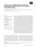
Swelling behavior
A first hint that the surrounding medium influences
the response of RLM to MC came from the observa-
tion that MC induced a swelling response that differed
with the incubation medium used (Fig. 1A). Thus,
RLM suspended in the sucrose-based medium did not
swell upon treatment with MC, even at concentrations
up to 100 lm. In contrast, MC at concentrations
‡ 25 lm triggered a rapid decrease in light absorbance
in the KCl-based medium, indicating expansion of the
mitochondrial matrix volume. The extent of swelling
was concentration-dependent and was not affected by
the potent mPTP inhibitor cyclosporin A (CsA, not
shown). Swelling was paralleled by a release of cyto-
chrome c from RLM into the medium, which also
could not be blocked by CsA (Fig. 1B). In the sucrose-
based medium, MC did not induce the translocation of
cytochrome c (Fig. 1C). In contrast, cytochrome c
release was similar in both media when the trans-
location was triggered by activating the mPTP using
calcium ions.
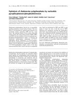
Membrane potential and mitochondrial
respiration
The MC-induced swelling of RLM in KCl-based med-
ium was associated with depolarization of the IMM
(Fig. 2A). When RLM were suspended in medium
supplemented with safranine O as a Dw
m
probe, they
rapidly accumulated the cationic dye, as indicated by
the dramatic decrease of the safranine O fluorescence
upon addition of RLM (Fig. 2A). In the KCl-based
medium, MC in concentrations found to induce
A
B
C
Fig. 1. Effect of MC on swelling behavior and cytochrome c
release. RLM (0.5 mgÆmL
)1
protein) were suspended in either KCl-
based (black traces) or sucrose-based (gray trace) medium. (A) RLM
were treated with MC as indicated. Data shown are mean ± stan-
dard error of the mean (SEM) of three independent preparations.
(B, C) After treatment of RLM (0.5 mgÆmL
)1
protein) incubated in
KCl-based (B) or sucrose-based (C) medium with 100 l
M MC or
200 l
M CaCl
2
, cytochrome c was measured in the mitochondrial
pellet and the supernatant as described in Experimental procedures.
Representative blots of three preparations are shown.
Minocycline and mitochondria K. Kupsch et al.
1730 FEBS Journal 276 (2009) 1729–1738 ª 2009 The Authors Journal compilation ª 2009 FEBS
swelling triggered a strong increase in fluorescence,
reflecting the release of safranine O from the mito-
chondria due to a decreased Dw
m
. In contrast, when
RLM were incubated in the sucrose-based medium,
MC-induced depolarization of the IMM was minor,
even at relatively high concentrations of MC (100 lm).
Furthermore, MC affected the respiration of RLM
when they were suspended in KCl-based medium, but
not in sucrose-based medium. In the absence of MC,
the rates of state 3 respiration were 56 ± 7 (1 mm
Mg
2+
) and 62 ± 6 (Mg
2+
-free) nmol O
2
Æmin
)1
Æmg
)1
protein in KCl-based medium, or 51 ± 6 (1 mm
Mg
2+
) and 49 ± 3 (Mg
2+
-free) nmol O
2
Æmin
)1
Æmg
)1
protein in sucrose-based medium. As shown in
Fig. 2B, RLM exposed to MC (25–100 lm) exhibited a
concentration-dependent decrease of state 3 respira-
tion. At the highest concentration applied (100 lm),
MC decreased state 3 respiration to 60% of the con-
trol (without MC). The decline of state 3 respiration
was dependent on the Mg
2+
concentration in the med-
ium, with MC being more effective in the absence of
Mg
2+
.InMg
2+
-free medium, state 3 respiration
decreased to about 40% of the control upon treatment
with 100 lm MC. Mg
2+
did not affect state 3 respira-
tion in the absence of MC. MC also inhibited the
carbonyl cyanide p-(trifluoromethoxy)-phenylhydraz-
one-uncoupled respiration of RLM in KCl-based
medium (not shown). In contrast, state 3 respiration of
RLM suspended in sucrose-based medium was not
affected by MC (Fig. 2C). The decline of state 3 and
carbonyl cyanide p-(trifluoromethoxy)-phenylhydraz-
one-dependent respiration was found not only when
mitochondria oxidized NAD-dependent substrates
(glutamate and malate), but also with succinate (plus
rotenone). Thus, 100 lm MC decreased succinate-
supported state 3 respiration from 95 ± 7 nmol
O
2
Æmin
)1
Æmg
)1
protein to 68 ± 5 nmol O
2
Æmin
)1
Æmg
)1
protein.
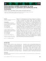
CRC
As recent results concerning the role of MC in Ca
2+
-
triggered permeability transition were controversial
[20–22], we studied the effect of MC on the mitochon-
drial CRC, which indicates the susceptibility of mito-
chondria to undergo permeability transition upon
Ca
2+
uptake into the mitochondrial matrix. Low
concentrations of MC (10 lm) completely abolished
the ability of RLM to accumulate Ca
2+
from the KCl-
based medium (Fig. 3A). The inability of MC-treated
RLM to accumulate Ca
2+
in the KCl-based medium
can simply be explained by the MC-induced swelling
and concomitant decrease of Dw
m
, the driving force
for Ca
2+
uptake.
However, Ca
2+
uptake was also suppressed by MC
in the sucrose-based medium, where MC did not initi-
ate swelling or a collapse of Dw
m
. Under these condi-
tions, slightly higher concentrations of MC were
needed to completely abolish Ca
2+
uptake (‡ 50 lm;
Fig. 3B). How can we explain the MC-induced reduc-
tion in CRC in the absence of mitochondrial depolar-
ization? In order to clarify this issue, RLM were
incubated in sucrose-based medium supplemented with
A
B
C
Fig. 2. Effect of MC on the membrane potential and respiration.
(A) In order to follow changes in Dw
m
, RLM (0.5 mgÆmL
)1
protein)
were suspended in either KCl-based (black traces) or sucrose-based
(gray trace) medium supplemented with 5 l
M safranine O as D w
m
probe. After the uptake of safranine O by energized RLM, MC was
added as indicated. Representative traces of a single preparation
out of three preparations are shown. (B, C) RLM (1 mgÆmL
)1
protein) suspended in either KCl-based (B) or sucrose-based (C)
medium were pretreated for 2 min with 25, 50, 75 or 100 l
M MC.
State 3 respiration was stimulated by the addition of 2 m
M ADP.
Respiration was also measured in the absence of Mg
2+
. Data
shown (mean ± SEM) were obtained from three to four pre-
parations.
K. Kupsch et al. Minocycline and mitochondria
FEBS Journal 276 (2009) 1729–1738 ª 2009 The Authors Journal compilation ª 2009 FEBS 1731
CsA to prevent Ca
2+
-triggered mPTP opening
(Fig. 4A, ‘control’ trace). We found that addition of
MC (25 lm) to RLM preloaded with Ca
2+
(100 nmo-
lÆmg
)1
protein) initiated the release of Ca
2+
. MC-
induced Ca
2+
release was paralleled by depolarization
of the IMM (Fig. 4B).
MC initiated the oxidation of external NADH
We were now interested to understand why MC
decreased the state 3 respiration of RLM in KCl-based
medium (Fig. 2B). As uncoupled respiration was also
sensitive to MC (not shown), we can exclude the possi-
bility that MC decreased state 3 respiration by block-
ing the F
1
F
0
-ATPase or ADP ⁄ ATP exchange across
the IMM. The observed loss of cytochrome c from
RLM upon treatment with MC, however, could con-
tribute to the MC-induced decline in respiration.
Therefore, we examined the effect of added cyto-
chrome c on the respiration of MC-treated RLM.
Addition of cytochrome c (5 lm) increased the respira-
tion only moderately (Fig. 5A). Surprisingly, subse-
quent addition of 200 lm NADH (substrate of
complex I) strongly increased the respiration of
MC-treated RLM, which was not sensitive to CsA
(not shown). In the absence of MC, addition of cyto-
chrome c and NADH only slightly affected mitochon-
drial respiration (Fig. 5B). However, subsequent
addition of MC dramatically stimulated O
2
consump-
tion. Stimulation of respiration by external NADH
suggests that MC permeabilized the IMM to NADH;
the mechanism of this remains unclear.
MC depleted mitochondria of endogenous Mg
2+
MC is a highly lipophilic TC derivative, and it is worth
recalling that TCs are able to chelate polycharged
A
B
Fig. 3. Effect of MC on CRC. RLM (0.5 mgÆmL
)1
protein) were
suspended in either KCl-based (A) or sucrose-based (B) medium
supplemented with 200 l
M ADP and 1 lgÆmL
)1
of the F
1
F
0
-ATPase
inhibitor oligomycin. Aliquots (5 lL) of a 5 m
M CaCl
2
solution were
added. The extramitochondrial Ca
2+
concentration was measured
with CaG as Ca
2+
fluorochrome. Representative traces of a single
preparation out of three preparations are shown.
A
B
Fig. 4. Effect of MC on CRC and Dw
m
of Ca
2+
-loaded mitochondria.
RLM (0.5 mg of protein) were suspended in sucrose-based med-
ium supplemented with 1 l
M CsA. (A) MC at 25 lM was added to
RLM loaded with 100 nmol Ca
2+
⁄ mg protein (indicated by two
25 l
M Ca
2+
additions; solid line). The increase of the Ca
2+
–CaG flu-
orescence observed after addition of MC indicates Ca
2+
release
from RLM. Ca
2+
uptake by RLM in the absence of MC (solid and
dotted lines) is shown for comparison. (B) The traces show the cor-
responding responses of Dw
m
to the addition of Ca
2+
and ⁄ or MC.
Representative traces of a single preparation out of three prepara-
tions are shown.
Minocycline and mitochondria K. Kupsch et al.
1732 FEBS Journal 276 (2009) 1729–1738 ª 2009 The Authors Journal compilation ª 2009 FEBS
cations, including Mg
2+
[23,24]. Therefore, there is
reason to assume that MC could extract Mg
2+
from
RLM. In order to investigate this, we tested the effect
of MC on the matrix Mg
2+
content using the Mg
2+
-
specific dye Magnesium Green (MgG). Figure 6A
shows that addition of MC decreased the fluorescence
of the matrix Mg
2+
–MgG complex in a concentration-
dependent manner (Fig. 6A). This fluorescence
decrease suggests that MC has the capability to deplete
RLM of Mg
2+
. This view is supported by the finding
that the bivalent cation ionophore A23187 induced a
similar decrease in Mg
2+
–MgG fluorescence. It should
be noted that MC also decreased the fluorescence of
the Mg
2+
–MgG complex when RLM were suspended
in the sucrose-based medium (Fig. 6B). In addition, we
observed that TC induced a much smaller decrease in
Mg
2+
–MgG fluorescence than did MC (Fig. 6B).
Inhibition of MC-induced swelling
MC-induced Mg
2+
depletion of RLM was paralleled
by mitochondrial swelling in KCl-based medium, but
not in sucrose-based medium (Fig. 1A). What could be
the mechanism underlying MC-triggered swelling of
RLM in the KCl-based medium? Mg
2+
depletion is
known to activate ion-conducting pathways within the
IMM, such as the inner membrane anion channel
(IMAC), the K
+
-uniporter, and the K
+
⁄ H
+
-antiport-
er [25–27]. Therefore, we studied a possible effect of
inhibitors of these ion-conducting pathways. Indeed,
N,N¢-dicyclohexylcarbodiimide (1 lm), a nonspecific
inhibitor of both the mitochondrial K
+
-uniporter [28]
and the mitochondrial K
+
⁄ H
+
-antiporter [29], moder-
Fig. 5. Effect of cytochrome c and NADH on mitochondrial respira-
tion in the presence and absence of MC. RLM (1 mgÆmL
)1
protein)
were suspended in KCl-based medium. Traces of the oxygen con-
centration in the medium (trace a) and its first derivative (d[O
2
] ⁄ dt;
trace b) are shown. (A, B) The respiration of control and MC-treated
(100 l
M) RLM in response to additions of 5 lM cytochrome c
(Cyt c) and 100 l
M NADH is shown. Rates of respiration are given
as numbers (in nmol O
2
Æmin
)1
Æmg
)1
protein) at the d[O
2
] ⁄ dt traces.
Representative experiments obtained from four mitochondrial prep-
arations are shown.
A
B
Fig. 6. Effect of MC on mitochondrial Mg
2+
content. RLM
(1 mgÆmL
)1
protein) loaded with MgG were suspended either in
KCl-based or sucrose-based medium supplemented with 1 m
M
EDTA. MC was added at the indicated concentrations. (A) The
decrease of the Mg
2+
–MgG fluorescence indicates release of
endogenous Mg
2+
from RLM incubated in KCl-based medium.
A23187 (1 l
M) was applied to induce complete depletion of matrix
Mg
2+
. Representative traces obtained from four mitochondrial prep-
arations are shown. (B) The graph summarizes the MC-induced
Mg
2+
release from RLM in KCl-based and sucrose-based medium.
Mg
2+
depletion triggered by TC is also included for comparison
(n = 3). Data are mean ± SEM of four preparations (*P < 0.05,
**P < 0.01, ***P < 0.001).
K. Kupsch et al. Minocycline and mitochondria
FEBS Journal 276 (2009) 1729–1738 ª 2009 The Authors Journal compilation ª 2009 FEBS 1733
ately reduced the MC-induced swelling of RLM
(Fig. 7A,D). Similarly, treatment of RLM with 1 lm
of the IMAC inhibitor tributyltin chloride (TBT) [30]
inhibited MC-induced swelling (Fig. 7B,D). Finally,
when RLM were suspended in choline chloride (Chol-
Cl)-based medium, only minor swelling was observed
upon addition of 100 lm MC (Fig. 7C,D). This obser-
vation might indicate that the choline cation is a poor
substrate of the K
+
-uniporter [31].
Discussion
We have demonstrated here that MC impairs mito-
chondrial energy metabolism. Our results support
recent reports proposing that MC most likely has no
beneficial effects on mitochondria [21,32]. Further-
more, we show that the response of energy-linked
parameters (state 3 respiration, Dw
m
) to MC depends
on the mitochondrial environment. When RLM were
suspended in KCl-based medium, MC triggered swell-
ing and decreased state 3 respiration as well as Dw
m
.A
similar observation has been reported after treatment
of rat brain mitochondria with MC in KCl-based med-
ium [21,22]. In sucrose-based medium, however, we
did not observe any effect of MC on swelling behavior,
state 3 respiration or Dw
m
of RLM. Conflicting with
our results, MC-induced swelling of RLM suspended
in mannitol⁄ sucrose-based medium has been reported
elsewhere [32]. The reason for this discrepancy remains
unclear.
What could be the mechanism underlying the MC-
induced decline of state 3 respiration, breakdown of
Dw
m
and swelling of RLM suspended in KCl-based
medium? There is reason to assume that these changes
are associated with depletion of mitochondrial matrix
Mg
2+
. Depletion of Mg
2+
from RLM could be
explained by the high lipophilicity of MC (chloro-
form ⁄ water partition coefficient of 30 at pH 7.4 [24])
and the ability of MC to chelate bivalent cations such
as Mg
2+
[23]. Mg
2+
depletion is known to activate the
IMAC, the mitochondrial K
+
-uniporter, and the mito-
chondrial K
+
⁄ H
+
-antiporter, ion-conducting path-
ways that are normally blocked in vitro by Mg
2+
binding [25–27,33]. These well-known observations
Fig. 7. Effect of N,N¢-dicyclohexylcarbodiimide, TBT and CholCl on MC-induced swelling. RLM (0.5 mgÆmL
)1
protein) were suspended in
KCl-based or CholCl-based medium. Data shown are mean ± SEM (n = 3). (A) MC at 100 l
M was added to RLM pretreated in KCl-based
medium with N,N¢-dicyclohexylcarbodiimide (an inhibitor of the K
+
-uniporter and the K
+ ⁄
H
+
-antiporter) for 10 min. (B) The suppression of the
MC-induced swelling by 1 l
M TBT (an inhibitor of the IMAC) is shown. (C) MC was added to RLM suspended in CholCl-based medium. (D)
Statistical analysis of the absorbance values 2 min after addition of MC (dotted line in A–C): (A) the MC-induced absorbance decrease was
significantly smaller in CholCl-based medium (82.4 ± 2.6%) than in KCl-based medium (65.1 ± 3.8% of baseline; P < 0.05, n = 4); (B, C)
Both TBT and N,N¢-dicyclohexylcarbodiimide significantly restored the absorbance of MC-treated RLM (for TBT, 87.4 ± 0.7 versus
75.6 ± 2.7, P < 0.01, n = 4; for N,N¢-dicyclohexylcarbodiimide, 82.8 ± 1.9 versus 75.1 ± 2.6, P < 0.05, n = 4).
Minocycline and mitochondria K. Kupsch et al.
1734 FEBS Journal 276 (2009) 1729–1738 ª 2009 The Authors Journal compilation ª 2009 FEBS
inspired us to assume that MC unmasks these ion-con-
ducting pathways, thereby enabling the uptake of KCl
by RLM. This hypothesis is in line with our finding
that MC depletes mitochondria of Mg
2+
in both ionic
and nonionic media equally, whereas MC-induced
swelling occurred only in KCl-based medium. Addi-
tionally, we found that TC, which has a much smaller
effect on matrix Mg
2+
concentration than does MC,
does not trigger swelling (not shown). Furthermore, it
is known that bivalent cations react with TCs to form
fluorescent chelates [34], which are mainly found in the
mitochondrial and in the microsomal fractions [35].
TCs preferentially bind to cations on membrane sur-
faces [34]. It is also worth mentioning that other
reagents, such as mercurials (p-hydroxymercuribenzo-
ate) and nonesterified long chain fatty acids, deplete
RLM of endogenous Mg
2+
as well, and hence induce
large-amplitude swelling in KCl-based medium
[31,36,37].
In addition to the observed partial loss of the elec-
tron carrier cytochrome c from RLM, limitation of
NADH oxidation could contribute to the decline in
state 3 respiration. Such a possibility is suggested
from our finding that the basal respiration of MC-
treated RLM strongly responds to external NADH.
Keeping in mind that the IMM of intact RLM is
impermeable to NADH, this surprising observation
could indicate that MC induced leakage of NADH
from the matrix.
There are controversial reports on whether or not
MC protects mitochondria against Ca
2+
-triggered
opening of the mPTP. It is known that MC fails to
protect mitochondria against toxin-stimulated perme-
ability transition [32]. It has also been shown that MC
cannot prevent Ca
2+
-triggered swelling when energized
RBM are in sucrose-based medium [21]. Similarly,
cytochrome c release initiated by Ca
2+
-triggered per-
meability transition was not prevented by MC [21]. In
contrast, other studies conclude that MC prevents the
Ca
2+
-dependent permeability transition irrespective of
the medium used [20,22]. This conclusion was derived
from the observation that MC suppressed Ca
2+
-depen-
dent swelling. However, suppression of swelling was
associated with deficient Ca
2+
uptake [20,22] and a
collapse of Dw
m
[22]. Therefore, the suppression of
swelling might be due to the lower sensitivity of
de-energized mitochondria to undergo Ca
2+
-triggered
permeability transition. Here, we confirm that MC
abolishes the Ca
2+
uptake of RLM suspended in KCl-
based or sucrose-based medium. We also demonstrate
that RLM preloaded with Ca
2+
release the accumu-
lated Ca
2+
upon addition of MC, even in the presence
of CsA. The release of Ca
2+
is paralleled by depolar-
ization. Taken together, these observations suggest
that mitochondrial Ca
2+
uptake is not primarily inhib-
ited by MC. Instead, MC seems to trigger a CsA-
insensitive permeability transition in Ca
2+
-loaded
RLM incubated in sucrose-based medium. This effect
might be explained by the ability of MC to deplete
mitochondria of endogenous Mg
2+
,asMg
2+
is a
powerful inhibitor of mPTP formation [38].
Our data suggest that prior results concerning the
action of MC on the mPTP might have been misinter-
preted. Thus, we show that the previously reported
MC-related inhibition of Ca
2+
-triggered swelling in
sucrose-based medium does not reflect inhibition of
the mPTP. Furthermore, our results demonstrate that
the effects of a drug on mitochondrial parameters can
depend on the incubation medium used. For instance,
the operation of K
+
-dependent ion-conducting
pathways embedded in the IMM is excluded when
sucrose-based medium is applied. Hence, the medium
composition should be more carefully considered in
future studies. In general, a KCl-based medium mimics
the in vivo situation much better than a sucrose-based
medium.
In summary, we propose that MC impairs the
function of isolated mitochondria by two distinct
mechanisms: (a) it depletes mitochondria of endoge-
nous Mg
2+
, thereby inducing permeability of the
IMM to K
+
and Cl
)
; and (b) it activates the mPTP
in the presence of external Ca
2+
or in Ca
2+
-loaded
RLM, thereby inducing permeability of the IMM to
nonionic, low-molecular solutes, such as sucrose.
These detrimental activities might contribute to the
harmful effects of MC, as recently reported from a
phase III clinical trial in patients with amyotrophic
lateral sclerosis [39]. In the study cited, doses of
400 mgÆday
)1
were administered. Assuming a body
weight of 70 kg and a body water content of 60%,
the concentration of MC in the aqueous phase can
be calculated to be about 20 lm at most after a
bolus administration. Considering that MC easily
permeates the blood–brain barrier [9] and has high
solubility in membranes [24], there is good reason to
assume that mitochondria can be affected by harmful
activities of MC in vivo.
Experimental procedures
Chemicals
CsA was obtained from Alexis (Lausanne, Switzerland).
CaG and MgG were from Molecular Probes (Karlsruhe,
Germany). All other biochemicals were purchased from
Sigma (Steinheim, Germany).
K. Kupsch et al. Minocycline and mitochondria
FEBS Journal 276 (2009) 1729–1738 ª 2009 The Authors Journal compilation ª 2009 FEBS 1735
Animals
Procedures for animal use were in strict accordance with
the Animal Health and Care Committee of the State Sach-
sen-Anhalt, Germany. Male Wistar rats (Harlan–Winkel-
mann, Borchen, Germany) were single-housed and
maintained under a 12 : 12 h light ⁄ dark cycle. Before being
killed, rats were allowed a 2 week acclimation period and
had free access to standard food and water ad libitum.
Isolation of RLM
RLM were prepared from Harlan-Winkelmann male Wistar
rats (Borchen, Germany) with a wet-liver mass of about 8 g
as described previously [40]. The final mitochondrial pellet
was suspended in isolation medium (250 mm sucrose,
0.5 mm EDTA; pH 7.4). Mitochondrial protein in the stock
suspension was determined using the Biuret method. The
total yield of isolated mitochondria per liver was about
120 mg of protein. The respiratory control ratio was
routinely measured to be in the range 6–12.
Incubations
Experiments were performed in two different incubation
media, a KCl-based (125 mm KCl, 20 mm Tris, 1 mm
MgCl
2
,10lm EGTA, 5 mm glutamate, 5 mm malate, and
1mm P
i
; pH 7.2) or a sucrose-based (200 mm sucrose,
10 mm Tris, 1 mm MgCl
2
,10lm EGTA, 5 mm glutamate,
5mm malate, and 1 mm P
i
; pH 7.2) medium. Further addi-
tions are specified in the figure legends. MC was added
from an aqueous stock solution (10 mm). Incubations were
routinely performed at 30 °C.
Measurement of swelling and cytochrome c
release
Swelling of mitochondria was measured as decrease in light
absorbance at 620 nm using a multiplate reader (Titertek
Plus MS212; ICN, Frankfurt, Germany). RLM were sus-
pended in 200 lL of the indicated incubation medium
(0.5 mgÆmL
)1
protein). For determination of cytochrome c
release, RLM (0.5 mgÆmL
)1
protein) were preincubated in
1 mL of the incubation medium for 5 min. Subsequently,
MC or Ca
2+
was added as indicated, and mitochondria
were incubated at room temperature for an additional
5 min. After centrifugation of the incubation mixture
(4000 g, 5 min), the supernatants (extramitochondrial frac-
tions) were collected. The pellets (mitochondrial fractions)
were resuspended in 250 lL of 10% SDS and incubated at
95 °C for 10 min. All fractions were diluted 1 : 4 in Roti-
Load 4x and denatured at 95 °C for 5 min. Equal volumes
were applied to a 5–20% SDS gel. After electrophoresis
(20 mAÆgel
)1
, 90 min), proteins were transferred to a
Hybond-c Extra nitrocellulose membrane (Amersham Bio-
sciences, Little Chalfont, UK). Immunostaining was per-
formed using the primary 7H8.2C12 mouse antibody against
cytochrome c (1 : 500; BD Pharmingen) plus secondary
goat anti-mouse IgG + IgM conjugated to peroxidase
(1 : 10 000; Jackson ImmunoResearch Laboratories Inc.,
Westgrove, USA). Protein bands were visualized by chemilu-
minescence (Immobilon Western, Millipore, Billerica, USA).
Membrane potential and CRC
Membrane potential (Dw
m
) and extramitochondrial Ca
2+
concentration were recorded using safranine O (Dw
m
probe) and the membrane-impermeant Ca
2+
-sensitive dye
Calcium Green-5N (CaG). RLM (0.5 mg of protein) were
suspended in 1 mL of the indicated incubation medium
supplemented with 5 lm safranine O or 100 nm CaG. Flu-
orescence intensities were measured at excitation wave-
lengths of 525 and 506 nm and emission wavelengths of
587 and 532 nm for safranine O and CaG, respectively,
using a Cary Eclipse fluorometer (Varian, Darmstadt,
Germany).
Respiration
Oxygen consumption was measured in 2 mL of incubation
medium (1 mgÆmL
)1
protein) at 30 °C using the high-reso-
lution OROBOROS Oxygraph (Anton Paar KG, Graz,
Austria). State 3 respiration and uncoupled respiration was
adjusted by addition of 2 mm ADP and 0.7 lm p-(trifluoro-
methoxy)-phenylhydrazone, respectively.
Determination of matrix Mg
2+
Free matrix Mg
2+
was monitored fluorimetrically using
MgG as described previously [36]. Briefly, mitochondria
suspended in isolation medium (10 mgÆmL
)1
) were loaded
with MgG-acetoxymethylester (2 lm) for 5 min at room
temperature. After centrifugation of the mitochondrial sus-
pension (10 000 g for 2 min), the mitochondrial pellet was
resuspended in 450 lL of the isolation medium. Aliquots of
MgG-loaded RLM (0.2 mg of protein) were added to 1 mL
of the indicated assay medium supplemented with 1 mm
EDTA. The fluorescence was recorded using a PerkinElmer
Luminescence Spectrophotometer LS50B at 510 nm excita-
tion and 535 nm emission.
Statistical analysis
All experiments were replicated in at least three indepen-
dent mitochondrial preparations. Values obtained were
compared by one-way ANOVA followed by Dunnett
post-test using graphpadprism (version 3.02; GraphPad
Software, San Diego, CA, USA).
Minocycline and mitochondria K. Kupsch et al.
1736 FEBS Journal 276 (2009) 1729–1738 ª 2009 The Authors Journal compilation ª 2009 FEBS
Acknowledgements
This work was supported by funding from Magde-
burger Forschungsverbund NBL3 (to G. Wolf and
D. Siemen) and from the BMBF (to D. Siemen).
References
1 Craven GR, Gavin R & Fanning T (1969) The transfer
RNA binding site of the 30 S ribosome and the site of
tetracycline inhibition. Cold Spring Harb Symp Quant
Biol 34, 129–137.
2 Vandekerckhove BN, Quirynen M & van Steenberghe
D (1998) The use of locally delivered minocycline in the
treatment of chronic periodontitis. A review of the liter-
ature. J Clin Periodontol 25, 964–968.
3 Zhu S, Stavrovskaya IG, Drozda M, Kim BY, Ona V,
Li M, Sarang S, Liu AS, Hartley DM, Wu DC et al.
(2002) Minocycline inhibits cytochrome c release and
delays progression of amyotrophic lateral sclerosis in
mice. Nature 417, 74–78.
4 Wang X, Zhu S, Drozda M, Zhang W, Stavrovskaya
IG, Cattaneo E, Ferrante RJ, Kristal BS & Friedlander
RM (2003) Minocycline inhibits caspase-independent
and -dependent mitochondrial cell death pathways in
models of Huntington’s disease. Proc Natl Acad Sci
USA 100, 10483–10487.
5 Haroon MF, Fatima A, Scholer S, Gieseler A, Horn
TF, Kirches E, Wolf G & Kreutzmann P (2007) Mino-
cycline, a possible neuroprotective agent in Leber’s
hereditary optic neuropathy (LHON): studies of cybrid
cells bearing 11778 mutation. Neurobiol Dis 28, 237–
250.
6 Stack EC, Smith KM, Ryu H, Cormier K, Chen M,
Hagerty SW, Del Signore SJ, Cudkowicz ME, Fried-
lander RM & Ferrante RJ (2006) Combination therapy
using minocycline and coenzyme Q10 in R6 ⁄ 2 trans-
genic Huntington’s disease mice. Biochim Biophys Acta
1762, 373–380.
7 Koistinaho M, Malm TM, Kettunen MI, Goldsteins G,
Starckx S, Kauppinen RA, Opdenakker G & Koisti-
naho J (2005) Minocycline protects against permanent
cerebral ischemia in wild type but not in matrix metal-
loprotease-9-deficient mice. J Cereb Blood Flow Metab
25, 460–467.
8 Scarabelli TM, Stephanou A, Pasini E, Gitti G,
Townsend P, Lawrence K, Chen-Scarabelli C,
Saravolatz L, Latchman D, Knight R et al. (2004)
Minocycline inhibits caspase activation and reactiva-
tion, increases the ratio of XIAP to smac ⁄ DIABLO,
and reduces the mitochondrial leakage of cyto-
chrome C and smac ⁄ DIABLO. J Am Coll Cardiol 43,
865–874.
9 Jordan J, Fernandez-Gomez FJ, Ramos M, Ikuta I,
Aguirre N & Galindo MF (2007) Minocycline and
cytoprotection: shedding new light on a shadowy con-
troversy. Curr Drug Deliv 4, 225–231.
10 Yrjanheikki J, Keinanen R, Pellikka M, Hokfelt T &
Koistinaho J (1998) Tetracyclines inhibit microglial acti-
vation and are neuroprotective in global brain ischemia.
Proc Natl Acad Sci USA 95, 15769–15774.
11 Kraus RL, Pasieczny R, Lariosa-Willingham K, Turner
MS, Jiang A & Trauger JW (2005) Antioxidant proper-
ties of minocycline: neuroprotection in an oxidative
stress assay and direct radical-scavenging activity.
J Neurochem 94, 819–827.
12 Wang J, Wei Q, Wang CY, Hill WD, Hess DC & Dong
Z (2004) Minocycline up-regulates Bcl-2 and protects
against cell death in mitochondria. J Biol Chem 279,
19948–19954.
13 Castanares M, Vera Y, Erkkila K, Kyttanen S, Lue Y,
Dunkel L, Wang C, Swerdloff RS & Hikim AP (2005)
Minocycline up-regulates BCL-2 levels in mitochondria
and attenuates male germ cell apoptosis. Biochem
Biophys Res Commun 337, 663–669.
14 Alano CC, Kauppinen TM, Valls AV & Swanson RA
(2006) Minocycline inhibits poly(ADP-ribose) polymer-
ase-1 at nanomolar concentrations. Proc Natl Acad Sci
USA 103, 9685–9690.
15 Brundula V, Rewcastle NB, Metz LM, Bernard CC &
Yong VW (2002) Targeting leukocyte MMPs and trans-
migration: minocycline as a potential therapy for multi-
ple sclerosis. Brain 125 , 1297–1308.
16 Chen M, Ona VO, Li M, Ferrante RJ, Fink KB, Zhu
S, Bian J, Guo L, Farrell LA, Hersch SM
et al. (2000)
Minocycline inhibits caspase-1 and caspase-3 expression
and delays mortality in a transgenic mouse model of
Huntington disease. Nat Med 6, 797–801.
17 Teng YD, Choi H, Onario RC, Zhu S, Desilets FC,
Lan S, Woodard EJ, Snyder EY, Eichler ME & Fried-
lander RM (2004) Minocycline inhibits contusion-trig-
gered mitochondrial cytochrome c release and mitigates
functional deficits after spinal cord injury. Proc Natl
Acad Sci USA 101, 3071–3076.
18 Fuks B, Talaga P, Huart C, Henichart J-P, Bertrand K,
Grimee R & Lorent G (2005) In vitro properties of
5-(benzylsulfonyl)-4-bromo-2-methyl-3(2H)-pyridazi-
none: a novel permeability transition pore inhibitor.
Eur J Pharmacol 519, 24–30.
19 Leung AW & Halestrap AP (2008) Recent progress in
elucidating the molecular mechanism of the mitochon-
drial permeability transition pore. Biochim Biophys Acta
1777, 946–952.
20 Theruvath TP, Zhong Z, Pediaditakis P, Ramshesh VK,
Currin RT, Tikunov A, Holmuhamedov E & Lemasters
JJ (2008) Minocycline and N-methyl-4-isoleucine cyclo-
sporin (NIM811) mitigate storage ⁄ reperfusion injury
after rat liver transplantation through suppression of
the mitochondrial permeability transition. Hepatology
47, 236–246.
K. Kupsch et al. Minocycline and mitochondria
FEBS Journal 276 (2009) 1729–1738 ª 2009 The Authors Journal compilation ª 2009 FEBS 1737
21 Mansson R, Hansson MJ, Morota S, Uchino H,
Ekdahl CT & Elmer E (2007) Re-evaluation of mito-
chondrial permeability transition as a primary
neuroprotective target of minocycline. Neurobiol Dis 25,
198–205.
22 Fernandez-Gomez FJ, Galindo MF, Gomez-Lazaro M,
Gonzalez-Garcia C, Cena V, Aguirre N & Jordan J
(2005) Involvement of mitochondrial potential and cal-
cium buffering capacity in minocycline cytoprotective
actions. Neuroscience 133, 959–967.
23 Durckheimer W (1975) Tetracyclines: chemistry, bio-
chemistry, and structure–activity relations. Angew Chem
Int Ed Engl 14, 721–734.
24 Barza M, Brown RB, Shanks C, Gamble C & Weinstein
L (1975) Relation between lipophilicity and pharmaco-
logical behavior of minocycline, doxycycline, tetracy-
cline, and oxytetracycline in dogs. Antimicrob Agents
Chemother 8, 713–720.
25 Beavis AD & Powers MF (1989) On the regulation of
the mitochondrial inner membrane anion channel by
magnesium and protons. J Biol Chem 264, 17148–
17155.
26 Nakashima RA, Dordick RS & Garlid KD (1982) On
the relative roles of Ca2 + and Mg2 + in regulating
the endogenous K+ ⁄ H+ exchanger of rat liver mito-
chondria. J Biol Chem 257, 12540–12545.
27 Bernardi P, Angrilli A, Ambrosin V & Azzone GF
(1989) Activation of latent K+ uniport in mitochondria
treated with the ionophore A23187. J Biol Chem 264,
18902–18906.
28 Gauthier LM & Diwan JJ (1979) Inhibition of K+ flux
into rat liver mitochondria by dicyclohexylcarbodiimide.
Biochem Biophys Res Commun 87, 1072–1079.
29 Martin WH, DiResta DJ & Garlid KD (1986) Kinetics
of inhibition and binding of dicyclohexylcarbodiimide
to the 82,000-dalton mitochondrial K+ ⁄ H+ antiporter.
J Biol Chem 261, 12300–12305.
30 Powers MF & Beavis AD (1991) Triorganotins inhibit
the mitochondrial inner membrane anion channel.
J Biol Chem 266, 17250–17256.
31 Scho
¨
nfeld P, Gerke S, Bohnensack R & Wojtczak L
(2003) Stimulation of potassium cycling in mitochondria
by long-chain fatty acids. Biochim Biophys Acta 1604,
125–133.
32 Cornet S, Spinnewyn B, Delaflotte S, Charnet C, Rou-
bert V, Favre C, Hider H, Chabrier PE & Auguet M
(2004) Lack of evidence of direct mitochondrial involve-
ment in the neuroprotective effect of minocycline. Eur J
Pharmacol 505, 111–119.
33 Beavis AD (1992) Properties of the inner membrane
anion channel in intact mitochondria. J Bioenerg Bio-
membr 24, 77–90.
34 Caswell AH & Hutchison JD (1971) Visualization of
membrane bound cations by a fluorescent technique.
Biochem Biophys Res Commun 42, 43–49.
35 Rall DP, Loo TL, Lane M & Kelly MG (1957) Appear-
ance and persistence of fluorescent material in tumor
tissue after tetracycline administration. J Natl Cancer
Inst 19, 79–85.
36 Scho
¨
nfeld P, Schu
¨
ttig R & Wojtczak L (2002) Rapid
release of Mg(2 + ) from liver mitochondria by none-
sterified long-chain fatty acids in alkaline media. Arch
Biochem Biophys
403, 16–24.
37 Bogucka K & Wojtczak L (1979) On the mechanism of
mercurial-induced permeability of the mitochondrial
membrane to K
+
. FEBS Lett 100 , 301–304.
38 Novgorodov SA, Gudz TI, Brierley GP & Pfeiffer DR
(1994) Magnesium ion modulates the sensitivity of the
mitochondrial permeability transition pore to cyclospo-
rin A and ADP. Arch Biochem Biophys 311, 219–228.
39 Gordon PH, Moore DH, Miller RG, Florence JM,
Verheijde JL, Doorish C, Hilton JF, Spitalny GM,
Macarthur RB, Mitsumoto H et al. (2007) Efficacy of
minocycline in patients with amyotrophic lateral sclero-
sis: a phase III randomised trial. Lancet Neurol 6, 1045–
1053.
40 Kupsch K, Parvez S, Siemen D & Wolf G (2007) Mod-
ulation of the permeability transition pore by inhibition
of the mitochondrial K(ATP) channel in liver vs. brain
mitochondria. J Membr Biol 215, 69–74.
Minocycline and mitochondria K. Kupsch et al.
1738 FEBS Journal 276 (2009) 1729–1738 ª 2009 The Authors Journal compilation ª 2009 FEBS