Báo cáo khoa học: Engineering thermal stability of L-asparaginase by in vitro directed evolution ppt
Bạn đang xem bản rút gọn của tài liệu. Xem và tải ngay bản đầy đủ của tài liệu tại đây (641.2 KB, 12 trang )
Engineering thermal stability of L-asparaginase by in vitro
directed evolution
Georgia A. Kotzia and Nikolaos E. Labrou
Laboratory of Enzyme Technology, Department of Agricultural Biotechnology, Agricultural University of Athens, Greece
Bacterial l-asparaginases (l-ASNases, EC 3.5.1.1)
have been used as therapeutic agents in the treatment
of lymphoblastic leukaemia [1,2]. l-ASNase exerts its
antitumor activity by depleting the nonessential
amino acid l-Asn from human blood and other
extracellular fluids [3,4]. Certain malignant cells,
unlike normal cells, are unable to synthesize l-Asn
due to the lack of l-asparagine synthetase activity
[5,6]. These cells are dependent on extracellular
sources of l-Asn in order to complete protein synthe-
sis [1,7]. Therefore, administration of l-ASNase selec-
tively destroys the neoplastic cells by starving them
of l-Asn [8,9].
To date, l-ASNases of Erwinia chrysanthemi and
Escherichia coli are in clinical use, as effective drugs in
the treatment of acute lymphoblastic leukaemia, Hodg-
kin’s disease, acute myelocytic leukaemia, acute myelo-
monocytic leukemia, lymphosarcoma, melanosarcoma,
etc. [10,11]. The main restrictions to the therapeutic
use of l-ASNase include its premature inactivation,
thus necessitating frequent injections to maintain ther-
apeutic levels, and several types of side reactions from
mild allergies and the development of immune
responses to dangerous anaphylactic shock [9,12–14].
Over the years, many homologous l-ASNases have
been cloned and characterized to find enzymes with
Keywords
directed evolution; enzyme engineering;
leukaemia; saturation mutagenesis; thermal
stability
Correspondence
N. E. Labrou, Laboratory of Enzyme
Technology, Department of Agricultural
Biotechnology, Agricultural University of
Athens, Iera Odos 75, 11855 Athens,
Greece
Fax: +30 210 5294308
Tel: +30 210 5294308
E-mail:
(Received 19 September 2008, revised 2
December 2008, accepted 19 January 2009)
doi:10.1111/j.1742-4658.2009.06910.x
l-Asparaginase (EC 3.5.1.1, l-ASNase) catalyses the hydrolysis of l-Asn,
producing l-Asp and ammonia. This enzyme is an anti-neoplastic agent; it
is used extensively in the chemotherapy of acute lymphoblastic leukaemia.
In this study, we describe the use of in vitro directed evolution to create a
new enzyme variant with improved thermal stability. A library of enzyme
variants was created by a staggered extension process using the genes that
code for the l-ASNases from Erwinia chrysanthemi and Erwinia carotovora.
The amino acid sequences of the parental l-ASNases show 77% identity,
but their half-inactivation temperature (T
m
) differs by 10 °C. A thermo-
stable variant of the E. chrysamthemi enzyme was identified that contained
a single point mutation (Asp133Val). The T
m
of this variant was 55.8 °C,
whereas the wild-type enzyme has a T
m
of 46.4 °C. At 50 °C, the half-life
values for the wild-type and mutant enzymes were 2.7 and 159.7 h, respec-
tively. Analysis of the electrostatic potential of the wild-type enzyme
showed that Asp133 is located at a neutral region on the enzyme surface
and makes a significant and unfavourable electrostatic contribution to
overall stability. Site-saturation mutagenesis at position 133 was used to
further analyse the contribution of this position on thermostability. Screen-
ing of a library of random Asp133 mutants confirmed that this position is
indeed involved in thermostability and showed that the Asp133Leu muta-
tion confers optimal thermostability.
Abbreviations
Eca
L-ASNase, L-asparaginase from Erwinia carotovora; ErL-ASNase, L-asparaginase from Erwinia chrysanthemi 3937; L-ASNase,
L-asparaginase; StEP, staggered extension process; T
m,
half-inactivation temperature.
1750 FEBS Journal 276 (2009) 1750–1761 ª 2009 The Authors Journal compilation ª 2009 FEBS
less toxic effects [15,16]. In addition, many attempts
have been made with a view to extending the half life
of l-ASNase in the blood circulation [17,18], with the
most promising approach being its covalent coupling
to poly(ethylene glycol) [19–21].
Unfortunately, naturally available enzymes are usu-
ally not optimally suited for therapeutic purposes.
This incompatibility often relates to the stability of
the enzymes under body conditions. In the case of
engineering proteins for thermostability, researchers
are in the enviable position of being able to choose
between three different, apparently equally successful,
strategies: rational design, directed evolution and the
construction of (semirational) synthetic consensus
genes. Although there are many examples of enzymes
that have been stabilized by the introduction of only
one or two mutations [22] and despite many success-
ful efforts to understand the structural basis of pro-
tein stability, there is still no universal strategy to
stabilize ‘any’ protein by a limited number of ratio-
nally designed mutations. Well-known and reasonably
successful types of rational engineering work inc-
lude rigidifying mutations (e.g. Xxx fi Pro or
Gly fi Xxx or the introduction of disulfides), work-
ing primarily through their effect on the entropy of
the unfolded state, improvement of molecular packing
(e.g. shortening of loops, improvement of interactions
in the hydrophobic core, for example, by the removal
of internal cavities), modification of surface charge
networks or reinforcement of a higher oligomerization
state [23]. Because the structure–function relationship
is not known or fully understood for the majority of
proteins, directed evolution provides a powerful
approach for improving thermostability [24–27]. This
method, in combination with high-throughput screen-
ing, can be used even in cases where no information
on the 3D structure exists, and a single experiment
may provide enough variants to obtain the best
thermostable mutant [28].
In this study, l-ASNases from Erwinia carotovora
(Ecal-ASNase) [15] and Erwinia chrysanthemi (Erl-
ASNase) [16] were subjected to directed evolution,
with a view to find variants with improved thermosta-
bility. Enhancing the stability of l-ASNase by protein
engineering improves its body-residence time, and
thereby minimizes immunosuppressive effects by lower-
ing the therapeutic dose. After one round of directed
evolution one variant with a point mutation
(Asp133Val) was found. An extensive search for the
best-fit residue at position 133 was made using satura-
tion mutagenesis, which revealed that this site plays an
important role in the thermal stability of the enzyme.
The present work represents the first experimental
approach for improving the thermal stability of this
therapeutic important enzyme.
Results and Discussion
Identification and kinetic characterization of a
thermostable mutant
One of the most important goals of protein engineer-
ing is to produce modified enzymes with improved
thermostability. However, the thermal stability of a
protein is not readily predictable from its 3D structure.
Thus, directed evolution is by far the best way to alter
this enzyme property [24–28].
In this study, we used in vitro directed evolution
using two homologous l-ASNase sequences from
E. carotovora and E. chrysanthemi (77% sequence
identity). The library of mutated genes was generated
using the staggered extension process (StEP) and
expressed in E. coli. Wild-type and mutant enzymes
were expressed as non-tagged proteins. The diversity
of the DNAs in the resulting library was examined by
sequence analysis of 10 randomly picked clones, and
the results showed a satisfactory variability in recombi-
nations. The thermal stability of the enzyme variants
was evaluated at 55 °C. Under these conditions, wild-
type enzymes from E. carotovora and E. chrysanthemi
displayed 19.9% and 37.2% remaining activity, respec-
tively. One clone that showed 87.3% remaining activity
was identified and selected for further study. This
enzyme variant was sequenced and found to be a
single point mutant of the E. chrysanthemi enzyme.
The mutation was at position 133 of the amino acid
sequence, with Asp replaced by Val (codon
GAC fi GTC). Shis mutation was the result of an
error introduced by the DNA polymerase.
Following sequencing, the mutant enzyme was sub-
jected to purification according to a purification proce-
dure established for the wild-type enzyme using an
S-Sepharose FF column [16]. The results showed that
the mutant enzyme hardly bound to the cation exchan-
ger. For example, the dynamic capacities for the wild-
type and mutant enzymes were determined to be
8.9 UÆmL
)1
adsorbent and 0.1 UÆmL
)1
adsorbent,
respectively. Thus, a new purification protocol was
developed using a two-step chromatographic procedure
involving a negative purification step using DEAE–
Sepharose CL6B followed by immobilized metal
chelate-affinity chromatography on a Ni-NTA column
(Fig. 1). It is of particular importance to point out that
the Asp133Val mutant enzyme showed unusual
chromatographic behaviour on ion exchangers. In
particular, at pH 5.5, the enzyme did not bind to the
G. A. Kotzia and N. E. Labrou Engineering thermal stability of L-asparaginase
FEBS Journal 276 (2009) 1750–1761 ª 2009 The Authors Journal compilation ª 2009 FEBS 1751
cation exchanger (S-Sepharose FF column) or to the
anion exchanger (DEAE–Sepharose CL6B), although
its theoretical isoelectric point is 8.57. This was the
first indication that the mutant enzyme exhibits unu-
sual electrostatic potential.
The basis of thermal stability of the Asp133Val
mutant
The thermal stability of the purified Asp133Val mutant
enzyme was assessed by measuring its residual activity
after heat treatment for 7.5 min at various tempera-
tures (Fig. 2). The half-inactivation temperature (T
m
)
of the mutant enzyme was found to be 55.8 °C, which
is almost 9.4 °C higher than that of the wild-type Erl-
ASNase [16]. Kinetic analysis of the thermal inactiva-
tion of the wild-type and Asp133Val mutant enzymes
at 50 °C gave linear plots (Fig. 3). For both enzymes,
the inactivation process followed first-order kinetics
[29]. The inactivation rate constant was calculated
using the following equation:
log(% remaining activity) = 2.303 Á k
in
Á t
where k
in
is the inactivation constant. k
in
for the
Asp133Val mutant enzyme was found to be
4.34 · 10
)3
h
)1
, which is 59-fold lower than that of
wild-type Erl-ASNase. The half-lives (t
1 ⁄ 2
)at50°Cof
the wild-type and mutant enzymes were determined to
be 2.7 and 159.7 h, respectively.
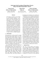
To gain a deeper insight into the structural basis of
thermal stability, a molecular model of the mutant was
constructed (Fig. 4). Analysis of the structure showed
that Asp133 lies at the external surface of Erl-ASNase,
in particular at a loop region formed by residues
Thr129–Lys134 between a4 and b4 (Fig. 4A). It forms
a salt bridge and two hydrogen bonds with Arg169.
The first hydrogen bond is formed between the OD1
atom of Asp133 (2.87 A
˚
) and the side chain NH
2
of
Arg169, whereas the OD2 atom forms a weak H-bond
(3.47 A
˚
) with the side chain NH
2
of Arg169 (Fig. 4B).
In the Asp133Val model, these two hydrogen bonds
are lost and the side chain of Val133 no longer inter-
acts with Arg169 (Fig. 4C).
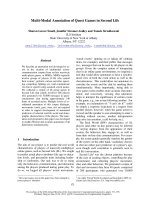
Fig. 2. Thermal inactivation curves. The residual activities of the
wild-type (h) and mutant Asp133Val (
) enzymes were measured
after heat treatment at various temperatures (30–70 °C) for
7.5 min.
Fig. 1. SDS ⁄ PAGE of Asp133Val mutant enzyme purification. Pro-
tein bands were stained with Coomassie Brilliant Blue R-250. Lane
A, molecular mass markers; lane B, E. coli BL21 (DE3)pLysS crude
extract after induction with 1 m
M isopropyl thio-b-D-galactoside;
lane C, unbound fraction from DEAE-Sepharose CL6B, pH 7.5;
lane D, eluted Asp133Val mutant enzyme from the Ni-NTA affinity
adsorbent.
Fig. 3. Kinetics of thermal inactivation of the wild-type and
Asp133Val mutant enzyme. The residual activities of the wild-type
(
) and mutant Asp133Val (¤) were measured at various times
after incubation at 50 °C. The results are presented as plot of
log(% remaining activity) versus time (h).
Engineering thermal stability of
L-asparaginase G. A. Kotzia and N. E. Labrou
1752 FEBS Journal 276 (2009) 1750–1761 ª 2009 The Authors Journal compilation ª 2009 FEBS
Thus, any attempt to understand the greater thermal
stability of the Asp133Val mutant enzyme in terms of
intramolecular interactions (e.g. H-bonding, ionic
bonds) in the structure was unsuccessful. Instead, the
explanation appears to lie in the much greater entropy
of activation in the case of the wild-type enzyme. In
principle, the entropy of inactivation consists of two
parts: the increased configurational entropy due to
partial unfolding of the protein in the transition state
and the decrease in entropy of the solvent due to expo-
sure of hydrophobic side chains. The latter effect is
likely to be small at elevated temperatures [30] and the
major contribution must then be the increase in config-
urational entropy. Thus, the greater rate of thermal
inactivation of the wild-type enzyme than of the
mutant is probably mainly due to greater flexibility or
disordering of the transition state with some contribu-
tion from a lower degree of exposure of hydrophobic
residues. To assess whether Asp133 contributes to
structural flexibility, we analysed the plots of the crys-
tallographic B-factors along the polypeptide chain of
the enzyme structure. This plot can give an indica-
tion of the relative flexibility of portions of the
protein [15,31]. As shown in Fig. 5, the structure dis-
plays a well-defined flexibility pattern. Several highly
mobile regions throughout the entire sequence can be
identified, and these are separated by a number of
segments with low mobility. Asp133 displays high
Fig. 4. Structural representations of the
wild-type and Asp133Val mutant enzyme.
(A) Diagram of the modelled Asp133Val
mutant enzyme subunit with succinamic
acid bound to the active site. The bound
ligand and the mutated residue (Val133) are
shown in a stick representation and are
labelled. (B) Structural representation of the
mutation site 133 of the wild-type ErL
-ASN-
ase. (C) Structural representation of the
mutation site 133 of the Asp133Val mutant.
Hydrogen bonds are shown as dashed lines
and residues are labelled. The model of the
mutated enzyme was constructed using
WHAT IF [51]. (D) A closer view of the electro-
static potential of the mutation site 133.
Negative, positive and neutral values of
electrostatic potential are indicated by
shades of red, blue and white colour,
respectively. (E) Superposition of the Pois-
son–Boltzmann electrostatic potential of the
mutant and the wild-type enzymes. Asp113
is shown as a spacefill representation (col-
oured black) and labelled. The colour code
utilized to represent the electrostatic poten-
tial along with the potential range is: red:
)1.8; white: 0; blue: 1.8. All figures were
created using
PYMOL [54], except (E) which
was created using
SWISS-PDB VIEWER [53].
G. A. Kotzia and N. E. Labrou Engineering thermal stability of
L-asparaginase
FEBS Journal 276 (2009) 1750–1761 ª 2009 The Authors Journal compilation ª 2009 FEBS 1753
crystallographic B-factors located at a region with high
mobility, indicating that this residue undergoes large
fluctuations. This may cause local disordering with
increased flexibility, which may contribute to the
increase in configurational entropy.
Erl-ASNase is composed of four identical subunits,
and the active enzyme is always a tetramer. To deter-
mine whether the higher thermal stability observed for
the Asp133Val mutant enzyme is due to higher stabil-
ity of the quaternary or tertiary structure, subunit-
dissociation experiments were carried out. Shifrin et al.
[32] showed that guanidinium chloride dissociates the
enzyme tetramer in 50 mm phosphate buffer at pH 7.5.
The dissociation is accompanied by the appearance of
an ultraviolet difference spectrum with a maximum at
288 nm. The band at 288 nm in the difference spec-
trum has been ascribed to tyrosyl residues [32]. In this
study, the rate of subunit dissociation in the wild-type
and Asp133Val mutant enzymes was monitored by
following the rate of appearance of the 288 nm band
in the ultraviolet difference spectrum. As illustrated in
Fig. 6, the rate of dissociation of the tetramer was
found to be approximately equal for both enzymes
(first-order rate constants: 0.011 and 0.013 min
)1
for
the wild-type and Asp133Val mutant enzymes, respec-
tively), indicating that the stabilizing effect of the
mutation is not due to quaternary structure stabiliza-
tion. Instead, it is the result of stabilization of the
tertiary structure.
Taking into account the unusual chromatographic
behaviour of the mutant enzyme on ion exchangers,
and because in silico structural analysis of the interac-
tions in the microenvironment of Val133 in the mutant
enzyme did not provide adequate explanations for its
higher thermostability, electrostatic potential analysis
was performed. Analysis of the wild-type enzyme
showed that Asp133 is located at an uncharged (neu-
tral) region of the enzyme (Fig. 4D). Replacement of a
surface charge (Asp) with a hydrophobic residue (Val)
would not normally be expected to increase the stabil-
ity of the protein, because for a hydrophobic side
chain it is more favourable to be buried within the
core of the protein than to be exposed to solvent. It
has been suggested, however, that in some cases there
can be residues located on the surface of a protein that
provide an unfavourable electrostatic contribution to
the overall stability of the domain, due to the asymme-
try of the electrostatic potential mapped on the surface
of the protein [33]. In our case, because Asp133 lies in
a neutral environment, its charge is unfavourable and
it may therefore destabilize the overall structure. Com-
parison of the Poisson–Boltzmann electrostatic poten-
tial of the mutant and the wild-type (Fig. 4E) showed
that greater symmetric positive potential was observed
in the mutant enzyme than in the wild-type enzyme.
This may cause less structural perturbations in the
mutant enzyme.
Effects of the Asp133Val mutation on kinetic
parameters
The mutant enzyme was subjected to steady-state
kinetic analysis using three different substrates: l-Asn,
l-Gln and N
a
-acetyl-Asn (Table 1). The results showed
that the k
cat
and K
m
values for l-Asn were increased
by 1.6- and 2.9-fold, respectively, compared with the
wild-type enzyme. By contrast, the K
m
values for the
Fig. 5. The dynamics of ASNase. A plot of the crystallographic
B-factors along the polypeptide chain obtained from the crystal
structure of E. chrysanthemi ASNase, (PDB code 1O7J). The plot
was produced using
WHAT IF software package [51]. The height at
each residue position indicates the average B-factor of all atoms in
the residue. B-factors are available in the PDB file 1O7J.
Fig. 6. Guanidinium chloride induced subunit dissociation. Rate of
appearance of the 288 nm band of the wild-type (s) and mutant
Asp133Val (d), as a function of time in 0.05
M phosphate buffer,
pH 7.5, containing 3
M guanidine chloride. Changes in absorbance
at 288 nm were monitored for 5 min.
Engineering thermal stability of
L-asparaginase G. A. Kotzia and N. E. Labrou
1754 FEBS Journal 276 (2009) 1750–1761 ª 2009 The Authors Journal compilation ª 2009 FEBS
l-Gln and N
a
-acetyl-l-Asn were reduced and k
cat
values were increased (Table 1). The increased K
m
value for l-Asn, although undesirable, is still within
the range of acceptability for therapeutic applications
[35].
The results of the kinetic analysis indicate that
although the site of the mutation is distant from the
active site, it makes a significant contribution to
catalysis (k
cat
and K
m
), suggesting that long-range
effects play an important role. Considering the inter-
action of the enzyme with the substrate, it is reason-
able to propose that there may be a long-range effect
in the active site. In particular, the mutation at posi-
tion 133 may destabilize the crucial hydrogen bond
formed between the side chain of the substrate and
the main chain oxygen of Ala139. This interaction
has been shown to be responsible for the higher
mobility of the active-site loop and of the flexible cat-
alytic region 120–140, and it is believed to contribute
to substrate specificity and to the rate-limiting step
[15,16,36]. These structural observations are consistent
with the results of the kinetic analysis (Table 1) and
presumably explain the effect of the mutation on
kinetic constants. Similar long-range effects have been
found in the serine protease subtilisin BPN’ [37]. For
example, charged residues on the surface of the
enzyme some 13–15 A
˚
from the active site have been
found to modulate enzyme–substrate complex forma-
tion and catalysis. Also, long-range interactions have
been found in the case of aminoacyl-tRNA synthetase
and 4-chlorobenzoyl-CoA dehalogenase catalysis
[38,39].
Site-saturation mutagenesis at position 133
In vitro site-directed evolution (saturation mutagenesis)
can be used to advantage during protein engineering
to explore additional evolution pathways and enable
rapid diversification in protein traits [40]. This method
makes possible the creation of a library of mutants
containing all possible mutations at one or more pre-
determined target positions, in order to determine the
best-fit residue at that position. It has been used suc-
cessfully for rapid improvement of various protein
functions [40,41].
In this study, site-saturation mutagenesis at posi-
tion 133 was used to investigate in greater depth the
contribution of this position to the thermostability. A
library of enzyme variants was created by overlap
extension PCR using two degenerate synthetic oligo-
nucleotides in which the mutation site (position 133)
was diversified using a randomized NNN codon. The
library was subsequently screened for clones with
improved stability at 60 °C for 5 min. Under these
conditions, the mutant Asp133Val shows 21.3% resid-
ual activity. Four clones that showed ‡ 45% residual
activity (compared to the Asp133Val mutant) were
selected and sequenced. Three clones were found to
have single point mutations at position 133:
Asp133Leu, Asp133Ile and Asp133Thr. In addition,
Table 1. Kinetic parameters of the wild-type and mutant enzymes. Steady-state kinetic measurements were performed at 37 °C in 0.1 M
Tris ⁄ HCl, pH 8 (or pH 8.2 for L-Gln). All initial velocities were determined in triplicate. The kinetic parameters k
cat
and K
m
were calculated by
nonlinear regression analysis of experimental steady-state data using the
GRAFIT (Erythacus Software Ltd, Staines, UK) program [34].
Enzymes Substrates K
m
(mM) k
cat
(s
)1
)(· 10
3
)
k
cat
ÆK
m
)1
(mM
)1
Æs
)1
)
(· 10
3
)
Wild-type
a
L-Asn 0.058 ± 0.013 23.8 ± 1.1 411.8
L-Gln 6.7 ± 1.1 4.3 ± 0.5 0.6
N
a
-Acetyl-L-Asn 0.80 ± 0.09 10.8 ± 0.2 13.4
L-Asn 0.153 ± 0.021 37.94 ± 1.83 247.7
Asp133Val
L-Gln 1.999 ± 0.236 16.03 ± 0.69 8.018
N
a
-Acetyl-L-Asn 0.597 ± 0.096 22.71 ± 0.66 38.04
L-Asn 0.160 ± 0.051 4.708 ± 0.64 29.425
Asp133Leu
L-Gln 1.677 ± 0.278 0.815 ± 0.05 0.486
N
a
-Acetyl-L-Asn 1.788 ± 0.322 3.103 ± 0.21 1.735
L-Asn 0.097 ± 0.022 2.467 ± 0.19 25.433
Asp133Ile
L-Gln 1.099 ± 0.094 0.677 ± 0.02 0.616
N
a
-Acetyl-L-Asn 5.613 ± 1.426 2.576 ± 0.35 0.459
L-Asn 0.038 ± 0.004 1.686 ± 0.03 44.368
Asp133Thr
L-Gln 1.008 ± 0.061 0.646 ± 0.01 0.641
N
a
-Acetyl-L-Asn 2.096 ± 0.293 2.139 ± 0.12 1.021
a
Data for the wild-type enzyme were from Labrou & Kotzia [16] and are included for comparison.
G. A. Kotzia and N. E. Labrou Engineering thermal stability of
L-asparaginase
FEBS Journal 276 (2009) 1750–1761 ª 2009 The Authors Journal compilation ª 2009 FEBS 1755
one clone had a double mutation: Asp133Leu ⁄
Asp103Thr. The spontaneous Ala103Thr mutation
(codon GCG fi ACG) was due to random error
introduced by Pfu DNA polymerase. The four most
thermostable clones have an uncharged substitution at
position 133 (Leu, Ile, Thr). This further supports the
finding that position 133 contributes significantly to
the electrostatic potential of the enzyme, and it pro-
vides evidence for the necessity of uncharged ⁄ hydro-
phobic residue at position 133 for attainment high
thermostability.
The selected saturation variants were purified using
the same procedure that was used for the Asp133Val
mutant. Subsequently, their thermal stabilities were
evaluated using heat-inactivation studies at 57.5 °C
(Fig. 7) and the results are listed in Table 2. All
mutants showed reduced k
in
values (from 1.75- to
3.2-fold) compared with the Asp133Val mutant, indi-
cating further improvement in their thermal stability.
The Asp133Leu mutant appeared to be the most
thermostable, with a k
in
of 0.040 min
)1
. The double
mutant Asp133Leu ⁄ Ala103Thr showed a k
in
compa-
rable with that of the single point mutant
Asp133Leu.
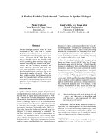
Structural analysis of 3D models of the mutant
enzymes showed the formation of additional non-
covalent interactions in the mutated enzymes
(Fig. 8). In particular, the side chain hydroxyl group
of the Asp133Thr mutant is involved in an H-bond
with the side chain of Arg169, similarly to the wild-
type Erl-ASNase (Fig. 4C). By contrast, the
Asp133Leu and Asp133Ile mutants appear to form
additional van der Waals interactions. The side chain
of Leu133 interacts (van der Waals contacts) with
the side chain of Asn226, and Ile133 interacts with
Arg169 and Gly170. All residues are parts of loops;
therefore, the additional stabilizing effects of the
mutations, compared with the Asp133Val enzyme,
may be due either to restriction of the conforma-
tional freedom of the protein or to the energetic
contribution of the newly formed H-bond and van
der Waals contacts.
Variants bearing a single mutation (Asp133Leu,
Asp133Ile, Asp133Thr) were subjected to kinetic
analysis using l-Asn, l-Gln and N
a
-acetyl-l-Asn as
substrates. The results showed little to moderate effect
of the Leu, Ile and Thr substitutions on the K
m
values
compared with the Asp133Val mutant (Table 1). It is
interesting to note that for the Asp133Thr mutant, the
K
m
value for l-Asn was reduced fourfold compared
with the Asp133Val mutant and by 1.5-fold compared
with the wild-type Erl-ASNase. By contrast, the k
cat
values were significantly reduced although the
enhanced specificity (k
cat
ÆK
m
)1
) towards l-Asn was
maintained in all mutants.
Conclusions
The results of this study provide new data on the
structural basis of the thermal stability of E. chry-
santhemi l-ASNase, and provide a basis for the design
of new, improved forms of the enzyme for future ther-
apeutic use. It has been shown that the hydrophobic
effect, hydrogen bonding and packing interactions
between residues in the interior of the protein are
dominant factors that define protein stability. The role
of surface residues in protein stability has received
much less attention. The stability of the Asp113Val
mutant enzyme is a particularly interesting rare case
in which replacement of a surface charge with a
hydrophobic residue leads to an increase in the stabil-
ity of the protein. Whereas conventional chemical
intuition would expect that salt bridges should
contribute favourably to protein stability, recent
Fig. 7. Kinetics of thermal inactivation of the mutant Asp133Val
and its saturation variants. The residual activities of the mutant
Asp133Val (e) and of the saturation variants were measured at var-
ious times after incubation at 57.5 °C. The results are presented as
plot of log(% remaining activity) versus time (min). Asp133Leu (
),
Asp133Ile (d) and Asp133Thr (D).
Table 2. First-order inactivation rate constants (k
in
, min
)1
) of the
mutant enzymes at 57.5 °C.
Enzymes Rate constants k
in
(min
)1
)
Asp133Val 0.123 ± 0.0053
Asp133Leu 0.043 ± 0.0004
Asp133Ile 0.071 ± 0.0015
Asp133Thr 0.055 ± 0.0007
Engineering thermal stability of
L-asparaginase G. A. Kotzia and N. E. Labrou
1756 FEBS Journal 276 (2009) 1750–1761 ª 2009 The Authors Journal compilation ª 2009 FEBS
computational and experimental evidence has shown
that salt bridges can be either stabilizing or destabiliz-
ing [41]. For example, alleviation of unfavourable sur-
face charge can increase the stability of proteins [42].
Many proteins contain clusters of positively or nega-
tively charged residues, and optimization of the
surface electrostatic potential may enhance protein
stability. For example, in recent studies of ribonucle-
ase T1 and ubiquitin, it was shown that relieving
surface charge through mutation increased protein
stability [42–44].
During the development of a therapeutic protein, it
is important to improve its long-term stability. Under-
standing the stability parameters and the factors that
affect them are critical steps. The thermostable enzyme
variants reported in here may be tested further on ani-
mals and ⁄ or humans in order to create a new drug for
future therapeutic use.
Experimental procedures
l-Asn and l-Gln were obtained from Serva (Heidelberg,
Germany). a-Ketoglutaric acid and Sepharose CL6B from
Sigma (St Louis, MO, USA). N
a
-Acetyl-l-Asn was
obtained from Sigma-Aldrich, (Milwaukee, WI, USA).
NADH (disodium salt, grade II, $ 98%) and crystalline
bovine serum albumin (fraction V) were purchased from
Boehringer Mannheim (Mannheim, Germany). Nessler’s
reagent and glutamate dehydrogenase were obtained from
Fluka (Taufkirchen, Germany). All primers were synthe-
sized and purified by MWG-biotech AG (Ebersberg,
Germany). TOPO cloning kit and all other molecular
biology reagents were from Invitrogen (Carlsbad, CA,
USA).
Directed evolution of L-ASNase
Directed evolution of l-ASNase was carried out using the
StEP [45]. Plasmids (pCR
Ò
T7 ⁄ CT-TOPO
Ò
) containing the
nucleotide sequences of l-ASNase from E. carotovora
(Ecal-ASNase; NCBI accession number: AY560097, the
enzyme was cloned as a non-tagged protein) [15] and
E. chrysanthemi 3937 (ErL-ASNase; NCBI accession num-
ber AY560098, the enzyme was cloned as a non-tagged pro-
tein) [16] were used as parental sequences in the PCR.
The forward primers used in the reaction were the
5¢-ATGGAACGATGGTTTAAATCTCTG-3¢ and 5¢-ATG
TTTAACGCATTATTCGTTGTTGTTTTTG-3¢, and the
reverse primers were the 5¢ -TCAATAGGTGTGGAAATA
GTCCTGG-3¢ and 5¢-TTAAGCTTTTAATAAGCGTGG
AAGTAATCC-3¢. The PCR was carried out in a total vol-
ume of 50 lL containing 25 ng of each primer, 10 ng of
template plasmid DNA, 0.2 mm of each dNTP, 5 lLof
10· Taq buffer, 1.5 mm MgCl
2
buffer and 2.5 units of Taq
DNA polymerase (Stratagene, La Jolla, CA, USA). The
PCR procedure comprised 99 cycles of 30 s at 94 °C and
10 s at 46 °C.
Fig. 8. Structural representations of the mutant enzymes. A closer
view of the mutation site of Asp133Thr, (A); Asp133Ile, (B); and
Asp133Leu, (C). Hydrogen bonds are shown as dashed lines and
residues are labelled. The models of the mutated enzymes were
constructed using
WHAT IF [51]. The figure was created using PYMOL
[54].
G. A. Kotzia and N. E. Labrou Engineering thermal stability of
L-asparaginase
FEBS Journal 276 (2009) 1750–1761 ª 2009 The Authors Journal compilation ª 2009 FEBS 1757
Cloning, expression and screening for
thermostable mutants
Following completion of StEP, the resulting PCR amplicon
was treated with the restriction enzyme DpnI to eliminate
parental plasmid DNA and was TOPO ligated to
pCR
Ò
T7 ⁄ CT-TOPO
Ò
expression vector. The presence of
the stop codon in the 5¢-end of the reverse primers allowed
the expression of non-tagged enzyme variants. The resulting
expression constructs pT7Mut-ASNase were used to trans-
form competent BL21(DE3)pLysS E. coli cells. E. coli cells,
harbouring plasmids pT7MutASNase, were grown at 37 °C
in 30 mL of Luria–Bertani medium containing 100 lgÆmL
)1
ampicillin and 34 lg Æ mL
)1
chloramphenicol. The synthesis
of l-ASNases was induced by the addition of 1 m m isopro-
pyl thio-b-d-galactoside when the absorbance at 600 nm
was 0.6–0.8. Five hours after induction, cells were harvested
by centrifugation at 4000 g and 4 °C for 20 min. In order
to find thermotolerant mutants, cell-free extracts were incu-
bated at 55 °C for 7.5 min and were then used to measure
the residual activities using the coupled enzyme assay
method described below.
Saturation mutagenesis, library creation and
screening
Saturation mutagenesis at amino acid position 133 was per-
formed by overlap extension using PCR [46]. Mutations
were introduced using a set of degenerate synthetic oligonu-
cleotides, in which the mutation site was diversified using a
randomized NNN codon. The pairs of oligonucleotide
primers used in the PCR for the saturation mutagenesis
were as follows: the first pair 5¢-ATGGAACGATG
GTTTAAATCTCTG-3¢ (P
1
) and 5¢-GTGAAAAGCNNN
AAGCCGGTAGTG-3¢ (P
2
), and the second pair 5¢-CACT
ACCGGCTTNNNGCTTTTCAC-3¢ (P
3
) and 5¢-TCAAT
AGGTGTGGAAATAGTCCTGG-3¢ (P
4
). Sites of muta-
tion are indicated in italics. The expression construct encod-
ing the wild-type ErL-ASNase [16] was used as template
DNA. After completion of the PCR (using primers P1, P2
and P3, P4), the PCR products were digested with DpnIto
eliminate parental DNA, and were then used in another
PCR as templates using P
1
and P
4
primers to amplify the
entire mutated gene. The latter was TOPO ligated into a T7
expression vector (pEXP5-CT ⁄ TOPO
Ò
) and recombinant
plasmids were isolated and were used to transform compe-
tent BL21(DE3)pLysS E. coli cells. E. coli cells were grown
at 37 °C in 30 mL of Luria–Bertani medium containing
100 lgÆmL
)1
ampicillin and 34 lgÆmL
)1
chloramphenicol.
The synthesis of the mutated enzymes was induced by the
addition of 1 mm isopropyl thio-b-d-galactoside when the
absorbance at 600 nm was 0.6–0.8. Four hours after induc-
tion, cells were harvested by centrifugation at 4000 g and
4 °C for 20 min. Hundreds of transformants of the library
were examined for their thermotolerance at 60 °C for
5 min, in order to identify promising mutants showing
increased thermotolerance compared with the Asp133Val.
The thermotolerance was estimated by measuring the
residual activities using the coupled enzyme assay
method described below. For the variants showing ‡ 45%
residual activity, the mutations were determined by DNA
sequencing.
Purification of the wild-type and mutant enzymes
Purification of the wild-type enzymes was carried out
according to published methods [15,16]. The purification of
mutants was accomplished by a two-step procedure,
comprising a negative purification step using a DEAE–
Sepharose CL6B, followed by an immobilized metal che-
late-affinity chromatography on a Ni-NTA column. This
was carried out as follows: cell paste was suspended in
potassium phosphate buffer (5 mm, pH 7.5), sonicated and
centrifuged at 10 000 g for 5 min. The supernatant was col-
lected and applied to DEAE–Sepharose CL6B (1 mL,
0.5 · 2 cm i.d.) column, previously equilibrated with potas-
sium phosphate buffer (5 mm, pH 7.5). Nonadsorbed pro-
tein was washed off with 3 mL equilibration buffer. The
flow-through and the first fraction (1 mL) of the washing
step were mixed, adjusted to pH 8 (with the addition of
0.1 m potassium phosphate buffer, pH 8) and 0.1 m NaCl
(by the addition of 5 m NaCl) before being applied to
Ni-NTA (0.5 mL, 0.5 · 1 cm i.d.) column. The column was
previously equilibrated with potassium phosphate buffer
(50 mm, pH 8) containing 0.3 m NaCl. Nonadsorbed
protein was washed off with 8 mL equilibration buffer,
followed by 8 mL potassium phosphate buffer (50 mm,
pH 7.5) containing 0.3 m NaCl, and 8 mL potassium phos-
phate buffer (50 mm, pH 7) containing 0.3 m NaCl. Bound
l-ASNase was eluted with potassium phosphate buffer
(50 mm, pH 6.2) containing 0.3 m NaCl. Collected fractions
(2 mL) were assayed for l-asparaginase activity and pro-
tein. Following purification, the wild-type enzyme as well
as the mutant enzymes were dialysed against 1000 vol. of
0.1 m Tris ⁄ HCl pH 8.0 (for kinetic analysis; see below) or
against 10 mm KH
2
PO
4
buffer pH 7 (for thermal inactiva-
tion studies; see below).
Assay of enzyme activity and protein
Enzyme assays were performed at 37 °C at a Hitachi
U-2000 double beam UV ⁄ Vis spectrophotometer carrying
a thermostated cell holder (10 mm path length). Activities
were measured by determining the rate of ammonia forma-
tion, by coupling with glutamate dehydrogenase, according
to Balcao et al. [47]. The final assay volume of 1 mL con-
tained 71 mm Tris ⁄ HCl buffer, pH 8.0, 1 mm Asn, 0.15 mm
a-ketoglutaric acid, 0.15 mm NADH, 4 units glutamate
dehydrogenase and sample containing l-ASNase activity.
Alternatively, the rate of ammonia formation was measured
Engineering thermal stability of L-asparaginase G. A. Kotzia and N. E. Labrou
1758 FEBS Journal 276 (2009) 1750–1761 ª 2009 The Authors Journal compilation ª 2009 FEBS
at 37 °C using the Nessler’s reagent [48]. One unit of
l-ASNase activity is defined as the amount of enzyme that
liberates 1 lmol of ammonia from l-Asn per min at 37 °C.
Protein concentrations were determined at 25 °C using
the method of Bradford [49] using BSA (fraction V) as
standard.
Kinetic analysis
Steady-state kinetic measurements were performed as
described previously [15,16,50]. The kinetic parameters k
cat
and K
m
were calculated by nonlinear regression analysis of
experimental steady-state data. Turnover numbers were cal-
culated on the basis of one active site per subunit. Kinetic
data were analysed using the computer program grafit
(Erythacus Software Ltd, Staines, UK) [34].
Molecular modelling and computational analysis
Molecular modelling for creating the model of E. chrysant-
hemi l-ASNase was carried out as described by Kotzia &
Labrou [16]. The molecular modelling program what if
[51] was used to predict the conformation of the mutant
enzymes. Prediction of the conformation of the new side
chains was performed as described by Chinea et al. [52]
Poisson–Boltzmann electrostatic potential analysis of the
mutant and the wild-type enzymes was carried out using
swiss-pdb viewer [53]. The parameters utilized to calculate
the electrostatic potential were: dielectric constant (protein)
4, dielectric constant (solvent) 80, solvent ionic strength
0.00, partial charged ‘on’. Nonbonded interactions were
analysed by moltalk (). The program
pymol was used for inspection of models and crystal struc-
tures [54].
Thermal stability of the wild-type, Asp133Val and
saturation variants
Thermal inactivation of the wild-type and Asp133Val and
saturation variants was monitored by activity measure-
ments. Samples of the enzymes, in 10 mm KH
2
PO
4
buffer
pH 7, were incubated at a range of temperatures from 30
to 70 °C for 7.5 min. Subsequently, the samples were
assayed for residual activity, using the coupled enzyme
assay method described above. The T
m
values were deter-
mined from the plots of relative inactivation (%) versus
temperature (°C). The T
m
value is the temperature at
which 50% of the initial enzyme activity is lost after heat
treatment. The kinetics of thermal inactivation of the
wild-type and Asp133Val was monitored at 50 °C. The
kinetics of thermal inactivation of saturation variants was
monitored at 57.5 °C. The rates of inactivation were
followed by periodically removing samples for assay of
enzymatic activity. Observed rates of inactivation (k
in
)
were deduced from plots of log (% remaining activity)
versus time.
Guanidinium chloride induced subunit
dissociation
Guanidinium chloride induced dissociation of the wild-type
and mutant Asp133Val enzymes was carried out according
to Shifrin et al. [32] in 50 mm KH
2
PO
4
pH 7.5. Guanidinium
chloride treatments were performed in the presence of 3 m
guanidinium chloride and measurements were taken for
5 min. The rate of dissociation was determined by the
increase of the absorbance at 288 nm as described by Shifrin
et al. [32].
Electrophoresis
SDS ⁄ PAGE was performed according to the method of
Laemmli [55] on a slab gel containing 12.5% (w ⁄ v) poly-
acrylamide (running gel) and 2.5% (w ⁄ v) stacking gel. The
protein bands were stained with Coomassie Brilliant Blue
R-250.
Acknowledgements
This work was financially supported by the Hellenic
General Secretariat for Research and Technology:
Operational Program for Competitiveness, Joint
Research and Technology Program.
References
1 Keating MJ, Holmes R, Lerner S & Ho DH (1993)
l-Asparaginase and PEG asparaginase – past, present,
and future. Leuk Lymphoma 10, 153–157.
2 Muller HJ & Boos J (1998) Use of l-asparaginase in
childhood ALL. Crit Rev Oncol Hematol 28, 97–113.
3 Goldberg DM (1992) Enzymes as agents for the treat-
ment of disease. Clin Chim Acta 206, 45–76.
4 Baran ET, Ozer N & Hasirci V (2002) In vivo half life
of nanoencapsulated l-asparaginase. J Mater Sci Mater
Med 13, 1113–1121.
5 Prager MD & Bachynsky N (1968) Asparagine synthe-
tase in normal and malignant tissues: correlation with
tumor sensitivity to asparaginase. Arch Biochem Biophys
127, 645–654.
6 Vieira Pinheiro JP, Wenner K, Escherich G, Lanvers-
Kaminsky C, Wurthwein G, Janka-Schaub G & Boos J
(2006) Serum asparaginase activities and asparagine
concentrations in the cerebrospinal fluid after a single
infusion of 2,500 IU ⁄ m(2) PEG asparaginase in children
with ALL treated according to protocol COALL-06-97.
Pediatr Blood Cancer 46, 18–25.
G. A. Kotzia and N. E. Labrou Engineering thermal stability of L-asparaginase
FEBS Journal 276 (2009) 1750–1761 ª 2009 The Authors Journal compilation ª 2009 FEBS 1759
7 Wriston JC Jr (1985) Asparaginase. Meth Enzymol 113,
608–618.
8 Mitchell L, Hoogendoorn H, Giles AR, Vegh P &
Andrew M (1994) Increased endogenous thrombin gen-
eration in children with acute lymphoblastic leukemia:
risk of thrombotic complications in l-asparaginase-
induced antithrombin III deficiency. Blood 83, 386–391.
9 Moola ZB, Scawen MD, Atkinson T & Nicholls DJ
(1994) Erwinia chrysanthemi l-asparaginase: epitope
mapping and production of antigenically modified
enzymes. Biochem J 302, 921–927.
10 Stecher AL, de Deus PM, Polikarpov I &
Abrahao-Neto J (1999) Stability of l-asparaginase: an
enzyme used in leukemia treatment. Pharm Acta Helv
74, 1–9.
11 Khushoo A, Pal Y, Singh BN & Mukherjee KJ (2004)
Extracellular expression and single step purification of
recombinant Escherichia coli l-asparaginase II. Protein
Expr Purif 38, 29–36.
12 Mashburn LT & Wriston JC Jr (1964) Tumor inhibi-
tory effect of l-asparaginase from Escherichia coli. Arch
Biochem Biophys 105, 450–452.
13 Marlborough DI, Miller DS & Cammack KA (1975)
Comparative study on conformational stability and sub-
unit interactions of two bacterial asparaginases. Biochim
Biophys Acta 386, 576–589.
14 Soares AL, Guimaraes GM, Polakiewicz B, de Moraes
Pitombo RN & Abrahao-Neto J (2002) Effects of poly-
ethylene glycol attachment on physicochemical and bio-
logical stability of E. coli l-asparaginase. Int J Pharm
237, 163–170.
15 Kotzia GA & Labrou NE (2005) Cloning, expression
and characterization of Erwinia carotovora l-asparagin-
ase. J Biotechnol 119, 309–323.
16 Kotzia GA & Labrou NE (2007) l-Asparaginase from
Erwinia chrysanthemi 3937: cloning, expression and
characterization. J Biotechnol 127, 657–669.
17 Boyse EA, Old LJ, Campbell HA & Mashburn LT
(1967) Suppression of murine leukemias by l-asparagin-
ase. Incidence of sensitivity among leukemias of various
types: comparative inhibitory activities of guinea pig
serum l-asparaginase and Escherichia coli l-asparagin-
ase. J Exp Med 125, 17–31.
18 Fernandes AI & Gregoriadis G (1997) Polysialylated
asparaginase: preparation, activity and pharmacokinet-
ics. Biochim Biophys Acta 1341, 26–34.
19 Burnham NL (1994) Polymers for delivering peptides
and proteins. Am J Hosp Pharm 51, 210–218.
20 Veronese FM & Pasut G (2005) PEGylation, successful
approach to drug delivery. Drug Discov Today 10,
1451–1458.
21 Kotzia GA, Lappa K & Labrou NE (2007) Tailoring
structure–function properties of l-asparaginase: engi-
neering resistance to trypsin cleavage. Biochem J 404,
337–343.
22 Bjork A, Dalhus B, Mantzilas D, Sirevag R & Eijsink
VGH (2004) Large improvement in the thermal stability
of a tetrameric malate dehydrogenase by single point
mutations at the dimer–dimer interface. J Mol Biol 341,
1215–1226.
23 Polizzi KM, Bommarius AS, Broering JM & Chaparro-
Riggers JF (2007) Stability of biocatalysts. Curr Opin
Chem Biol 11, 220–225.
24 Giver L, Gershenson A, Freskgard PO & Arnold FH
(1998) Directed evolution of a thermostable esterase.
Biochemistry 95, 12809–12813.
25 Oh KH, Nam SH & Kim HS (2002) Improvement of
oxidative and thermostability of N-carbamyl-d-amino
acid amidohydrolase by directed evolution. Protein Eng
15, 689–695.
26 Hao J & Berry A (2004) A thermostable variant of fruc-
tose bisphosphate aldolase constructed by directed evo-
lution also shows increased stability in organic solvents.
Protein Eng Des Sel 17, 689–697.
27 Eijsink VGH, Ga
˚
seidnes S, Borchert TV & Van den
Burg B (2005) Directed evolution of enzyme stability.
Biomol Engin 22, 21–30.
28 Bommarius AS, Broering JM, Chaparro-Riggers JF &
Polizzi KM (2006) High-throughput screening for
enhanced protein stability. Curr Opin Biotechnol 17 ,
606–610.
29 Freire E, van Osdol WW, Mayorga OL & Sanchez-Ruiz
JM (1990) Calorimetrically determined dynamics of
complex unfolding transitions in proteins. Annu Rev
Biophys Chem 19, 159–188.
30 Privalov PL & Gill SJ (1988) Stability of protein struc-
ture and hydrophobic interaction. Adv Protein Chem 39,
191–234.
31 Ricci G, Caccuri AM, Lo Bello M, Parker MW,
Nuccetelli M, Turella P, Stella L, Di Iorio EE &
Federici G (2003) Glutathione transferase P1-1: self-
preservation of an anti-cancer enzyme. Biochem J 376,
71–76.
32 Shifrin S, Solis BG & Chaiken IM (1973) l-Asparagin-
ase from Erwinia carotovora. Physicochemical properties
of the native and succinylated enzyme. J Biol Chem
248, 3464–3469.
33 Spector S, Wang M, Carp SA, Robblee J, Hendsch ZS,
Fairman R, Tidor B & Raleigh DP (2000) Rational
modification of protein stability by the mutation of
charged surface residues. Biochemistry 39, 872–879.
34 Leatherbarrow RJ (1998) GraFit, Version 3. Erythacus
Software Ltd, Staines.
35 Asselin BL, Lorenson MY, Whitin JC, Coppola DJ,
Kende AS, Blakley RL & Cohen HJ (1991) Measure-
ment of serum l-asparagine in the presence of l-aspara-
ginase requires the presence of an l-asparaginase
inhibitor. Cancer Res 51, 6568–6573.
36 Aghaiypour K, Wlodawer A & Lubkowski J (2001b)
Structural basis for the activity and substrate specificity
Engineering thermal stability of L-asparaginase G. A. Kotzia and N. E. Labrou
1760 FEBS Journal 276 (2009) 1750–1761 ª 2009 The Authors Journal compilation ª 2009 FEBS
of Erwinia chrysanthemi l-asparaginase. Biochemistry
40, 5655–5664.
37 Jackson SE & Fersht AR (1993) Contribution of long-
range electrostatic interactions to the stabilization of
the catalytic transition state of the serine protease sub-
tilisin BPN’. Biochemistry 32, 13909–13916.
38 Thompson D, Plateau P & Simonson T (2006) Free-
energy simulations and experiments reveal long-range
electrostatic interactions and substrate-assisted specific-
ity in an aminoacyl-tRNA synthetase. ChemBioChem 7,
337–344.
39 Wu J, Xu D, Lu X, Wang C, Guo H & Dunaway-
Mariano D (2006) Contributions of long-range electro-
static interactions to 4-chlorobenzoyl-CoA dehalogenase
catalysis: a combined theoretical and experimental
study. Biochemistry 45, 102–112.
40 Miyazaki K & Arnold FH (1999) Exploring nonnatural
evolutionary pathways by saturation mutagenesis: rapid
improvement of protein function. J Mol Evol 49, 716–
720.
41 Zheng L, Baumann U & Reymond JL (2004) An effi-
cient one-step site-directed and site-saturation mutagen-
esis protocol. Nucleic Acids Res 32, e115.
42 Kumar S & Nussinov R (2002) Close-range electrostatic
interactions in proteins. ChemBioChem 3, 604–617.
43 Grimsley GR, Shaw KL, Fee LR, Alston RW, Huygh-
ues-Despointes BMP, Thurlkill RL, Scholtz JM & Pace
CN (1999) Increasing protein stability by altering long-
range columbic interactions. Protein Sci 8, 1843–1849.
44 Loladze VV, Ibarra-Molero B, Sanchez-Ruiz JM &
Makhatadze GI (1999) Engineering a thermostable
protein via optimization of charge–charge interactions
on the protein surface. Biochemistry 38, 16419–16423.
45 Zhao H, Giver L, Shao Z, Affholter JA & Arnold FH
(1998) Molecular evolution by staggered extension
process (StEP) in vitro recombination. Nat Biotechnol
16, 258–261.
46 Ho SN, Hunt HD, Horton RM, Pullen JK & Pease
LR (1989) Site-directed mutagenesis by overlap exten-
sion using the polymerase chain reaction. Gene 77,
51–59.
47 Balcao VM, Mateo C, Fernandez-Lafuente R, Malcata
FX & Guisan JM (2001) Structural and functional sta-
bilization of l-asparaginase via multisubunit immobili-
zation onto highly activated supports. Biotechnol Prog
17, 537–542.
48 Wriston JC Jr & Yellin TO (1973) l-Asparaginase: a
review. Adv Enzymol Relat Areas Mo Biol 39, 185–
248.
49 Bradford MA (1976) A rapid and sensitive method for
the quantitation of microgram quantities of protein
utilizing the principle of protein–dye binding. Anal
Biochem 72, 248–254.
50 Kelo E, Noronkoski T, Stoineva IB, Petkov DD &
Mononen I (2002) Beta-aspartylpeptides as substrates
of l-asparaginases from Escherichia coli and Erwi-
nia chrysanthemi. FEBS Lett 528, 130–132.
51 Vriend G (1990)
what if: a molecular modeling and
drug design program. J Mol Graph 8, 52–56.
52 Chinea G, Padron G, Hooft RWW, Sander C & Vriend
G (1995) The use of position specific rotamers in model
building by homology. Proteins 23, 415–421.
53 Guex N & Peitsch MC (1997) SWISS-MODEL and the
Swiss-PdbViewer: An environment for comparative pro-
tein modeling. Electrophoresis 18 , 2714–2723.
54 DeLano WL (2002) The PyMOL Molecular Graphics
System. DeLano Scientific, San Carlos, CA.
55 Laemmli UK (1970) Cleavage of structural proteins
during the assembly of the head of bacteriophage T4.
Nature 227, 680–685.
G. A. Kotzia and N. E. Labrou Engineering thermal stability of L-asparaginase
FEBS Journal 276 (2009) 1750–1761 ª 2009 The Authors Journal compilation ª 2009 FEBS 1761