Báo cáo khoa học: Metabolic fate of L-lactaldehyde derived from an alternative L-rhamnose pathway ppt
Bạn đang xem bản rút gọn của tài liệu. Xem và tải ngay bản đầy đủ của tài liệu tại đây (394.51 KB, 11 trang )
Metabolic fate of L-lactaldehyde derived from an
alternative
L-rhamnose pathway
Seiya Watanabe
1,2,3
, Sommani Piyanart
1
and Keisuke Makino
1,2,3,4
1 Institute of Advanced Energy, Kyoto University, Japan
2 New Energy and Industrial Technology Development Organization, Kyoto, Japan
3 CREST, JST (Japan Science and Technology Agency), Japan
4 Innovative Collaboration Center, Kyoto University, Japan
l-Rhamnose (l-6-deoxymannose) is a constituent of
glycolipids and glycosides, such as plant pigments,
pectic polysaccharides, gums and biosurfactants, and
can be utilized as the sole carbon and energy source by
most bacteria, including Escherichia coli and Salmonella
typhimurium. In this pathway, l-rhamnose is converted
into dihydroxyacetone phosphate and l-lactaldehyde
via l-rhamnulose and l-rhamnulose l-phosphate by the
Keywords
Azotobacter vinelandii;
L-lactaldehyde
dehydrogenase;
L-rhamnose metabolism;
molecular evolution; Pichia stipitis
Correspondence
S. Watanabe, Institute of Advanced Energy,
Kyoto University, Gokasho, Uji, Kyoto
611-0011, Japan
Fax: +81 774 38 3524
Tel: +81 774 38 3596
E-mail:
(Received 8 July 2008, revised 9 August
2008, accepted 15 August 2008)
doi:10.1111/j.1742-4658.2008.06645.x
Fungal Pichia stipitis and bacterial Azotobacter vinelandii possess an alter-
native pathway of l-rhamnose metabolism, which is different from the
known bacterial pathway. In a previous study (Watanabe S, Saimura M
& Makino K (2008) Eukaryotic and bacterial gene clusters related to an
alternative pathway of non-phosphorylated l-rhamnose metabolism.
J Biol Chem 283, 20372–20382), we identified and characterized the gene
clusters encoding the four metabolic enzymes [l-rhamnose 1-dehydrogenase
(LRA1), l-rhamnono-c-lactonase (LRA2), l-rhamnonate dehydratase
(LRA3) and l-2-keto-3-deoxyrhamnonate aldolase (LRA4)]. In the known
and alternative l-rhamnose pathways, l-lactaldehyde is commonly pro-
duced from l-2-keto-3-deoxyrhamnonate and l-rhamnulose 1-phosphate by
each specific aldolase, respectively. To estimate the metabolic fate of l-lact-
aldehyde in fungi, we purified l-lactaldehyde dehydrogenase (LADH) from
P. stipitis cells l-rhamnose-grown to homogeneity, and identified the gene
encoding this enzyme (PsLADH) by matrix-assisted laser desorption ioniza-
tion-quadruple ion trap-time of flight mass spectrometry. In contrast,
LADH of A. vinelandii (AvLADH) was clustered with the LRA1–4 gene on
the genome. Physiological characterization using recombinant enzymes
revealed that, of the tested aldehyde substrates, l-lactaldehyde is the best
substrate for both PsLADH and AvLADH, and that PsLADH shows
broad substrate specificity and relaxed coenzyme specificity compared with
AvLADH. In the phylogenetic tree of the aldehyde dehydrogenase super-
family, PsLADH is poorly related to the known bacterial LADHs, includ-
ing that of Escherichia coli (EcLADH). However, despite its involvement in
different l-rhamnose metabolism, AvLADH belongs to the same subfamily
as EcLADH. This suggests that the substrate specificities for l-lactaldehyde
between fungal and bacterial LADHs have been acquired independently.
Abbreviations
ALDH, aldehyde dehydrogenase; AvLADH, Azotobacter vinelandii LADH; EcLADH, Escherichia coli LADH; GAPDH, glyceraldehyde 3-
phosphate dehydrogenase; LADH,
L-lactaldehyde dehydrogenase; LAR, L-lactaldehyde reductase; L-KDR, L-2-keto-3-deoxyrhamnonate; LRA1,
L-rhamnose 1-dehydrogenase; LRA2, L-rhamnono-c-lactonase; LRA3, L-rhamnonate dehydratase; LRA4, L-2-keto-3-deoxyrhamnonate aldolase;
MjLADH, Methanocaldococcus jannaschii LADH; PsLADH, Pichia stipitis LADH.
FEBS Journal 275 (2008) 5139–5149 ª 2008 The Authors Journal compilation ª 2008 FEBS 5139
sequential action of l-rhamnose isomerase (RhaA,
EC 5.3.1.14), rhamnulokinase (RhaB, EC 2.7.1.5) and
l-rhamnulose l-phosphate aldolase (RhaD, EC 4.1.2.19)
(Fig. 1A). Most fungi, including Saccharomyces cerevi-
siae, cannot grow on d-xylose, l-arabinose and
l-rhamnose as the sole carbon source [1]. However,
Pichia stipitis possesses the ability to metabolize these
sugars through alternative pathways different from
L-Rhamnose
L-Rhamnono-γ-lactone
L-Rhamnonate
L
-2-Keto-3-deoxyrhamnonate
(
L
-KDR)
L-Rhamnose
1-dehydrogenase
( LRA1, EC 1.1.1.173)
L-Rhamnono-γ-lactonase
( LRA2, EC 3.1.1.65)
L-Rhamnonate dehydratase
( LRA3, EC 4.2.1.90)
NAD(P)
+
NAD(P)H
H
2
O
H2O
Pyruvate
L-Lactaldehyde
L-KDR aldolase
( LRA4, EC 4.2.1 )
L
-Rhamnose
L-Rhamnulose
L-Rhamnulose 1-P
ATP
ADP
L-Rhamnose isomerase
(RhaA, EC 5.3.1.14)
L
-Rhamnulokinase
(RhaB, EC 2.7.1.5)
L-Rhamnulose 1-P aldolase
(RhaD, EC 4.1.2.19)
Dihydroxyacetone-P
RhaD RhaA RhaB RhaS RhaR RhaT
AAC76884 AAC76885 AAC76886 AAC76887 AAC76888 AAC76889
E. coli
P. stipitis
(L-Rhamnose:H
+
symporter)
EAM07803 EAM07804 EAM07805 EAM07806 EAM07807 EAM07808
EAM07809 EAM07810
(Sugar transporter)(Sugar channel)
A. vinelandii
ABN68602ABN68405ABN68404 ABN68603Chr 8 Chr 2 ABN64318
AAC74497
Methylglyoxal
NADPH
NADP
+
Glutathione
L-Lactaldehyde
S-Lactoyl glutathione
NAD(P)
+
NAD(P)H
Lactate
Glutathione
NAD
+
NADH
Pyruvate
Dihydroxyacetone-P
NADH NAD
+
1,2-Propanediol
P
NAD
+
NADH
L-Lactaldehyde dehydrogenase
( LADH, EC 1.2.1.22)
Lactaldehyde:propanediol
oxidoreductase
( EC 1.1.1.77(55))
FucO FucA FucP FucI FucK FucU
AAC75841 AAC75842 AAC75843 AAC75844 AAC75845 AAC75846
OH
H
H
HO
OH
H H
O
H
3
C
HO
H
H
HO
OH
H H
O
H
3
C
HO
H
O
CH
3
H
OH
H
OH
OH
H
OH
H
HOOC
CH
3
H
OH
H
OH
H
H
O
HOOC
3
CH
H
OH
OHC
CH
3
O
HOOC
L
-Rhamnose
D
-Xylose
L
-Arabinose
OH
H
H
HOH
2
C
H
OH
HO H
O
OH
H
HOH
2
C
H
H
OH
HO H
O
HOH
2
C
OH
H
H
OH
CH
2
OPO
3
2-
O
A
C
D
B
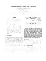
Fig. 1. (A) Known bacterial L-rhamnose pathway. (B) Novel non-phosphorylating L-rhamnose pathway. In addition to L-rhamnose, Pichia stipi-
tis (but not Saccharomyces cerevisiae) can metabolize
D-xylose and L-arabinose to yield a common phosphorylated end-product, xylulose
5-phosphate. (C) Schematic gene clusters related to
L-rhamnose metabolism. Chr 8 and Chr 2 in P. stipitis indicates chromosome number.
Homologous genes are indicated in the same colour. Fungal and bacterial LRA4 enzymes are not related evolutionally [3]. LADH enzymes of
P. stipitis and Azotobacter vinelandii (orange) were characterized in this study.
L-Fucose is converted to pyruvate and L-lactaldehyde through
the analogous pathway to
L-rhamnose, and metabolic genes, including FucO, are also clustered on the Escherichia coli genome. (D)
Metabolic network around
L-lactaldehyde. In this study, we focused on LADH (black line).
L-Lactaldehyde dehydrogenase S. Watanabe et al.
5140 FEBS Journal 275 (2008) 5139–5149 ª 2008 The Authors Journal compilation ª 2008 FEBS
the well-known bacterial pathways. Although both
d-xylose and l-arabinose are converted into a com-
mon end-product, xylulose 5-phosphate, as in the
bacterial pathway, it is believed that l-rhamnose is
metabolized via non-phosphorylated intermediates
(Fig. 1B) [2]. In this pathway, l-rhamnose is oxidized
to l-rhamnono-c-lactone by NAD(P)
+
-dependent
dehydrogenase. The lactone is cleaved by a lactonase
to l-rhamnonate, followed by a dehydration reaction
forming l-2-keto-3-deoxyrhamnonate (l-KDR). The
last step is the aldol cleavage of l-KDR to pyruvate
and l-lactaldehyde. We are in the process of enzy-
matically and genetically characterizing the alterna-
tive l-rhamnose pathway of P. stipitis, and recently
identified four metabolic enzymes: l-rhamnose
1-dehydrogenase (LRA1, EC 1.1.1.173), l-rhamnono-
c-lactonase (LRA2, EC 3.1.1.65), l-rhamnonate
dehydratase (LRA3, EC 4.2.1.90) and l-KDR aldol-
ase (LRA4) [3]. The LRA1–4 genes were clustered on
the P. stipitis genome (Fig. 1C), and the homologous
gene cluster was found on the genomes of many
fungi as well as several bacteria, including Azoto-
bacter vinelandii.
In the known and alternative l-rhamnose pathways,
the final reaction step is catalysed by each specific
aldolase to commonly yield l-lactaldehyde as one of the
products. There are two known enzymes for l-lact-
aldehyde in bacteria (Fig. 1D). The first is oxidation
by NAD
+
-dependent l-lactaldehyde dehydrogenase
(EC 1.2.1.22, LADH) to produce l-lactate [4–6]. In
E. coli, the enzyme is commonly responsible for both
l-rhamnose and l-fucose metabolism, and is also iden-
tical to the glycolaldehyde dehydrogenase (EC 1.2.1.21)
involved in ethylene glycol metabolism and glyoxylate
biosynthesis [4,5]. Under anaerobic conditions, l-lactal-
dehyde is reduced by NADH-dependent l-lactaldehyde
reductase (LAR, EC 1.1.1.77) and the l-1,2-propane-
diol obtained is excreted in the medium. In an E. coli
mutant that can grow on l-1,2-propanediol as a sole
carbon source, LAR also functions as l-1,2-propanediol
dehydrogenase, so-called ‘lactaldehyde : propanediol
oxidoreductase’ [7]. In contrast with bacteria, the
correct physiological role of l-lactaldehyde and related
enzymes in fungi has not yet been clarified. Chen et al.
[8] reported that the Gre2 (YOL151W) gene from
S. cerevisiae encodes a NADPH-dependent methyl-
glyoxal reductase (EC 1.1.1.283) catalysing the reduc-
tion of methylglyoxal to d- and ⁄ or l-lactaldehyde.
Furthermore, Inoue et al. [9] identified an aldehyde
dehydrogenase (ALDH) with specificity for l-lact-
aldehyde enzymatically but not genetically. However, it
is well known that a toxic methylglyoxal is neutralized
to lactate via lactoylglutathione (but not l-lactaldehyde)
by glyoxalase I (EC 4.4.1.5, YML004C) and gly-
oxalase II (EC 3.1.2.5, YDR272W).
In this regard, the alternative l-rhamnose pathway
is the significant physiological origin of l-lactaldehyde
in fungi. In this study, we first identified a fungal
LADH from P. stipitis. Furthermore, phylogenetic
comparison with the LADH of A. vinelandii revealed
that the same alternative l-rhamnose pathways
appeared by convergent evolution between fungi and
bacteria.
Results
Metabolic fate of L-lactaldehyde in P. stipitis
When compared with d-glucose medium, approxi-
mately 30-fold higher NAD
+
-dependent dehydroge-
nase activity for l-lactaldehyde was observed in the
cell-free extract from P. stipitis cells grown on l-rham-
nose as the sole carbon source (Fig. 2A). Similar
results were observed when d-lactaldehyde was used as
a substrate instead of l-lactaldehyde. In Zymogram
staining analysis, active bands of NAD
+
-dependent
dehydrogenases for l-lactaldehyde and d-lactaldehyde
appeared in the same position (Fig. 2B), and no active
GR GR GR GR
LD LD
P. stipitis A. vinelandii
Band A
Band B
NAD
AC
B
+
NADP
+
0.5
0.4
0.3
0.2
0.1
0
0.06
0.04
0.02
0
GRGR
GRGR
L D L D
Specific activity
(unit mg
–1
protein)
P. stipitis
A. vinelandii
PsLADH
PsALDH*
AvLADH
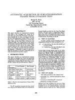
Fig. 2. Translational and transcriptional regulation of LADH. Pichi-
a stipitis and Azotobacter vinelandii cells were cultured in synthetic
medium containing
D-glucose (G) or L-rhamnose (R) (2%, w ⁄ v).
(A) NAD
+
- and NADP
+
-dependent dehydrogenase activity for L-lact-
aldehyde (L) or
D-lactaldehyde (D) in the cell-free extract. Values are
the means ± SD, n = 3. (B) Zymogram staining. Fifty micrograms
of the cell-free extract were applied to a 6% (w ⁄ v) non-denaturing
PAGE gel. After electrophoresis, the gel was soaked in staining
solution in the presence of 10 m
ML-orD-lactaldehyde and 10 mM
NAD
+
. (C) Transcriptional effect of carbon source on PsLADH,
PsALDH* and AvLADH genes. Total RNAs (4 lg per lane) were
isolated from microorganism cells grown on the indicated carbon
sources.
S. Watanabe et al.
L-Lactaldehyde dehydrogenase
FEBS Journal 275 (2008) 5139–5149 ª 2008 The Authors Journal compilation ª 2008 FEBS 5141
band was observed in the presence of NADP
+
(data
not shown), suggesting that the l-rhamnose-inducible
NAD
+
-dependent (or preferring) dehydrogenase for
l-lactaldehyde and d-lactaldehyde seems to derive
from the same enzyme, and that NADP
+
-dependent
activity may be derived from the concomitant activity
of other constitutively expressed ALDH(s). Under
anaerobic conditions, P. stipitis could metabolize
l-rhamnose (data not shown). These results indicate
that the metabolic fate of l-lactaldehyde derived from
the alternative l-rhamnose pathway in P. stipitis is
dehydrogenation by LADH.
Purification of LADH from P. stipitis (PsLADH)
PsLADH was purified from P. stipitis cells grown on
l-rhamnose as a sole carbon source in four chromato-
graphic steps (Fig. 3A). During the purification proce-
dure, the ratio of NAD
+
- to NADP
+
-linked activity
remained almost constant (2.2–3.0), suggesting the
presence of only one protein as LADH. The purified
enzyme exhibited a clear preference for NAD
+
over
NADP
+
, with NAD
+
- and NADP
+
-dependent spe-
cific activities of 6.85 and 2.26 unitsÆ(mg protein)
)1
,
respectively. SDS-PAGE revealed only one subunit
with an apparent M
r
value of 55 kDa. As it was
impossible to determine the N-terminal sequence
because of blocking, the peptide mass fingerprinting of
trypsin-digested fragments was alternatively performed
by MALDI-TOF MS, and LADH was identified as a
protein annotated as a putative ALDH of P. stipitis
CBS 6054 (ABN64318): 63% sequence coverage
(Table S1). This protein consisted of a polypeptide of
495 amino acids with a calculated M
r
of 53 488.85 Da,
comparable with that of the purified LADH deter-
mined by SDS-PAGE.
For the known dehydrogenases for l-lactaldehyde,
the reaction product of the enzymes from E. coli [4,5],
Methanocaldococcus jannaschii [10] and S. cerevisiae [9]
is l-lactate (EC 1.2.1.22), whereas that from rat liver is
methylglyoxal (EC 1.1.1.78) [11]. In HPLC analysis,
the retention time of the reaction product for
PsLADH (13.32 min) was almost the same as that
of l-lactate (13.35 min), but not methylglyoxal
(12.36 min); therefore, the enzyme catalyses the
NAD(P)
+
-linked oxidation of l-lactaldehyde into
l-lactate. The amino acid sequence of PsLADH was
most closely related to E. coli LADH (EcLADH) of the
ALDH-like proteins on the P. stipitis genome (34.5%
identity), whereas the protein annotated as a putative
mitochondrial ALDH (ABN68636) also showed similar
homology to EcLADH (32.2% identity), indicating the
possibility that the latter is an LADH isozyme (referred
to as PsALDH*); therefore, both enzymes were
expressed in E. coli cells (see below).
Candidate of LADH gene from A. vinelandii
As described in the Introduction, we have previously
identified the gene cluster related to the alternative
l-rhamnose pathway of A. vinelandii [3]. The LRA1–4
genes are clustered together with putative sugar trans-
porters and the ALDH gene (EAM07810) (Fig. 1C).
This ALDH showed highest sequential similarity to
EcLADH (61.7% identity) of all the putative ALDHs
in the A. vinelandii genome, indicating that the protein
may function as LADH (referred to as AvLADH).
Two active bands corresponding to NAD
+
-dependent
LADH were found in Zymogram staining analysis
using the cell-free extract prepared from A. vinelandii
cells grown on l-rhamnose: strict l-rhamnose-inducible
enzyme with l-lactaldehyde specificity (band A); mod-
erate l-rhamnose-inducible enzyme that utilizes both
d- and l-lactaldehyde (band B) (Fig. 2B). Subsequent
characterization revealed that ALDH with EAM07810
may correspond to band A, a major LADH in l-rham-
nose-grown cells (see below).
Functional expression of LADH in E. coli
PsLADH, PsALDH* and AvLADH genes were overex-
pressed in E. coli cells as a His6-tagged enzyme and
purified homogeneously with a nickel-chelating affinity
1 2 345M
AB
M1234
19.5 kDa
119 kDa
91 kDa
65 kDa
48 kDa
37 kDa
28 kDa
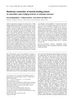
Fig. 3. (A) SDS-PAGE purification of native PsLADH in 10% (w ⁄ v) gel.
Lane 1, cell-free extracts (50 lg); lane 2, HiPrep 16 ⁄ 10 Q FF (50 lg);
lane 3, HiLoad 16 ⁄ 60 Superdex 200 pg (20 lg); lane 4, CHT
Ceramic Hydroxyapatite (20 lg); lane 5, Blue Sepharose Fast Flow
(10 lg). (B) SDS-PAGE of native and His6-tagged recombinant
enzymes. Lane 1, native PsLADH; lane 2, His6-tagged PsLADH;
lane 3, His6-tagged PsALDH*; lane 4, His6-tagged AvLADH. Ten
micrograms of the purified enzyme were applied. Bottom panel:
immunoblot analysis using anti-His6-tag IgG. One microgram of
the purified enzyme was applied.
L-Lactaldehyde dehydrogenase S. Watanabe et al.
5142 FEBS Journal 275 (2008) 5139–5149 ª 2008 The Authors Journal compilation ª 2008 FEBS
column (Fig. 3B). Western blot analysis with anti-
His6-tag IgG confirmed the His6 tag in the enzyme
(bottom panel in Fig. 3B).
Substrate specificity
Generally, ALDHs show relatively broad substrate
specificity in addition to the physiological substrate;
therefore, various aldehydes, including l-lactaldehyde,
were tested as substrates for dehydrogenation by
purified proteins in the presence of NAD
+
, and the
activity values for the tested aldehydes relative to
l-lactaldehyde are summarized in Table 1. l-Lactalde-
hyde was the best substrate for PsLADH, and the
specific activity [6.95 unitsÆ(mg protein)
)1
] was compa-
rable with that of native enzyme [6.85 unitsÆ(mg pro-
tein)
)1
]. Only five other aldehydes showed more than
50% activity relative to l-lactaldehyde. The significant
utilization of d-lactaldehyde conformed to the preli-
minary Zymogram staining analysis using the cell-free
extract (Fig. 2B). By contrast, PsALDH* utilized C2,
C3 and C4 aldehydes more efficiently than l-lactalde-
hyde, and most of the remaining aldehydes were also
good substrates at varying rates up to about one-half
the rate with l-lactaldehyde. Overall, the specificity for
l-lactaldehyde of PsLADH was significantly higher
than that of PsALDH*, conforming to the physiologi-
cal role as a LADH involved in the alternative l-rham-
nose pathway. Comparable dehydrogenase activity of
AvLADH with PsLADH was found only for l-lactal-
dehyde and glycolaldehyde, and activities with d-lactal-
dehyde and C7 aldehyde were only 10% less than
those with l-lactaldehyde: band A in Zymogram stain-
ing may correspond to AvLADH (Fig. 2B). These
results suggest that the enzyme should be assigned to
LADH, as expected from the sequential similarity to
EcLADH.
Kinetic analysis
EcLADH functions as a glycolaldehyde dehydro-
genase involved in ethylene glycol metabolism and
glyoxylate biosynthesis [4,5]. PsLADH, PsALDH*
Table 1. Substrate specificity of PsLADH, PsALDH* and AvLADH.
Substrate
a
Relative activity (%)
b
PsLADH PsALDH* AvLADH
L-Lactaldehyde 100 100 100
D-Lactaldehyde 75 54 8.6
Formaldehyde (C1) 13 13 0
Acetaldehyde (C2) 67 309 0
Propionaldehyde (C3) 81 350 0
Butylaldehyde (C4) 39 175 0
Valeraldehyde (C5) 38 105 0
Hexylaldehyde (C6) 41 82 0
Heptylaldehyde (C7) 28 70 7.4
Octylaldehyde (C8) 22 57 0
Isobutylaldehyde 82 54 0
Glutaraldehyde 47 251 0
Glycolaldehyde 74 70 91
Benzaldehyde 27 23 0
Betaine aldehyde 13 15 0
Glyceraldehyde 30 27 0
Glyceraldehyde 3-phosphate 12 11 0
a
The assay was performed with standard assay solution containing
10% (v ⁄ v) ethanol, 1 m
M aldehyde and 1.5 mM NAD
+
using purified
His6-tagged recombinant enzymes.
b
Relative values were
expressed as a percentage of the values obtained in
L-lactaldehyde.
Table 2. Kinetic parameters of PsLADH, PsALDH*, AvLADH and EcLADH.
Enzyme Substrate Coenzyme Specific activity [unitÆ(mg protein)
)1
]
a
K
m
(lM) k
cat
(min
)1
) k
cat
⁄ K
m
(min
)1
ÆlM
)1
)
PsLADH
L-Lactaldehyde
b
NAD
+
6.95 ± 0.10 42.8 ± 4.2 1390 ± 127 32.4 ± 0.2
NADP
+
1.65 ± 0.04 9.79 ± 0.74 195 ± 8 20.0 ± 0.3
D-Lactaldehyde
b
NAD
+
4.43 ± 0.05 52.9 ± 3.4 1460 ± 79 27.5 ± 0.3
Glycolaldehyde
c
NAD
+
8.94 ± 0.40 78.0 ± 1.6 469 ± 8 6.01 ± 0.03
PsALDH*
L-Lactaldehyde
b
NAD
+
3.64 ± 0.10 350 ± 62 651 ± 112 1.88 ± 0.01
NADP
+
0.355 ± 0.007 131 ± 9 15.4 ± 0.7 0.119 ± 0.001
D-Lactaldehyde
b
NAD
+
2.10 ± 0.04 32.6 ± 5.6 89.5 ± 11.9 2.76 ± 0.10
Glycolaldehyde
c
NAD
+
4.28 ± 0.30 287 ± 12 137 ± 5 0.478 ± 0.003
AvLADH
L-Lactaldehyde
b
NAD
+
17.2 ± 0.9 35.5 ± 0.8 554 ± 19 15.6 ± 0.2
D-Lactaldehyde
b
NAD
+
2.82 ± 0.04 167 ± 5 47.0 ± 1.5 0.281 ± 0.001
Glycolaldehyde
c
NAD
+
11.5 ± 0.4 274 ± 41 307 ± 44 1.12 ± 0.01
EcLADH
d
L-Lactaldehyde NAD
+
5.73 40 418 10.5
Glycolaldehyde NAD
+
14.7 380 993 2.61
a
Under standard assay conditions in Experimental procedures.
b
Eight different concentrations of aldehyde between 2 and 100 lM were
used.
c
Eight different concentrations of glycolaldehyde between 10 and 100 lM were used.
d
Calculation from data in [5].
S. Watanabe et al.
L-Lactaldehyde dehydrogenase
FEBS Journal 275 (2008) 5139–5149 ª 2008 The Authors Journal compilation ª 2008 FEBS 5143
and AvLADH also utilize glycolaldehyde efficiently as a
substrate (Table 1); therefore, these enzymes were sub-
jected to further kinetic analysis with l-lactaldehyde,
d-lactaldehyde and glycolaldehyde, and the parameters
determined are listed in Table 2. The catalytic efficiency
(k
cat
⁄ K
m
) with l-lactaldehyde of PsLADH in the pres-
ence of NAD
+
(32.4 min
)1
Ælm
)1
) was 17.2-fold higher
than that of PsALDH* (1.88 min
)1
Ælm
)1
), caused by
both higher K
m
and lower k
cat
values. However, by
contrast with l-lactaldehyde, the two fungal enzymes
possessed similar k
cat
⁄ K
m
values with d-lactaldehyde,
but their values with glycolaldehyde were significantly
lower, mainly caused by decreased k
cat
values. When
NADP
+
was used as a coenzyme, the k
cat
⁄ K
m
value
with l-lactaldehyde of PsALDH* decreased 15.8-fold
compared with that in the presence of NAD
+
because
of a decreased k
cat
value, whereas that of PsLADH
decreased only 1.6-fold. These results suggest that
PsLADH possesses a stricter substrate specificity for
l-lactaldehyde and a more relaxed coenzyme specificity
than does PsALDH*. Furthermore, the PsLADH gene
was significantly induced by l-rhamnose in P. stipitis
cells, but the PsALDH* gene was not (Fig. 2C). These
results strongly suggest the physiological function of
PsLADH in the alternative l-rhamnose metabolism.
The k
cat
⁄ K
m
value with l-lactaldehyde of AvLADH
(15.6 min
)1
Ælm
)1
) in the presence of NAD
+
was
55.5-fold higher than that with d-lactaldehyde
(0.281 min
)1
Ælm
)1
), and no activity was observed in
the presence of NADP
+
, in contrast with fungal
enzymes. The kinetic parameters of l-lactaldehyde and
glycolaldehyde were similar to those of EcLADH [5].
Furthermore, the AvLADH gene was up-regulated dur-
ing growth on l-rhamnose (Fig. 2C). As the activities
of LRA1–4 proteins were also significantly induced by
l-rhamnose-grown A. vinelandii cells (data not shown),
the gene cluster containing LRA1–4 and AvLADH
genes may be strictly regulated by l -rhamnose as a
single transcriptional unit (Fig. 1C).
Amino acid sequence analysis of LADH
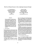
In the phylogenetic tree of the ALDH superfamily,
PsLADH and PsALDH* fall into the fungal ALDH
subfamily, one of the 14 ALDH subfamilies compiled
by Perozich et al. [12] (Fig. 4), confirming the micro-
organism source. The fungal ALDH subfamily belongs
to the Class 1 ⁄ 2 branch of ALDHs, which consists of
tetrameric ALDH subfamilies with variable substrate
specificity, as well as two P. stipitis enzymes (Table 1).
In S. cerevisiae, there is biochemical evidence of two
types of ALDH [13,14]. The mitochondrial ALDHs,
ScALDH4 and ScALDH5, show dual coenzyme speci-
ficity between NAD
+
and NADP
+
and are activated
by K
+
. The cytosolic ALDHs, ScALDH2, ScALDH3
and ScALDH6, are specific to NADP
+
; only ALDH6
is activated by Mg
2+
. Higher degrees of similarity to
PsLADH (probably cytosolic enzyme because of no
mitochondrial leader sequence) were found in the
mitochondrial ALDHs of S. cerevisiae, confirming the
enzyme properties, including coenzyme specificity.
Indeed, the activity of PsLADH is also absolutely
dependent on K
+
(data not shown). However,
PsALDH* (cytosolic enzyme as well as PsLADH) is
more closely related than PsLADH to cytosolic
ALDHs, indicating that this enzyme may be assigned
as an acetaldehyde dehydrogenase rather than LADH,
based on substrate specificity (Table 1). A branch of
AvLADH and EcLADH was located on the root of
the non-phosphorylating glyceraldehyde 3-phosphate
dehydrogenase (GAPDH, EC 1.2.1.9.) subfamily in the
Class 3 branch, consisting of substrate-specific ALDH
subfamilies (Fig. 4), confirming their enzyme proper-
ties of high specificity with l-lactaldehyde (Tables 1
and 2). (dl-)Lactaldehyde dehydrogenase of archaeal
M. jannaschii (MjLADH) is also a member of this
subfamily, and is involved in the production of lactate
for coenzyme F
420
biosynthesis [10].
Discussion
In this study, we have identified the LADHs involved
in the alternative l-rhamnose pathways of fungi and
bacteria. In particular, although fungi possess multiple
ALDH genes, only one physiological substrate, acetal-
dehyde, has been identified in fermentation and ⁄ or
growth on ethanol. To our knowledge, this is the
second report of fungal ALDH as an aldehyde
substrate in addition to acetaldehyde; the other stated
that ScALDH2 and ScALDH3 play a role as 3-amino-
propionaldehyde dehydrogenases in pantothenic acid
(vitamin B
5
) and coenzyme A biosynthesis [15].
Enzyme catalysis of LADH
Hempel et al. [16] proposed several characteristic con-
served regions containing almost all active amino acid
residues in ALDHs. In particular, glutamate in the
motif of LELGGKSP participates as a general base
for the activation of catalytic cysteine and deacylation
of the enzyme, and cysteine in the motif of
FXNXGQXCIA (where X is any amino acid) acts as a
nucleophile. These motifs are also conserved in
PsLADH and AvLADH with a few modifications
(Fig. 5), indicating that the overall structure and
fundamental catalytic mechanism may be similar to
L-Lactaldehyde dehydrogenase S. Watanabe et al.
5144 FEBS Journal 275 (2008) 5139–5149 ª 2008 The Authors Journal compilation ª 2008 FEBS
those in known ALDHs. Based on structural studies of
other ALDHs, amino acid residues at equivalent
positions of 190 and 193 in PsLADH are involved in the
distinction between NAD
+
and NADP
+
. The structur-
ally equivalent lysine residue to Lys190 is conserved in
all ALDHs, and is unlikely to influence directly coen-
zyme specificity. A glutamate residue at an equivalent
position to 193 interacts with 2¢- and 3¢-hydroxyl groups
of the ribose of the adenine moiety in strict NAD
+
-
preferring enzymes, such as EcLADH, AvLADH and
PsALDH*. However, the structurally equivalent gluta-
mate is found not only in PsLADH with significant
NADP
+
-dependent activity, but also in NADP
+
-
preferring ScALDH4 and ScALDH5. However, it is
Fig. 5. Partial alignment of amino acid sequences around several active sites. Open and filled circles indicate NAD
+
- and NADP
+
-dependent
enzymes. ScALDH4 (grey circle) utilizes both NAD
+
and NADP
+
as a coenzyme. Grey-shaded letters are highly conserved. In the crystal
structure of EcLADH (PDB ID, 2IMP), open and filled stars indicate amino acid residues bound to
L-lactate and 2¢- and 3¢-hydroxyl groups of
NADH, respectively. The catalytic glutamate and cysteine residues are indicated by grey stars.
Class 1
Class 2
Fungal ALDH
FTDH
HMSALDH
Group X
BALDH
SSALDH
GAPDH
Aromatic
ALDH
MMSALDH
Turgor ALDH
GGSALDH
Class 3 ALDH
Class 1/2 branch
Class 3 branch
PsALDH*
ScALDH3
ScALDH2
ScALDH6
PsLADH
ScALDH5
ScALDH4
Pichia angusta
Alternaria alternata
Cladosporium herbarum
Aspergillus nidulans
Aspergillus niger
Ustilago maydis
EcLADH
AvLADH
MjLADH
Fig. 4. The overall phylogenetic tree of
known ALDHs, including LADHs. Sequence
names and references of ALDHs are avail-
able on the ALDH website described in
Experimental procedures. The three
enzymes in the boxes were characterized in
this study.
S. Watanabe et al.
L-Lactaldehyde dehydrogenase
FEBS Journal 275 (2008) 5139–5149 ª 2008 The Authors Journal compilation ª 2008 FEBS 5145
known that ALDH isozyme B of E. coli (AldB, 33%
identity to EcLADH) is a strict NADP
+
-dependent
enzyme and possesses the structural equivalent arginine
residue at this position (Arg197) [17]. When compared
with the wild-type enzyme, the R197E mutant shows
10% NADP
+
-dependent activity, together with no
detection of NAD
+
-dependent activity. In PsLADH
and PsALDH*, each E193R mutant expressed a simi-
lar level to the wild-type enzyme in E. coli cells as
an inclusion body; we did not perform further enzy-
matic characterization (data not shown). This indicates
that, although the glutamate residue in PsLADH (and
fungal ALDHs) should play a role in coenzyme bind-
ing and ⁄ or structural maintenance, other amino acid
residues may also influence coenzyme specificity.
Inoue et al. [9] purified a NAD
+
-dependent dehydro-
genase for l-lactaldehyde from S. cerevisiae cells cul-
tured in a nutrient medium. Although the enzyme has
not yet been characterized genetically, the molecular
structures (monomeric form consisting of the subunit
with M
r
of 40 kDa) are clearly different from those of
general ALDH enzymes, including fungal ALDHs
(tetrameric or dimeric form consisting of the subunit
with M
r
of 50–55 kDa, see Fig. 3). Furthermore, the
activity with d-lactaldehyde is only 0.2% of that with
l-lactaldehyde, and acetaldehyde, dl-glyceraldehyde
and propionaldehyde are inactive substrates, in contrast
with PsLADH (Table 1); acetaldehyde is a common
active substrate for the known ScALDH2–6. Therefore,
although the genetic background and physiological
functions of LADH in S. cerevisiae have not been eluci-
dated so far, it has been reported recently that the Gre2
(YOL151w) gene encodes methylglyoxal reductase,
related to the detoxification of methylglyoxal [8], in
which the LADH(-like) enzyme may also be involved.
Convergent evolution of LADHs in fungi
and bacteria
In the phylogenetic tree, substrate-specific ALDHs
have a tendency to belong to subfamilies in the Class 3
branch, whereas ALDH families with broad substrate
specificity are more often found in the Class 1 ⁄ 2
branch (Fig. 4). PsLADH (and also PsALDH*) shows
significant activity for several aldehydes in addition
to l-lactaldehyde (Table 1), and the fungal ALDH
subfamily containing this enzyme belongs to the
Class 1 ⁄ 2 branch. However, AvLADH, which shows
high specificity to l-lactaldehyde, is similar to the
GAPDH subfamily in the Class 3 branch. It is note-
worthy that, although l-lactaldehyde is produced by
the same alternative pathway of l-rhamnose in
P. stipitis and A. vinelandii, their LADHs are classified
into different subfamilies, strongly suggesting that their
substrate specificities have been acquired by ‘conver-
gent evolution’ rather than divergence from a common
ancestor. Indeed, four ligands for the substrate (l-lac-
tate) are not conserved between PsLADH and
EcLADH (Fig. 5). PsLADH seems to have evolved
from an ancestor with broader substrate specificity,
such as PsALDH*, because PsALDH* is located at
the root of the fungal ALDH subfamily (Fig. 4).
It is certain that AvLADH and EcLADH, which are
involved in different pathways of the same l-rhamnose
metabolism, evolved from a common ancestor.
EcLADH is responsible for not only l-rhamnose but
also l-fucose metabolism [5], whereas the LRA1–4
proteins, components of the gene cluster containing
the AvLADH gene (Fig. 1C), show no significant activ-
ity with l-fucose-related intermediates [3]. Therefore, it
is probable that the gene cluster is involved in l-rham-
nose metabolism only, but not l-fucose. MjLADH is
involved in different metabolism from l-rhamnose
(coenzyme F
420
biosynthesis) [10] and is similar to
EcLADH and AvLADH phylogenetically (Fig. 5).
Although glyceraldehyde 3-phosphate is commonly
an inactive substrate for AvLADH (see Table 1),
EcLADH and MjLADH, MjLADH is capable of
utilizing several aldehydes, such as glycolaldehyde,
dl-glyceraldehyde, formaldehyde, acetaldehyde and
propionaldehyde (the last three are inactive substrates
for AvLADH and EcLADH). Furthermore, substrate-
binding sites of bacterial LADHs are not completely
conserved in MjLADH (Fig. 5). These results suggest
that substrate specificity for l-lactaldehyde has also
been acquired for bacteria and Archaea independently,
similar to fungi.
Of the 14 subfamilies in the ALDH superfamily, some
subfamilies, such as c-glutamyl semialdehyde dehydro-
genase, methylmalonyl semialdehyde dehydrogenase
and succinic semialdehyde dehydrogenase, include
sequences from organisms ranging from bacteria to
mammals (Fig. 4) [12]. In contrast, the fungal ALDH
subfamily (consisting of only fungal sequences) appears
to have diverged much later in evolution, indicating that
the acquisition of substrate specificity for l-lactaldehyde
might have occurred after divergence between bacteria
and eukaryotes (fungi). Four metabolic enzymes (genes)
that convert l-rhamnose into pyruvate and l-lactalde-
hyde are found on the genomes of several fungi, includ-
ing P. stipitis, Debaryomyces hansenii, Candida species
and Aspergillus species [3], but not S. cerevisiae, which is
not capable of growth on l-rhamnose [1]. Therefore, the
acquisition of these l-rhamnose metabolic genes might
have led to the appearance of LADH from a common
ancestor of fungal ALDHs under evolutionary pressure.
L-Lactaldehyde dehydrogenase S. Watanabe et al.
5146 FEBS Journal 275 (2008) 5139–5149 ª 2008 The Authors Journal compilation ª 2008 FEBS
Experimental procedures
Microorganism strains, culture conditions and
preparation of cell-free extracts
Pichia stipitis CBS 6054 was kindly provided by T. W. Jef-
fries (University of Wisconsin, Milwaukee, WI, USA).
A. vinelandii NBRC 102612 was purchased from the
National Institute of Technology and Evaluation (Chiba,
Japan). P. stipitis and A. vinelandii were grown at 30 °Cin
yeast nitrogen broth and Burk’s nitrogen-free medium
supplemented with 20 gÆL
)1
d-glucose or l-rhamnose as
carbon source, respectively. Usually, l-rhamnose was sterili-
zed separately by filtration and added to each medium. The
grown cells were harvested by centrifugation at 30 000 g
for 20 min, washed with 20 mm potassium phosphate
(pH 7.5) containing 1 mm EDTA and 10 mm 2-mercapto-
ethanol (referred to as Buffer A), and stored at )35 °C until
use. Fungal cells were suspended in Buffer A, homogenized
with an equal volume of glass beads (0.5 mm diameter,
Sigma, St Louis, MO, USA) for 30 min with appropriate
intervals on ice using a TORNADO
Ò
Laboratory Power
Mixer (AS ONE Co., Ltd., Osaka, Japan) and then
centrifuged at 108 000 g for 1 h at 4 °C to obtain cell-free
extracts. Bacterial cells were suspended in Buffer A,
disrupted by sonication for 20 min with appropriate
intervals on ice using an ASTRASON
Ò
Ultrasonic Liquid
Processor XL2020 (Misonix Incorporated, New York, NY,
USA) and then centrifuged.
Enzyme activity assay
l- and d-lactaldehyde were chemically synthesized from
l- and d-threonine, respectively, according to the method
of Huff and Rudney [18] with a few modifications. LADH
activity was assayed routinely in the direction of aldehyde
oxidation by measuring the reduction of NAD(P)
+
at
340 nm and 30 °C. The standard assay mixture contained
1mml-lactaldehyde, 1 mm EDTA and 10 mm 2-mercapto-
ethanol in 66.7 mm potassium phosphate (pH 7.5) buffer.
The reaction was started by the addition of 15 mm
NAD(P)
+
solution (100 lL) with a final reaction volume of
1 mL. In the case of water-insoluble aldehyde, 10 mm
substrates in ethanol (0.1 volume) were added to the
standard assay solution. Protein concentrations were
determined by the method of Lowry et al. [19], with bovine
serum albumin as a standard.
Zymogram staining analysis for LADH
Cell-free extracts were separated by non-denaturing PAGE
with a 12% gel at 4 °C. The gels were then soaked in
10 mL of staining solution [20] consisting of 100 mm
Tris ⁄ HCl (pH 9.0), 10 mmd-orl-lactaldehyde, 0.25 mm
nitroblue tetrazolium, 0.06 mm phenazine methosulfate and
15 mm NAD(P)
+
at 30 °C for 15 min. Dehydrogenase
activity appeared as a dark band.
Purification of native LADH from P. stipitis
All purification steps were performed below 4 °C. All
chromatography was carried out using an A
¨
KTA purifier
system (Amersham Pharmacia Biotech, Little Chalfont,
UK) and ⁄ or BioAssist eZ system (TOSOH, Tokyo, Japan).
Cell-free extracts prepared from l-rhamnose-grown P.
stipitis cells were loaded onto a HiPrep 16 ⁄ 10 Q FF column
(1.6 · 10 cm, Amersham Biosciences, Uppsala, Sweden)
equilibrated with Buffer A, and washed thoroughly with
the same buffer. The column was developed with 300 mL
of a linear gradient of 0–0.5 m NaCl in Buffer A. Active
fractions containing LADH were combined and concen-
trated by ultrafiltration with a Centriplus YM-30
(Millipore, Bedford, MA, USA) at 18 000 g for approxi-
mately 2 h. The enzyme solution was loaded onto a column
of HiLoad 26 ⁄ 60 Superdex 200 pg (2.6 · 60 cm, Amersham
Biosciences) equilibrated with Buffer A. The active
fractions were pooled, concentrated and applied to a
column of Ceramic Hydroxyapatite Type I (1.6 · 5 cm,
Bio-Rad Laboratories, Hercules, CA, USA) equilibrated
with Buffer A. The column was washed thoroughly with
the same buffer and developed with 150 mL of a linear
gradient of 0–0.3 m potassium phosphate in Buffer A. The
fractions with high enzymatic activity were combined, con-
centrated and loaded onto a column of Blue-SepharoseÔ
6 Fast Flow (1.6 · 5 cm, Amersham Biosciences) equili-
brated with Buffer A. The column was washed with
Buffer A containing 50 mm NaCl, and then the enzyme
was eluted with Buffer A containing 1 m NaCl. The elutant
was concentrated, dialysed against 50 mm potassium
phosphate, pH 7.5, containing 1 mm EDTA, 1 mm
dithiothreitol and 50% (v ⁄ v) glycerol, and stored at )35 °C
until use.
Determination of internal amino acid sequences
Purified PsLADH ( 50 lg) was separated by SDS-PAGE
with a 10% (w ⁄ v) gel. In-gel digestion by trypsin was
performed according to a standard protocol [21] with a
few modifications. The peptide masses were analysed
using a matrix-assisted laser desorption ionization-
quadruple ion trap-mass spectrometer (AXIMA QIT,
Shimadzu, Kyoto, Japan) with 2,5-dihydroxybenzoic acid
(Shimadzu GLC Ltd, Tokyo, Japan) as a matrix in
positive ion mode.
Identification of enzyme reaction product
HPLC analysis was performed using a Multi-Station
LC-8020 model II system (TOSOH). Purified native
S. Watanabe et al. L-Lactaldehyde dehydrogenase
FEBS Journal 275 (2008) 5139–5149 ª 2008 The Authors Journal compilation ª 2008 FEBS 5147
PsLADH ( 1 mg) was added to a reaction mixture
(500 lL) consisting of 20 mm potassium phosphate,
pH 7.5, 10 mml-lactaldehyde and 10 mm NAD
+
. After
incubation at 30 °C for 1 h, 12% (w ⁄ v) trichloroacetic
acid was added to the samples (0.1 volume) to remove
proteins. The filtrate (100 lL) was applied at 35 °Ctoan
Aminex HPX-87H Organic Analysis column (300 ·
7.8 mm, Bio-Rad) linked to an RID-8020 refractive index
detector (TOSOH), and eluted with 5 mm H
2
SO
4
at a
flow rate of 0.6 mLÆmin
)1
.
Functional expression and purification of
His6-tagged proteins
Genomic DNA of P. stipitis and A. vinelandii was prepared
using a DNeasy
Ò
Boold & Tissue Kit (Qiagen, Tokyo,
Japan). To introduce the restriction sites for BamHI and
PstI at the 5¢- and 3¢-termini of PsLADH, PsALDH* and
AvLADH genes, respectively, genomic PCR was carried out
using Ex Taq
Ò
DNA polymerase (TaKaRa, Otsu, Shiga,
Japan) and appropriate primers (Table S2). Each amplified
DNA fragment was introduced into BamHI-PstI sites in
pQE-80L (Qiagen), a plasmid vector for conferring the
N-terminal His6 tag on expressed proteins. E. coli DH5a
harbouring the expression plasmid was grown at 37 °Ctoa
turbidity of 0.6 at 600 nm in Super broth medium contain-
ing 50 mgÆL
)1
ampicillin. After the addition of 1 mm iso-
propyl thio-b -d-galactopyranoside, the culture was grown
for a further 6 h to induce the expression of His6-tagged
protein. Cells were harvested and resuspended in Buffer B
(pH 8.0, 50 mm sodium phosphate containing 300 mm
NaCl, 10 mm 2-mercaptoethanol and 10 mm imidazole).
The cells were then disrupted by sonication, and the
solution was centrifuged. The supernatant was loaded onto
a column of Ni-NTA Super Flow (Qiagen) equilibrated
with Buffer B, using an A
¨
KTA purifier system and ⁄ or Bio-
Assist eZ system. The column was washed with Buffer C
(pH 8.0, Buffer D containing 10% (v ⁄ v) glycerol and
50 mm imidazole instead of 10 mm imidazole). The enzymes
were then eluted with Buffer C containing 250 mm imid-
azole instead of 50 mm imidazole. All His6-tagged enzymes
were used within 1 week in further experiments.
Amino acid sequence alignment and
phylogenetic analysis
For phylogenetic analysis, 145 ALDH sequences were
obtained from a website devoted to ALDHs: http://www.
psc.edu/biomed/pages/research/Col_HBN_ALDH.html [12].
The sequences were aligned using the program clustalw ,
distributed by GenomeNet (Bioinformatics Center, Kyoto
University, Kyoto, Japan) (). The
phylogenetic tree was produced using the treeview 1.6.1
program.
Northern blot analysis
Pichia stipitis and A. vinelandii cells were cultured at 30 ° C
to the mid-logarithmic phase (A
600
= 0.6–0.8) and har-
vested by centrifugation. Total RNAs were prepared using
an RNeasy
Ò
Mini Kit (Qiagen). Northern hybridization
was carried out using a standard method. The PCR prod-
ucts of PsLADH, PsALDH* and AvLADH genes, amplified
by PCR using appropriate DNA primers, were labelled
with [a-
32
P]dCTP using the Random Primer Labelling Kit
(TaKaRa) and used as probes for hybridization.
Acknowledgements
This work was supported by a Grant-in-Aid for
Young Scientists (B) (No. 18760592) from the Ministry
of Education, Culture, Sports, Science and Technology
of Japan (to S.W.), by the Fermentation and Meta-
bolism Research Foundation, by the Japan Bioindustry
Association (to S.W.), by the Research Foundation
from the Association for the Progress of New Chemis-
try (to S.W.), by the New Energy and Industrial Tech-
nology Development Organization (to S.W.) and by
CREST, JST (to K.M.). We thank Dr T. W. Jeffries
(University of Wisconsin, Milwaukee, WI, USA) for
the gift of P. stipitis CBS 6054. We are especially
grateful to Dr M. Yamada (Shimadzu Corporation,
Kyoto, Japan) for his help with MALDI-TOF MS
analysis.
References
1 van Maris AJ, Abbott DA, Bellissimi E, van den Brink
J, Kuyper M, Luttik MA, Wisselink HW, Scheffers
WA, van Dijken JP & Pronk JT (2006) Alcoholic
fermentation of carbon sources in biomass hydrolysates
by Saccharomyces cerevisiae: current status. Antonie
Van Leeuwenhoek 90, 391–418.
2 Twerdochlib AL, Pedrosa FO, Funayama S & Rigo LU
(1994) l-Rhamnose metabolism in Pichia stipitis and
Debaryomyces polymorphus. Can J Microbiol 40, 896–
902.
3 Watanabe S, Saimura M & Makino K (2008) Eukary-
otic and bacterial gene clusters related to an alternative
pathway of non-phosphorylated l-rhamnose metabo-
lism (2008). J Biol Chem 283, 20372–20382.
4 Caballero E, Baldoma
´
L, Ros J, Boronat A &
Aguilar J (1983) Identification of lactaldehyde
dehydrogenase and glycolaldehyde dehydrogenase as
functions of the same protein in Escherichia coli.
J Biol Chem 258, 7788–7792.
5 Baldoma
´
L & Aguilar J (1987) Involvement of
lactaldehyde dehydrogenase in several metabolic
L-Lactaldehyde dehydrogenase S. Watanabe et al.
5148 FEBS Journal 275 (2008) 5139–5149 ª 2008 The Authors Journal compilation ª 2008 FEBS
pathways of Escherichia coli K12. J Biol Chem 262,
13991–13996.
6 Di Costanzo L, Gomez GA & Christianson DW (2007)
Crystal structure of lactaldehyde dehydrogenase from
Escherichia coli and inferences regarding substrate and
cofactor specificity. J Mol Biol 366, 481–493.
7 Boronat A & Aguilar J (1979) Rhamnose-induced pro-
panediol oxidoreductase in Escherichia coli: purification,
properties, and comparison with the fucose-induced
enzyme. J Bacteriol 140, 320–326.
8 Chen CN, Porubleva L, Shearer G, Svrakic M, Holden
LG, Dover JL, Johnston M, Chitnis PR & Kohl DH
(2003) Associating protein activities with their genes:
rapid identification of a gene encoding a methylglyoxal
reductase in the yeast Saccharomyces cerevisiae. Yeast
20, 545–554.
9 Inoue Y, Watanabe K, Shimosaka M, Saikusa T,
Fukuda Y, Murata K & Kimura A (1985) Metabolism of
2-oxoaldehydes in yeasts. Purification and characteriza-
tion of lactaldehyde dehydrogenase from Saccharomyces
cerevisiae. Eur J Biochem 153, 243–247.
10 Grochowski LL, Xu H & White RH (2006) Identifica-
tion of lactaldehyde dehydrogenase in Methanocaldococ-
cus jannaschii and its involvement in production of
lactate for F
420
biosynthesis. J Bacteriol 188, 2836–
2844.
11 Ting SM, Miller ON & Sellinger OZ (1965) The
metabolism of lactaldehyde. VII. The oxidation of
d-lactaldehyde in rat liver. Biochim Biophys Acta 97,
407–415.
12 Perozich J, Nicholas H, Wang BC, Lindahl R &
Hempel J (1999) Relationships within the aldehyde
dehydrogenase extended family. Protein Sci 8,
137–146.
13 Tessier WD, Meaden PG, Dickinson FM & Midgley M
(1998) Identification and disruption of the gene encod-
ing the K
+
-activated acetaldehyde dehydrogenase of
Saccharomyces cerevisiae. FEMS Microbiol Lett 164,
29–34.
14 Wang X, Mann CJ, Bai Y, Ni L & Weiner H (1998)
Molecular cloning, characterization, and potential roles
of cytosolic and mitochondrial aldehyde dehydrogenases
in ethanol metabolism in Saccharomyces cerevisiae.
J Bacteriol 180, 822–830.
15 White WH, Skatrud PL, Xue Z & Toyn JH (2003)
Specialization of function among aldehyde dehydro-
genases: the ALD2 and ALD3 genes are required for
beta-alanine biosynthesis in Saccharomyces cerevisiae.
Genetics 163, 69–77.
16 Hempel J, Nicholas H & Lindahl R (1993) Aldehyde
dehydrogenases: widespread structural and functional
diversity within a shared framework. Protein Sci 2,
1890–1900.
17 Ho KK & Weiner H (2005) Isolation and characteriza-
tion of an aldehyde dehydrogenase encoded by the aldB
gene of Escherichia coli. J Bacteriol 187, 1067–1073.
18 Huff E & Rudney H (1959) The enzymatic oxidation of
1,2-propanediol phosphate to acetol phosphate. J Biol
Chem 234, 1060–1064.
19 Lowry OH, Rosebrough NJ, Farr AL & Randall RJ
(1951) Protein measurement with the folin phenol
reagent. J Biol Chem 193, 265–275.
20 Gennady PM (2002) Handbook of Detection of Enzymes
on Electrophoretic Gels, 2nd edn. pp. 27–33. CRC Press,
Inc., Boca Raton, FL.
21 Toda T & Kimura N (1997) Standardization of proto-
col for Immobiline 2-D PAGE and construction of 2-D
PAGE protein database on World Wide Web home
page. Jpn J Electrophoresis 41, 13–20.
Supporting information
The following supplementary material is available:
Table S1. Tryptic peptide sequences from PsLADH by
MALDI-TOF MS.
Table S2. Primer list used in this study.
This supplementary material can be found in the
online version of this article.
Please note: Wiley-Blackwell is not responsible for
the content or functionality of any supplementary
materials supplied by the authors. Any queries (other
than missing material) should be directed to the
corresponding author for the article.
S. Watanabe et al. L-Lactaldehyde dehydrogenase
FEBS Journal 275 (2008) 5139–5149 ª 2008 The Authors Journal compilation ª 2008 FEBS 5149