Báo cáo khoa học: Differential binding of factor XII and activated factor XII to soluble and immobilized fibronectin – localization of the Hep-1/Fib-1 binding site for activated factor XII pdf
Bạn đang xem bản rút gọn của tài liệu. Xem và tải ngay bản đầy đủ của tài liệu tại đây (423.97 KB, 12 trang )
Differential binding of factor XII and activated factor XII
to soluble and immobilized fibronectin – localization of
the Hep-1/Fib-1 binding site for activated factor XII
Inger Schousboe
1
, Birthe T. Nystrøm
1
and Gert H. Hansen
2
1 Department of Biomedical Sciences, The Panum Institute, University of Copenhagen, Denmark
2 Department of Cellular and Molecular Medicine, The Panum Institute, University of Copenhagen, Denmark
Several studies have suggested that in the cardio-
vascular system, the interaction between the vessel wall
and the contact activation system of blood coagula-
tion, including factor XII (FXII), high molecular mass
kininogen (HK) and prekallikrein, involves Zn
2+
-
dependent and receptor-mediated binding of FXII and
HK. Thus, investigations of FXII and HK binding to
endothelial cells in the vascular wall mimicked by
Keywords
association; factor XII; factor XIIa;
fibronectin
Correspondence
I. Schousboe, Department of Biomedical
Sciences, Heart and Circulatory Research
Section, The Panum Institute, University of
Copenhagen, Blegdamsvej 3C, DK-2200
Copenhagen, Denmark
Fax: +45 35367980
Tel: +45 35327800
E-mail:
(Received 7 May 2008, revised 8 July 2008,
accepted 18 August 2008)
doi:10.1111/j.1742-4658.2008.06647.x
Fibronectins (FNs) are dimeric glycoproteins that adopt a globular con-
formation when present in plasma and solution and an extended confor-
mation in the extracellular matrix. Factor XII (FXII) is a zymogen of
the proteolytically active FXIIa that plays a role in thrombus stabiliza-
tion by enhancing clot formation and in inflammation by enhancing
bradykinin formation. To investigate whether the extracellular matrix
could play a role in these events, we have recently shown that FXIIa,
but not FXII, binds to the extracellular matrix (ECM), and suggested
that FN may be the target for the binding. Immunofluorescence micros-
copy has in the present investigation confirmed that FXIIa added to the
ECM colocalizes with FN deposited during growth of human umbilical
vein endothelial cells. The aim of the present study, therefore, was to fur-
ther elucidate the interaction between FXIIa and FN by the use of a
solid face binding assay. This showed, like the binding to the ECM, that
FXIIa, but not FXII, binds in a Zn
2+
-independent manner to immo-
bilized FN. The K
D
for the binding was 8.5 ± 0.9 nm (n = 3). The
binding was specific for the immobilized FN, as the binding could not be
inhibited by soluble FN. Furthermore, soluble FN did not bind to
immobilized FXIIa. However, soluble FN could bind to FXII, and this
binding inhibited the surface-induced autoactivation of FXII and subse-
quent binding of the generated FXIIa to immobilized FN. The presence
of FXII in an anti-FN immunoprecipitate of plasma indicated that some
FXII in plasma circulates bound to FN. The binding of FXIIa to FN
was inhibited by gelatine and fibrin but not by heparin, indicating that
FXIIa binds to immobilized FN through the type I repeat modules.
Accordingly, FXIIa was found to bind to immobilized fragments of FN
containing the type I repeat modules in the N-terminal domain to which
fibrin and gelatine bind.
Abbreviations
CTI, corn trypsin inhibitor; DS, dextran sulfate; ECM, extracellular matrix; Fib-1, the N-terminal fibrinogen binding domain on fibronectin; FN,
fibronectin; FXII, factor XII; FXIIa, activated factor XII; Hep-1, the N-terminal heparin binding domain on fibronectin; Hep-2, the C-terminal
heparin binding domain on fibronectin; HK, high molecular mass kininogen; HRP, horseradish peroxidase; HUVEC, human umbilical vein
endothelial cell; OPD, o-phenylenediamine.
FEBS Journal 275 (2008) 5161–5172 ª 2008 The Authors Journal compilation ª 2008 FEBS 5161
cultures of human umbilical vein endothelial cells
(HUVECs) have shown that FXII and HK interact by
multiprotein assembly [1–3].
FXII is a precursor of the proteolytically active acti-
vated FXII (FXIIa). FXII and FXIIa bind equally well
to a confluent layer of HUVECs [4]. However, a recent
investigation has shown that the binding might have
been artefactual, and that FXII in the presence but
not in the absence of a negatively charged surface
bound rather to the extracellular matrix (ECM) gener-
ated during growth of HUVECs. The presence of
negatively charged surfaces appeared to serve two
purposes: (a) it induced and enhanced the autoactiva-
tion of FXII, generating FXIIa; and (b) it abrogated
nonspecific binding of FXIIa [5].
The binding of FXIIa to the ECM showed several
differences from the binding to HUVECs. Thus, the
binding to the ECM was: (a) specific for FXIIa;
(b) Zn
2+
-independent; (c) not inhibited by HK; and
(d) nonelectrostatic. As proteolytic degradation of the
ECM abrogated the binding of FXIIa, it was assumed
that a matrix protein was the target for the binding.
Therefore, it was tentatively analyzed and found that
FXIIa binds to fibronectin (FN) [5].
FN is a dimeric high molecular mass glycoprotein
that is found both as a circulating soluble molecule in
the blood and as insoluble molecules forming elon-
gated multimeric structures in the ECM [6,7]. The
monomer of the dimeric soluble molecule is a mosaic
protein composed of modular subunits generating
different domains [8], which harbor binding sites for
glycosaminoglycans, collagen or gelatine, fibrin, and
integrin receptors. Some of these binding sites become
available only in the multimeric, elongated forms in
which internal sequences of amino acid residues
become exposed [9,10]. Several factors mediate the
transition from the soluble to an elongated form,
including adsorption of FN to plastic surfaces [11–14].
As our previous studies have shown that the binding
of FXIIa to the ECM could be due to binding of
FXIIa to FN generated during growth of HUVECs
[5], we here report on studies of the interactions of
FXIIa with FN using a solid-phase binding assay in
which either FN or FXIIa is immobilized.
Results
FXII/FXIIa binding to FN
The association of FXIIa with the ECM was assumed
to take place through binding to FN. Therefore, inves-
tigations were first performed to determine whether it
could be shown that FXIIa associated with FN depos-
ited on the surface of the culture dish after depletion
of HUVECs by EDTA extraction. Immunofluores-
cence clearly showed that FXIIa bound to the depos-
ited FN (Fig. 1). No FN was deposited on and no
FXIIa bound to surfaces incubated with growth
medium under the same conditions and for the same
periods of time as the cells but in the absence of cells.
To obtain more information about this association,
the interaction between FXIIa and FN was subse-
quently analyzed by measuring the binding of FXIIa
to FN immobilized on a plastic surface.
Using a solid-phase binding assay, the binding
of FXIIa to FN was visualized by reactions with an
A
B
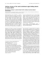
Fig. 1. Colocalization of the ECM-bound FXII and FN. HUVECs
were grown to near confluence, and the generated ECM was
exposed by detaching the cells with EDTA. After washing, the
ECM was incubated for 1 h with 20 n
M FXIIa. The ECM was then
washed again and incubated first with a mixture of goat anti-FXII
IgG (1 : 100) and rabbit anti-FN IgG (1 : 100) for 1 h, and second
with a mixture of Alexa 594-conjugated donkey anti-(goat IgG)
(1 : 800) and Alexa 488-conjugated goat anti-(mouse IgG) (1 : 800).
(A) Red indicates the presence of FXIIa. (B) Green indicates the
presence of FN. Bar: 20 lm.
Factor XII binding to fibronectin I. Schousboe et al.
5162 FEBS Journal 275 (2008) 5161–5172 ª 2008 The Authors Journal compilation ª 2008 FEBS
antibody against FXII and a horseradish peroxidase
(HRP)-labeled secondary antibody. Neither the anti-
body against FXII nor the secondary antibody was
observed to bind to FN in the absence of FXIIa. This
excludes the possibility that the response was nonspe-
cific and due to a direct interaction between the immo-
bilized FN and the immunoglobulins, as previously
noted [15]. Furthermore, preincubation of FXIIa for
1 h with a two-fold molar excess of the antibody
against FXII prior to incubation with FN abolished
the binding. Surprisingly, the binding could not be
inhibited if the immobilized FN had been preincubated
with a polyclonal antibody against soluble FN (data
not shown). This could be due to lack of recognition
of the binding site on the immobilized FN for FXIIa,
but it could also be due to a nonspecific interaction
between FXIIa and the plastic surface. However, very
little FXIIa bound to wells devoid of FN (controls).
Moreover, nonspecific binding is nonsaturable. The
binding of FXIIa to immobilized FN was saturable
even at low concentrations of FXIIa. This was demon-
strated by analyzing the binding of varying concentra-
tions of FXIIa. At low concentrations of FXIIa,
considerably more FXIIa bound to FN than to control
wells. At high concentrations of FXIIa, the binding to
FN increased linearly with the concentration of FXIIa,
and in parallel with the binding of FXIIa to control
wells. After subtraction of nonspecific binding from
the total binding, saturated binding to immobilized
FN was observed at FXIIa concentrations ‡ 20 nm
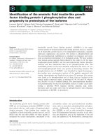
(Fig. 2). Linear transformation of the the binding iso-
therm (Fig. 2 insert) obtained in one of three indepen-
dent experiments, each performed in triplicate, showed
high-affinity binding, the K
D
of which was estimated
to be 8.5 ± 0.9 nm, using all available data.
To determine whether the binding of FXIIa to
immobilized FN was mediated through the N-terminal
surface binding sequence in FXIIa, investigations were
performed to determine whether the presence of nega-
tively charged compounds such as sulfatides would
affect the binding of FXIIa to FN. This showed that
sulfatides neither inhibited nor enhanced the binding
to immobilized FN. The apparently higher-affinity
binding of FXIIa in the present experiment in the
absence than in the presence of sulfatides was due to
parallel higher nonspecific binding. However, if FXIIa
was exchanged with FXII, the presence of sulfatides
induced binding of FXII, which in the absence of
0
0.5
1
1.5
2
2.5
3
3.5
20010 304050
60
Concentration of FXIIa, n
M
FXIIa bound, absorbance units
FN
Control
FN - Control
y = 1.1214x + 9.7607
R
2
= 0.993
0
10
20
30
40
50
60
70
Concentration of FXIIa/ absorbance units
200 10 304050
60
Concentration of FXIIa, n
M
Fig. 2. Concentration-dependent binding of FXIIa to FN. The microtiter plate was coated overnight with FN (10 lgÆmL
)1
) and NaCl ⁄ P
i
(con-
trol), respectively, and subsequently blocked with blocking buffer. Then, it was incubated for 1 h with increasing concentrations of FXIIa in
blocking buffer. The amount of bound FXIIa was determined by sequential incubation with goat anti-FXII IgG and HRP-conjugated rabbit anti-
(goat IgG) and visualized by reactions with OPD as described in Experimental procedures. d, total amount of FXIIa bound to wells coated
with FN; s, total amount of FXIIa bound to control wells (devoid of FN but ‘coated’ overnight with NaCl ⁄ P
i
; , binding of FXIIa to FN, calcu-
lated as the difference between binding of FXIIa to the former and the latter. Linear transformation of the results shown in the figure, which
is representative of three experiments performed in triplicate, gave a K
D
of 8.7 nM. Results are means ± SD (n = 3), shown by vertical bars
when extending beyond the symbols.
I. Schousboe et al. Factor XII binding to fibronectin
FEBS Journal 275 (2008) 5161–5172 ª 2008 The Authors Journal compilation ª 2008 FEBS 5163
sulfatides was negligible (Fig. 3). The sulfatide-depen-
dent binding of FXII was most likely due to a sulfat-
ide-induced and sulfatide-enhanced autoactivation of
FXII [16,17]. Accordingly, the presence of corn trypsin
inhibitor (CTI), which inhibits the activity of FXIIa,
and thus the autoactivation of FXII, almost com-
pletely blocked the sulfatide-induced binding of FXII
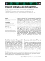
to FN. As compared to FXIIa, a small amount of
FXII bound to FN. Binding of the activated form of
FXII was shown by western blots of extracts of immo-
bilized FN incubated with FXII in the presence of sulf-
atides (Fig. 4).
As FXII and FXIIa bind equally well to sulfatides
[5], the lack of binding to immobilized FN of FXII
and the lack of inhibition of FXIIa by sulfatides indi-
cate that the binding is not brought about by the
N-terminal surface-binding region in FXIIa. To confirm
this, it was investigated whether the binding of FXIIa
to FN could be inhibited by the nine amino acid pep-
tide YHKCTHKGR(39–47), containing the surface-
binding sequence [18]. The presence of this peptide did
not inhibit the binding of FXIIa to immobilized FN
(data not shown).
In plasma and in solution, FN adopts a compact
soluble conformation in which the two subunits of the
dimer are thought to be folded upon each other [7].
Several studies have reported a change in the FN con-
formation upon binding to plastic [11–14], exposing a
cryptic binding site by transition from the soluble to
the immobilized form [19,20]. To determine whether
these conformational changes were of significance for
the binding of FXIIa, subsequent investigations were
performed to determine whether the presence of solu-
ble FN could inhibit the binding to immobilized FN.
This was shown not to be the case. The amount of
FXIIa that bound to immobilized FN was the same
regardless of the presence of soluble FN. In contrast,
the presence of soluble FN reduced the sulfatide-
induced binding of FXII (P < 0.001) (Fig. 5). How-
ever, as sulfatides had hardly any effect on the binding
of FXIIa to immobilized FN, and FXII did not bind
to immobilized FN in the absence of sulfatides
(Fig. 3), the inhibition could be due to an inhibition of
the interaction between FXII and sulfatides. To inves-
tigate this further, the solid-phase binding assay was
turned around and the microtiter plate was coated
0
0.5
1
1.5
2
2.5
FXII –
sulfatide
FXII +
sulfatide
FXII +
sulfatide +
CTI
FXIIa –
sulfatide
FXIIa +
sulfatide
FXIIa + anti-
FXII
antibody
Block buffer
FXIIa bound, absorbance units
Fig. 3. The effect of sulfatide on the binding of FXIIa to immobilized FN. The microtiter plate, coated overnight with FN (10 lgÆmL
)1
) and
NaCl ⁄ P
i
(control), respectively, was blocked with blocking buffer and incubated for 1 h with FXII (20 nM) and FXIIa (20 nM) in the presence
(+sulfatide) and absence ()sulfatide) of sulfatides (20 lgÆmL
)1
). To ensure that possible sulfatide-dependent binding of FXII could not be
explained by autoactivation of FXII, incubation of FXII in the presence of sulfatides was additionally performed in the presence of CTI
(10 lgÆmL
)1
). FN was also incubated for 1 h with FXIIa, which had been preincubated for 1 h with a twofold molar excess of goat anti-FXII
IgG. At the end of the incubation, the incubation mixtures were removed, and the microtiter plate was washed extensively. Then, the
microtiter plate was incubated sequentially with goat anti-FXII IgG, and HRP-conjugated rabbit anti-(goat IgG) in 1% skimmed milk, and the
amount of bound FXIIa was visualized by reaction with OPD. The combination of primary and secondary antibodies did not bind to either
FN-coated or control wells in the absence of FXIIa ⁄ FXII + sulfatides, as indicated by the column showing the binding of blocking buffer. The
total amount of FXIIa bound to FN and control wells is indicated by gray and white, respectively. Results are means ± SD (n = 3), shown by
vertical bars.
Factor XII binding to fibronectin I. Schousboe et al.
5164 FEBS Journal 275 (2008) 5161–5172 ª 2008 The Authors Journal compilation ª 2008 FEBS
with FXII and FXIIa instead of FN. Then, the bind-
ing of soluble FN to immobilized FXII and FXIIa was
visualized by incubation with rabbit anti-(soluble FN)
IgG as the primary antibody and HRP-conjugated
swine anti-(rabbit IgG) as secondary antibody.
Figure 6 shows that whereas almost no FN could bind
to immobilized FXIIa, it could bind to FXII. The
presence of sulfatides increased only slightly the bind-
ing to both FXII and FXIIa. Although it seemed most
unlikely, these differences in the amount of bound FN
could be due to differences in the amount of FXII and
FXIIa coated on the plate. This was found not to be
the case, as the immunochemical response was
analyzed and observed to be identical using goat
anti-FXII IgG. Moreover, to ensure that FXII had not
been activated during the coating period, the wells
were coated in the presence of CTI, which inhibits the
activity of FXIIa and thus the conversion of FXII to
FXIIa. Furthermore, in order to prevent FXII from
activation during the incubation with FN, CTI was
added to the incubation mixture. This did not affect
the binding of FN (results not shown). Thus, these
results clearly show that soluble FN interacts directly
with FXII in the absence of sulfatides. To determine
whether this interaction also occurs in plasma, the
presence of FXII was analyzed in immunoprecipitates
of FN. Plasma was immunoprecipitated with antibod-
ies against FN and adsorbed to protein G–Sepharose,
from which FXII was extracted. The plasma was not
preabsorbed to protein G–Sepharose, as binding of
FXII to the Sepharose could disturb the equilibrium
for the binding of FXII to FN. Instead, the amount of
FXII bound to protein G–Sepharose in the absence of
antibodies against FN was simultaneously analyzed
(Fig. 7). A much greater amount of FXII could be
21FXIIaFXII 43
80
50
Fig. 4. Western blot of extracts of bound protein after incubation
of FXII on immobilized FN in the absence and presence of sul-
fatides. The microtiter plate was coated overnight with FN
(10 lgÆmL
)1
) and subsequently blocked with blocking buffer. Then,
it was incubated for 1 h with 20 n
M FXII in blocking buffer in the
presence and absence of 20 lgÆmL
)1
sulfatide. After washing, the
proteins bound to immobilized FN were extracted with SDS under
reducing conditions (SDS containing dithiothreitol) and subjected to
reduced SDS ⁄ PAGE and western blotting. FXII, FXIIa and standard
samples of molecular mass markers were run simultaneously. Anti-
body-reacting bands were visualized by sequential incubation with
goat anti-(human FXII) IgG (1 : 2500), HRP-conjugated rabbit anti-
(goat IgG) (1 : 2500) and SuperSignal West Femto Maximum Sensi-
tivity Substrate. FXII; FXIIa; Lane 1: proteins extract from control
wells devoid of FN in which FXII had been incubated in the
absence of sulfatides. Lane 2: proteins extracted from immobilized
FN after incubation with FXII in the absence of sulfatides. Lane 3:
proteins extracted from control wells in which FXII had been incu-
bated in the presence of sulfatides. Lane 4: proteins extracted from
immobilized FN after incubation with FXII in the presence of sulfati-
des. The positions of 50 kDa and 80 kDa proteins are indicated to
the left. The blot shows that only FXIIa binds to FN.
0
0.2
0.4
0.6
0.8
1
1.2
1.4
1.6
1.8
2
FXII – sulf
FXII – sulf + globular FN
FXII + sulf
FXII + sulf + globular FN
FXIIa – sulf
FXIIa – sulf + globular FN
FXIIa bound, absorbance units
FN
Control
**
*
Fig. 5. The effect of soluble FN on the binding of FXII and FXIIa to
immobilized FN. The microtiter plate was coated overnight with FN
(10 lgÆmL
)1
) and NaCl ⁄ P
i
, respectively. Then, it was blocked with
blocking buffer and incubated for 1 h with FXII (20 n
M) or FXIIa
(20 n
M) in blocking buffer in the absence ()sulf) and presence (+sulf)
of sulfatides (20 lgÆmL
)1
) and in the absence and presence of solu-
ble FN (10 lgÆmL
)1
), as indicated. The amount of FXIIa bound to FN
was measured by sequential incubation with goat anti-FXII IgG and
HRP-conjugated rabbit anti-(goat IgG) as described in Experimental
procedures. The amounts of FXIIa bound to FN and control wells are
indicated by gray and white, respectively. Statistically significant dif-
ferences in binding of FXIIa to FN coated on the microtiter plate
when incubated in the absence and presence of soluble FN are indi-
cated by asterisks (*not significant and **P < 0.001). Results are
means ± SD (n = 3), shown by vertical bars.
I. Schousboe et al. Factor XII binding to fibronectin
FEBS Journal 275 (2008) 5161–5172 ª 2008 The Authors Journal compilation ª 2008 FEBS 5165
extracted from FN immunoprecipitates of plasma than
from the plasma alone. This indicates that FXII also
forms a complex with FN in plasma.
Further characterization of FXIIa binding to
immobilized FN
The high-affinity interaction between FXIIa and
immobilized FN and the lack of interference by solu-
ble FN indicated that the binding site on FN for
FXIIa may be buried in the compact soluble form of
FN [7]. FN has binding sites for a series of ligands
such as glycosaminoglycans, collagens or gelatine,
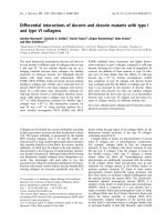
fibrin and integrins [21–27]. Figure 8 shows a sketch
of FN and the localization of the different binding
sites used in our attempt to identify the binding site
for FXIIa. Thus, concentration-dependent inhibition
of FXIIa binding to immobilized FN was observed
with gelatine and high concentrations of dextran sul-
fate (DS) but not with heparin (Fig. 9). As shown in
Fig. 8, FN has two binding sites for heparin. The
Hep-1-binding site is a low-affinity binding site, and
the Hep-2-binding site is a high-affinity binding site
[21–23]. If FXIIa bound to the C-terminal high-affin-
ity Hep-2-binding site, it would have been expected
that its interaction with immobilized FN would be
inhibited by heparin. Thus, the lack of inhibition by
heparin indicated that FXIIa did not bind to the
C-terminal high-affinity heparin-binding domain in
FN (Hep-2). However, the inhibition by high concen-
trations of DS and gelatine may indicate that FXIIa
binds to the N-terminal region of FN, including the
low-affinity Hep-1-binding domain. DS is a heparin-
like molecule and may, as such, be assumed to bind
to the heparin-binding sites on FN. To investigate
this further, the binding of FXIIa to commercially
available proteolytic fragments of FN was analyzed.
Each of these fragments contains binding domains
for heparin, gelatine and cells, respectively. Surpris-
ingly, the binding of FXIIa to these fragments
showed that although heparin was unable to inhibit
the binding of FXIIa to intact FN, FXIIa bound pri-
marily to the 30 kDa low-affinity heparin-binding
fragment (Hep-1), less to the 45 kDa gelatine-binding
fragment, and not at all to the 120 kDa fragment
containing the cell-binding domain (Fig. 10). The
amount of FXIIa that bound to the 30 kDa Hep-1-
binding fragment was similar to the amount of
FXIIa bound to FN. The N-terminal 30 kDa Hep-1-
binding domain has also been identified as a binding
site for fibrinogen and fibrin [25,26]. Further evidence
for FXIIa binding to this domain was therefore
provided, showing that the binding of FXIIa to
immobilized FN was inhibited in a concentration-
dependent manner by both fibrin generated by incu-
bation of fibrinogen with thrombin and fibrinogen.
As compared to the inhibition by fibrin, however, an
approximately 100-fold higher fibrinogen concentra-
tion was needed to yield an identical amount of
inhibition (Fig. 11).
Discussion
Although the presence in the blood of FXII has been
known for more than 50 years, its physiological func-
0
0.5
1
1.5
2
2.5
3
FN FN + Sulf FN FN + Sulf
Globular FN bound, absorbance units
FXIIFXIIa
Fig. 6. Binding of soluble FN to immobilized FXII and FXIIa. The
microtiter plate was coated overnight as indicated with FXII (20 n
M)
and FXIIa (20 n
M), respectively, diluted in NaCl ⁄ P
i
. Then, the micro-
titer plate was blocked with blocking buffer and incubated for 1 h
with FN (10 lgÆmL
)1
) in blocking buffer or in blocking buffer con-
taining sulfatides (+sulf; 20 lgÆmL
)1
). The amounts of FN bound to
FXII and FXIIa, respectively, were determined by sequential incuba-
tion with rabbit anti-FN IgG, HRP-conjugated swine anti-(rabbit IgG)
and OPD, as described in Experimental procedures. Results are
means ± SD (n = 3), shown by vertical bars.
Fig. 7. Western blots of FXII present in FN immunoprecipitates of
plasma. FN was isolated from plasma by immunoprecipitation with
a rabbit antibody against FN and protein G–Sepharose. The pres-
ence of FXII in the immunoprecipitate (lane 2) was analyzed by
western blotting using goat anti-FXII IgG as primary antibodies and
HRP-conjugated rabbit anti-(goat IgG) as secondary antibody. To
assure that the presence of FXII in the immunoprecipitate was not
due to adsorption of FXII to the protein G–Sepharose, the amount
of adsorbed FXII in the absence of the antibody against FN was
analyzed simultaneously (lane 1).
Factor XII binding to fibronectin I. Schousboe et al.
5166 FEBS Journal 275 (2008) 5161–5172 ª 2008 The Authors Journal compilation ª 2008 FEBS
tion is still not known. For the past 15 years it has
been assumed that its function is connected with
Zn
2+
-dependent binding to a surface or a receptor.
The present study has demonstrated that in purified
systems, activated FXII (FXIIa), but not its zymogen
(FXII), binds with high affinity to immobilized FN.
The binding is independent of the presence of Zn
2+
,is
not affected by the presence of a negatively charged
surface represented by sulfatides, and is not inhibited
by soluble FN. Accordingly, soluble FN did not
bind to immobilized FXIIa. The binding of FXIIa to
immobilized FN occurs through type I modules in the
30 kDa N-terminal heparin (Hep-1)-binding and fibrin
(Fib-1)-binding domain of FN.
Immunohistochemical visualization of the interac-
tion between FXIIa and FN deposited on the surface
of the culture dish during 3 days of growth of
HUVECs clearly showed that FXIIa associated with
FN left behind on the plastic surface after removal of
the cells. The visualization showed that FN had been
deposited in a sparse and patchy manner, which may
reflect the conditions under which the cells had been
cultivated and subsequently removed by EDTA extrac-
tion. Indeed, the majority of the deposited FN was
attached to the cells and was thus removed during
extraction of the cells. Furthermore, experiments with
cultures of arterial endothelial cells have shown that
the amount of FN deposited on the surface of the cells
varied dramatically when preconfluent, newly confluent
and postconfluent cultures were analyzed. Thus,
whereas sparse patches of FN were generated in pre-
confluent and newly confluent cultures, a massive net
Fib-1/Hep-1
S
FXIIa binding
COOH
NH
2
Gelatine Cell Hep-2 Fib-2
S
Type I
Type IIIType II
30 kDa 120 kDa45 kDa
Fig. 8. Schematic diagram of the modular
structure of the FN monomer. The FN dimer
is formed through interchain disulfide bonds
at the C-terminus. Each subunit consists of
type I, type II and type III repeating
modules. Sets of repeats form domains of
regions implicated in adhesion of different
ligands. The squares show the positions and
the sizes of the different fragments.
0.0
0.5
1.0
1.5
2.0
Block buffer
Heparin, 20 µg·mL
–1
Heparin, 40 µg·mL
–1
Gelatine, 33 µg·
mL
–1
Gelatine, 330 µg·mL
–1
DS, 2
0 µg·mL
–1
DS, 40 µg·mL
–1
FXIIa bound, absorbance units
*
*
*
Fig. 9. The effect of gelatine and heparin on binding of FXIIa to immobilized FN. The microtiter plate was incubated overnight with FN
(10 lgÆmL
)1
) and NaCl ⁄ P
i
, respectively, and blocked with blocking buffer. Then, it was incubated for 1 h with FXIIa (20 nM) in blocking buffer
containing heparin (20 and 40 lgÆmL
)1
), gelatine (33 and 330 lgÆmL
)1
) or DS (20 and 40 lgÆmL
)1
). The amount of bound FXIIa was deter-
mined by sequential incubation with goat anti-FXII IgG and HRP-conjugated rabbit anti-(goat IgG) and visualized by reactions with OPD as
described in Experimental procedures. Results are mean ± SD (n = 3), shown by vertical bars. Statistically significant differences between
FXIIa bound to FN incubated in the presence and in the absence of effectors are indicated by asterisks (*P < 0.001).The binding to control
wells was less than 0.05 absorbance units.
I. Schousboe et al. Factor XII binding to fibronectin
FEBS Journal 275 (2008) 5161–5172 ª 2008 The Authors Journal compilation ª 2008 FEBS 5167
of FN covering the entire surface of the cells was
formed only in postconfluent cultures [28]. It may be
claimed that the deposited FN originates from the
serum present in the cell culture medium. However,
the lack of appearance of deposited FN on culture
dishes incubated with the medium using the same con-
ditions and periods of time as in the presence of cells
but in their absence showed that the deposited FN in
the present investigation was generated by a cell-medi-
ated process. This process induces conformational
changes in FN, exposing cryptic sites of importance
for fibril generation and elongation [28–30].
The high-affinity binding of FXIIa to the ECM with
a K
D
of 12.8 nm [5] and the binding of FXIIa to the
immobilized FN with a K
D
of 8.5 nm make it probable
that FN, whether deposited during growth of
HUVECs or coated on a plastic surface, constitutes a
binding site for FXIIa. Indeed, this binding site was
found not to be present in soluble FN, as soluble FN
was unable to inhibit the binding of FXIIa to immobi-
lized FN. Together with the observed lack of inhibi-
tion by an antibody against soluble FN, this suggests
that the association between FXIIa and FN involves a
cryptic site in FN. Such a binding site has been shown
to be also responsible for the interaction of FN with
fibrinogen and fibrin [27]. Hence, fibrinogen and fibrin
inhibited the binding of FXIIa. The binding of fibrino-
gen and fibrin has been mapped to type I modules of
FN present both N-terminally and C-terminally
(Fig. 8). Binding of FXIIa to the 30 kDa N-terminal
fragment of FN indicates that FXIIa binds to FN
through the type I modules in the cryptic N-terminal
end of FN but does not exclude the possibility that
FXIIa may also interact with the C-terminal Fib-2-
binding site.
The binding site in FXIIa is unknown, but lack of
inhibition of the binding of FXIIa to FN by sulfatides
and the surface-binding peptide of FXII strongly indi-
cates that the binding does not involve the surface-
binding region in FXIIa [18]. The lack of inhibition of
FXIIa binding to immobilized FN by the surface-bind-
ing peptide strengthens the statement that FN is the
target for the binding of FXIIa to the ECM, as this
binding also could not be inhibited by the peptide [5].
Thus, the affinities for FXIIa binding to ECM and to
immobilized FN were the same, and neither one of
the binding events could be inhibited by the surface-
binding peptide of FXIIa.
The binding to immobilized FN was specific for
FXIIa, as FXII did not bind. This indicates that the
binding is of no physiological relevance for the
0.0
0.2
0.4
0.6
0.8
1.0
1.2
1.4
1.6
1.8
PBS buffer 30 kDa 45 kDa
coatin
g
120 kDa FN
Ab
sor
b
ance un
it
s
Fig. 10. The binding of FXIIa to immobilized fragments of FN. The
microtiter plate was incubated overnight with the 30 kDa heparin-
and fibrin-binding fragment, the 45 kDa gelatine-binding fragment,
the 120 kDa cell-binding fragment, and FN, respectively. The frag-
ments, as well as FN, were coated at a concentration of
10 lgÆmL
)1
in NaCl ⁄ P
i
. The plate was then washed, blocked with
blocking buffer, and incubated for 1 h with FXIIa (20 n
M) in blocking
buffer. The amount of bound FXIIa was determined by sequential
incubation with goat anti-FXII IgG and HRP-conjugated rabbit anti-
(goat IgG) and visualized by reactions with OPD, as described in
Experimental procedures. Results are means ± SD (n = 3), shown
by vertical bars.
0.0
0.5
1.0
1.5
2.0
0 100 200 300 400 500 600 700
Concentration of fibrino
g
en/fibrin, nM
FXIIa bound, absorbance units
Fig. 11. Fibrin inhibition of FXIIa binding to immobilized FN. The
microtiter plate was incubated overnight with FN (10 lgÆmL
)1
) and
NaCl ⁄ P
i
, respectively, and blocked with blocking buffer. Mean-
while, 1.74 l
M fibrinogen dissolved in blocking buffer was incu-
bated overnight with 90 mUÆmL
)1
thrombin or blocking buffer at
room temperature, and subsequently diluted with blocking buffer
containing hirudin (100 UÆmL
)1
) to give the indicated final concen-
trations of fibrinogen and fibrin after mixing with FXIIa (final con-
centration: 20 n
M). The presence of hirudin did not affect the
binding of FXIIa to FN, and the concentration of hirudin was suffi-
ciently high to completely block the activity of thrombin. The
amounts of FXIIa bound to FN in the presence of fibrinogen (
)
and fibrin (d), and the amount of FXIIa bound to control wells (s),
were determined by sequential incubation with goat anti-FXII IgG
and HRP-conjugated rabbit anti-(goat IgG) and visualized by reac-
tions with OPD, as described in Experimental procedures. Results
are mean ± SD (n = 3), shown by vertical bars when extending
beyond the symbols.
Factor XII binding to fibronectin I. Schousboe et al.
5168 FEBS Journal 275 (2008) 5161–5172 ª 2008 The Authors Journal compilation ª 2008 FEBS
activation of FXII. The binding of FXIIa to the
same domain as fibrin and fibrinogen indicates, how-
ever, that FXIIa may interfere with fibril formation
and elongation during fibrillogenesis and not with the
binding of FN to its cellular receptors. Further stud-
ies are needed to determine whether and how the
binding of FXIIa to immobilized FN regulates these
processes.
FXII was observed not to bind to immobilized FN,
but soluble FN bound to immobilized FXII, and
immunoprecipitates of plasma FN revealed the pres-
ence of FXII. This indicates a role of FN in the activa-
tion and function of FXII. The general concept of the
function of FXII is connected to its binding to a sur-
face. This generates FXIIa, which circumstantially can
cleave FXI and prekallikrein. However, the mechanism
of this activation in vivo has still not been elucidated.
Furthermore, the significance of FXIIa for the activa-
tion of FXI and prekallikrein in vivo has been ques-
tioned, as FXII deficiency is not associated with
hemophilia. In addition, FXI can be activated by
thrombin [31], and prekallikrein by a prolylcarboxy-
peptidase [32] and the HSP90 protein [33]. However,
recent investigations have shown that FXII in vivo
plays an important role in thrombus formation, being
activated on the surface of activated platelets [34] to
which FN binds [35]. The mechanism for this activa-
tion is unknown, but although speculative, the present
investigation may be of importance in understanding
the impact of FXII in thrombus formation. Thus, the
binding of FXII to soluble FN may be of relevance
for the activation of FXII on the surface of activated
platelets, but this remains to be established.
Experimental procedures
Materials
FXII and thrombin were obtained as 50% glycerol solu-
tions from Haematologic Technologies Inc. (Essex Junction,
VT, USA) and stored at )20 °C; FXII appeared as a single
band with a molecular mass of 80 kDa in reduced
SDS ⁄ PAGE (Fig. 4). Lyophilized FXIIa was obtained from
Enzyme Research Laboratories (Swansea, UK). FXIIa was
dissolved in water as recommended by the company,
and stored in aliquots in siliconized test tubes at )80 °C.
Siliconized test tubes were likewise used for subsequent
dilutions of FXII and FXIIa, and excess dilutions were dis-
carded. Human plasma FN was from Gibco (Invitrogen,
Carlsbad, CA, USA). CTI, hirudin, the N-terminal 29 kDa
heparin-binding fragment and the 45 kDa gelatine-binding
fragment were from Sigma Chemicals (St Louis, MO,
USA). The 120 kDa cell-binding fragment was obtained
from Chemicon (AH Diagnostics, Aarhus, Denmark).
YHKCTHKGR(39–47), the surface-binding region of
FXII ⁄ FXIIa, was a gift from A. H. Schmaier (Case Wes-
tern Reserve University, Cleveland, OH, USA). Fibrinogen
from bovine serum was obtained lyophilized from citrate
buffer (pH 7.4). It was purchased from Calbiochem (La
Jolla, CA, USA). The concentration of fibrinogen in
solution was determined at 280 nm absorbance using an
extinction coefficient (E
1%
280 nm
) of 15.1. Heparin [sodium
salt; H3125; Grade 1 from porcine intestinal mucosa
(181 USP unitsÆmg
)1
)] was from Sigma Chemicals, and DS
(sodium salt; M
r
500 000) was from Pharmacia Fine
Chemicals (Uppsala, Sweden). All other chemicals were of
the purest grade commercially available.
Affinity-purified goat anti-(human FXII) IgG (GAFXII-
AP) was from Affinity Biologicals Inc. (Hamilton, ON,
Canada). Rabbit anti-FN IgG (ab 299) and monoclonal
antibody to FN, (Fn-3, ab 18265), which reacts with human
cellular fibronectin but not with plasma fibronectin, were
from Abcam (Cambridge, UK). HRP-conjugated rabbit
anti-(goat IgG) (P-0449), HRP-conjugated swine anti-
(rabbit IgG) (P-0399) and o-phenylenediamine (OPD) were
from DAKOCytomation (Ejby, Denmark). Secondary
Alexa 488 ⁄ 594-conjugated antibodies for immunofluores-
cence microscopy were from Invitrogen (Copenhagen,
Denmark).
Solid-phase binding assay
The solid-phase binding assay was performed in 96-well
maximum-binding polystyrene microtiter plates (NUNC,
Roskilde, Denmark). The plates were coated with 150 lL
per well of either 10 lgÆmL
)1
FN or FN fragments in
NaCl ⁄ P
i
(0.1 m sodium phosphate, pH 7.4) and incubated
overnight at 4 °C. Control wells were coated concurrently
with NaCl ⁄ P
i
. This was followed by two washing cycles
with Locke’s buffer (154 mm NaCl, 5.6 mm KCl, 3.6 mm
NaHCO
3
, 2.3 mm CaCl
2
, 5.6 mm glucose, 5 mm Hepes,
pH 7.4), and unoccupied binding sites were blocked by
incubation for a minimum of 30 min at room temperature
or overnight at 4 °C with 200 l L per well of blocking buf-
fer [0.35% (w ⁄ v) of essentially fatty acid-free BSA (A7030;
Sigma Chemicals) dissolved in Locke’s buffer]. The wells
were then incubated for 60 min with FXII or FXIIa added
in a final volume of 100 lL in blocking buffer in the
presence or absence of 20 l gÆmL
)1
sulfatides. Bound FXII
antigens were measured following washing of the wells with
washing buffer [Tween-20 0.05% v ⁄ v in NaCl ⁄ Tris (50 mm
Tris, 0.15 mm NaCl, pH 8.0)]. The wells were then incu-
bated for 1 h with goat anti-(human FXII) IgG, diluted
1 : 2000 in 1% (w ⁄ v) skimmed milk in washing buffer, and
for 1 h with HRP-conjugated secondary antibodies diluted
1 : 2500 in the skimmed milk solution. Extensive washing
with washing buffer was performed between each change of
I. Schousboe et al. Factor XII binding to fibronectin
FEBS Journal 275 (2008) 5161–5172 ª 2008 The Authors Journal compilation ª 2008 FEBS 5169
incubation conditions. Finally, the plates were incubated
for 10–30 min with OPD, dissolved in water according to
the manufacturer’s recommendations. The peroxidase reac-
tion was stopped by twofold dilution with 0.5 m H
2
SO
4
,
and the relative amount of bound FXII antigen was
determined as absorbance units at 490 nm. All experiments
were performed in triplicate and repeated at least twice. To
obtain estimates of affinity constants, the data were
analyzed according to the isotherm
A ¼ A
max
[FXIIa]/(K
D
þ [FXIIa])
where [FXIIa] is the molar concentration of FXIIa, A is
the absorbance of the oxidized HRP substrate, which is
assumed to be proportional to the amount of FXIIa bound,
and A
max
represents the absorbance at saturating concen-
trations of FXIIa.
Alternatively, the microtiter plate was coated with 20 nm
FXII or FXIIa in NaCl⁄ P
i
, and incubated with FN
(10 lgÆmL
)1
). The amount of soluble FN bound to immobi-
lized FXII or FXIIa was visualized by sequential incubation
with rabbit anti-FN IgG and HRP-conjugated swine anti-
(rabbit IgG), both diluted 1 : 2000 in 1% (w ⁄ v) skimmed
milk in washing buffer, and OPD, as described above.
Immunoprecipitation
Ten microliters of rabbit anti-FN IgG was added to one of
two aliquots containing 200 lL of plasma, 2 lL of hirudin
(10 UÆmL
)1
) and 0.4 lL of CTI (10 lgÆmL
)1
), and the mix-
tures were rotated overnight at 4 °C. Then 200 lLofa1:1
slurry of protein G–Sepharose (Sigma-Aldrich, St Louis,
MO, USA) was added to each aliquot, and the rotation was
continued for another night. Following centrifugation
(1 min, 2000 g) and 10-fold washing of the precipitate with
0.5 mL of NaCl ⁄ Tris (10 mm Tris, 1 mm EDTA, 1 mm
EGTA, 0.2 m NaCl, pH 7.4), the protein adsorbed to the
protein G–Sepharose was extracted by boiling for 10 min
with 100 lL of SDS ⁄ glycerol ⁄ dithiothreitol according to the
standard procedure for SDS ⁄ PAGE electrophoresis.
SDS/PAGE and immunoblotting
For western blot analysis, bound proteins were extensively
washed with Locke’s buffer and then extracted with electro-
phoresis buffer containing 2% (w ⁄ v) SDS and 0.1 m dith-
iothreitol. Aliquots of the extracts and FXII, FXIIa and
molecular weight markers were run simultaneously. Pro-
teins were separated on 4–12% SDS ⁄ polyacrylamide gels,
and transferred to poly(vinylidene difluoride) membranes
according to standard procedures. The membrane was then
incubated for 1 h with NaCl ⁄ Tris blocking buffer (50 mm
Tris, 0.15 mm NaCl, pH 8.0, containing 0.1% v ⁄ v Tween-
20 and 0.1% w ⁄ v BSA) and probed with goat anti-FXII
IgG (diluted 1 : 5000) ⁄ HRP-conjugated rabbit anti-(goat
IgG) (diluted 1 : 5000). Dilutions of antibodies were per-
formed in 1% nonfat skimmed milk in NaCl ⁄ Tris blocking
buffer. Detection was carried out using the chemilumines-
cence enhancer SuperSignal West Femto Maximum Sensi-
tivity Substrate (Pierce Biotechnology, Rockford, IL, USA)
as recommended by the manufacturer, and the results were
monitored on a Las Chemiluminator.
Immunofluorescence microscopy
For immunofluorescence microscopy of FXIIa bound to
the ECM, HUVECs were plated on eight chamber slides
(Nalgene Nunc International Corp., Roskilde, Denmark) at
a density of 10
4
cellsÆcm
)2
, and grown with a change of
medium on the second day. On day 4, the cells were
detached by EDTA. The ECM was incubated with 20 n m
FXIIa in blocking buffer for 1 h. After the washing proce-
dure described above for the solid-phase binding assay, the
slides were incubated with antibodies. The primary anti-
bodies were a mixture of goat anti-FXII IgG (diluted
1 : 100) and mouse anti-FN IgG (Fn-3) (diluted 1 : 100).
The secondary antibodies were a mixture of Alexa 594-con-
jugated donkey anti-(goat IgG) (diluted 1 : 800) and
Alexa 588-conjugated goat anti-(mouse IgG) (1 : 800).
Finally, the slides were mounted in antifade medium
(DAKOCytomation, Ejby, Denmark) and examined in a
Leica DM 4000 B microscope equipped with a Leica
DC 300 FX digital camera.
Specificity analyses of the antibodies showed no reaction
of the secondary antibodies with the ECM incubated in the
absence of the primary antibodies.
Sulfatide preparation
Sulfatides extracted from bovine brain were from Sigma
Chemicals. Vesicles of sulfatides were prepared as previ-
ously described [5].
Statistics
The results are shown as means ± SD, and statistically
significant differences were calculated using Student’s t-test.
Acknowledgements
The work was supported by grants 2005-1-192 and
2006-1-0247 from the Carlsberg Foundation.
References
1 Hasan AA, Cines DB, Ngaiza JR, Jaffe EA & Schmaier
AH (1995) High-molecular-weight kininogen is exclu-
sively membrane bound on endothelial cells to influence
activation of vascular endothelium. Blood 85, 3134–
3143.
Factor XII binding to fibronectin I. Schousboe et al.
5170 FEBS Journal 275 (2008) 5161–5172 ª 2008 The Authors Journal compilation ª 2008 FEBS
2 Motta G, Rojkjaer R, Hasan AA, Cines DB &
Schmaier AH (1998) High molecular weight kininogen
regulates prekallikrein assembly and activation on
endothelial cells: a novel mechanism for contact
activation. Blood 91, 516–528.
3 Joseph K, Ghebrehiwet B & Kaplan AP (1999) Cyto-
keratin 1 and gC1qR mediate high molecular weight
kininogen binding to endothelial cells. Clin Immunol 92,
246–255.
4 Schousboe I (2001) Rapid and cooperative binding of
factor XII to human umbilical vein endothelial cells.
Eur J Biochem 268, 3958–3963.
5 Schousboe I (2006) Endothelial cells express a matrix
protein which binds activated factor XII in a zinc-
independent manner. Thromb Haemost 95, 312–
319.
6 Erickson HP & Carrell NA (1983) Fibronectin in
extended and compact conformations. Electron micro-
scopy and sedimentation analysis. J Biol Chem 258,
14539–14544.
7 Johnson KJ, Sage H, Briscoe G & Erickson HP (1999)
The compact conformation of fibronectin is determined
by intramolecular ionic interactions. J Biol Chem 274,
15473–15479.
8 Petersen TE, Thøgersen HC, Skorstengaard K, Vibe-
Pedersen K, Sahl P, Sottrup-Jensen L & Magnusson S
(1983) Partial primary structure of bovine plasma fibro-
nectin: three types of internal homology. Proc Natl
Acad Sci USA 80, 137–141.
9 Hocking DC, Sottile J & McKeown-Longo PJ (1994)
Fibronectin’s III-1 module contains a conformation-
dependent binding site for the amino-terminal region of
fibronectin. J Biol Chem 269, 19183–19187.
10 Oberhauser AF, Badilla-Fernandez C, Carrion-Vazquez
M & Fernandez JM (2002) The mechanical hierarchies
of fibronectin observed with single-molecule AFM.
J Mol Biol 319, 433–447.
11 Narasimhan C & Lai CS (1989) Conformational
changes of plasma fibronectin detected upon adsorption
to solid substrates: a spin-label study. Biochemistry 28,
5041–5046.
12 Lhoest J, Detrai E, van den Bosch de Aguilar P &
Bertrand P (1998) Fibronectin adsorption, conforma-
tion, and orientation on polystyrene substrates studied
by radiolabeling, XPS, and ToF SIMS. J Biomed Mater
Res 41, 95–103.
13 Baugh L & Vogel V (2004) Structural changes of
fibronectin adsorbed to model surfaces probed by
fluorescence resonance energy transfer. J Biomed Mater
Res 69A, 525–534.
14 Mao Y & Schwarzbauer JE (2005) Fibronectin fibrillo-
genesis, a cell mediated matrix assembly process. Matrix
Biol 24, 389–399.
15 Rostagno A, Williams M, Frangione B & Gold LI
(1996) Biochemical analysis of the interaction of
fibronectin with IgG and localization of the respective
binding sites. Mol Immunol 33, 561–572.
16 Tans G, Rosing J & Griffin JH (1983) Sulfatide-depen-
dent autoactivation of human blood coagulation fac-
tor XII (Hageman factor). J Biol Chem 258, 8215–8222.
17 Rojkjaer R & Schousboe I (1997) The surface-depen-
dent autoactivation mechanism of factor XII. Eur J
Biochem 243, 160–166.
18 Citarella F, te Velthuis H, Helmer-Citterich M & Hack
CE (2000) Identification of a putative binding site for
negatively charged surfaces in the fibronectin type II
domain of human factor XII – an immunochemical and
homology modeling approach. Thromb Haemost 84,
1057–1065.
19 Zenhausen F, Adrian M & Descouts P (1993) Solution
structure and direct imaging of fibronectin adsorption
to solid surfaces by scanning force microscopy and
cryo-electron microscopy.
J Electron Microsc (Tokyo)
42, 378–388.
20 Ingham KC, Brew SA & Erickson HP (2004) Localiza-
tion of a cryptic binding site for tenascin on fibronectin.
J Biol Chem 279, 28132–28135.
21 Gold LI, Frangione B & Pearlstein E (1983) Biochemi-
cal and immunological characterization of three binding
sites on human plasma fibronectin with different affini-
ties for heparin. Biochemistry 22, 4113–4119.
22 Yamada KM, Kennedy DW, Kimata K & Pratt RM
(1980) Characterization of fibronectin interactions with
glycosaminoglycans and identification of active proteo-
lytic fragments. J Biol Chem 255, 6055–6063.
23 Ingham KC, Brew SA & Atha DH (1990) Interaction
of heparin with fibronectin and isolated fibronectin
domains. Biochem J 272, 605–611.
24 Rostagno A, Williams MJ, Baron M, Campbell ID &
Gold LI (1994) Further characterization of the NH
2
-
terminal fibrin-binding site on fibronectin. J Biol Chem
269, 31938–31945.
25 Matsuka YV, Medved LV, Brew SA & Ingham KC
(1994) The NH
2
-terminal fibrin-binding site of fibronec-
tin is formed by interacting fourth and fifth finger
domains. Studies with recombinant finger fragments
expressed in Escherichia coli. J Biol Chem 269, 9539–
9546.
26 Pickford AR, Potts JR, Bright JR, Phan I & Camp-
bell ID (1997) Solution structure of a type 2 module
from fibronectin: implications for the structure and
function of the gelatine-binding domain. Structure 5,
359–370.
27 Makogonenko E, Tsurupa G, Ingham K & Medved L
(2002) Interaction of fibrin(ogen) with fibronectin:
further characterization and localization of the
fibronectin-binding site. Biochemistry 41, 7907–7913.
28 Magnusson MK & Mosher DF (1998) Fibronectin:
structure, assembly, and cardiovascular implications.
Arterioscler Thromb Vasc Biol 18, 1363–1370.
I. Schousboe et al. Factor XII binding to fibronectin
FEBS Journal 275 (2008) 5161–5172 ª 2008 The Authors Journal compilation ª 2008 FEBS 5171
29 Schwarzbauer JE & Sechler JL (1999) Fibronectin fibril-
logenesis: a paradigm for extracellular matrix assembly.
Curr Opin Cell Biol 11, 622–627.
30 Kowalczyk AP, Tulloh RH & McKeown-Longo PJ
(1990) Polarized fibronectin secretion and localized
matrix assembly sites correlate with subendothelial
matrix formation. Blood 75, 2335–2342.
31 Gailani D & Broze GJ Jr (1991) Factor XI activation in
a revised model of blood coagulation. Science 253, 909–
912.
32 Shariat-Mada Z, Mahdi F & Schmaier AH (2002) Iden-
tification and characterization of prolylcarboxypeptidase
as an endothelial cell prekallikrein activator. J Biol
Chem 277, 17962–17969.
33 Joseph K, Tholanikunnel BG & Kaplan AP (2002)
Activation of the bradykinin-forming cascade on
endothelial cells: a role for heat shock protein 90. Int
Immunopharmacol 2, 1851–1859.
34 Renne
´
T, Nieswandt B & Gailani D (2006) The intrinsic
pathway of coagulation is essential for thrombus stabil-
ity in mice. Blood Cells Mol Dis 36, 148–151.
35 Cho J & Mosher DF (2006) Characterization of
fibronectin assembly by platelets adherent to adsorbed
laminin-111. J Thromb Haemost 4, 943–951.
Factor XII binding to fibronectin I. Schousboe et al.
5172 FEBS Journal 275 (2008) 5161–5172 ª 2008 The Authors Journal compilation ª 2008 FEBS