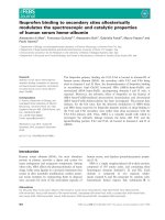
Báo cáo khoa học: Structural adaptation to low temperatures ) analysis of the subunit interface of oligomeric psychrophilic enzymes pdf
Bạn đang xem bản rút gọn của tài liệu. Xem và tải ngay bản đầy đủ của tài liệu tại đây (670.58 KB, 14 trang )
Structural adaptation to low temperatures
)
analysis of
the subunit interface of oligomeric psychrophilic enzymes
Daniele Tronelli
1
, Elisa Maugini
1
, Francesco Bossa
1
and Stefano Pascarella
1,2
1 Dipartimento di Scienze Biochimiche ‘A. Rossi Fanelli’, Universita
`
degli Studi di Roma ‘La Sapienza’, Rome, Italy
2 Centro Interdipartimentale di Ricerca per la Analisi dei Modelli e dell’Informazione nei Sistemi Biomedici (CISB), Universita
`
degli Studi di
Roma ‘La Sapienza’, Rome, Italy
Many terrestrial environments present physical and
chemical conditions that can be defined as extreme
from a human point of view. Among these, permanent
cold environments are the most common. In fact,
about 70% of the earth’s surface is covered by the
oceans, whose temperature is constantly at 4–5 °C
below a depth of 1000 m, regardless of the latitude.
Moreover, polar regions constitute a further 15% of
the earth, and there are also alpine regions and gla-
ciers. Ectothermic organisms that have colonized such
environments are called psychrophiles, and, consider-
ing their spread, represent a considerable component
of the biosphere, in terms of species diversity and
biomass. Psychrophilic organisms include eubacteria,
archaea, protozoa, fungi and multicellular eukaryotes
such as algae, invertebrates and fish [1,2].
Keywords
cold-adapted enzymes; electrostatic and
hydrophobic interactions; interface; protein
quaternary structure; psychrophiles
Correspondence
S. Pascarella, Dipartimento di Scienze
Biochimiche, Universita
`
‘La Sapienza’,
P. le A. Moro 5, 00185 Rome, Italy
Fax: +39 06 49917566
Tel: +39 06 49917574
E-mail:
Website: />(Received 8 June 2007, revised 12 July
2007, accepted 13 July 2007)
doi:10.1111/j.1742-4658.2007.05988.x
Enzymes from psychrophiles show higher catalytic efficiency in the 0–20 °C
temperature range and often lower thermostability in comparison with
meso ⁄ thermophilic homologs. Physical and chemical characterization of
these enzymes is currently underway in order to understand the molecular
basis of cold adaptation. Psychrophilic enzymes are often characterized by
higher flexibility, which allows for better interaction with substrates, and
by a lower activation energy requirement in comparison with meso ⁄ ther-
mophilic counterparts. In their tertiary structure, psychrophilic enzymes
present fewer stabilizing interactions, longer and more hydrophilic loops,
higher glycine content, and lower proline and arginine content. In this
study, a comparative analysis of the structural characteristics of the inter-
faces between oligomeric psychrophilic enzyme subunits was carried out.
Crystallographic structures of oligomeric psychrophilic enzymes, and their
meso ⁄ thermophilic homologs belonging to five different protein families,
were retrieved from the Protein Data Bank. The following structural
parameters were calculated: overall and core interface area, characteriza-
tion of polar ⁄ apolar contributions to the interface, hydrophobic contact
area, quantity of ion pairs and hydrogen bonds between monomers, inter-
nal area and total volume of non-solvent-exposed cavities at the interface,
and average packing of interface residues. These properties were compared
to those of meso ⁄ thermophilic enzymes. The results were analyzed using
Student’s t-test. The most significant differences between psychrophilic and
mesophilic proteins were found in the number of ion pairs and hydrogen
bonds, and in the apolarity of their subunit interface. Interestingly, the
number of ion pairs at the interface shows an opposite adaptation to those
occurring at the monomer core and surface.
Abbreviations
CS, citrate synthase; MDH, malate dehydrogenase; TIM, triose phosphate isomerase.
FEBS Journal 274 (2007) 4595–4608 ª 2007 The Authors Journal compilation ª 2007 FEBS 4595
To survive at temperatures close to the freezing
point of water, psychrophiles have evolved some
important cellular adaptations, including mechanisms
to maintain membrane fluidity [3,4], synthesis of cold-
acclimation proteins [5], freeze tolerance strategies [6],
and cold-active enzymes. Psychrophilic enzymes are of
great interest in the scientific community, and are cur-
rently under study to characterize their physical and
chemical properties in an attempt to understand the
molecular basis of cold adaptation.
Low temperatures have a negative effect on enzyme
kinetics: any decrease in temperature results in an
exponential decrease in reaction rate. For example,
lowering the temperature by 10 C° causes a two-fold
to four-fold decrease in enzyme activity [1,7]. There-
fore, enzymes from psychrophiles show high catalytic
efficiency in the 0–20 °C temperature range, tempera-
tures at which counterparts from mesophilic or ther-
mophilic organisms do not allow adequate metabolic
rates to support life or cellular growth. Such high
activity balances the cold-induced inhibition of reac-
tion rates. However, the structure of cold-adapted
enzymes is also heat-labile. Indeed, low stability at
moderate temperatures (usually > 40 °C) is the other
peculiar characteristic of psychrophilic enzymes [8,9].
This trend was revealed by calorimetric analysis of
residual enzyme activities after incubation at increasing
temperatures (it should be pointed out, however, that
the loss of activity at moderate temperatures might not
be always directly related to the loss of enzyme struc-
ture). It is generally believed that cold adaptation
results from a combination of lack of selective pressure
for thermostability and strong selection for high activ-
ity at low temperatures [1].
Psychrophilic enzymes are often characterized by high
flexibility [10], which allows better interaction with sub-
strates, and by lower activation energy requirements in
comparison with their mesophilic and thermophilic
counterparts. Hence, the presence of high flexibility
could explain both thermolability and high catalytic effi-
ciency at low temperatures [11]. The higher structural
flexibility of psychrophilic enzymes, as compared to
their mesophilic and thermophilic counterparts, could
be the result of a combination of several features: weak-
ening of intramolecular bonds (fewer hydrogen bonds
and salt bridges as compared to mesophilic and thermo-
philic homologs have been shown); a decrease in com-
pactness of the hydrophobic core; an increase in the
number of hydrophobic side chains that are exposed to
the solvent; longer and more hydrophilic loops; a
reduced number of proline and arginine residues; and a
higher number of glycine residues [12–15]. However,
each protein family adopts its own strategy to increase
its overall or local structural flexibility by using one or a
combination of these structural modifications.
Earlier studies on the structural adaptation of
extremophilic enzymes [16–19] were based on compara-
tive analysis, also using homology modeling in cases
where no experimental three-dimensional structures
were available [20,21]. These approaches could give
valuable information on rules to be followed by pro-
tein engineers to produce modified enzymes with suit-
able features for biotechnological applications [22]. In
fact, because of their high catalytic efficiency at low
temperatures, psychrophilic enzymes are investigated
for their high potential economic benefit: in particular,
they could be utilized in industrial processes as energy
savers, and in the detergent industry as additives
[23,24]. Also, the possibility of selecting and rapidly
inactivating these enzymes, due to their high thermola-
bility, makes psychrophilic enzymes extremely useful in
biomolecular applications [25].
Previous comparative studies investigated factors
governing cold adaptation occurring in the protein
structure core, in the enzyme active site, and in the
overall protein structure. However, although the
molecular adaptation of enzymes to extreme conditions
has been intensively studied, not very much is known
about the adaptations that have occurred at the inter-
face of oligomeric enzymes. Even less is known regard-
ing the adaptation of the psychrophilic interface of
oligomeric enzymes. Intersubunit interactions have spe-
cial importance in the stability of oligomeric psychro-
philic enzymes and their function. Indeed, interface
regions between protein monomers are mainly respon-
sible for the maintenance of the quaternary structure
in oligomeric enzymes. The hydrophobic interaction is
at the base of the process of folding and the stabiliza-
tion of protein association [26,27]. The hydrophobic
interaction occurs when apolar residues aggregate in
an aqueous environment, achieving a loss in free
energy that stabilizes the protein structure. During
association of monomers, hydrophobic residues are
buried in the interface region, minimizing the number
of thermodynamically unfavorable solute–solvent inter-
actions. Other important features, such as interface
extension, residue packing, hydrogen bonds, salt
bridges and internal cavities, can play a significant role
in the stability of the quaternary structure in cold-
adapted enzymes.
Our work is aimed at detecting the structural varia-
tion related to the cold adaptation of the subunit inter-
face of oligomeric psychrophilic enzymes. To our
knowledge, this is the first study entirely focused on
the analysis of the molecular adaptations that have
occurred at the level of subunit interfaces of psychro-
Analysis of the oligomeric psychrophilic interface D. Tronelli et al.
4596 FEBS Journal 274 (2007) 4595–4608 ª 2007 The Authors Journal compilation ª 2007 FEBS
philic enzymes. Some of the key questions are as fol-
lows. Are these interfaces significantly different from
the interfaces of mesophilic enzymes? Which structural
features are mostly variable? Are the interface adapta-
tions different from those occurring at the level of
monomer hydrophobic core and surface? The answers
to these questions can give indications about aspects
of protein–protein interaction at low temperatures and
suggest rules for interface engineering.
Results
The main dataset utilized for the analysis (Table 1)
contained five psychrophilic, 20 mesophilic, four
thermophilic and six hyperthermophilic structures,
accounting for a total of 35 oligomeric enzymes corre-
sponding to a total of 30 pairwise comparisons. The
proteins belong to five families: citrate synthase (CS)
[17], triose phosphate isomerase (TIM) [28], malate
dehydrogenase (MDH) [18], alkaline phosphatase [29]
and glyceraldehyde-3-phosphate dehydrogenase [30]. In
total, 21 incomplete side chains distributed in six pro-
teins (1cer, 1a59, 1ixe, 1ew2, 4gpd, 1bmd) were rebuilt
as described in Experimental procedures. The careful
reconstruction of the 21 incomplete side chains was nec-
essary to include in the working dataset as much infor-
mation as possible. Ideally, only complete coordinate
sets should be used, to avoid any artefact. However, the
Table 1. List of enzymes used in the work (main dataset). AP, alkaline phosphatase; GAPDH, glyceraldehyde-3-phosphate dehydrogenase;
PDB, Protein Data Bank.
Family Source organism
Growth
temperature
(°C)
Structure
resolution
(A
˚
)
Identity
a
(%)
PDB
ID
Sequence
length
(monomer) No. of subunits
1. CS Antarctic bacterium DS2–3R 5 2.09 Ref. 1a59 377 2
Escherichia coli 37 2.20 31 1k3p 426 2
Sulfolobus solfataricus 85 2.70 32 1o7x 379 2
Thermus thermophilus 85 2.30 41 1ixe 376 2
Pyrococcus furiosus 100 1.90 40 1aj8 371 2
2. GAPDH Homarus americanus 20 2.80 Ref. 4gpd 333 4
Leishmania mexicana 37 2.80 58 1a7k 358 4
Oryctolagus cuniculus 37 2.40 73 1j0x 332 4
Achromobacter xylosoxidans 30 1.70 45 1obf 335 4
Trypanosoma cruzi 37 2.75 54 1qxs 359 4
Escherichia coli 37 1.80 65 1gad 330 4
Bacillus stearothermophilus 55 1.80 53 1gd1 334 4
Thermus aquaticus 72 2.50 49 1cer 331 4
Thermotoga maritima 85 2.50 50 1hdg 332 4
Thermus thermophilus 85 2.60 48 1vc2 331 4
3. MDH Aquaspirillium arcticum 4 1.90 Ref. 1b8p 327 2
Sus scrofa 37 2.40 51 5mdh 333 2
Thermus flavus 72 1.90 62 1bmd 327 2
4. AP Pandalus borealis 5 1.92 Ref. 1k7h 476 2
Homo sapiens 37 1.82 42 1ew2 479 2
Escherichia coli 37 1.75 33 1ed9 449 2
5. TIM Vibrio marinus 15 2.65 Ref. 1aw2 255 2
Saccharomices cerevisiae 27 1.90 43 1ypi 248 2
Homo sapiens 37 2.80 41 1hti 248 2
Gallus gallus 37 1.80 41 1tph 245 2
Oryctolagus cuniculus 37 1.50 41 1r2r 248 2
Trypanosoma cruzi 37 1.83 41 1tcd 248 2
Caenorhabditis elegans 22 1.70 46 1mo0 249 2
Escherichia coli 37 2.60 65 1tre 255 2
Entamoeba histolytica 37 1.50 41 1m6j 261 2
Plasmodium falciparum 37 2.20 38 1ydv 246 2
Leishmania mexicana 37 1.83 39 1amk 250 2
Trypanosoma brucei 41 1.80 39 1tpf 250 2
Bacillus stearothermophilus 55 2.40 42 2btm 250 2
Thermotoga maritima 85 2.85 39 1b9b 252 2
a
Percentage residue identity to the psychrophilic reference (Ref.) homolog sequence.
D. Tronelli et al. Analysis of the oligomeric psychrophilic interface
FEBS Journal 274 (2007) 4595–4608 ª 2007 The Authors Journal compilation ª 2007 FEBS 4597
21 side chains represent in this case only 0.3% of all the
interface residues contained in the working dataset.
Consequently, even if, in the worst case, the reconstruc-
tion was not correct, the potential effect on the final
statistics would be negligible. The average sequence
identity was calculated between enzyme pairs of the
same family, and gave a value of 49.0% over all protein
families.
The differences in structural features observed
between the psychrophilic and mesophilic enzymes
were compared with the differences between the same
properties calculated from an oligomeric mesophilic
reference dataset (Table 2). This nonredundant refer-
ence dataset contained 148 protein structures belonging
to 43 oligomeric enzyme families, with an average
sequence identity between enzyme pairs of the same
family of 52.4%. The dataset generated a total of 514
pairwise comparisons. The whole dataset included 10
mainly-a domains, two mainly-b domains, and 53
a–b domains. The taxonomic composition of the
Table 2. List of mesophilic enzymes used in the reference dataset.
Family Protein Data Bank IDs
01. Alcohol dehydrogenase 1deh, 1p1r, 1p0f, 1e3i, 1cdo
02. Glycine-N-methyltransferase 1d2c, 1r8x, 1r74
03. Glutathione-disulfide reductase 1get, 1bwc, 1onf, 1gxf
04. Glutathione transferase 1xwk, 1b4p, 1c72, 1dug
05. Aspartate transaminase 7aat, 1ajs, 1yaa, 1art
06. Serine protease 1dsu, 3rp2
07. AICAR transformylase 1m9n, 1pkx
08. Methylenetetrahydrofolate dehydrogenase 1a4i, 1b0a
09. Phospholipase A
2
1ijl, 1pp2, 1vip, 1cL5, 1kvo
10. 3-Dehydroquinate dehydratase 1gqn, 1sfl
11. Adenosylhomocysteinase 1a7a, 1v8b
12. Thymidylate synthase 2tdm, 1tjs, 1sej, 2tsr, 1hw4, 1f28
13. Phosphorylase 1qm5, 1xoi, 1lwo, 1ygp
14. Creatine kinase 2crk, 1vrp, 1qh4, 1g0w
15. 3-Isopropylmalate dehydrogenase 1cm7, 1cnz
16.
L-lactate dehydrogenase 1lld, 1ez4, 9ldb, 1i0z, 1v6a
17. Fructose bisphosphate aldolase 1qo5, 1fba, 1epx, 1f2j, 1zah, 1a5c
18. Xylose isomerase 1xim, 1s5n, 1qt1
19. 2-Haloacid dehalogenase 1aq6, 1jud
20. Superoxide dismutase (iron ⁄ manganese) 1isa, 3sdp, 2awp, 2a03, 1uer, 1y67
21. Superoxide dismutase (copper ⁄ zinc) 1hL4, 1q0e, 1xso, 1to5, 1f1g
22. dTDP-glucose-4,6-dehydratase 1bxk, 1g1a, 1ket, 1r66
23. Citrate synthase 1csh, 2cts
24. Ribonuclease 1bsr, 1z7x, 1rra
25. Adenylosuccinate synthase 1ade, 1p9b
26. Adenylate kinase 2ar7, 3adk, 4ake
27. Lactoylglutathione lyase 1fro, 1fa8
28. Inositol phosphate phosphatase 1imb, 2bji
29. dTMP kinase 1e98, 1tmk
30. Phosphopyruvate hydratase 1ebh, 1te6, 1pdz, 1e9i, 1iyx
31. NADPH dehydrogenase (quinone) 1qr2, 1qrd, 1dxq, 1d4a
32. Ribonucleoside diphosphate reductase 1r2f, 1uzr
33. Carboxypeptidase C 1ivy, 1wpx
34. Transketolase 1trk, 1r9j, 1qgd
35. Protein kinase 1a06, 1h1w
36. Hypoxanthine phosphoribosyltransferase (dimeric) 1tc1, 1pzm, 1hgx
37. Hypoxanthine phosphoribosyltransferase (tetrameric) 1grv, 1z7g, 1cjb
38. 6-Phosphofructokinase 1pfk, 1zxx
39. Steroid D-isomerase 1opy, 8cho
40. Glycine hydroxymethyltransferase 1dfo, 1ls3, 2a7v, 1eji
41. Alkaline phosphatase 1ew2, 1ed9
42. Triose phosphate isomerase 1ypi, 1hti, 1tph, 1r2r, 1tcd, 1mo0, 1tre, 1m6j, 1ydv, 1amk, 1tpf
43. Glyceraldehyde-3-phosphate dehydrogenase 1a7k, 1j0x, 1obf, 1qxs,1gad
Analysis of the oligomeric psychrophilic interface D. Tronelli et al.
4598 FEBS Journal 274 (2007) 4595–4608 ª 2007 The Authors Journal compilation ª 2007 FEBS
dataset included species belonging to the prokaryotes
and eukaryotes (comprising protozoa and multicellular
organisms such as invertebrates, fish, and mammals).
The significance of the observed differences in
structural properties, calculated as described in
Experimental procedures, was measured by a t-value.
t-values ¼ +1.96 or t-values ¼ )1.96 with a number
of degrees of freedom > 500 correspond to a
P-value ¼ 5% that the null hypothesis is true.
This value represents the significance threshold
adopted in our analyses.
The structural properties tested at the subunit
interface were: interface and core interface extension;
number of ion pairs and hydrogen bonds; fraction of
apolar contact surface; atomic packing; volume and
internal surface area of interface cavities; and fraction
of apolar surface in the interface and in the core inter-
face.
Whenever applicable, the properties were normalized
by interface extension and number of interface resi-
dues. However, as there was no difference between the
two normalizations, only the former is considered
here.
Table 3 shows the t-values and the percentage prob-
abilities relative to the structural differences in the
number of strong, weak and total ion pairs and hydro-
gen bonds between psychrophilic and meso⁄ thermo-
philic homologs. The t-value relative to the strong ion
pairs at the interface indicates a significant increase in
these electrostatic interactions in the psychrophilic
enzymes as compared to the mesophilic enzymes. The
same results were found for weak and total ion pairs.
The significance of this trend decreased in the compari-
son of psychrophilic with both mesophilic and thermo-
philic enzymes. Figure 1 shows the normalized mean
number of total ionic interactions at the interface,
calculated from psychrophilic, mesophilic and thermo-
philic protein structures for each family of the main
dataset. The number of total ion pairs at the interface
was normalized by the number of residues composing
the interface. For each one of the five enzyme families
considered, the number of ion pairs was higher in psy-
chrophilic proteins than in mesophilic ones, whereas in
three cases out of four (atomic coordinates of thermo-
philic AP are not available), the number of ion pairs
was higher in thermophilic proteins than in mesophilic
ones, with the exception of the TIM enzyme family.
No significant trend was found in the comparison of
strong ion pairs between psychrophilic and meso ⁄ ther-
mophilic homologs (Table 3). The t-value for hydrogen
bonds (Table 3) showed a significant decrease in these
interactions at the oligomeric interface of psychrophilic
enzymes when compared to their mesophilic counter-
parts. The trend held for the number of hydrogen
bonds per unitary surface and per interface residue.
The inclusion of thermophilic enzymes in the compari-
son increased both tendencies.
The volume of the interface internal cavities, as well
as the amino acid packing at the interface, did not
show any measurable difference between psychrophilic
and mesophilic proteins, and therefore the results are
not shown.
Psychrophilic enzymes (Table 4) showed a significant
decrease in the apolar fraction of interface in compari-
son with their mesophilic counterparts (t-value of
) 2.12, P ¼ 3.44). The trend was strengthened by the
inclusion of thermophilic enzymes in the comparison.
Figure 2 shows the percentage of apolar interface,
Table 3. Statistical analysis of the differences in the number of
interface ion pairs and hydrogen bonds. The electrostatic interaction
quantities showed were normalized by mean interface surface.
psy, psychrophilic; mes, mesophilic; therm, thermophilic.
Interface structural property t-value P-value (%)
Strong ion pairs
psy versus mes 2.69 7.4 · 10
)1
psy versus mes + therm 0.85 39.57
Weak ion pairs
psy versus mes 5.85 8.75 · 10
)7
psy versus mes + therm 4.62 4.83 · 10
)4
Total ion pairs
psy versus mes 6.12 1.86 · 10
)7
psy versus mes + therm 4.20 3.13 · 10
)3
Hydrogen bonds
psy versus mes ) 2.60 9.6 · 10
)1
psy versus mes + therm ) 3.02 2.6 · 10
)1
0
0.1
0.2
0.3
0.4
0.5
0.6
0.7
Salt bridges per interface residue
GAPDHAPTIM CS MDH
Fig. 1. Normalized mean number of psychrophilic (white), mesophilic
(gray) and thermophilic (black) interface total ionic interactions, calcu-
lated from protein structures for each family of the main dataset. The
number of total ion pairs at the interface was normalized by the num-
ber of residues composing the interface.
D. Tronelli et al. Analysis of the oligomeric psychrophilic interface
FEBS Journal 274 (2007) 4595–4608 ª 2007 The Authors Journal compilation ª 2007 FEBS 4599
calculated from psychrophilic, mesophilic and thermo-
philic protein structures for each family of the main
dataset. Each apolar interface area was normalized by
the total interface area. With the exception of the CS
enzyme family, the percentage of apolar interface was
lower in cold-adapted enzymes than in mesophilic
ones, whereas in three cases out of four, the percentage
of apolar interface was higher in thermophilic proteins
than in mesophilic ones, with the exception of the
MDH enzyme family. A similar significant trend was
found for the hydrophobic contact area at the interface
(Table 4).
No significant trend was detected in the comparison
of the percentage of overall interface area between
psychrophilic and mesophilic proteins. However, this
trend became significant upon inclusion of thermophilic
enzymes (t-value of ) 2.19) in the comparison (Table 4).
Psychrophilic enzymes (Table 4) did not show signif-
icant variation of the core interface area and of the
core interface apolar atomic composition when com-
pared to mesophilic counterparts and to meso ⁄ thermo-
philic counterparts.
Table 5 reports the results of the validation of the ref-
erence mesophilic dataset to exclude potential statistical
bias on the t-tests applied to the psychrophilic proteins.
The number of randomized tests out of 1000 trials that
resulted in a nonsignificant t-value were recorded for
each structural properties. On average, a structural
property obtained a nonsignificant t-value in 820 out of
1000 randomized tests. This suggests that the reference
dataset appropriately represents the mesophilic proteins
in the main dataset.
Discussion
This research was aimed at elucidating the adaptations
that have occurred at the interface of oligomeric
enzymes synthesized by psychrophilic microorganisms.
We analyzed the structural differences between the
oligomeric interfaces of psychrophilic and meso ⁄ ther-
mophilic homologs. Psychrophilic oligomeric enzymes
must maintain high structural flexibility and, at the
same time, the correct quaternary structure. Hence, the
comparison was focused on those physicochemical
characteristics of the interface that are related to struc-
tural stability, namely: apolar contact surface; number
of ionic interactions and hydrogen bonds; atomic
packing; presence of cavities; percentage of apolar
Table 4. Statistical analysis of the differences in the percentage of
apolar interface, the hydrophobic contact area of interface, and the
percentage of overall interface area. psy, psychrophilic; mes, meso-
philic; therm, thermophilic.
Interface structural property t-value P-value (%)
Percentage of apolar interface
psy versus mes ) 2.12 3.44
psy versus mes + therm ) 3.29 1.1 · 10
)1
Hydrophobic contact area
in the interface
psy versus mes ) 2.34 1.96
psy versus mes + therm ) 3.55 4.0 · 10
)2
Percentage of overall
interface area
psy versus mes ) 1.01 31.29
psy versus mes + therm ) 2.19 2.90
Percentage of apolar
core interface
psy versus mes 1.66 9.75
psy versus mes + therm 1.00 31.77
Percentage of overall
core interface area
psy versus mes ) 1.11 26.75
psy versus mes + therm ) 1.83 6.78
0
10
20
30
40
50
60
70
80
Apolar interface area / Total interface area
GAPDHAPTIM CS MDH
Fig. 2. Percentage of psychrophilic (white), mesophilic (gray) and
thermophilic (black) apolar interface, calculated from protein struc-
tures for each family of the main dataset. Each apolar interface
area was normalized by the total interface area.
Table 5. Statistical validation of the reference mesophilic dataset.
Interface structural property
% not
significant
a
Strong ion pairs 79.1
Weak ion pairs 84.7
Total ion pairs 82.8
Hydrogen bonds 85.3
Percentage of apolar interface 74.0
Hydrophobic contact area in the interface 83.7
Percentage of overall interface area 78.1
Percentage of apolar core interface 85.5
Percentage of overall core interface area 84.5
a
Fraction of randomized trials that resulted in a nonsignificant
t-value.
Analysis of the oligomeric psychrophilic interface D. Tronelli et al.
4600 FEBS Journal 274 (2007) 4595–4608 ª 2007 The Authors Journal compilation ª 2007 FEBS
surface; and interface and core interface extension. It
could be argued that cold interface adaptations may
be similar to those occurring at the level of the mono-
mer hydrophobic core and surface, which have already
been thoroughly studied. However, it was previously
shown that hydrophobic interactions play a less rele-
vant role in protein binding than in protein folding
[31]. Furthermore, in 1997, Xu and colleagues found
that hydrophilic bridges, established between charged
or polar atoms, can result in stronger stabilization in
monomer–monomer binding than in the interior of
monomers, due to the different environments to which
such interactions are exposed [32]
2
. All of these findings
prompted one of our initial questions: are the psychro-
philic interface adaptations different from those occur-
ring at the level of the protein hydrophobic core and
surface? We believe that our comparative analysis can
correctly answer this question.
However, one of the criticisms of such comparative
studies is the way in which the statistical significance
of the differences is assessed. Therefore, in this analy-
sis, for each structural feature, a robust reference dis-
tribution of the differences observed in the comparison
of 148 mesophilic protein interfaces belonging to 43
oligomeric enzyme families was calculated. Such a dis-
tribution answers the question of what difference
should be expected for the structural property if the
interfaces of two homologous mesophilic enzymes were
compared. The t-test should then establish whether the
magnitude of the differences detected between the psy-
chrophilic enzyme and the meso ⁄ thermophilic counter-
parts is significantly different from that expected from
a mesophilic–mesophilic comparison.
The features that showed a significant difference
from the reference sample were: (a) increase in the
number of ionic interactions; (b) decrease in the num-
ber of hydrogen bonds; (c) decrease in the fraction of
apolar interface; and (d) decrease in the apolar contact
surface.
However, the cold-adapted enzymes considered here
also showed a significant decrease in the overall inter-
face area, when compared with both mesophilic and
thermophilic homologs, but this trend disappeared
after comparison with the sole mesophilic oligomers
(Table 4). This suggests that the thermophilic enzymes
need a wider interface to maintain oligomer stability,
but psychrophilic counterparts obtain no advantage
from the shrinkage of the interface extension.
The increase in the number of strong, weak and total
ion pairs at the interface of psychrophilic enzymes is
important in maintaining the quaternary structure,
whereas the strength of hydrophobic interactions is
diminished at low temperature. It should be considered
that, at moderate temperatures, hydrophobic interac-
tions are the most relevant forces for the preservation
of enzyme structure. On the other hand, the trend for
apolar surfaces to interact with other apolar surfaces,
rather than with water, decreases at low temperatures,
because solvation of nonpolar surface is thermodynam-
ically favored at low temperatures. This effect can lead
to cold-induced denaturation, particularly of the most
hydrophobic proteins, as well as oligomeric enzymes
[33]. Moreover, it was previously observed [34] that
psychrophilic enzymes are affected by the weakening of
hydrophobic interactions at low temperatures. Hence,
in these conditions, hydrophobic interactions are less
relevant in maintaining the quaternary structure, and
this phenomenon is reflected in the significant decrease
in apolar components at the interface that we have
found for oligomeric psychrophilic enzymes. Moreover,
our analysis underlines a significant decrease in the
interface apolar contact area when comparing psychro-
philic and mesophilic enzymes or psychrophilic and
mesophilic plus thermophilic enzymes. The same trend
was previously found in the analysis of hydrophobicity
in core residues of psychrophilic proteins [35]. Indeed,
buried residues in psychrophilic enzymes show weaker
hydrophobicity than those in their mesophilic homo-
logs, making the protein interior less compact and
more flexible. Therefore, a lower degree of hydropho-
bic interaction renders the role of salt bridges more
relevant in stabilizing the protein quaternary structure
of oligomeric cold-adapted enzymes, particularly if we
consider that, as the formation of ion pairs is an exo-
thermic electrostatic interaction, they are particularly
strong at low temperatures. A similar hypothesis was
put forward by Russell et al. [17] with regard to cold-
active CS in comparison with the hyperthermophilic
homolog. The authors observed an increase in psychro-
philic intramolecular ion pairs, but paradoxically also a
reduced number of interface ion pairs. They concluded
that a large number of intramolecular ion pairs
may serve to counteract the reduced thermodynamic
stabilization due to hydrophobic interaction at low
temperatures, preventing the cold denaturation of psy-
chrophilic CS. However, the psychrophilic enzyme
showed a reduction in the extent of intersubunit ion
pairs in comparison with the hyperthermophilic homo-
log. Another comparative study of psychrophilic MDH
and its thermophilic counterpart revealed the same
trend: the cold-adapted enzyme had more intrasubunit
and fewer intersubunit ion pairs [18]. This is in appar-
ent contrast with our results. Indeed, in our analysis,
we observed a significant increase in interface ion pairs
for psychrophilic enzymes when compared exclusively
to mesophilic homologs (Fig. 1). The significance of
D. Tronelli et al. Analysis of the oligomeric psychrophilic interface
FEBS Journal 274 (2007) 4595–4608 ª 2007 The Authors Journal compilation ª 2007 FEBS 4601
this trend decreased when psychrophilic enzymes were
compared to mesophilic plus thermophilic proteins.
This suggests that, in general, psychrophilic enzymes
establish, on average, a number of ionic interactions at
the subunit interface that is slightly lower or compara-
ble with that observed in thermo ⁄ hyperthermophilic
counterparts, but definitely higher than in mesophilic
proteins.
It should be noted, therefore, that although psychro-
philic and thermophilic enzymes are adapted to oppo-
site temperature conditions, the structural adaptation
strategies, relying on ionic interactions, appear to be
similar. A consistently higher number of interface salt
bridges, in comparison to that present in mesophilic
oligomers (Fig. 1), could therefore be useful to
improve the cohesion between monomers and to avoid
both cold-induced and heat-induced unfolding in psy-
chrophilic and thermophilic enzymes, respectively.
These results underline the fact that the comparative
analyses for determining the structural differences
related to thermal adaptation of psychrophiles should
always include both mesophilic and thermophilic coun-
terparts to ensure that the significant structural differ-
ences are appreciated.
The role of hydrogen bonds in psychrophilic protein
adaptation is widely accepted. Previous studies showed
that cold-adapted enzymes have fewer total hydrogen
bonds than their meso ⁄ thermophilic homologs.
Accordingly, we found a significant decrease in the
number of hydrogen bonds at the interface of psychro-
philic oligomers when compared with mesophilic
enzymes. This trend increased when thermo ⁄ hyper-
thermophilic enzymes were included in the working
dataset. Our findings underline the role of this kind of
electrostatic interaction in determining greater stability
of the quaternary structure in mesophilic and thermo-
philic proteins; in heat-labile cold-adapted enzymes,
the number of interface hydrogen bonds is lower. At
the moment, no satisfactory mechanistic explanation
for the decrease in the number of interface hydrogen
bonds has been proposed.
Other structural features analyzed did not show any
significant trend. In particular, no significant trend was
detected in the comparison of the percentage of core
interface area and in the comparison of the core inter-
face apolar atomic composition between psychrophilic
and mesophilic proteins. These results, showing that
the percentage of core interface area does not show a
significant difference, could be interpreted in the light
of the work of Bahadur et al. [36]. These authors stud-
ied the subunit interfaces of 122 homodimers, and
showed that the distribution of the area between the
rim and core interface varies widely from one oligomer
to another. This could lead to a large value for the
standard deviation of distributions of the rim and core
interface extension, and, as a consequence, could lead
to the small t-value.
An analysis of amino acid packing in mesophilic and
thermophilic enzymes was performed by Karshikoff &
Ladenstein [37] to determine the role of packing density
in thermostability. They concluded that mesophilic and
thermophilic proteins do not differ in the degree of
packing. Likewise, our analysis did not find any mea-
surable difference in the amino acid packing at the
interface of psychrophilic enzymes and in the total vol-
ume of internal interface cavities, and for this reason
the results are not shown.
In conclusion, the answers to our initial questions
reveal that the interfaces of oligomeric psychrophilic
enzymes are significantly different from those of their
mesophilic and thermophilic homologs. The most vari-
able features are the increase in the number of ionic
interactions, the decrease in the number of hydrogen
bonds, the decrease in the fraction of apolar interface,
and the decrease in the apolar contact surface. There-
fore, the structural adaptations observed are similar to
those occurring at the monomer core and surface, with
the notable exception of the increase in the number of
ionic interactions. Indeed, it has been reported that, in
general, the flexibility of the monomeric structure is
often achieved via a reduction of electrostatic inter-
actions in psychrophiles. Our results suggest that the
interfaces of oligomeric psychrophilic enzymes need to
be stabilized by the introduction of additional ion
pairs.
It should be considered that relatively few structures
of oligomeric psychrophilic enzymes are presently
available. Therefore, although the conclusions reported
here are correct from the statistical point of view, par-
ticularly considering the robust testing procedure
adopted, the results may change with the availability
of significantly more data. To confirm the results
described here, the analysis should be repeated when
more structures of psychrophilic oligomeric enzymes
are available.
Several other analyses of the structural basis of
enzyme cold adaptation have recently appeared in the
literature. For example, Jahandideh et al. [38] reported
a statistical analysis of the sequence and structural
parameters enhancing adaptation of proteins to low
temperatures. Their work was aimed at the detection
of variations in structural properties for the entire
enzyme molecule, without any focus on the subunit
interface. Indeed, they considered both monomeric and
oligomeric enzymes in their dataset, which included
13 pairs of homologous psychrophilic and mesophilic
Analysis of the oligomeric psychrophilic interface D. Tronelli et al.
4602 FEBS Journal 274 (2007) 4595–4608 ª 2007 The Authors Journal compilation ª 2007 FEBS
proteins. The structural properties tested were residue
frequencies, helical and tight turn content, backbone
hydrogen bonds, and disulfide bonds. They assessed
the significance of the differences between the average
values of each structural property taken into account,
calculated over 13 psychrophilic and mesophilic homo-
logs. Moreover, they utilized a t-test with a significance
threshold lower than that which we used in our analy-
sis, corresponding to a P-value equal to 0.1 with 24
degrees of freedom. These differences make it difficult
to relate the results presented here to those reported
by Jahandideh et al. [38], as well as to those of previ-
ous analyses. Indeed, to our knowledge, the analysis
described here is the first systematic study of cold
adaptation at the level of the subunit interface. None-
theless, Jahandideh et al. [38] came to the conclusion
that the number of hydrogen bonds and, generally, the
number of electrostatic interactions are decreased in
psychrophilic proteins.
It should be noted that, although each enzyme fam-
ily has its own strategy to increase flexibility by using
one or a combination of the above alterations in struc-
tural features [1], even with a relatively limited number
of psychrophilic enzyme structures available, some
general trends involved in the maintenance of both
structural flexibility and quaternary stucture in oligo-
meric psychrophilic enzymes can be appreciated by
comparative analysis.
In conclusion, this kind of comparative analysis can
contribute to the elucidation of structural determinants
of adaptation of proteins to extreme conditions, and
can give useful hints on how to modulate, through
protein engineering, the stability and catalytic features
of enzymes of biotechnological interest.
Experimental procedures
Collection of main dataset
The crystallographic structures of the available cold-active
oligomeric enzymes were found in the Brookhaven Protein
Data Bank [39]. The search was carried out with the key-
words ‘psychro’, ‘cold’, ‘arctic’, ‘antarctic’ and the like.
Only psychrophilic enzyme structures, for which exceptional
high cold activity and low thermostability have previously
been shown, were considered. The protein structures corre-
sponding to the biological units were collected from the
Protein Quaternary Structure databank [40]. Homologous
structures from mesophilic and thermophilic organisms
were subsequently retrieved from the Protein Data Bank
and Protein Quaternary Structure databank by means of
the program blast [41]. To ensure structural homology,
only sequences sharing ‡ 30% residue identity to the
psychrophilic sequence were considered. Only unique struc-
tures were retrieved, and when there were alternative struc-
tures for the same protein, only those displaying the best
resolution and without point mutations were collected. Pro-
teins from plants were not taken into consideration, owing
to the ambiguous definition of ‘optimum temperature’ for
such organisms.
In order to assess the structural similarity within each
collected family, we performed a structural alignment using
the ce-mc program [42]. Sequences of the selected proteins
were aligned to each psychrophilic homolog. The align-
ments were then manually corrected by inspection of the
superimposed structures.
All the programs were written in PERL language and
run under IRIX 6.5 or RED HAT ENTERPRISE
LINUX 4.0 operating systems.
Crystallographic structure quality assessments
All structures showing a resolution worse than 2.85 A
˚
were
excluded from the main dataset. All the incomplete interface
side chains were rebuilt using the program biopolymer of
the insightii package (version 2005; Accelrys, San Diego,
CA, USA). The side chain rotamer displaying the lowest
nonbond energy was kept and treated as experimental.
Ligands (cofactors, inhibitors, substrate analogs, etc.) and
solvent molecules were always removed from the structures.
The quality assessment of the crystallographic structures
was carried out using procheck software [43]. Only two
structures, 4gpd and 1ypi, did not pass the procheck
stereochemical quality check, showing an overall average
G-factor below ) 0.5, which is the lowest threshold for
acceptable quality. For these, an energetic minimization
was performed using the program modeller [44] of the
insightll package, using the highest optimization level.
After the energy minimization, the quality of the two struc-
tures was evaluated using the program prosaII [45]. The
refined structures showed overall average G-factors of
) 0.47 and ) 0.02, respectively.
Identification of interface residues
In each oligomeric enzyme, the interface region was defined
as being composed of those residues that change their
solvent accessibility area in the monomeric and in the oligo-
meric state (Fig. 3). Solvent accessibility computation [47]
was performed with naccess [48]. The change in solvent
accessibility area for each residue in the monomeric state
and in the oligomeric state was calculated using a PERL
script. The interface residues were defined as those residues
that show a change in solvent accessibility area upon
monomer association. Those residues for which the change
was more than 90% were defined as composing the core
interface [36].
D. Tronelli et al. Analysis of the oligomeric psychrophilic interface
FEBS Journal 274 (2007) 4595–4608 ª 2007 The Authors Journal compilation ª 2007 FEBS 4603
The structural similarity of the subunit interfaces within
each protein family was evaluated on the basis of the multi-
ple structure alignment. To ensure that the interface was
structurally conserved within each family and the selected
structural data were comparable, the interface C
a
carbons
of each mesophilic and thermophilic member were superim-
posed on the equivalent atoms from the psychrophilic
homolog. Only interfaces showing rmsd £ 1.3 A
˚
were con-
sidered to be similar (Fig. 4). This threshold is within the
expected structural variation corresponding to the range of
sequence similarities of the multiple structure alignments
[49]. Indeed, the expected value of rmsd for a pair of
homologous proteins whose sequence identity is 30% is
equal to 1.42 A
˚
. rmsds were calculated using the deepview–
swiss-pdbviewer iterative magic fit tool [50] and the
insightII package (version 2005; Accelrys).
Surface characteristics
naccess was utilized to calculate the percentages of the
overall surface composing the interface and the core inter-
face, the percentages of polar and nonpolar atomic contri-
butions to the interface, and the percentages of polar and
nonpolar atomic contributions to the core interface.
The overall hydrophobic contact area between residues of
different monomers was calculated using the program
pdb_np_cont [51] with the aid of a PERL script. The
pdb_np_cont program calculates pairwise atom contact
areas between apolar atoms using a set of 512 points located
on a sphere around each atom. The sphere interaction radius
of each atom is equal to the sum of the van der Waals radius
of the atom type plus the radius of a water molecule. Then,
for each atom, the closest interacting atom is found for every
point that is not buried by other atoms of the same residue.
Hydrogen bonds and ion pairs
Hydrogen bonds were calculated using hbplus [52] with
the default parameters, except for the maximum distance
between donor and acceptor, which was set to 3.5 A
˚
instead of 3.9 A
˚
, to be closer to that proposed in 1984 by
Baker & Hubbard (3.1–3.2 A
˚
) [53].
Ion pairs at the interface were identified using a PERL
script, on the basis of calculation of atomic distance. Two
residues with opposite charges are considered a strong ion
pair if the distance between charged atoms is less than 4 A
˚
.
This distance threshold is generally accepted after system-
atic analysis on a sample of protein structures [54]. Weak
ion pairs, which are established up to a distance of 8 A
˚
[16], were also considered. Positively charged atoms were
arginine and lysine side chain nitrogens. Negatively charged
atoms were side chain carboxylate oxygens of glutamate
and aspartate. Complex salt bridges, defined as ion pair
interactions joining more than two side chains, and simple
salt links involving two side chains but more than two
charged atoms were not considered in the calculations as a
single interaction; rather, each individual atomic interaction
(single ion pair) was counted [14]. Ion pairs involving histi-
dine were not considered because of the ambiguous assign-
ment of its protonation state in the proteins. Once all
interactions were found, only those between residues of dif-
ferent monomers were considered.
Packing density, cavity volume and cavity
internal surface
The atomic packing of interface residues was computed
using the os program [55]. The os package calculates the
Fig. 3. Interface and core interface of the dimer of thymidylate syn-
thase from Lactobacillus casei (2tdm). The rim interface residues,
in green, show a change in solvent accessibility area upon mono-
mer association that is smaller than 90%. Core interface residues,
in red, show a change greater than 90%. The remaining solvent-
accessible residues are shown in gray. Drawn with
PYMOL [46].
Fig. 4. Structural superimposition of 14 TIMs. Interface regions are
in red. Drawn with
INSIGHTII (version 2005, Accelrys, Cambridge, UK).
Analysis of the oligomeric psychrophilic interface D. Tronelli et al.
4604 FEBS Journal 274 (2007) 4595–4608 ª 2007 The Authors Journal compilation ª 2007 FEBS
occluded surface, defined as the molecular surface that is
less than 2.8 A
˚
from the surface of neighboring nonbonded
atoms. If a water molecule cannot fit between two atoms,
they occlude each other. To calculate the occluded surface,
normals at the molecular surface were extended outwards
until they intersected neighboring van der Waals surfaces.
The collection of extended normals and their respective
lengths were used to define the packing of each atom in an
enzyme structure.
A combination of occluded surface area and average
length of the normals was used to calculate the occluded
surface packing value for each residue. These residue
occluded surface packing values were used to calculate the
average occluded surface packing value for interface and
core interface residues.
The total surface area and the overall volume of internal
cavities between different monomers in each oligomer were
calculated using the program castp [56], which detects pro-
tein pockets and cavities using a water probe of 1.4 A
˚
radius. The detection of internal cavities was carried out
using atomic coordinates of the residues previously identi-
fied as composing the subunit interface.
Data analysis
For each oligomer, the number of hydrogen bonds, and
strong, weak and total ion pairs at the interface, were nor-
malized either by the mean interface area or by the number
of residues composing the interface. The hydrophobic con-
tact area at the interface was normalized by the overall
monomeric surface as calculated by naccess. The overall
volume of internal cavities was normalized by the interface
residue volume calculated using the VADAR server [57]
The total sur-
face area of cavities was normalized by the mean interface
area.
The structural parameters calculated on the main data-
set for each psychrophilic enzymes were compared to the
same property observed in the homologous mesophilic
and thermophilic enzymes. The comparison was carried
out with and without the thermophilic homologs in order
to assess the impact of the latter enzymes on the final
statistics.
To test whether the observed differences were statistically
significant or were instead within the expected range of var-
iation, a new reference dataset of mesophilic oligomeric
protein families was collected. To ensure a nonredundant
reference dataset, the cathsolid hierarchical domain classi-
fication tool available at the CATH server [58] (http://
www.cathdb.info/latest/index.html) was used. Only families
with different homologous superfamily domains (H-level)
were considered. Multidomain protein families were
rejected for cases when all the homologous superfamily
domains within the same family have been already encoun-
tered in other families. In each protein family, enzymes
shared ‡ 30% and < 90% sequence identity. Moreover, in
each protein family, the sequence residue identity was cal-
culated for every enzyme pair, in order to determine the
sequence identity distribution and its mean.
The differences in the values of the same structural fea-
tures considered in the analysis were calculated between all
the possible pairs of homologous enzymes in this dataset,
and a reference distribution was built for each structural
property. Likewise, the reference mean and the standard
deviation of the distributions were calculated. Each
observed difference was taken twice, once with a positive
and once with a negative sign, as there is an equal probabil-
ity of subtracting a property of enzyme A from a property
of enzyme B or a property of enzyme B from a property of
enzyme A if they are both mesophilic. Hence, the mean of
the distribution is, by definition, null. The mean and the
standard deviation (Eqns 1–3) of the distribution of the dif-
ferences of the structural properties calculated between each
possible pair of psychrophilic and homologous mesophilic
and thermophilic enzymes were calculated according to the
following equations:
DX
i
pj
¼ X
i
p
À X
i
j
ð1Þ
DX
p
¼
P
F
i¼1
P
N
i
j¼2
DX
i
pj
P
F
i¼1
ðN
i
À 1Þ
ð2Þ
rðDX
p
Þ¼
ffiffiffiffiffiffiffiffiffiffiffiffiffiffiffiffiffiffiffiffiffiffiffiffiffiffiffiffiffiffiffiffiffiffiffiffiffiffiffiffi
P
F
i¼1
P
N
i
j¼2
ðDX
i
pj
À DX
p
Þ
2
P
F
i¼1
ðN
i
À 1Þ
v
u
u
u
u
u
u
t
ð3Þ
where X is a generic property (e.g. number of hydrogen
bonds, percentage of apolar interface, etc.), F is the number
of families collected, X
i
p
is the property calculated at the
interface of the psychrophilic enzymes of the ith family, X
i
j
is the interface property of the jth homolog in the same
family (X
i
1
coincides with X
i
p
), N
i
is the number of members
in the ith family,
DX
p
is the average difference, and r(DX
p
)
is the standard deviation of the differences. Under these
conditions, a Student’s unpaired two-tailed t-test could be
applied to assess whether the distribution of the differences
of the structural features observed in the main dataset
between the psychrophilic and mesophilic enzymes was sig-
nificantly different from the distribution of the reference
differences of the same properties observed between meso-
philic enzymes of the reference dataset. In these t-tests, the
null hypothesis was that there was no difference in a given
property between psychrophilic and mesophilic structures
(Eqn 4).
DX and r(DX) were calculated both in the main
dataset containing cold-active enzymes,
DX
p
and r(DX
p
),
and in the reference mesophilic dataset
DX
m
and r(DX
m
).
D. Tronelli et al. Analysis of the oligomeric psychrophilic interface
FEBS Journal 274 (2007) 4595–4608 ª 2007 The Authors Journal compilation ª 2007 FEBS 4605
t-values were calculated according to:
t ¼
ðDX
p
À DX
m
Þ
ffiffiffiffiffiffiffiffiffiffiffiffiffiffiffiffiffiffiffiffiffiffiffiffiffiffiffiffiffiffiffiffiffiffi
½rðDX
p
Þ
2
N
p
þ
½rðDX
m
Þ
2
N
m
r
ð4Þ
where N
p
and N
m
are the numbers of differences calculated
in the main dataset containing cold-active enzymes and in
the reference mesophilic dataset; therefore, the number of
degrees of freedom corresponds to N
p
+ N
m
) 2.
Assessment of the reference dataset
To test whether the mesophilic reference dataset adequately
represents the properties of the mesophilic proteins included
in the main dataset, the following procedure was applied.
All the extremophilic proteins in the main dataset were
removed. Subsequently, one of the mesophilic proteins in
each of the main dataset family containing at least two
members was randomly labeled as ‘psychrophilic’. Then, all
the calculations involving Eqns (1–4) were repeated for
each structural property considered. The entire procedure
was repeated 1000 times. The number of times that each
trial resulted in a t-value outside the one-sided tail of the
distribution where the t-value from the real experiment fell
was then counted. For example, if the t-value for property
X was ) 5.0, then the trial t-value was considered to be not
significant if greater than ) 1.96. Finally, the percentage of
randomized tests that gave a nonsignificant t-value were
recorded for each structural property.
Acknowledgements
This work was partially supported by the funds from
‘Progetto Nazionale di Ricerche in Antartide’ of the
Italian ‘Ministero dell’Istruzione, dell’Universita
`
e della
Ricerca’. The authors are grateful to Dr Giulio Gianese
and Dr Alessandro Paiardini for help and discussion.
References
1 Feller G & Gerday C (2003) Psychrophilic enzymes: hot
topics in cold adaptation. Nat Rev Microbiol 1,
200–208.
2 Verde C, De Rosa MC, Giordano D, Mosca D, De
Pascale D, Raiola L, Cocca E, Carratore V, Giardina B
& Di Prisco G (2005) Structure, function and molecular
adaptations of haemoglobins of the polar cartilaginous
fish Bathyraja eatonii and Raja hyperborea. Biochem J
389, 297–306.
3 Russell NJ (1997) Psychrophilic bacteria: molecular
adaptations of membrane lipids. Comp Biochem Physiol
118, 489–493.
4 Jagannadham MV, Chattopadhyay MK, Subbalakshmi C,
Vairamani M, Narayanan K, Mohan RC & Shivaji S
(2000) Carotenoids of an Antarctic psychrotolerant
bacterium, Sphingobacterium antarcticus, and a meso-
philic bacterium, Sphingobacterium multivorum. Arch
Microbiol 173, 418–424.
5 Hebraud M & Potier P (1999) Cold shock response and
low temperature adaptation in psychrotrophic bacteria.
J Mol Microbiol Biotechnol 1, 211–219.
6 Jia Z, De Luca CI, Chao H & Davies PL (1996)
Structural basis for the binding of a globular antifreeze
protein to ice. Nature 384, 285–288.
7 Lonhienne T, Gerday C & Feller G (2000) Psychrophilic
enzymes: revisiting the thermodynamic parameters of
activation may explain local flexibility. Biochim Biophys
Acta 1543, 1–10.
8 D’Amico S, Marx JC, Gerday C & Feller G (2003)
Activity–stability relationships in extremophilic
enzymes. J Biol Chem 278, 7891–7896.
9 Fields PA & Somero GN (1998) Hot spots in cold
adaptation: localized increases in conformational flexi-
bility in lactate dehydrogenase A
4
orthologs of Antarc-
tic notothenioid fishes. Proc Natl Acad Sci USA 95,
11476–11481.
10 Papaleo E, Riccardi L, Villa C, Fantucci P &
De Gioia L (2006) Flexibility and enzymatic cold-
adaptation: a comparative molecular dynamics investi-
gation of the elastase family. Biochim Biophys Acta
1764, 1397–1406.
11 Feller G & Gerday C (1997) Psychrophilic enzymes:
molecular basis of cold adaptation. Cell Mol Life Sci
53, 830–841.
12 Smala
˚
s AO, Leiros HK, Os V & Willassen NP
(2000) Cold adapted enzymes. Biotechnol Annu Rev 6,
1–57.
13 Russell RJM (2000) Toward a molecular understanding
of cold activity of enzymes from psychrophiles.
Extremophiles 4, 83–90.
14 Gianese G, Bossa F & Pascarella S (2002) Comparative
structural analysis of psychrophilic and meso- and ther-
mophilic enzymes. Proteins 47, 236–249.
15 Georlette D, Blaise V, Collins T, D’Amico S, Gratia E,
Hoyoux A, Marx JC, Sonan G, Feller G & Gerday C
(2004) Some like it cold: biocatalysis at low tempera-
tures. FEMS Microbiol Rev 28, 25–42.
16 Szilagyi A & Zavodszky P (2000) Structural differences
between mesophilic, moderately thermophilic and
extremely thermophilic protein subunits: results of a
comprehensive survey. Structure Fold Des 8, 493–
504.
17 Russell RJM, Gerike U, Danson MJ, Hough DW &
Taylor GL (1998) Structural adaptations of the cold-
active citrate synthase from an Antarctic bacterium.
Structure 6, 351–361.
18 Kim SY, Hwang KY, Kim SH, Sung HC, Han YS &
Cho Y (1999) Structural basis for cold adaptation.
Sequence, biochemical properties, and crystal structure
Analysis of the oligomeric psychrophilic interface D. Tronelli et al.
4606 FEBS Journal 274 (2007) 4595–4608 ª 2007 The Authors Journal compilation ª 2007 FEBS
of malate dehydrogenase from a psychrophile Aquaspi-
rillium arcticum. J Biol Chem 274, 11761–11767.
19 Aghajari N, Feller G, Gerday C & Haser R (1998)
Crystal structures of the psychrophilic alpha-amylase
from Alteromonas haloplanctis in its native form
and complexed with an inhibitor. Protein Sci 7,
564–572.
20 Gianese G, Argos P & Pascarella S (2001) Structural
adaptation of enzymes to low temperatures. Protein Eng
14, 141–148.
21 Paiardini A, Gianese G, Bossa F & Pascarella S (2003)
Structural plasticity of thermophilic serine hydroxy-
methyltransferases. Proteins 50, 122–134.
22 Cavicchioli R, Siddiqui KS, Andrews D & Sowers KR
(2002) Low-temperature extremophiles and their appli-
cations. Curr Opin Biotechnol 13, 253–261.
23 Marshall CJ (1997) Cold-adapted enzymes. Trends
Biotechnol 15, 359–364.
24 Russell NJ (1998) Molecular adaptations in
psychrophilic bacteria: potential for biotechnological
applications. Adv Biochem Eng Biotechnol 61, 1–21.
25 Kobori H, Sullivan CW & Shizuya H (1984) Heat-labile
alkaline phosphatase from Antarctic bacteria: Rapid 5¢
end-labeling of nucleic acids. Proc Natl Acad Sci USA
81, 6691–6695.
26 Jones S & Thornton JM (1996) Principles of
protein–protein interactions. Proc Natl Acad Sci USA
93, 13–20.
27 Chothia C & Janin J (1975) Principles of protein–pro-
tein recognition. Nature 256, 705–708.
28 Alvarez M, Zeelen JP, Mainfroid V, Rentier-Delrue F,
Martial JA, Wyns L, Wierenga RK & Maes D (1998)
Triose-phosphate isomerase (TIM) of the psychrophilic
bacterium Vibrio marinus. Kinetic and structural proper-
ties. J Biol Chem 273, 2199–2206.
29 De Backer M, McSweeney S, Rasmussen HB, Riise
BW, Lindley P & Hough E (2002) The 1.9 A crystal
structure of heat-labile shrimp alkaline phosphatase.
J Mol Biol 318, 1265–1274.
30 Tanner JJ, Hecht RM & Krause KL (1996) Determi-
nants of enzyme thermostability observed in the molecu-
lar structure of Thermus aquaticus d-glyceraldehyde-3-
phosphate dehydrogenase at 2.5 Angstroms resolution.
Biochemistry 35, 2597–2609.
31 Tsai CJ, Lin SL, Wolfson HJ & Nussinov R (1996)
Studies of protein–protein interfaces: statistical analysis
of the hydrophobic effect. Protein Sci 6, 53–64.
32 Xu D, Lin SL & Nussinov R (1997) Protein binding
versus protein folding: the role of hydrophilic bridges
in protein associations. J Mol Biol 265, 68–84.
33 Creighton T (1991) Stability of folded conformations.
Curr Opin Struct Biol 1, 5–16.
34 Feller G, D’Amico D & Gerday C (1999) Thermody-
namic stability of a cold-active alpha-amylase from
the Antarctic bacterium Alteromonas haloplanctis.
Biochemistry 38, 4613–4619.
35 Siddiqui KS & Cavicchioli R (2006) Cold adapted
enzymes. Annu Rev Biochem 75, 403–433.
36 Bahadur RP, Chakrabarti P, Rodier F & Janin J (2003)
Dissecting subunit interfaces in homodimeric proteins.
Proteins 53, 708–719.
37 Karshikoff A & Ladenstein R (1998) Proteins from
thermophilic and mesophilic organisms essentially do
not differ in packing. Protein Eng 11, 867–872.
38 Jahandideh S, Abdolmaleki P, Jahandideh M & Asada-
badi EB (2007) Sequence and structural parameters
enhancing adaptation of proteins to low temperatures.
J Theor Biol 246, 159–166.
39 Berman HM, Westbrook J, Feng Z, Gilliland G, Bhat TN,
Weissig H, Shindyalov IN & Bourne PE (2000) The Pro-
tein Data Bank. Nucleic Acids Res 28, 235–242.
40 Henrick K & Thornton JM (1998) PQS: a protein qua-
ternary structure file server. Trends Biochem Sci 23,
358–361.
41 Altschul SF, Gish W, Miller W, Myers EW & Lipman
DJ (1990) Basic local alignment search tool. J Mol Biol
215, 403–410.
42 Guda C, Lu S, Scheeff ED, Bourne PE & Shindyalov
IN (2004) CE-MC: a multiple protein structure align-
ment server. Nucleic Acids Res 32, W100–W103.
43 Laskowski RA, Moss DS & Thornton JM (1993) Main-
chain bond lengths and bond angles in protein struc-
tures. J Mol Biol 231, 1049–1067.
44 Sali A & Blundell TL (1993) Comparative protein mod-
elling by satisfaction of spatial restraints. J Mol Biol
234, 779–815.
45 Sippl MJ (1993) Boltzmann’s principle, knowledge-
based mean fields and protein folding. An approach to
the computational determination of protein structures.
J Comput Aided Mol Des 7, 473–501.
46 DeLano WL (2002) The PyMOL User’s Manual.
DeLano Scientific, San Carlos, CA.
5
47 Connolly ML (1983) Solvent-accessible surfaces of pro-
teins and nucleic acids. Science 221, 709–713.
48 Hubbard SJ & Thornton JM (1993) NACCESS,
Computer Program. Department of Biochemistry and
Molecular Biology, University College, London.
49 Chothia C & Lesk AM (1986) The relation between the
divergence of sequence and structure in proteins. EMBO
J 5, 823–826.
50 Guex N & Peitsch MC (1997) Swiss-model and the
swiss-pdbviewer: an environment for comparative pro-
tein modeling. Electrophoresis 18, 2714–2723.
51 Drabløs F (1999) Clustering of non-polar contacts in
proteins. Bioinformatics 15, 501–509.
52 McDonald IK & Thornton JM (1994) Satisfying hydro-
gen bonding potential in proteins. J Mol Biol 238,
777–793.
D. Tronelli et al. Analysis of the oligomeric psychrophilic interface
FEBS Journal 274 (2007) 4595–4608 ª 2007 The Authors Journal compilation ª 2007 FEBS 4607
53 Baker EN & Hubbard RE (1984) Hydrogen bonding
in globular proteins. Prog Biophys Mol Biol 44,
97–179.
54 Barlow DJ & Thornton JM (1983) Ion-pairs in proteins.
J Mol Biol 168, 867–885.
55 Pattabiraman N, Ward KB & Fleming PJ (1995)
Occluded molecular surface: analysis of protein packing.
J Mol Recognit 8, 334–344.
56 Liang J, Edelsbrunner H & Woodward C (1998) Anat-
omy of protein pockets and cavities: measurement of
binding site geometry and implications for ligand
design. Protein Sci 7, 1884–1897.
57 Willard L, Ranjan A, Zhang H, Monzavi H, Boyko RF,
Sykes BD & Wishart DS (2003) VADAR: a web server
for quantitative evaluation of protein structure quality.
Nucleic Acids Res 31, 3316–3319.
58 Orengo CA, Michie AD, Jones S, Jones DT, Swindells
MB & Thornton JM (1997) CATH. A hierarchic classi-
fication of protein domain structures. Structure 5,
1093–1108.
4608 FEBS Journal 274 (2007) 4595–4608 ª 2007 The Authors Journal compilation ª 2007 FEBS
Analysis of the oligomeric psychrophilic interface D. Tronelli et al.