Báo cáo khoa học: Myocyte enhancer factor 2B is involved in the inducible expression of NOX1⁄ NADPH oxidase, a vascular superoxide-producing enzyme ppt
Bạn đang xem bản rút gọn của tài liệu. Xem và tải ngay bản đầy đủ của tài liệu tại đây (708.51 KB, 9 trang )
Myocyte enhancer factor 2B is involved in the inducible
expression of NOX1
⁄
NADPH oxidase, a vascular
superoxide-producing enzyme
Masato Katsuyama*, Muhammer Ozgur Cevik*, Noriaki Arakawa, Tomoko Kakehi, Toru Nishinaka,
Kazumi Iwata, Masakazu Ibi, Kuniharu Matsuno and Chihiro Yabe-Nishimura
Department of Pharmacology, Kyoto Prefectural University of Medicine, Japan
Reactive oxygen species including hydrogen peroxide,
hydroxyl radical, singlet oxygen, peroxynitrite, and
superoxide (O
2
–
) have been documented as intrinsic
signaling molecules that modulate multiple cellular
responses. The major source of O
2
–
in vascular cells
and cardiac myocytes is the NADPH oxidase family
[1–3]. NADPH oxidase catalyzes the reduction of
molecular oxygen to O
2
–
using NADPH as an electron
donor. A wealth of data has been collected on the
phagocyte NADPH oxidase, an essential component of
the host antimicrobial defense system [4]. The phago-
cyte oxidase consists of the catalytic subunit gp91phox
(NOX2), the regulatory subunits p22phox, p47phox,
p40phox and p67phox, and the small GTPase Rac.
Recent studies in nonphagocytic cells identified several
homologs of the catalytic subunit NOX2. NOX1 is
one of these homologs predominantly expressed in
colon epithelial cells (CEC) and in vascular smooth
muscle cells (VSMC).
The expression of NOX1 in VSMC is induced by
various vasoactive factors, such as angiotensin II,
platelet-derived growth factor (PDGF), phorbol ester,
and fetal bovine serum [5,6]. Among these factors,
prostaglandin (PG) F
2a
, one of the primary prostanoids
Keywords
activating transcription factor-1; myocyte
enhancer factor 2; NADPH oxidase; NOX1;
vascular smooth muscle cells
Correspondence
C. Yabe-Nishimura, Department of
Pharmacology, Kyoto Prefectural University
of Medicine, Kyoto 602–8566, Japan
Fax: +81 75 251 5348
Tel: +81 75 251 5333
E-mail:
*These authors contributed equally to this
work
(Received 2 June 2007, revised 26 July
2007, accepted 7 August 2007)
doi:10.1111/j.1742-4658.2007.06034.x
NADPH oxidase is a major source of the superoxide produced in cardio-
vascular tissues. Expression of NOX1, a catalytic subunit of NADPH oxi-
dase, is induced by various vasoactive factors, including angiotensin II,
prostaglandin (PG) F
2a
and platelet-derived growth factor (PDGF). To
clarify the molecular basis of this transcriptional activation, we delineated
the promoter region of the NOX1 gene. RT-PCR and 5¢-rapid amplifica-
tion of cDNA ends-based analyses revealed a novel 5¢-terminal exon of the
rat NOX1 gene located approximately 28 kb upstream of the exon contain-
ing the start codon. Both PGF
2a
and PDGF enhanced the transcriptional
activity of the ) 3.6 kb 5¢-flanking region of the NOX1 gene in A7r5 cells,
a rat vascular smooth muscle cell line. A PGF
2a
-response element was
located between )146 and )125 in the 5¢-flanking region containing a
consensus binding site for myocyte enhancer factor 2 (MEF2), to which
binding of MEF2 was augmented by PGF
2a
. Gene silencing of MEF2B
by RNA interference significantly suppressed the expression of NOX1,
while silencing of activating transcription factor (ATF)-1, previously impli-
cated in up-regulation of NOX1, abolished the PGF
2a
- or PDGF-induced
expression of MEF2B. These results indicate that superoxide production in
vascular smooth muscle cells is regulated by the ATF-1–MEF2B cascade
by induction of the expression of the NOX1 gene.
Abbreviations
ATF, activating transcription factor; CEC, colon epithelial cells; CRE, cAMP response element; CREB, cAMP response element-binding
protein; dsRNA, double-stranded RNA; EMSA, electrophoresis mobility shift assay; MEF, myocyte enhancer factor; PDGF, platelet-derived
growth factor; PG, prostaglandin; PI3, phosphoinositide 3; VSMC, vascular smooth muscle cells.
5128 FEBS Journal 274 (2007) 5128–5136 ª 2007 The Authors Journal compilation ª 2007 FEBS
generated in the vascular tissue, was also shown to
induce the expression of NOX1 mRNA and cause
hypertrophy of VSMC through increased generation of
O
2
–
[7]. In PGF
2a
-induced as well as PDGF-induced
NOX1 expression, transactivation of the epidermal
growth factor receptor, which depends on protein
kinase C d, elicits activation of extracellular signal-
regulated kinase 1 ⁄ 2 as well as of phosphoinositide 3
(PI3) kinase. Downstream of PI3 kinase, a transcrip-
tion factor of the cAMP response element (CRE)-
binding protein (CREB) ⁄ activating transcription factor
(ATF) family, ATF-1, was suggested to take part in the
induction of NOX1 expression [8–10].
Except for the involvement of ATF-1, the entire
picture of the transcriptional regulation of the NOX1
gene has not been clarified yet. In the upstream
region of the human NOX1 gene, an interferon-c-
responsive element was recently identified, which reg-
ulates the expression of NOX1 in CEC [11]. On the
other hand, we recently depicted novel transcripts of
the mouse NOX1 gene that were induced to express
under phenotypic modulation of VSMC. Of particular
interest is that these transcripts were governed by
promoters different from the one utilized for expres-
sion of the NOX1 transcript in CEC [12]. To clarify
the molecular basis of the transcriptional activation
of NOX1 in vascular tissue, we delineated the pro-
moter region implicated in the up-regulation of
NOX1 gene expression. We report here the predomi-
nant role of the ATF-1-myocyte enhancer factor 2B
(MEF2B) cascade in superoxide production in vascu-
lar smooth muscle cells by inducing the expression of
the NOX1 gene.
Results
Determination of the 5¢-end of the NOX1 mRNA
expressed in VSMC
We reported the expression of novel NOX1 transcripts
(c- and f-types) in mouse VSMC, which encoded an
extended N-terminal peptide sequence upstream of the
form expressed in CEC (a-type) [12]. We searched the
rat genomic database and found the counterpart of
the mouse NOX1 exon 1c, the 5¢-terminal exon of the
c-type mRNA. RT-PCR was then performed in primary
VSMC or PGF
2a
-stimulated A7r5 cells, a rat vascular
smooth muscle cell line, using a forward primer for the
rat counterpart (ex1c2F in Fig. 1A), and a reverse
primer covering the start codon of the NOX1 mRNA
(NR3 in Fig. 1A). Amplified products were sequenced
and the 5¢-terminus of the NOX1 transcript was
determined by 5¢-RACE. The 5¢-terminus encoded
433 bp of exon 1c, and it was placed upstream of
exon 1a containing the start codon (Fig. 1B). Unlike
the mouse c-type mRNA, however, the rat c-type-like
NOX1 mRNA neither contained the counterpart of the
mouse exon 1b nor encoded an additional N-terminal
peptide. Although the counterpart of the mouse exon 1f
was found in the genomic database, an f-type-like
transcript was not detected by RT-PCR in A7r5 cells.
PGF
2a
- and PDGF-induced transcriptional
activation of the NOX1 promoter
To examine whether the promoter region of the rat
NOX1 gene contains elements responsive to vasoactive
NOX1 mRNA
1a 2
chrX
3 k28 k
1c
ATG
209 96433
ATG
Intron
Exon
A
B
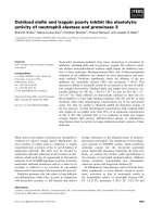
Fig. 1. Structure of the rat NOX1 gene and
mRNA. (A) 5¢-Nucleotide sequences of the
rat NOX1 cDNA and deduced amino acid
sequences at the beginning of the open
reading frame. Primers used for RT-PCR
and 5¢-RACE are indicated with arrows.
(B) The exon ⁄ intron structure of the NOX1
gene and its splicing pathway. Open boxes
indicate exons. Numbers in the box denote
the size of each exon (bp). The size of the
intron is indicated under the broken lines
(bp). Closed boxes show the open reading
frame of the transcript.
M. Katsuyama et al. MEF2B regulates vascular NOX1 ⁄ NADPH oxidase
FEBS Journal 274 (2007) 5128–5136 ª 2007 The Authors Journal compilation ª 2007 FEBS 5129
factors, approximately 3.6 kb of the 5¢-flanking region
of exon 1c was isolated and subcloned into a luciferase
vector. As demonstrated in Fig. 2A, the transcriptional
activity of the NOX1 promoter, r3642Luc, in A7r5
cells was significantly enhanced when cells were treated
with PGF
2a
or PDGF. These findings suggest that the
3.6 kb of the 5¢-flanking region contains sequences
responsible for PGF
2a
-induced as well as PDGF-
induced transcriptional activation. As the activation by
PGF
2a
was more prominent than that by PDGF,
PGF
2a
was used for the subsequent promoter analyses.
MEF2-binding site was essential for transcriptional
activation of the NOX1 promoter
To identify the region responsible for the transcrip-
tional activation, a series of deletion mutants of the
NOX1 promoter-luciferase chimera plasmids were con-
structed. As shown in Fig. 2B, deletion up to )489, but
not to )930, reduced PGF
2a
-induced transcriptional
activation, suggesting the existence of an enhancer
binding site between )930 and )489. Deletion up to
)125, but not to )146, completely abolished the
PGF
2a
-induced transcriptional activation. Between
)146 and )125, a consensus sequence of the MEF2-
binding site, 5¢-CTA(A ⁄ T)
4
TAG ⁄ A-3¢, was located
(Fig. 3A). The introduction of mutations at this
site (5¢-CTATAAATAG-3¢ to 5¢-CTATAgccAG-3¢)
abolished PGF
2a
-induced transcriptional activation
(Fig. 3B). These findings clearly indicate that the
MEF2-binding site between )139 and )130 is essential
for PGF
2a
-induced activation of the NOX1 promoter.
Binding of MEF2 to the consensus sequence in
the NOX1 promoter
To verify whether the transcription factor MEF2 actu-
ally binds to the consensus sequence in the NOX1 pro-
moter, an electrophoretic mobility shift assay (EMSA)
was carried out using nuclear extracts obtained from
A7r5 cells (Fig. 4). With the probe containing the con-
sensus MEF2-binding site of the NOX1 promoter,
several bands were observed (lane 1, Fig. 4A). Among
these bands, those indicated by arrowheads were mark-
edly diminished when the mutated probe was utilized
(lane 5). Stimulation of A7r5 cells with PGF
2a
increased
the intensity of these bands (lane 2), whereas the bands
were undetectable in the presence of an excess amount
of the unlabeled wild-type probe (lane 3). By contrast,
the bands persisted in the presence of an excess amount
of the mutated probe (lane 4). As shown in Fig. 4B,
pre-incubation of the nuclear extract with an anti-
MEF2 IgG generated supershifted bands as indicated
by arrowheads (lane 3). On the other hand, pre-incuba-
tion with an anti-ATF-1 IgG did not affect the mobility
of the specific bands (lane 4, Fig. 4B). These results
suggest that PGF
2a
increases the binding of MEF2
to the consensus-binding site located between )139
and )130 of the NOX1 promoter.
Gene silencing of MEF2B attenuated PGF
2a
-or
PDGF-induced NOX1 expression
There are four types of MEF2 – MEF2A, MEF2B,
MEF2C, and MEF2D – and these subtypes are enco-
ded by distinct genes [13]. In A7r5 cells, MEF2A,
MEF2C and MEF2D were constitutively highly
expressed, whereas the expression level of MEF2B was
very low (see Fig. 5A, Cycles of PCR). Upon stimula-
tion with PGF
2a
or PDGF, however, the expression of
0123
0123
Relative Luciferase Activity
–91
–125
–146
–235
–323
–489
–930
1c
luc
pGL3basic
MEF2
–3642
AP-1
TATA-like
Relative Luciferase Activity
PGF
2
α
A
B
PGDF
*
*
*
*
*
*
*
*
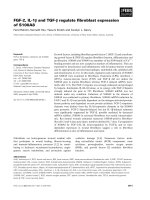
Fig. 2. Analyses of the NOX1 promoter activity in A7r5 cells.
(A) PGF
2a
- or PDGF-induced transcriptional activation of the NOX1
promoter. The 3.6 kb of the 5¢-flanking region of exon 1c was
cloned into pGL3-basic and the reporter construct was transfected
into A7r5 cells. The b-galactosidase-expression vector was cotrans-
fected as an internal control. Serum-starved cells were incubated
with 100 n
M PGF
2a
or 20 ngÆmL
)1
PDGF-BB for 24 h. The relative
luciferase activity is denoted as the fold-increase induced by PGF
2a
or PDGF. Bars represent means ±SE of three experiments.
Open bar, pGL3-basic; closed bar, r3642Luc. *P < 0.01 versus
pGL3-basic. (B) Deletion of the MEF2-binding site abolished
PGF
2a
-induced transcriptional activation. A schematic diagram of
the promoter-luciferase fusion plasmids is shown on the left,
where the 5¢⁄3¢ ends of the construct relative to the transcription
initiation site are indicated. The relative luciferase activity is
denoted as the fold-increase induced by PGF
2a
. Bars represent
means ±SE of three experiments. *P < 0.01 versus pGL3-basic.
MEF2B regulates vascular NOX1 ⁄ NADPH oxidase M. Katsuyama et al.
5130 FEBS Journal 274 (2007) 5128–5136 ª 2007 The Authors Journal compilation ª 2007 FEBS
MEF2B was markedly augmented (Fig. 6A, mock).
We therefore focused on the role of MEF2B in the
PGF
2a
- or PDGF-induced up-regulation of NOX1
gene expression. An expression vector coding a dou-
ble-stranded RNA (dsRNA) targeting nucleotides 470–
494 of the rat MEF2B mRNA sequence was intro-
duced into A7r5 cells. Following single cell cloning of
the transfectants, two clones stably expressing the
dsRNA, MEF2B-RNAi-1 and MEF2B-RNAi-2, were
isolated. In these clones, mRNA levels of MEF2B, but
not those of other types of MEF2, were reduced com-
pared to levels in the mock-transfected cells (Fig. 5A).
As shown in Fig. 5B, induction of NOX1 mRNA
expression by PGF
2a
or PDGF was almost completely
abolished in MEF2B knocked-down cells. In addition,
a PGF
2a
-induced increase in O
2
–
production as well as
the basal level of cellular O
2
–
was reduced in these
clones compared with the mock-transfected cells
(Fig. 5C). These results highlight the pivotal role of
MEF2B in the PGF
2a
- or PDGF-induced up-regula-
tion of NOX1 expression.
Gene silencing of ATF-1 attenuated PGF
2a
-or
PDGF-induced MEF2B expression
We previously reported the involvement of ATF-1, a
transcription factor of the CREB ⁄ ATF family, in the
up-regulation of NOX1 expression in VSMC [8].
ATF-1 elicits transcriptional activation by binding to
the cAMP response element (CRE). As CRE was not
found in the promoter region of the NOX1 gene,
ATF-1 was assumed to indirectly regulate the expres-
sion of the NOX1 mRNA. We therefore elucidated the
role of ATF-1 in the MEF2B-dependent activation of
NOX1 expression. As shown in Fig. 6, the expression
of MEF2B mRNA induced by PGF
2a
or PDGF was
almost completely abolished in ATF-1 knocked-down
clones that we previously isolated [8]. These findings
provide a clear link between ATF-1 and MEF2B, in
that expression of MEF2B in VSMC is governed by
ATF-1 itself.
Discussion
The major lines of evidence provided by this study are
as follows: (a) the 5¢-terminus of the rat NOX1 mRNA
expressed in VSMC was a counterpart of the mouse
c-type mRNA induced under phenotypic modulation
of VSMC; (b) the promoter region of the NOX1 gene
contained a consensus MEF2-binding site that confers
the responsiveness to PGF
2a
; (c) stimulation with
PGF
2a
enhanced the binding of MEF2 to its consensus
binding site in the NOX1 promoter; (d) RNA interfer-
ence targeted at MEF2B abolished expression of the
0123
Relative Luciferase Activity
–125
–146
luc
pGL3basic
–146mut
1c
B
A
MEF2
AP-1
TATA-like
*
Fig. 3. PGF
2a
-induced transcriptional activation of the NOX1 promoter was dependent on the MEF2-binding site. (A) A consensus sequence
of the MEF2-binding site and the AP-1 site, and a TATA-like sequence located upstream of the transcription initiation site. (B) Introduction of
mutations at the MEF2-binding site (5¢-CTATAAATAG-3¢ to 5¢-CTATAgccAG-3¢) abolished PGF
2a
-induced transcriptional activation. A sche-
matic diagram of the promoter-luciferase fusion plasmids is shown on the left, where the 5¢⁄3¢ ends of the construct relative to the tran-
scription initiation site are indicated. The relative luciferase activity is denoted as the fold-increase induced by PGF
2a
. Bars represent means
±SE of three experiments. *P<0.01 versus pGL3-basic.
M. Katsuyama et al. MEF2B regulates vascular NOX1 ⁄ NADPH oxidase
FEBS Journal 274 (2007) 5128–5136 ª 2007 The Authors Journal compilation ª 2007 FEBS 5131
NOX1 mRNA induced by PGF
2a
or PDGF; (e) RNA
interference targeted at ATF-1 attenuated the induc-
tion of MEF2B expression by PGF
2a
or PDGF. Based
on these findings and those of our earlier studies
[8–10], it is reasonable to conclude that the ATF-1-
MEF2B cascade constitutes the major signaling path-
way that leads to the up-regulation of NOX1 gene
expression in VSMC.
The 5¢-terminus of the NOX1 mRNA identified in
rat VSMC was a counterpart of the mouse c-type
mRNA which was expressed in dedifferentiated
origin
probe
MEF2
PGF
2α
α
PGF
2
α
–++++
wt wt wt wt mut
x100 cold
–– –
wt mut
supershift
lane
A
B
12 345
anti MEF2 Ab
––+–
anti ATF-1 Ab
origin
MEF2
–+++
––
lane
1234
–+
free
probes
Fig. 4. PGF
2a
increased binding of MEF2 to the consensus
sequence. (A) Specific bands detected by EMSA. Nuclear extracts
were prepared from A7r5 cells incubated with 100 n
M PGF
2a
for
8 h. Binding specificity was evaluated with a 100-fold excess of
unlabeled oligonucleotide (lane 3), and with a mutated oligonucleo-
tide probe (lane 5). (B) Supershift bands demonstrated in the
presence of an anti-MEF2 IgG. Nuclear extracts were pre-incubated
in the presence or absence of an anti-MEF2 IgG or an anti-ATF-1
IgG.
0
2
4
6
NOX1
GAPDH
NOX1/GAPDH
mock
MEF2B
RNAi-1
MEF2B
RNAi-2
Control
PGF
2α
α
PGF
2
α
PGF
2
α
Control
Control
PDGF
PDGF
PDGF
Cycles of PCR
mock
RNAi-1
RNAi-2
anti-MEF2B
A
B
C
dsRNA
GAPDH
MEF2B
MEF2A
MEF2C
MEF2D
(40)
(40)
(25)
(25)
(25)
(25)
*
*
Mean Fluorescence Values
mock MEF2BRNAi-1 MEF2BRNAi-2
0
50
60
70
80
90
*
*
*
†
†
Fig. 5. Gene silencing of MEF2B attenuated PGF
2a
- or PDGF-
induced NOX1 expression. (A) Expression of anti-MEF2B dsRNA
precursors and silencing of MEF2B expression in the MEF2B
RNAi-1 and RNAi-2 clones. Total RNA was reverse-transcribed and
the cDNA fragments were amplified by PCR. (B) Induction of the
NOX1 mRNA expression by PGF
2a
or PDGF was suppressed in
RNAi-1 and RNAi-2. A representative northern blot is shown. Bars
represent the mean ±SE of three experiments. *P<0.01 versus
mock control. (C) Ethidium fluorescence in the cells untreated
(control; open bar) or treated with 100 n
M PGF
2a
(closed bar) for
24 h. Mean values of ethidium fluorescence were calculated
from four samples. *P<0.05 versus control mock-transfected
cells. P<0.05 versus mock-transfected cells treated with PGF
2a
.
MEF2B regulates vascular NOX1 ⁄ NADPH oxidase M. Katsuyama et al.
5132 FEBS Journal 274 (2007) 5128–5136 ª 2007 The Authors Journal compilation ª 2007 FEBS
VSMC. The rat NOX1 mRNA, however, did not con-
tain the counterpart of the mouse exon 1b, which
encoded an additional N-terminal peptide upstream of
exon 1a. Unlike the NOX1 mRNA expressed in
VSMC, the transcript identified in rat colon or intact
aorta did not contain exon 1c at the 5¢-terminus (data
not shown). The transcript expressed in VSMC con-
tained an extended 5¢-untranslated region that differed
from the colon-type NOX1 mRNA. Accordingly, the
5¢-flanking region of exon 1c appears to contain ele-
ments essential for the specific expression of the NOX1
mRNA in dedifferentiated VSMC. In the aorta or in a
vascular smooth muscle cell line (T ⁄ G HA-VSMC) of
human origin, the c-type-like mRNA has not been
identified so far (data not shown). Thus there seems to
be considerable species–specific differences in the regu-
lation of the NOX gene expression in VSMC. It should
be noted that another catalytic subunit of NADPH
oxidase, NOX5, is expressed in human VSMC [14],
but not in rodents.
The MEF2-binding site in the promoter region was
shown to be elemental for induction of NOX1 expres-
sion in VSMC. This site was also conserved in the
mouse NOX1 gene. To date the functional role of
MEF2 documented in VSMC has been somewhat con-
troversial. MEF2A was reported to be involved in an
angiotensin II-induced vascular hypertrophy [15], and
in inducing the expression of monocyte chemo-
attractant protein-1 [16], which plays a key role in the
development of atherosclerosis and restenosis after
angioplasty. Conversely, MEF2B was reported to bind
to the promoter region of the smooth muscle myosin
heavy chain gene to regulate the transcription of the
smooth muscle-specific gene expression [17]. Intrigu-
ingly, MEF2B was documented to be highly expressed
in neointima of balloon-injured carotid artery [18],
which suggests that the expression of MEF2B is up-
regulated under phenotypic modulation of VSMC.
Among four types of MEF2, MEF2A, MEF2C and
MEF2D were constitutively expressed in A7r5 cells,
and the levels of these transcripts were relatively high.
By contrast, the basal level of the MEF2B mRNA was
very low, but expression of MEF2B was increased
upon stimulation with PGF
2a
or PDGF. In MEF2B
knocked-down cells, the induction of NOX1 mRNA
expression by PGF
2a
or PDGF was almost completely
abolished, whereas it was unaffected in MEF2A
knocked-down clones (data not shown). Accordingly,
inducible expression of MEF2B appears to up-regulate
the NOX1 gene expression in VSMC.
Previously, the involvement of ATF-1, a transcrip-
tion factor of the CREB ⁄ ATF family, was suggested in
the up-regulation of NOX1 expression by various
vasoactive factors [8]. ATF-1 is known to activate
transcription of genes by binding to CRE. In the
3.6 kb promoter region of the rat NOX1 gene, how-
ever, a canonical CRE was not found. This suggests
an indirect involvement of ATF-1 in the NOX1 tran-
scription, though there might be other ATF-1 binding
sites elsewhere in the NOX1 gene. We also observed
that phosphorylation of ATF-1 occurred within 5 min
of stimulation with PGF
2a
[8], whereas expression of
the NOX1 mRNA was not observed until 3 h after the
stimulation [7]. These findings support the view that
ATF-1 indirectly regulates the NOX1 gene expression,
possibly by up-regulating the expression of the genes
encoding other transcription factors. It should be
pointed out that a consensus CRE sequence was
located approximately 2.6 kb upstream of the tran-
scription start site of the rat MEF2B gene (rat MEF2B
mRNA, GenBank BC079361; genomic sequence, rat
chromosome 16). In accord with this observation, the
PGF
2a
- or PDGF-induced increase in MEF2B mRNA
was almost completely abolished in ATF-1 knocked-
down clones. These results strongly suggest the
involvement of the ATF-1-MEF2B cascade in the reg-
ulation of vascular NOX1 gene expression.
We previously reported the pathophysiological
significance of NOX1-derived reactive oxygen species
in angiotensin II-induced chronic hypertension using
NOX1-deficient mice [19]. The basal level of the
NOX1 transcript is much lower than the levels of
other NOX isoforms expressed in vascular tissue,
whereas inducible expression of NOX1 has been docu-
mented in association with various vascular disorders.
In this context, identification of the ATF-1-MEF2B
cascade involved in the up-regulation of NOX1
MEF2B
GAPDH
mock
ATF-1
RNAi-5
ATF-1
RNAi-16
Control
PGF
2
α
α
Control
PGF
2
α
Control
PGF
2
α
PDGF
PDGF
PDGF
MEF2B/GAPDH
0
1
2
3
4
5
*
Fig. 6. Gene silencing of ATF-1 attenuated PGF
2a
- or PDGF-induced
MEF2B expression. Expression of the MEF2B transcript was exam-
ined by RT-PCR in the ATF-1 RNAi-5 and RNAi-16 clones. Bars rep-
resent the mean ±SE of three experiments. *P<0.05 versus
mock control.
M. Katsuyama et al. MEF2B regulates vascular NOX1 ⁄ NADPH oxidase
FEBS Journal 274 (2007) 5128–5136 ª 2007 The Authors Journal compilation ª 2007 FEBS 5133
gene expression may lead to a better understanding
of regulatory mechanisms in vascular superoxide
production.
Experimental procedures
Materials
PGF
2a
was purchased from Nacalai Tesque (Kyoto, Japan).
Antibodies against MEF2 and ATF-1 were obtained from
Santa Cruz Biotechnology (Santa Cruz, CA). PDGF-BB
was obtained from PEPROTECH (London, UK). [c-
32
P]-
ATP, [a-
32
P]-UTP, and [a-
32
P]-dCTP were from ICN Bio-
medicals (Costa Mesa, CA).
Cell culture
The A7r5 cell line, obtained from American Type Culture
Collection (Rockville, MD), was cultured in Dulbecco’s
modified Eagle’s medium (DMEM) supplemented with
10% fetal bovine serum. Primary VSMC were isolated from
Sprague-Dawley rats by a migration method [20].
5¢-RACE
5¢-RACE was carried out using a-3¢⁄5¢-RACE kit (Roche,
Basel, Switzerland). Total RNA isolated from either A7r5
cells or primary VSMC was reverse transcribed with the
primer NR1, which is complementary to nucleotides 207–
231 of the rat NOX1 mRNA (GenBank AF152963;
Fig. 1A). The cDNAs were tagged with dATP using termi-
nal deoxytransferase. The first round of amplification was
carried out using the oligo dT-anchor primer and the sec-
ond primer NR2, complementary to nucleotides 182–206 of
the rat NOX1 mRNA. The resulting PCR products served
as templates for the subsequent nested amplification of
cDNAs specific for NOX1. For this amplification, the
anchor primer and the primer cup1R or cup2R, comple-
mentary to the sequence in exon 1c, were utilized (Fig. 1A).
Based on the sequencing analyses, the 5¢-end of the longest
cDNA clones was regarded as the transcriptional start site
and denoted as +1.
Reporter constructs and luciferase assay
The rat genomic DNA was isolated from A7r5 cells with a
PUREGENE DNA Isolation Kit (Gentra SYSTEMS, Min-
neapolis, MN). The 5¢-flanking region of the rat NOX1
gene was amplified by PCR and cloned into the vector
pGL3-basic (Promega, Madison, WI). The 3.6 kb 5¢-flank-
ing region was cloned into the HindIII site of pGL3-basic.
A series of 5¢-deletion constructs were made by cleavage
with restriction enzymes or amplification by PCR. All con-
structs were subjected to sequencing analyses to verify the
orientation and fidelity of the insert. Luciferase plasmids
(0.75 lgÆwell
)1
) and a pSV-b-galactosidase control vector
(0.25 lgÆwell
)1
; Promega) were cotransfected into A7r5 cells
with FuGENE 6 Transfection Reagent (Roche). The cells
were then cultured for 24 h, and a further 24 h in serum-
free DMEM. They were subsequently incubated for 24 h in
the presence or absence of 100 nm PGF
2a
or 20 ngÆmL
)1
PDGF-BB. Luciferase activity in cell lysates was deter-
mined and normalized with b-galactosidase activity as
described previously [21].
EMSA
The EMSA was performed essentially as described
previously [22]. A double-stranded probe containing an
MEF2-binding site was prepared by annealing complemen-
tary synthetic oligonucleotides. The sense sequence was
5¢-GATTCTTCTATAAATAGGTACTTTCCCTCA-3¢. The
sequence of the mutated probe was 5¢-GATTCTTCT
ATAgccAGGTACTTTCCCTCA-3¢. Probes were labeled at
the 5¢-end with [c-
32
P]-ATP and T4 polynucleotide kinase.
Nuclear extracts of A7r5 cells were prepared as described
previously [8]. The nuclear extracts and the labeled probe
were incubated at 25 °C for 30 min, resolved in a 4%
polyacrylamide gel, and analyzed using a Fujix BAS 2000
Bio-imaging Analyzer (Fuji Film, Tokyo, Japan).
Gene silencing of MEF2B
The anti-MEF2B dsRNA was designed against nucleotides
470–494 of the rat MEF2B mRNA sequence (GenBank
BC079361). Sense and antisense oligonucleotides containing
the hairpin sequence, the terminator sequence, and over-
hanging sequences were synthesized. By annealing over-
hanging sequences of the synthetic oligonucleotides, PCR
was performed to amplify the sequence encoding the
dsRNA, which was inserted into pPUR-KE harboring a
tRNA
Val
promoter. A7r5 clones stably expressing the anti-
MEF2B dsRNA were obtained as described previously [7].
Cells incubated for 48 h in DMEM containing 0.5%
fetal bovine serum were exposed to 100 nm PGF
2a
or
20 ngÆmL
)1
PDGF-BB for 24 h, and used for the subse-
quent isolation of total RNA. Northern blot analysis and
measurement of intracellular O
2
–
production using a flow
cytometer were performed as described previously [7,8]. The
geometric mean of ethidium fluorescence intensity was used
for analysis.
Statistical analysis
Values were expressed as the mean ± SE. The statistical
analysis was performed with Student’s t-test. For multiple
treatment groups, a one-way anova followed by Bonferroni’s
t-test was applied.
MEF2B regulates vascular NOX1 ⁄ NADPH oxidase M. Katsuyama et al.
5134 FEBS Journal 274 (2007) 5128–5136 ª 2007 The Authors Journal compilation ª 2007 FEBS
Acknowledgements
This work was supported in part by Grant-in-Aid for
Young Scientists (B) 17790173 from The Ministry of
Education, Culture, Sports, Science and Technology of
Japan (MK). The nucleotide sequences reported in this
paper have been submitted to DDBJ ⁄ EMBL ⁄ GenBank
with accession number AB258525. We thank Dr
S. Tsuchiya, Graduate School of Pharmaceutical
Sciences, Kyoto University, for valuable discussions
and advice.
References
1 Griendling KK, Sorescu D & Ushio-Fukai M (2000)
NAD(P)H oxidase: role in cardiovascular biology and
disease. Circ Res 86, 494–501.
2 Irani K (2000) Oxidant signaling in vascular cell growth,
death, and survival: a review of the roles of reactive
oxygen species in smooth muscle and endothelial cell
mitogenic and apoptotic signaling. Circ Res 87, 179–
183.
3 Guzik TJ, West NE, Black E, McDonald D, Ratna-
tunga C, Pillai R & Channon KM (2000) Vascular
superoxide production by NAD(P)H oxidase: associa-
tion with endothelial dysfunction and clinical risk
factors. Circ Res 86, E85–E90.
4 Gallin JI (1993) Delineation of the phagocyte NADPH
oxidase through studies of chronic granulomatous dis-
eases of childhood. Int J Tissue React 15, 99–103.
5 Suh YA, Arnold RS, Lassegue B, Shi J, Xu X, Sorescu D,
Chung AB, Griendling KK & Lambeth JD (1999) Cell
transformation by the superoxide-generating oxidase
Mox1. Nature 401, 79–82.
6 Lassegue B, Sorescu D, Szocs K, Yin Q, Akers M,
Zhang Y, Grant SL, Lambeth JD & Griendling KK
(2001) Novel gp91 (phox) homologues in vascular
smooth muscle cells: nox1 mediates angiotensin II-
induced superoxide formation and redox-sensitive sig-
naling pathways. Circ Res 88, 888–894.
7 Katsuyama M, Fan C & Yabe-Nishimura C (2002)
NADPH oxidase is involved in prostaglandin F2alpha-
induced hypertrophy of vascular smooth muscle cells:
induction of NOX1 by PGF2alpha. J Biol Chem 277,
13438–13442.
8 Katsuyama M, Fan C, Arakawa N, Nishinaka T, Miy-
agishi M, Taira K & Yabe-Nishimura C (2005) Essen-
tial role of ATF-1 in induction of NOX1, a catalytic
subunit of NADPH oxidase: involvement of mitochon-
drial respiratory chain. Biochem J 386, 255–261.
9 Fan C, Katsuyama M, Nishinaka T & Yabe-Nishimura C
(2005) Transactivation of the EGF receptor and a PI3
kinase-ATF-1 pathway is involved in the upregulation of
NOX1, a catalytic subunit of NADPH oxidase. FEBS
Lett 579, 1301–1305.
10 Fan C, Katsuyama M & Yabe-Nishimura C (2005)
PKCdelta mediates up-regulation of NOX1, a catalytic
subunit of NADPH oxidase, via transactivation of
the EGF receptor: possible involvement of PKCdelta
in vascular hypertrophy. Biochem J 390, 761–767.
11 Kuwano Y, Kawahara T, Yamamoto H, Teshima-
Kondo S, Tominaga K, Masuda K, Kishi K, Morita K
& Rokutan K (2006) Interferon-gamma activates tran-
scription of NADPH oxidase 1 gene and upregulates
production of superoxide anion by human large intesti-
nal epithelial cells. Am J Physiol Cell Physiol 290,
C433–C443.
12 Arakawa N, Katsuyama M, Matsuno K, Urao N, Tab-
uchi Y, Okigaki M, Matsubara H & Yabe-Nishimura C
(2006) Novel transcripts of NOX1 are regulated by
alternative promoters and expressed under phenotypic
modulation of vascular smooth muscle cells. Biochem J
398, 303–310.
13 Black BL & Olson EN (1998) Transcriptional control of
muscle development by myocyte enhancer factor-2
(MEF2) proteins. Annu Rev Cell Dev Biol 14, 167–196.
14 Banfi B, Molnar G, Maturana A, Steger K, Hegedus B,
Demaurex N & Krause KH (2001) A Ca(2+)-activated
NADPH oxidase in testis, spleen, and lymph nodes.
J Biol Chem 276, 37594–37601.
15 Suzuki E, Nishimatsu H, Satonaka H, Walsh K, Goto A,
Omata M, Fujita T, Nagai R & Hirata Y (2002)
Angiotensin II induces myocyte enhancer factor 2- and
calcineurin ⁄ nuclear factor of activated T cell-dependent
transcriptional activation in vascular myocytes. Circ Res
90, 1004–1011.
16 Suzuki E, Satonaka H, Nishimatsu H, Oba S, Takeda R,
Omata M, Fujita T, Nagai R & Hirata Y (2004) Myocyte
enhancer factor 2 mediates vascular inflammation via the
p38-dependent pathway. Circ Res 95, 42–49.
17 Katoh Y, Molkentin JD, Dave V, Olson EN & Perias-
amy M (1998) MEF2B is a component of a smooth
muscle-specific complex that binds an A ⁄ T-rich element
important for smooth muscle myosin heavy chain gene
expression. J Biol Chem 273, 1511–1518.
18 Firulli AB, Miano JM, Bi W, Johnson AD, Casscells
W, Olson EN & Schwarz JJ (1996) Myocyte enhancer
binding factor-2 expression and activity in vascular
smooth muscle cells. Association with the activated
phenotype. Circ Res 78, 196–204.
19 Matsuno K, Yamada H, Iwata K, Jin D, Katsuyama M,
Matsuki M, Takai S, Yamanishi K, Miyazaki M,
Matsubara H et al. (2005) Nox1 is involved in
angiotensin II-mediated hypertension: a study in
Nox1-deficient mice. Circulation 112, 2677–2685.
20 Qin H, Ishiwata T, Wang R, Kudo M, Yokoyama M,
Naito Z & Asano G (2000) Effects of extracellular
matrix on phenotype modulation and MAPK transduc-
tion of rat aortic smooth muscle cells in vitro. Exp Mol
Pathol 69, 79–90.
M. Katsuyama et al. MEF2B regulates vascular NOX1 ⁄ NADPH oxidase
FEBS Journal 274 (2007) 5128–5136 ª 2007 The Authors Journal compilation ª 2007 FEBS 5135
21 Nishinaka T & Yabe-Nishimura C (2005) Transcription
factor Nrf2 regulates promoter activity of mouse aldose
reductase (AKR1B3) gene. J Pharmacol Sci 97, 43–51.
22 Nishinaka T, Fu YH, Chen LI, Yokoyama K & Chiu R
(1997) A unique cathepsin-like protease isolated from
CV-1 cells is involved in rapid degradation of retino-
blastoma susceptibility gene product, RB, and transcrip-
tion factor SP1. Biochim Biophys Acta 1351, 274–286.
MEF2B regulates vascular NOX1 ⁄ NADPH oxidase M. Katsuyama et al.
5136 FEBS Journal 274 (2007) 5128–5136 ª 2007 The Authors Journal compilation ª 2007 FEBS