Báo cáo khoa học: The phosphatidylethanolamine level of yeast mitochondria is affected by the mitochondrial components Oxa1p and Yme1p ppt
Bạn đang xem bản rút gọn của tài liệu. Xem và tải ngay bản đầy đủ của tài liệu tại đây (260.15 KB, 11 trang )
The phosphatidylethanolamine level of yeast mitochondria
is affected by the mitochondrial components Oxa1p and
Yme1p
Ruth Nebauer
1
, Irmgard Schuiki
1
, Birgit Kulterer
2
, Zlatko Trajanoski
2
and Gu
¨
nther Daum
1
1 Institute of Biochemistry, Graz University of Technology, Austria
2 Institute for Genomics and Bioinformatics and Christian-Doppler Laboratory for Genomics and Bioinformatics,
Graz University of Technology, Austria
Phosphatidylserine decarboxylases (PSDs) catalyze the
formation of phosphatidylethanolamine (PtdEtn) from
phosphatidylserine (PtdSer). These enzymes play a key
role in phospholipid metabolism from bacteria to
humans. In the yeast Saccharomyces cerevisiae there
are two different PSDs, Psd1p, which is associated
with the inner mitochondrial membrane (IMM) [1],
and Psd2p, which is a component of the Golgi [2].
Unlike bacteria, yeast and other eukaryotes can also
synthesize PtdEtn via a pathway that is independent
of PSDs and uses cytidine diphosphate-ethanolamine
and diacylglycerol as substrates [3,4].
PtdEtn is an essential component of yeast mitochon-
drial membranes. Depletion of PtdEtn in mitochondria
leads to dysfunctions in respiration, defects in the
assembly of mitochondrial protein complexes and loss
of mitochondrial DNA [5–7]. Deletion of the major
PtdEtn-synthesizing enzyme, Psd1p, causes a substan-
tial decrease in PtdEtn in cellular and mitochondrial
membranes, thereby conferring a petite phenotype
characterized by a loss of respiratory capacity [5]. The
link between cell respiration and PtdEtn homeostasis
in mitochondria tempted us to speculate that: (a) other
defects resulting in the depletion of mitochondrial
Keywords
mitochondria; Oxa1p;
phosphatidylethanolamine;
phosphatidylserine decarboxylase; yeast
Correspondence
G. Daum, Institute of Biochemistry, Graz
University of Technology, Petersgasse 12 ⁄ 2,
A-8010 Graz, Austria
Fax: +43 316 873 6952
Tel: +43 316 873 6462
E-mail:
(Received 27 August 2007, revised 10 Octo-
ber 2007, accepted 11 October 2007)
doi:10.1111/j.1742-4658.2007.06138.x
The majority of phosphatidylethanolamine, an essential component of
yeast mitochondria, is synthesized by phosphatidylserine decarboxylase 1
(Psd1p), a component of the inner mitochondrial membrane. Here, we
report that deletion of OXA1 encoding an inner mitochondrial membrane
protein translocase markedly affects the mitochondrial phosphatidyletha-
nolamine level. In an oxa1D mutant, cellular and mitochondrial levels of
phosphatidylethanolamine were lowered similar to a mutant with PSD1
deleted, and the rate of phosphatidylethanolamine synthesis by decarboxyl-
ation of phosphatidylserine in vivo and in vitro was decreased. This was
due to a lower PSD1 transcription rate in the oxa1D mutant compared
with wild-type and compromised assembly of Psd1p into the inner mito-
chondrial membrane. Lack of Mba1p, another component involved in the
assembly of mitochondrial proteins into the inner mitochondrial mem-
brane, did not affect the amount of phosphatidylethanolamine or the
assembly of Psd1p. Deletion of the inner membrane protease Yme1p
enhanced Psd1p stability suggesting that Yme1p contributed substantially
to the proteolytic turnover of Psd1p in wild-type. In summary, our results
demonstrate a link between the mitochondrial protein import machinery,
assembly and stability of Psd1p, and phosphatidylethanolamine homeo-
stasis in yeast mitochondria.
Abbreviations
IMM, inner mitochondrial membrane; PSD, phosphatidylserine decarboxylase; PtdCho, phosphatidylcholine; PtdEtn,
phosphatidylethanolamine; PtdIns, phosphatidylinositol; PtdSer, phosphatidylserine.
6180 FEBS Journal 274 (2007) 6180–6190 ª 2007 The Authors Journal compilation ª 2007 FEBS
PtdEtn may also cause the petite phenotype, and ⁄ or
(b) petite mutations may generally affect the formation
of mitochondrial PtdEtn. Based on this hypothesis, we
screened a yeast petite mutant collection [8] for strains
with abnormal phospholipid patterns. Among the
candidate strains identified (R. Nebauer, unpublished
results), a mutant with OXA1 deleted exhibited marked
PtdEtn depletion.
Oxa1p is a polypeptide involved in the insertion of
mitochondrially encoded proteins into the IMM, but it
also mediates the assembly of nuclear-encoded proteins
into this submitochondrial fraction [9]. The import of
proteins synthesized on cytoplasmic ribosomes into
mitochondria starts with translocation across the outer
mitochondrial membrane, mediated by a general
import machinery, the translocase of the outer mem-
brane complex. Assembly of polypeptides into the
IMM requires an energized IMM and another trans-
location machinery, the translocase of the inner
membrane complex [9–12]. IMM proteins are targeted
to mitochondria by N-terminal targeting signals,
imported into the mitochondrial matrix and sorted to
the IMM via a specific export pathway [9,13–15].
Proteins with their N-termini protruding into the inter-
membrane space attain their membrane orientation by
physical interaction with Oxa1p [16], although the
function of this protein is not limited to proteins that
undergo N-terminal tail export. Recently, Mba1p was
identified as a protein that interacts with the Oxa1p
insertion machinery of the IMM [17]. Mba1p binds to
the large subunit of mitochondrial ribosomes and
thereby cooperates with the C-terminal ribosome-bind-
ing domain of Oxa1p to ensure proper insertion of
proteins into the IMM.
Like the majority of mitochondrial proteins, the
mitochondrial PtdSer decarboxylase Psd1p is encoded
by a nuclear gene, synthesized as a larger precursor on
cytoplasmic ribosomes and imported post-translation-
ally into mitochondria [18]. As indicated in the Uni-
Prot knowledge base ( ⁄ ), the
yeast Psd1p proenzyme has one potential mitochon-
drial targeting sequence and an a-chain and b-chain
linked by a defined cleavage site [19]. According to
von Heijne [20] or applications available at ExPASy
( [21], Psd1p localized to the
IMM [1] has at least one transmembrane domain. The
N-terminus of Psd1p contains motifs for protein tar-
geting to mitochondria and specifically to the IMM ⁄
intermembrane space [18,22].
In this study, we analyzed the roles of Oxa1p,
Mba1p and the IMM protease Yme1p in the forma-
tion of PtdEtn by Psd1p. We demonstrate that in an
oxa1D mutant inefficient assembly of Psd1p into the
IMM leads to decreased PtdEtn levels in yeast mito-
chondria. No such effect could be observed in an
mba1D strain. Moreover, we show that lack of the
IMM protease Yme1p prevents degradation of Psd1p
resulting in partial protection of its enzymatic activity.
Thus, specific components of the mitochondrial bio-
synthetic machinery indirectly affect phospholipid
homeostasis in this organelle.
Results
The oxa1D mutant has an abnormal phospholipid
composition
Screening of a set of petite (respiratory-deficient) yeast
strains [8,23] for defects in the PtdEtn and phosphati-
dylcholine (PtdCho) biosynthetic pathways revealed a
number of candidate genes whose deletion caused
changes in the amounts of at least one of the major
phospholipids PtdCho, PtdEtn, and ⁄ or phosphatidyl-
inositol (PtdIns) in the cell homogenate and ⁄ or mito-
chondria (R. Nebauer, unpublished data). One of these
strains exhibiting decreased cellular PtdEtn levels com-
pared with wild-type was the oxa1D mutant (Table 1),
which is known to bear a defect in protein transloca-
tion from the mitochondrial matrix to the IMM (see
above). Fluorescence microscopic inspection employing
DAPI staining revealed mitochondrial DNA in wild-
type and oxa1D. The amount of mitochondrial DNA
appeared to be lower in oxa1D than in wild-type.
Thus, the petite phenotype of the mutant was not
caused by a rho°-mutation.
The decrease in cellular PtdEtn in oxa1D was com-
pensated by increased amounts of PtdIns, and also of
lysophospholipids, phosphatidic acid and, to a lesser
extent, PtdCho (Table 1). The decrease and compensa-
tion in oxa1D were similar to psd1D, which lacks the
major enzyme of cellular PtdEtn formation, mitochon-
drial Psd1p. In oxa1D mitochondria, the effect of
PtdEtn depletion was even more pronounced than in
total cell extracts. Depletion of mitochondrial PtdEtn
in oxa1D was mainly compensated by an increase in
PtdIns and, to a lesser extent, PtdCho. Although the
decrease in PtdEtn in oxa1D mitochondria was compa-
rable with that in psd1D, there was a difference in the
amount of mitochondrial PtdSer in these two strains.
In psd1D, PtdSer imported into mitochondria from the
endoplasmic reticulum [3] was not further converted to
PtdEtn and accumulated in this organelle to some
extent, whereas no such accumulation was observed
with oxa1D. Lack of such an accumulation appears to
be due to residual Psd1p activity in oxa1D mitochon-
dria, as shown below.
R. Nebauer et al. Phosphatidylethanolamine of yeast mitochondria
FEBS Journal 274 (2007) 6180–6190 ª 2007 The Authors Journal compilation ª 2007 FEBS 6181
During our studies of oxa1D-dependent PtdEtn
depletion in yeast mitochondria we also investigated
the effect on PtdEtn homeostasis of other yeast gene
products that are related or linked to the Oxa1p-
dependent protein translocation machinery. These
strains were mutants of MBA1, which encodes a com-
ponent involved in the Oxa1p-dependent export of
mitochondrially encoded proteins into the IMM [17],
and YME1, which encodes an intermembrane space-
located ATP-dependent AAA protease (ATPase associ-
ated with various cellular activities) [24]. The mba1D
deletion strain exhibited only a slight decrease in total
cellular PtdEtn and essentially the same mitochondrial
phospholipid pattern as wild-type (Table 1). The
yme1D and yme1D oxa1D mutants contained cellular
and mitochondrial amounts of PtdEtn (Table 1) that
exceeded wild-type levels.
Deletion of OXA1 affects the rate of PtdEtn
synthesis by Psd1p in vivo
Because mitochondrial Psd1p is the major producer of
cellular PtdEtn, we hypothesized that the decrease in
total cellular and mitochondrial PtdEtn levels in the
oxa1D mutant were due to reduced activity of this
enzyme. To test this hypothesis, we performed in vivo
experiments labeling PtdSer with [
3
H]serine and fol-
lowed its conversion to PtdEtn and PtdCho in a time-
dependent manner (see Experimental procedures). All
strains tested showed a linear increase in the formation
of the three aminoglycerophospholipids within the
selected timeframe, which enabled us to determine the
rate of formation, i.e. the incorporation of radiolabel
per period, for each phospholipid. The formation rates
for PtdSer, PtdEtn and PtdCho in wild-type cells were
set at 100%, and the corresponding rates for mutant
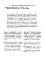
strains were calculated accordingly. As can be seen
from Fig. 1, deletion of OXA1 decreased the rate of
formation of all aminoglycerophospholipids. The rate
of PtdSer synthesis decreased to 80%, the rate of
PtdEtn formation to 70% and that of PtdCho synthesis
to 60% of wild-type. Because Oxa1p was assumed to
compromise only the mitochondrial PtdEtn-synthesiz-
ing Psd1p, leaving the Golgi-located Psd2p unaffected,
the decrease in the rate of PtdEtn synthesis in oxa1D
confirmed a defect in Psd1p-dependent PtdEtn forma-
tion. Under these circumstances, the decreased rate of
PtdCho formation seemed to be due to the lowered rate
of PtdEtn formation, whereas reduced PtdSer forma-
tion might reflect a response to a feedback regulatory
mechanism. It should be noted that the steady-state lev-
els of individual phospholipids do not necessarily reflect
the rates of synthesis of the components.
Table 1. Phospholipid composition of homogenate and mitochondria from cells grown on YPD. CF, cellular fraction; HOM, homogenate; MIT, mitochondria; LPL, lysophospholipids; DMPE,
dimethylphosphatidylethanolamine; PA, phosphatidic acid; CL, cardiolipin. Mean values of at least three independent measurements and standard deviations (SD) are shown.
Strain
% of total phospholipids
CF PtdCho PtdEtn PtdIns PtdSer LPL DMPE PA CL others
BY4742 HOM 44.13 ± 0.96 28.04 ± 0.48 14.96 ± 0.27 5.87 ± 0.06 0.98 ± 0.01 4.25 ± 0.10 1.03 ± 0.02 0.67 ± 0.01 0.07 ± 0.00
MIT 39.82 ± 1.13 30.37 ± 0.61 11.09 ± 0.16 3.83 ± 0.04 1.67 ± 0.04 4.64 ± 0.11 5.17 ± 0.08 3.15 ± 0.07 0.26 ± 0.01
psd1 HOM 49.21 ± 1.08 16.68 ± 0.23 17.55 ± 0.42 8.62 ± 0.06 2.62 ± 0.04 3.59 ± 0.08 1.53 ± 0.03 0.15 ± 0.00 0.05 ± 0.00
MIT 46.12 ± 1.09 18.67 ± 0.28 16.85 ± 0.28 7.77 ± 0.03 2.91 ± 0.07 3.07 ± 0.01 3.68 ± 0.06 0.79 ± 0.01 0.14 ± 0.00
oxa1 HOM 45.77 ± 0.81 20.93 ± 0.31 17.91 ± 0.25 6.13 ± 0.08 1.14 ± 0.01 2.96 ± 0.04 3.92 ± 0.06 1.15 ± 0.03 0.09 ± 0.00
MIT 42.15 ± 0.53 20.27 ± 0.30 16.71 ± 0.29 3.82 ± 0.04 3.48 ± 0.08 3.89 ± 0.06 4.21 ± 0.11 2.83 ± 0.02 2.64 ± 0.03
mba1 HOM 39.82 ± 0.95 24.83 ± 0.61 17.35 ± 0.28 7.64 ± 0.08 1.92 ± 0.03 5.04 ± 0.07 1.73 ± 0.05 1.42 ± 0.02 0.25 ± 0.01
MIT 41.10 ± 0.88 30.12 ± 0.79 10.56 ± 0.29 4.62 ± 0.08 1.72 ± 0.03 4.73 ± 0.08 3.59 ± 0.09 3.52 ± 0.05 0.04 ± 0.00
yme1 HOM 38.92 ± 0.75 35.58 ± 0.54 12.07 ± 0.28 6.59 ± 0.17 0.42 ± 0.01 2.75 ± 0.04 1.65 ± 0.02 1.86 ± 0.03 0.16 ± 0.00
MIT 31.27 ± 0.33 40.78 ± 0.55 12.82 ± 0.34 4.96 ± 0.08 2.17 ± 0.04 1.93 ± 0.03 2.04 ± 0.03 3.89 ± 0.04 0.14 ± 0.00
yme1 oxa1 HOM 39.32 ± 0.90 34.01 ± 0.82 12.99 ± 0.35 6.78 ± 0.16 0.26 ± 0.01 2.97 ± 0.04 2.34 ± 0.03 1.13 ± 0.02 0.20 ± 0.00
MIT 31.23 ± 0.60 39.14 ± 0.49 12.79 ± 0.18 4.12 ± 0.06 2.36 ± 0.02 2.76 ± 0.06 2.21 ± 0.03 5.24 ± 0.08 0.15 ± 0.00
Phosphatidylethanolamine of yeast mitochondria R. Nebauer et al.
6182 FEBS Journal 274 (2007) 6180–6190 ª 2007 The Authors Journal compilation ª 2007 FEBS
In contrast to psd1D, however, the oxa1D mutation
led to a smaller reduction of PtdEtn synthesis. That
the psd1D strain and the oxa1D psd1D double mutant
had comparable rates of PtdEtn formation suggests
that Oxa1p acted upstream of Psd1p. Not unexpect-
edly, rates of PtdEtn formation in the oxa1D psd2D
mutant were lower than in the psd2D mutant indicating
an additive effect of these two mutations acting on
two different pathways. Taken together, deletion of
OXA1 affected synthesis of PtdEtn by Psd1p, but did
not completely abolish the activity of this enzyme.
Activity of Psd1p in vitro is impaired in oxa1D
PtdEtn depletion in mitochondria and the decreased
rate of Psd1p-dependent PtdEtn formation in oxa1D
suggested a functional impairment of mitochondrial
Psd1p. To address the question of Psd1p enzyme activ-
ity we subjected subcellular fractions of an oxa1D
mutant to enzymatic analyses. As can be seen from
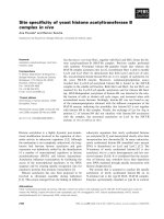
Fig. 2, the in vitro activity of Psd1p with oxa1D mito-
chondria was only 60% that of wild-type. In mito-
chondria from the psd1D mutant there was no
measurable Psd1p activity (data not shown). Psd1p
activity in mitochondria from mba1D was not
decreased, in line with the unchanged mitochondrial
level of PtdEtn in this strain (Table 1).
Studies on the stability of subunits of the mitochon-
drial membrane complexes Cox and ATPase revealed
that these proteins are degraded in the absence of
Oxa1p [25]. When functional Oxa1p is missing the
membrane subunits of these complexes cannot be
assembled and are cleaved by the intermembrane space
(i)-AAA protease Yme1p and ⁄ or by the matrix (m)-
AAA protease Afg3p ⁄ Yta12p. To test whether Psd1p
stability was also affected by the presence or absence
of these mitochondrial hydrolases, we analyzed Psd1p
activity in the respective single mutants or in double
mutants in combination with oxa1D. Deletion of
YME1 encoding the i-AAA protease led to a consider-
able increase in Psd1p activity (Fig. 2), which is in line
with the increased PtdEtn level in a deletion mutant
compared with wild-type (Table 1). This observation
was surprising because overexpression of the PtdEtn
biosynthetic pathway enzymes phosphatidylserine syn-
thase 1 (Pss1p) and ⁄ or Psd1p did not change the
PtdEtn level (R. Birner-Gruenberger, unpublished
results). The yme1D oxa1D double mutant showed an
intermediate value for the Psd1p activities from the
single mutants. Yme1p appears to contribute markedly
Fig. 1. Deletion of OXA1 causes a
decreased rate of PtdEtn synthesis in vivo.
Wild-type and mutant strains were labeled
for 0, 15, 30, 45 and 60 min with [
3
H]serine.
Incorporation of label into PtdSer, PtdEtn
and PtdCho was determined by liquid-scintil-
lation counting after separation of phospho-
lipids by TLC (see Experimental procedures).
The formation rate of PtdSer, PtdEtn and
PtdCho of wild-type (black bars) was set at
100%. Values are means from three inde-
pendent experiments with mean deviations
as indicated by the error bars.
Fig. 2. The oxa1D mutation affects Psd1p activity in vitro. Enzy-
matic assays were performed with isolated mitochondrial fractions
from wild-type BY4742, mba1D, oxa1D, yme1D and yme1D oxa1D.
Values are expressed relative to wild-type which was set at 100%
and are means from three independent experiments with mean
deviations as indicated by the error bars.
R. Nebauer et al. Phosphatidylethanolamine of yeast mitochondria
FEBS Journal 274 (2007) 6180–6190 ª 2007 The Authors Journal compilation ª 2007 FEBS 6183
to the proteolytic turnover of Psd1p. Deletions of
either subunit of the m-AAA protease yta12D and
afg3D, respectively, seemed to have no effects on
Psd1p turnover (data not shown) and were not investi-
gated further.
A decreased transcription rate for PSD1 and a
defect in Psd1p maturation are the molecular
basis of the decreased rate of PtdSer
decarboxylation in oxa1D mitochondria
One obvious explanation for the decreased amount of
Psd1p in oxa1D mitochondria was a possible reduction
in the PSD1 transcription rate in the mutant. To
address this question we performed RT-PCR analyses
of PSD1 mRNA with wild-type and oxa1D (see Exper-
imental procedures). These analyses revealed a reduc-
tion in PSD1 mRNA in the mutant. The transcription
rate for PSD1 was repressed in oxa1D to 50% that
of wild-type. Thus, downregulation of PSD1 expres-
sion at the transcriptional level appears to be one
reason for the decreased Psd1p activity in oxa1D.
Because Oxa1p had been shown to facilitate mem-
brane assembly in several mitochondrial proteins (see
above), it was tempting to speculate that it was also
necessary for correct insertion of Psd1p into the IMM.
To test this hypothesis, we performed import experi-
ments of radioactively labeled Psd1p into isolated
mitochondria. These in vitro assays (see Experimental
procedures) allowed analysis of protein assembly into
mitochondrial membranes independent of the tran-
scriptional level of a respective gene. The full-length
precursor form of Psd1p was synthesized by a coupled
transcription ⁄ translation reaction and incubated with
wild-type and oxa1D mitochondria. Complete process-
ing of Psd1p occurred in three proteolytic steps
(Fig. 3A). The primary translation product of 57 kDa
was cleaved to a first intermediate of 52 kDa, most
likely during or immediately after the import process.
This cleavage step is in agreement with the finding that
a positively charged amino acid stretch at the N-termi-
nus of Psd1p serves as a mitochondrial targeting
sequence. Processing of Psd1p was continued by cleav-
age of a 2 kDa fragment representing the inter-
membrane space sorting signal, yielding the second
intermediate of 50 kDa. Assembly of Psd1p into the
IMM was completed by (autocatalytic) cleavage of the
50 kDa intermediate to one a-chain and one b-chain
(4 and 46 kDa mature forms). The 4 kDa a-subunit
was not detected in electrophoretic analysis.
In oxa1D (Fig. 3B), import and processing of Psd1p
occurred more slowly than in wild-type resulting in
a lower ratio of mature form to precursors. Thus,
deletion of OXA1 decreased both the transcription rate
of PSD1 and the Psd1p assembly rate into the IMM.
Both effects appear to result in a reduced amount of
enzymatically active Psd1p in the IMM and thus in a
decreased capacity to form PtdEtn.
Discussion
The biosynthetic scheme shown in Fig. 4 summarizes
the possible ways in which the mitochondrial level of
PtdEtn can be affected. First, is the supply of PtdSer
to the mitochondria as a precursor for PtdEtn forma-
tion by Psd1p. This process includes synthesis of Ptd-
Ser in the endoplasmic reticulum by the PtdSer
synthase Pss1p and translocation of PtdSer to the site
of Psd1p-catalyzed decarboxylation in the IMM. Sec-
ond, mitochondrial factors may, directly or indirectly,
A
B
Fig. 3. Proteolytic processing of Psd1p. Maturation of Psd1p was
measured in wild-type BY4742 (A) and oxa1D (B). The primary
translation product of 57 kDa (not shown in the diagram) was
cleaved to a 52 kDa intermediate (d), which was further processed
to yield a 50 kDa polypeptide (h). The final processing step leads
to the formation of the mature 46 kDa b-subunit of Psd1p (*). For
each time point, the amount of every single processing intermedi-
ate was expressed as percent of the sum of all intermediates.
Values are means from three independent experiments with
mean deviations as indicated by the error bars.
Phosphatidylethanolamine of yeast mitochondria R. Nebauer et al.
6184 FEBS Journal 274 (2007) 6180–6190 ª 2007 The Authors Journal compilation ª 2007 FEBS
affect the activity of Psd1p, thereby decreasing or
increasing the efficiency of mitochondrial PtdEtn for-
mation. Third, import and export of PtdEtn may con-
tribute to a balance in the level of this phospholipid in
mitochondria. Finally, although not addressed specifi-
cally in this scheme, transcriptional ⁄ translational regu-
lation of PSD1 expression has to be taken into
account.
Similar to plants [26], increased levels of yeast mito-
chondrial Psd1p are not necessarily accompanied by
an increase in the amount of mitochondrial PtdEtn. In
strains overexpressing Pss1p and ⁄ or Psd1p neither the
PtdSer nor the PtdEtn level was markedly changed
compared with wild-type (R. Birner-Gruenberger,
unpublished data). These findings imply that the
amount of mitochondrial PtdEtn is tightly controlled
by one of the above-mentioned regulatory mechanisms.
Alternatively, the wild-type level of Psd1p may already
represent an excess of activity which cannot be
enhanced further by increasing the amount of protein.
A search for components affecting mitochondrial
PtdEtn levels led to the identification of mitochondrial
components interacting directly with mitochon-
drial Psd1p. One example of such a component is the
mitochondrial prohibitin, Phb1p ⁄ Phb2p. Recent studies
in our laboratory demonstrated the synthetic lethality
of a psd1D phb1 ⁄ 2D double mutant [7]. It was specu-
lated that the decreased PtdEtn level in mitochondria
caused by psd1D might be harmful in the phb1 ⁄ 2D back-
ground, which by itself causes an increase in mitochon-
drial PtdEtn. In view of the results of this study, this
hypothesis appears to be wrong, because depletion of
the mitochondrial PtdEtn level by oxa1D to an amount
comparable with that in psd1D did not lead to synthetic
lethality with phb1 ⁄ 2D (R. Nebauer, unpublished
results). Thus, it is the direct interaction of Psd1p
and Phb1 ⁄ 2p or even a more complex effect through
combination of the two gene products that may be
important for mitochondrial function.
In this study, we demonstrate another mode of
action that affects Psd1p activity in yeast mitochon-
dria, namely disturbance of the import and assembly
of this polypeptide into mitochondrial membranes. We
show that Oxa1p facilitates the import of Psd1p to its
proper destination in the IMM. Oxa1p has been char-
acterized previously as a helper protein for the
assembly of a number of other IMM proteins [9].
In wild-type yeast cells, import into mitochondria, pro-
cessing and assembly into mitochondrial membranes of
Psd1p is accomplished by a three-step mechanism simi-
lar to Chinese hamster ovary cells [27]. According to
Boeckmann et al. [28], Psd1p contains all the features
of a typical IMM ⁄ intermembrane space protein,
namely a positively charged N-terminal sequence fol-
lowed by a hydrophobic stretch. The three cleavage
steps are accomplished by the mitochondrial-process-
ing peptidase (MPP), the intermembrane space prote-
ase Imp1p and autocatalysis.
In oxa1D, the Psd1p processing rate was decreased
(Fig. 3). This resulted in slower utilization of the pre-
cursor polypeptide in the mutant than in wild-type,
delayed formation of intermediates and finally a
decreased appearance of the mature form. Although
only one intermediate step in the process, namely
translocation of the 50 kDa intermediate to the IMM,
appears to be directly affected by Oxa1p, the whole
process of Psd1p assembly into the IMM occurs more
slowly in the mutant than in the wild-type. The resid-
ual Psd1p activity in oxa1D appears to be due to alter-
native import pathways.
In addition to the reduced rate of Psd1p import into
mitochondria, the decreased transcription rate of
PSD1 in oxa1D seems to play a role in imbalanced
PtdEtn formation of the mutant. We can only specu-
Fig. 4. Factors affecting the PtdEtn level in mitochondria. Pss1p (phosphatidylserine synthase 1), Psd1p (phosphatidylserine decarboxylase
1), Psd2p (phosphatidylserine decarboxylase 2), Dpl1p (dihydrosphingosine 1-phosphate lyase 1), import of PtdSer into mitochondria (x),
export of PtdEtn from mitochondria (y), import of PtdEtn into mitochondria (z) and factors (F) affecting level and activity of Psd1p in mito-
chondria may contribute to the mitochondrial PtdEtn.
R. Nebauer et al. Phosphatidylethanolamine of yeast mitochondria
FEBS Journal 274 (2007) 6180–6190 ª 2007 The Authors Journal compilation ª 2007 FEBS 6185
late at present that a negative feedback control caused
by unassembled Psd1p precursor or intermediate pro-
teins might trigger this transcriptional regulation.
However, the additive effects of reduced PSD1 tran-
scription and Psd1p assembly are sufficient to cause a
limitation of active Psd1p being present in mitochon-
dria of oxa1D.
Another component that affects the mitochondrial
level of Psd1p activity is the intermembrane space
protease Yme1p. In a yme1D strain, Psd1p activity
exceeded the wild-type level, and in the oxa1D back-
ground, yme1D restored Psd1p activity to a higher
level than wild-type. Under the latter conditions,
Oxa1p-independent insertion of Psd1p seems to be suf-
ficient to ensure assembly of a functional enzyme
exhibiting activity higher than wild-type. We assume
from these results that Yme1p contributes to Psd1p
degradation and turnover. In a yme1D strain, an excess
of Psd1p appears to accumulate in the IMM leading
to the observed effects of enhanced enzyme activity
and increased PtdEtn levels.
In summary, our results demonstrate a link between
the mitochondrial machinery of protein assembly and
PtdEtn homeostasis in mitochondria and the whole
cell. We have to keep in mind, however, that depletion
of mitochondrial PtdEtn by the various possible effects
described appears to negatively affect proteins involved
in mitochondrial function or membrane properties and
may thus contribute to a petite phenotype (respiratory
defect). By contrast, it should be noted that not all
respiratory defects of mitochondria need to be linked
to lipid defects in mitochondrial membranes as docu-
mented by a recent screening of petite strains in our
laboratory (R. Nebauer, unpublished results). Rather
it appears that Psd1p-dependent PtdEtn formation is
affected by a distinct set of mitochondrial proteins,
e.g. Oxa1p, which are involved in the correct assembly
of Psd1p into the IMM.
Experimental procedures
Strains and culture conditions
The yeast strains used in this study are listed in Table 2.
Yeast mutants exhibiting a petite phenotype as described by
Dimmer et al. [8] were obtained from the Euroscarf strain
collection (Frankfurt, Germany). S. cerevisiae strains were
grown under aerobic conditions at 30 °C on YPD medium
containing 1% yeast extract, 2% peptone, and 2% glucose
as the carbon source. For large scale cultivation, inocula-
tions to a D
600
of 0.1 in fresh medium were made by
diluting precultures grown to the stationary phase. For
auxotrophy tests, yeast strains were cultivated on solid syn-
thetic medium [29].
Plasmid and strain constructions
Primers used in this study are listed in Table 3. The yeast
deletion mutants oxa1D::His3MX6 and psd1D::His3MX6
were constructed as described by Longtine et al. [30]. Prim-
ers OXA1-F1 and OXA1-R1 or PSD1-F1 and PSD2-F2,
respectively, were used to amplify the His3MX6 disruption
cassette. The cassette was introduced into the respective
strain by lithium acetate transformation [31]. Correct inser-
tion of the cassette was tested by growing strains on selec-
tive media without the respective amino acid and by colony
PCR with the appropriate primers. Double-deletion
mutants were constructed by mating the corresponding
single-deletion mutants, sporulation of zygotes, and tetrad
dissection using standard methods. Identity of strains was
confirmed by marker-dependent growth and colony PCR.
Fluorescence microscopy
Visualization of mitochondrial DNA in living cells was per-
formed using the fluorescent dye DAPI. In brief, cells were
grown in YPD medium over night at 30 °C. An inoculation
to a D
600
of 0.3 in fresh medium was made by diluting of
Table 2. Yeast strains used in this study.
Strain Genotype Source ⁄ Reference
Y00000 BY4741 Mata his3D1 leu2D0 met15D0 ura3D0 Euroscarf
Y00148 BY4741 Mata his3D1 leu2D0 met15D0 ura3D0 afg3D::KanMX4 Euroscarf
Y02043 BY4741 Mata his3D1 leu2D0 met15D0 ura3D0 psd1D::KanMX4 Euroscarf
Y04800 BY4741 Mata his3D1 leu2D0 met15D0 ura3D0 psd2D::KanMX4 Euroscarf
Y06224 BY4741 Mata his3D1 leu2D0 met15D0 ura3D0 yta12D::KanMX4 Euroscarf
Y07144 BY4741 Mata his3D1 leu2D0 met15D0 ura3D0 yme1D::KanMX4 Euroscarf
Y10000 BY4742 Mata his3D1 leu2D0 lys2D0 ura3D0 Euroscarf
Y13325 BY4742 Mata his3D1 leu2D0 lys2
D0 ura3D0 mba1D::KanMX4 Euroscarf
Y16151 BY4742 Mata his3D1 leu2D0 lys2D0 ura3D0 oxa1D::KanMX4 Euroscarf
YRN2 BY4742 Mata his3D1 leu2D0 lys2D0 ura3D0 oxa1D::KanMX4 psd1D::His3MX6 This study
YRN3 BY474X Mata his3D1 leu2D0 met15D0 ura3D0 oxa1D::KanMX4 psd2D::KanMX4 This study
YRN12 BY4741 Mata his3D1 leu2D0 met15D0 ura3D0 oxa1D::His3MX6 yme1D::KanMX4 This study
Phosphatidylethanolamine of yeast mitochondria R. Nebauer et al.
6186 FEBS Journal 274 (2007) 6180–6190 ª 2007 The Authors Journal compilation ª 2007 FEBS
an overnight culture and cells were harvested in the mid-log
phase. DNA was stained with 2.5 lgÆmL
)1
of DAPI dis-
solved in NaCl ⁄ P
i
at 30 °C for 30 min. After staining, cells
were rinsed once with NaCl ⁄ P
i
and then resuspended in
NaCl ⁄ P
i
. Suspensions were placed on a glass slide and cov-
ered with a cover slip. Cells were then visualized using a
fluorescence microscope (Axiovert 35, Carl Zeiss, Jena,
Germany) with the appropriate filter set for the blue-emit-
ting fluorochrome DAPI and a 100-fold oil immersion
objective. Mitochondrial DNA was visualized as smaller
spots distinct from larger nuclear DNA. At least 100 cells
from all strains to be tested were inspected.
Labeling of aminoglycerophospholipids in vivo
Labeling of aminoglycerophospholipids in vivo was deter-
mined by following the incorporation of [
3
H]serine into
PtdSer, PtdEtn and PtdCho as described by Birner et al.
[7]. For each time point, an equivalent of 10 D
600
from
an overnight culture ( 1 mL, corresponding to
1.45 · 10
8
cells) was harvested, washed once, suspended in
500 lL YPD and incubated for 30 min at 30 ° C. Cells were
labeled with 10 lCi [
3
H]serine (27 CiÆmmol
)1
, Perkin–
Elmer, Boston, MA) per time point. Samples were taken at
0, 15, 30 and 60 min, put on ice and harvested by centrifu-
gation. Chloroform ⁄ methanol (2 : 1, v ⁄ v) and glass beads,
3 mL each, were added to the cell pellets. For disintegra-
tion of cells samples were shock frozen in liquid nitrogen
and shaken vigorously on an IKAÒ Vibrax VXR for
15 min at 4 °C. Then, lipids were extracted for 30 min by
the method of Folch et al. [32]. Individual phospholipids
were separated by TLC on Silica gel 60 plates (Merck,
Darmstadt, Germany) with chloroform ⁄ methanol ⁄ 25%
ammonia (50 : 25 : 6, v ⁄ v ⁄ v) as a developing solvent. Spots
on TLC plates were stained with iodine vapor, scraped off
and suspended in 8 mL scintillation cocktail (Packard Bio-
Science, Groningen, the Netherlands) containing 5% water.
Radioactivity was determined by liquid scintillation count-
ing using a Packard TriCarbÒ Liquid Scintillation
Analyzer.
Preparation of subcellular fractions, protein
analysis, and enzymatic analysis
Mitochondria were prepared from spheroplasts by pub-
lished procedures [1,33]. Relative enrichment of markers
and cross-contamination of subcellular fractions were
assessed as described by Zinser and Daum [34]. Protein was
quantified by the method of Lowry et al. [35] by using BSA
as a standard. SDS–PAGE was carried out as published by
Laemmli [36]. Western blot analysis of proteins from
subcellular fractions prepared as described above was
performed as described by Haid and Suissa [37]. Immunore-
active bands were visualized by enzyme-linked immunosor-
bent assay using a peroxidase-linked secondary antibody
(Sigma-Aldrich, St Louis, MO) following the manufac-
turer’s instructions.
PtdSer decarboxylase activity was measured in isolated
mitochondria from yeast cells grown in YPD to the loga-
rithmic growth phase as reported by Kuchler et al. [38] with
minor modifications: 100 nmol [
3
H]PtdSer (specific activity
of 28 900 dpmÆnmol
)1
) was used as the substrate, and the
assay was performed in 0.1 m Tris ⁄ HCl, pH 7.2, containing
10 mm EDTA.
Import of Psd1p into mitochondria in vitro
Import, processing and assembly of Psd1p into mitochon-
dria in vitro were assayed following the protocol of Ryan
et al. [39]. The precursor Psd1p was synthesized in the
presence of [
35
S]methionine (15 mCiÆmL
)1
; Amersham Bio-
sciences, Chalfont, UK) by coupled transcription ⁄
translation in a reticulocyte lysate (Promega, Madison, WI)
following the manufacturer’s instructions. The T7 RNA
polymerase system with a PCR-generated DNA fragment
as a template was employed. Primers PSD1-T1 and PSD1-
U1 (see Table 2) were used to amplify PSD1 from genomic
DNA. Yeast mitochondria were isolated as described above
and aliquoted at 10 mgÆmL
)1
in SEM buffer containing
250 mm sucrose, 1 mm EDTA, 10 mm Mops-KOH, pH 7.2,
and stored at )70 °C. The import assay involved incuba-
Table 3. Primers used to construct strains for in vitro transcription ⁄ translation of PSD1 and RT-PCR described in this study. The underlined
sequences are homologous to the His3MX6 disruption cassette (OXA1-F1, OXA1-R1, PSD1-F1, PSD1-R1) or to the PSD1 ORF (PSD1-T1).
Primer PSD1-U1 is complementary to the region spanning the stop codon of the PSD1 ORF.
Primer Primer sequence (5¢-to3¢)
OXA1-F1 GTTCACGTACAAGCGGAGCCACAGAATAACCTCCCCGACG
CGGATCCCCGGGTTAATTAA
OXA1-R1 GTTTTATATTTTTATATTTACAGAGAGATATAGAGCCTTTAT
GAATTCGAGCTCGTTTAAAC
PSD1-F1 GCCAGTTAAGAACGCCTTGGCGCAAGGGAGGACGCTCCTC
CGGATCCCCGGGTTAATTAA
PSD1-R1 CAGGTATGTGGTTCCAAGTGTTTGTCGCTCTTTGAATTTG
GAATTCGAGCTCGTTTAAAC
PSD1-T1 TCTAATACGACTCACTATAGGGAGA
ATGTCAATTATGCCAGTTAAG
PSD1-U1 CTTTACATATGATTGCTTTCATTTTAAATCATTCTTTCC
PSD1-RT FW AGAACTGCGGTGCTATGGAATAGA
PSD1-RT REV TTTGGCACGATCCACAATCTC
R. Nebauer et al. Phosphatidylethanolamine of yeast mitochondria
FEBS Journal 274 (2007) 6180–6190 ª 2007 The Authors Journal compilation ª 2007 FEBS 6187
tion of the radiolabeled Psd1p precursor with isolated mito-
chondria in the presence of NADH (1.8 mm) and ATP
(1.8 mm) in a buffer containing 3% (w ⁄ v) fatty acid-free
BSA, 250 mm sucrose, 80 mm KCl, 5 mm MgCl
2
,2mm
KH
2
PO
4
,5mm methionine, 10 mm Mops-KOH, pH 7.2
[39]. After 2, 5, 10, and 15 min samples were withdrawn
and put on ice in the presence of valinomycin (final concen-
tration 0.5 lm) to stop the import reaction. Supernatants
were removed by centrifugation at 12 000 g and 4 °C for
5 min. Pellets were washed once in SEM buffer, recovered
by centrifugation and suspended in SDS ⁄ PAGE loading
buffer [36] prior to heating at 95 °C for 5 min. Analysis of
radioactively labeled translation products, intermediates
and mature polypeptides was performed employing stan-
dard methods of SDS–PAGE, autoradiography and densi-
tometric scanning.
Phospholipid quantification
For the analysis of total cellular phospholipids yeast cells
harvested from a 500 mL culture grown to the late logarith-
mic phase were disintegrated by shaking with glass beads in
a Merckenschlager homogenizer under CO
2
cooling in the
presence of 10 m m Tris ⁄ HCl, pH 7.2, and 1 mm phenyl-
methylsulfonyl fluoride (Calbiochem, La Jolla, CA). After
removal of the beads by centrifugation the supernatant rep-
resenting the total cell homogenate was aliquoted and
stored at )70 °C. Lipids from samples containing 3 mg
protein were extracted by the procedure of Folch et al. [32]
using 4 mL chloroform ⁄ methanol (2 : 1, v ⁄ v). Isolated
mitochondria (2 mg protein) were subjected to lipid extrac-
tion by the same method.
Individual phospholipids were separated by 2D TLC
using chloroform ⁄ methanol ⁄ 25% ammonia (70 : 35 : 5,
v ⁄ v ⁄ v) as first, and chloroform ⁄ acetone ⁄ methanol ⁄ acetic
acid ⁄ water (55 : 20 : 10 : 10 : 5, v ⁄ v ⁄ v ⁄ v ⁄ v) as second
developing solvent. Phospholipids were visualized on TLC
plates by staining with iodine vapor, scraped off and quan-
tified by the method of Broekhuyse [40].
RNA preparation and real-time PCR
Total RNA was isolated using phenol ⁄ chloroform extrac-
tion as described previously [29] and further purified by
RQ1 RNase-free DNase (Promega) treatment according to
the manufacturer’s instructions and subsequent ethanol pre-
cipitation. Integrity of RNA was tested by agarose gel elec-
trophoresis and determination of the 260 to 280 nm ratio
of the absorbencies. RNA concentration was determined by
measurement of the absorbance at 260 nm.
Total RNA was subjected to reverse transcription using
the SuperScript
TM
II First Strand Synthesis System (Invi-
trogen, Carlsbad, CA) for real-time PCR (RT-PCR).
Possible traces of contaminating genomic DNA were
removed by DNAse I digestion. In detail, 2.5 lg of RNA
with a concentration of 500 ng ÆlL
)1
were incubated with
10· DNAse I buffer, DNAse I amplification grade and
4 U RNaseOut
TM
ribonuclease for 15 min at room tem-
perature (all reagents from Invitrogen). DNA digestion
was stopped by adding 1 lL of EDTA (25 mm) and 2 lL
of H
2
O, incubating for 5 min at room temperature and
further 5 min at 70 °C. The DNase I treated RNA was
mixed with 0.5 lg of oligo-dT
2-18
,3lg of random primers,
and 4 U of RNaseOut
TM
ribonuclease, heated for 5 min
at 70 °C and left at room temperature for another 5 min.
The RNA sample was mixed with the cDNA synthesis
mix, consisting of 5 · RT buffer, dithiothreitol (0.1 m),
dNTP (10 mm), 4 U RNaseOut
TM
and 200 U Super-
Script
TM
II reverse transcriptase (all reagents from Invitro-
gen), and heated to 45 °C for 1 h. The reaction was
stopped by heating to 95 °C for 5 min. RT-PCR assays
were performed using the PlatinumÒ SYBRÒ Green Su-
perMix-UDG (Invitrogen) following the manufacturer’s
recommendations. Primers for RT-PCR were designed
using the software tool primer express
TM
(ABI). For a
25 lL RT-PCR reaction, 1 lL of primer pair (800 nm),
0.5 lL of diluted cDNA (10 ngÆlL
)1
) and 12.5 lL of Plati-
numÒ SYBRÒ Green SuperMix-UDG were applied. In
addition, no template controls (NTC) and no RT reaction
(No RT) controls were performed. The cycling conditions
on an ABI Prism 7000 were set for 2 min at 50 °C,
10 min at 95 °C and 40 cycles of 15 s at 95 °C and 1 min
at 60 °C. Data were analyzed using the ABI Prism 7000
sds software.
Acknowledgements
The authors wish to thank A. Hermetter for providing
access to the fluorescence microscope (FWF instru-
ment). This work was financially supported by the
FWF (Fonds zur Fo
¨
rderung der wissenschaftlichen
Forschung in O
¨
sterreich) projects 14468 and 17321 to
GD.
References
1 Zinser E, Sperka-Gottlieb CDM, Fasch E-V, Kohlwein
SD, Paltauf F & Daum G (1991) Phospholipid synthesis
and lipid composition of subcellular membranes in the
unicellular eukaryote Saccharomyces cerevisiae. J Bacte-
riol 173, 2026–2034.
2 Trotter PJ & Voelker DR (1995) Identification of a
non-mitochondrial phosphatidylserine decarboxylase
activity in the yeast Saccharomyces cerevisiae. J Biol
Chem 270, 6062–6070.
3 Nebauer R, Birner-Gru
¨
nberger R & Daum G (2003)
Biogenesis and cellular dynamics of glycerophospho-
lipids in the yeast Saccharomyces cerevisiae. Topics Curr
Genet 6, 125–168.
Phosphatidylethanolamine of yeast mitochondria R. Nebauer et al.
6188 FEBS Journal 274 (2007) 6180–6190 ª 2007 The Authors Journal compilation ª 2007 FEBS
4 Birner R & Daum G (2003) Biogenesis and cellular
dynamics of aminoglycerophospholipids. Int Rev Cytol
225, 273–323.
5 Birner R, Bu
¨
rgermeister M, Schneiter R & Daum G
(2001) Roles of phosphatidylethanolamine and of its
several biosynthetic pathways in Saccharomyces cerevisi-
ae. Mol Biol Cell 12, 997–1007.
6 Storey MK, Clay KL, Kutateladze T, Murphy RC,
Overduin M & Voelker DR (2001) Phosphatidylethanol-
amine has an essential role in Saccharomyces cerevisiae
that is independent of its ability to form hexagonal
phase structures. J Biol Chem 276, 48539–48548.
7 Birner R, Nebauer R, Schneiter R & Daum G (2003)
Synthetic lethal interaction of the mitochondrial phos-
phatidylethanolamine biosynthetic machinery with the
prohibitin complex of Saccharomyces cerevisiae. Mol
Biol Cell 14, 370–383.
8 Dimmer KS, Fritz S, Fuchs F, Messerschmitt M,
Weinbach N, Neupert W & Westermann B (2002)
Genetic basis of mitochondrial function and morphol-
ogy in Saccharomyces cerevisiae. Mol Biol Cell 13,
847–853.
9 Stuart RA (2002) Insertion of proteins into the inner
membrane of mitochondria: the role of the Oxa1 com-
plex. Biochim Biophys Acta 1592, 79–87.
10 Neupert W (1997) Protein import into mitochondria.
Annu Rev Biochem 66, 863–917.
11 Pfanner N & Geissler A (2001) Versatility of the mito-
chondrial protein import machinery. Nat Rev Mol Cell
Biol 2, 339–349.
12 Rehling P, Pfanner N & Meisinger C (2003) Insertion of
hydrophobic membrane proteins into the inner mitochon-
drial membrane – a guided tour. J Mol Biol 326, 639–657.
13 Herrmann JM & Neupert W (2003) Protein insertion
into the inner membrane of mitochondria. IUBMB Life
55, 219–225.
14 Stuart RA & Neupert W (1996) Topogenesis of inner
membrane proteins of mitochondria. Trends Biochem
Sci 21, 261–267.
15 Herrmann JM, Neupert W & Stuart RA (1997) Inser-
tion into the mitochondrial inner membrane of a poly-
topic protein, the nuclear-encoded Oxa1p. EMBO J 16,
2217–2226.
16 Hell K, Herrmann JM, Pratje E, Neupert W & Stuart
RA (1998) Oxa1p, an essential component of the N-tail
protein export machinery in mitochondria. Proc Natl
Acad Sci USA 95, 2250–2255.
17 Ott M, Prestele M, Bauerschmitt H, Funes S, Bonnefoy
N & Herrmann J (2006) Mba1, a membrane-associated
ribosome receptor in mitochondria. EMBO J 25, 1603–
1610.
18 Voelker DR (1997) Phosphatidylserine decarboxylase.
Biochimica Biophysica Acta 1348, 236–244.
19 The Uniprot Consortium (2007) The Universal Protein
Resource (UniProt). Nucleic Acids Res 35 , 193–197.
20 von Heijne G (1992) Membrane protein structure pre-
diction. Hydrophobicity analysis and the positive-inside
rule. J Mol Biol
20, 487–494.
21 Gasteiger E, Gattiker A, Hoogland C, Ivanyi I, Appel
RD & Bairoch A (2003) ExPASy: the proteomics server
for in-depth protein knowledge and analysis. Nucleic
Acids Res 31, 3784–3788.
22 Schatz G (1996) The protein import system of mito-
chondria. J Biol Chem 271, 31763–31766.
23 Atkinson KD, Jensen B, Kolat AI, Storm EM, Henry
SA & Fogel S (1980) Yeast mutants auxotrophic for
choline and ethanolamine. J Bacteriol 141, 558–564.
24 Leonhard K, Herrmann JM, Stuart RA, Mannhaupt G,
Neupert W & Langer T (1996) AAA proteases with cat-
alytic sites on opposite membrane surfaces comprise a
proteolytic system for the ATP-dependent degradation
of inner membrane proteins in mitochondria. EMBO J
15, 4218–4229.
25 Lemaire C, Hamel P, Velours J & Dujardin G (2000)
Absence of the mitochondrial AAA protease Yme1p
restores F
0
-ATPase subunit accumulation in an oxa1
deletion mutant of Saccharomyces cerevisiae. J Biol
Chem 275, 23471–23475.
26 Rontein D, Wu W-I, Voelker DR & Hanson AD (2003)
Mitochondrial phosphatidylserine decarboxylase from
higher plants. Functional complementation in yeast,
localization in plants, and overexpression in Arabidopsis.
Plant Physiol 132, 1678–1687.
27 Kuge O, Saito K, Kojima M, Akamatsu Y & Nishijima
M (1996) Post-translational processing of the phosphati-
dylserine decarboxylase gene product in Chinese ham-
ster ovary cells. Biochem J 319, 33–38.
28 Boeckmann B, Bairoch A, Apweiler R, Blatter MC,
Estreicher A, Gasteiger E, Martin MJ, Michoud K,
O’Donovan C, Phan I et al. (2003) The SWISS-PROT
protein knowledgebase and its supplement TrEMBL in
2003. Nucl Acids Res 31, 365–370.
29 Burke D, Dawson D & Stearns T (2000) Methods in
Yeast Genetics: A Cold Spring Harbor Laboratory
Course Manual. Cold Spring Harbor Laboratory Press,
Cold Spring Harbor, NY.
30 Longtine MS, McKenzie A III, Demarini DJ, Shah
NG, Wach A, Brachat A, Philippsen P & Pringle JR
(1998) Additional modules for versatile and economical
PCR-based gene deletion and modification in Saccharo-
myces cerevisiae. Yeast 14, 953–961.
31 Gietz D, St. Jean A, Woods RA & Schiestl RH (1992)
Improved method for high efficiency transformation of
intact yeast cells. Nucleic Acids Res 20, 1425.
32 Folch J, Lees M & Sloane Stanley GH (1957) A simple
method for the isolation and purification of total lipids
from animal tissues. J Biol Chem 226, 497–509.
33 Daum G, Bo
¨
hni PC & Schatz G (1982) Import of
proteins into mitochondria. J Biol Chem 257, 13028–
13033.
R. Nebauer et al. Phosphatidylethanolamine of yeast mitochondria
FEBS Journal 274 (2007) 6180–6190 ª 2007 The Authors Journal compilation ª 2007 FEBS 6189
34 Zinser E & Daum G (1995) Isolation and biochemical
characterization of organelles from the yeast. Yeast 11,
493–536.
35 Lowry OH, Rosebrough NJ, Farr AL & Randall RJ
(1951) Protein measurement with the Folin phenol
reagent. J Biol Chem 193, 265–275.
36 Laemmli UK (1970) Cleavage of structural proteins
during the assembly of the head of bacteriophage T4.
Nature 227, 680–685.
37 Haid A & Suissa M (1983) Immunochemical identifica-
tion of membrane proteins after sodium dodecyl
sulfate–polyacrylamide gel electrophoresis. Methods
Enzymol 96, 192–205.
38 Kuchler K, Daum G & Paltauf F (1986) Subcellular
and submitochondrial localization of phospholipid-syn-
thesizing enzymes in Saccharomyces cerevisiae. J Bacte-
riol 165, 901–910.
39 Ryan MT, Voos W & Pfanner N (2001) Assaying pro-
tein import into mitochondria. Methods Cell Biol 65,
189–215.
40 Broekhuyse RM (1968) Phospholipids in tissues of the
eye. I. Isolation, characterization and quantitative anal-
ysis by two-dimensional thin-layer chromatography of
diacyl and vinyl-ether phospholipids. Biochim Biophys
Acta 152, 307–315.
Phosphatidylethanolamine of yeast mitochondria R. Nebauer et al.
6190 FEBS Journal 274 (2007) 6180–6190 ª 2007 The Authors Journal compilation ª 2007 FEBS