Báo cáo khoa học: C-mannosylation in the hypertrehalosaemic hormone from the stick insect Carausius morosus doc
Bạn đang xem bản rút gọn của tài liệu. Xem và tải ngay bản đầy đủ của tài liệu tại đây (428.64 KB, 11 trang )
C-mannosylation in the hypertrehalosaemic hormone
from the stick insect Carausius morosus
Claudia E. Munte
1
, Gerd Ga
¨
de
2
, Barbara Domogalla
1
, Werner Kremer
1
, Roland Kellner
3
and
Hans R. Kalbitzer
1
1 Institute of Biophysics and Physical Biochemistry, University of Regensburg, Germany
2 Department of Zoology, University of Cape Town, Rondebosch, South Africa
3 Target Research and Biotechnology, Merck KGaA, Darmstadt, Germany
In insects, peptidergic regulation by neuropeptides is
the most important form of communication to con-
trol not only growth, development and reproduction,
but also metabolic homeostasis [1]. Fuel mobiliza-
tion, especially to fulfil the exceptionally high energy
demand during contraction of flight muscles, is
under neuroendocrine control by peptides of the so-
called adipokinetic hormone (AKH) family, as is the
case in all investigated insect orders [2,3]. These
short peptides of 8–10 amino acids in length
are produced in the retro-cerebral corpora cardiaca
from precursor polypeptides by proteolytic cleavage.
The sequence of AKH peptides is characterized
by phenylalanine, tryptophan and glycine residues
at positions 4, 8 and 9, respectively (Fig. 1). The
N-terminal glutamine of the precursor is transformed
into pGlu in the final product and the C-terminus is
amidated. Besides the general post-translational
modifications at both termini, some AKH peptides
are known to contain additional modifications. The
AKH peptide from the protea beetle Trichostheta
fascicularis can be phosphorylated at Thr6 [4]. AKH
peptides are responsible for the measurable increase
in haemolymph lipids, carbohydrates and proline
levels and are denoted accordingly as adipokinetic,
hypertrehalosaemic (trehalose instead of glucose is
the sugar circulating in the haemolymph of insects)
and hyperprolinaemic.
More interestingly in the context of the present
study, and as first demonstrated in 1992 [5], the stick
Keywords
Carausius morosus; hypertrehalosaemic
hormone; NMR; protein C-mannosylation;
a-mannosyltryptophan
Correspondence
H. R. Kalbitzer, Institute for Biophysics and
Physical Biochemistry, University of
Regensburg, D-93040 Regensburg,
Germany
Fax: +49 941 943 2479
Tel: +49 941 943 2595
E-mail: hans-robert.kalbitzer@biologie.
uni-regensburg.de
(Received 7 November 2007, revised 13
December 2007, accepted 8 January 2008)
doi:10.1111/j.1742-4658.2008.06277.x
The hypertrehalosaemic hormone from the stick insect Carausius morosus
(Cam-HrTH) contains a hexose covalently bound to the ring of the trypto-
phan, which is in the eighth position in the molecule. We show by solution
NMR spectroscopy that the tryptophan is modified at its C
d1
(C2) by an
a-mannopyranose. It is the first insect hormone to exhibit C-glycosylation
whose exact nature has been determined experimentally. Chemical shift
analysis reveals that the unmodified as well as the mannosylated Cam-
HrTH are not completely random-coil in aqueous solution. Most promi-
nently, C-mannosylation strongly influences the average orientation of the
tryptophan ring in solution and stabilizes it in a position clearly different
from that found in the unmodified peptide. NMR diffusion measurements
indicate that mannosylation reduces the effective hydrodynamic radius. It
induces a change of the average peptide conformation that also diminishes
the propensity for aggregation of the peptide.
Abbreviations
AKH, adipokinetic hormone; Cam-HrTH, hypertrehalosaemic hormone from Carausius morosus; DSS, 2,2-dimethyl-2-silapentane-5-sulfonate;
HSQC, heteronuclear single quantum coherence; IL, interleukin; TSR, thrombospondin type 1 repeats.
FEBS Journal 275 (2008) 1163–1173 ª 2008 The Authors Journal compilation ª 2008 FEBS 1163
insect Carausius morosus contains two hypertrehalosae-
mic decapeptides, Cam-HrTH-I and Cam-HrTH-II,
which differ only in the modification of Trp8 by a
hexose on peptide I. However, the exact nature of this
tryptophan glycosylation was previously unknown
because, due to the small amounts of naturally avail-
able peptide, only MS was feasible, which did not
allow determination of the exact type of the hexose
moiety. Two years later, the second example of a tryp-
tophan modification was reported in a mammalian
enzyme, human RNAse 2. Using MS and NMR spec-
troscopy, the hexose was shown to be connected to the
C
d1
of the tryptophan ring via a C-glycosidic linkage
[6], and could be unambiguously identified by subse-
quent studies as an a-d-mannopyranose [7]. This tryp-
tophan C-mannosylation was later found in another
mammalian protein, human interleukin (IL)-12 [8], and
was shown to be catalysed by a microsome-associated
transferase that uses dolychyl-phosphate-mannose as
donor of the glycosyl group [9]. The transferase recog-
nizes the motif WXXW (where X is any amino acid)
[9,10] and C-mannosylates the first tryptophan of this
sequence in RNAse 2 and IL-12. Subsequently, addi-
tional mammalian proteins with such a modification
have been identified, such as human terminal comple-
ment proteins C6, C7, C8a,C8b and C9 [11], proper-
din [12] and thrombospondin-1 [13,14]; all of them
containing thrombospondin type 1 repeats (TSR mod-
ules). In the TSR modules, WXXWXXX motifs are
found. Here, more than one tryptophan can be man-
nosylated. Variations of this motif can be found in C6,
C7 and in properdin [12], leading to the more general
recognition sequence (W ⁄ Y ⁄ F)XXWXX(W ⁄ C ⁄ V) in
the TSR modules. Since the number of modified tryp-
tophan residues varies in these sequences, it is assumed
that either features outside the motif determine the
degree of modification or more than one C-mannosyl-
transferase might exist.
It still remains unresolved as to whether the trypto-
phan glycosylation found in 1992 in the stick insect
hormone is identical to that found in mammalian
enzymes, especially because both differ significantly in
tryptophan-glycosylation motifs. In the present study,
we describe a detailed NMR analysis of the tryptophan
modification in the C. morosus hypertrehalosaemic pep-
tide Cam-HrTH-I, that was only possible with the high
sensitivity of a high field spectrometer (800 MHz)
equipped with a cryoprobe. In addition, NMR is used
to characterize the structure of Cam-HrTH in aqueous
solution at the atomic level, as well as to identify possi-
ble structural changes induced by tryptophan modifica-
tion that might play a role in receptor recognition.
Results
Assignment of peptide chemical shifts
As the modified peptide Cam-HrTH-I and the unmodi-
fied Cam -HrTH-II were obtained from natural sources
by isolating the proteins from the corpora cardiaca of
approximately 2000 stick insects, a
13
C and ⁄ or
15
N
enrichment was not feasible. The concentration of the
modified protein with approximately 60 lm was rather
low to conduct 2D NMR spectroscopy. Only the
high sensitivity of an 800 MHz-NMR spectrometer
equipped with a cryoprobe permitted a successful
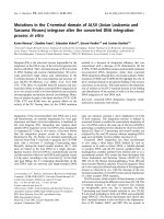
Fig. 1. Sequence comparison of AKH pre-
cursors. Only part of the N-terminal part of
the sequences is shown, corresponding to
the mature AKH (grey background) and the
cleavage site (K,R)R. The preceding glycine
residue provides the C-terminal NH
2
-group.
Conserved residues are shown in bold.
*, precursor not known.
C-mannosylation in HrTH from a stick insect C. E. Munte et al.
1164 FEBS Journal 275 (2008) 1163–1173 ª 2008 The Authors Journal compilation ª 2008 FEBS
assignment of the NMR lines. The assignment of the
1
H resonance lines was obtained by classical homo-
nuclear methods.
13
C NMR assignments were obtained
by
1
H,
13
C-heteronuclear single quantum coherence
(HSQC) spectra with different mixing times. To
achieve identical experimental conditions and to avoid-
ing any systematic chemical shift changes that could
arise from experimental differences such as buffer con-
ditions and temperature, the modified and unmodified
peptides were mixed in a 1 : 4 ratio. Because the two
peptides were added in different, well defined amounts,
the intensities of the resonance lines allowed the direct
identification of the two peptides in the NMR spectra
of the mixture. The assignments of the peptide reso-
nances are summarized in the supplementary Tables S1
and S2.
It had been previously shown by MS and amino
acid analysis that Cam-HrTH-I and Cam-HrTH-II
share the amino acid sequence pGlu-Leu-Thr-Phe-Thr-
Pro-Asn-Trp-Gly-Thr-NH
2
[5]. The only difference
found between both peptides is a modification of the
Cam-HrTH-I tryptophan residue at position 8 by an
unidentified hexose. We can expect that the assign-
ments of the two peptides mainly differ around this
residue. In agreement with this expectation, the first
four amino acids do not show significant distinctions
in their chemical shifts.
Identification of the sugar moiety and the
modification site in tryptophan
By theoretical considerations, it can be argued that the
glycosylation of the tryptophan ring system occurs via
an N–C or C–C bond; the formation of such a bond
would result in the disappearence of the corresponding
proton signal. Positions available for the glycosylation
of the ring system are N
e1
,C
d1
,C
f2
,C
g2
,C
e3
and C
f3
;
all of them were assigned in the unmodified peptide.
One likely attachment site is the indolic N
e1
atom of
tryptophan. As shown in Fig. 2, however, the corre-
sponding H
e1
diagonal peaks at 10.51 p.p.m. and
10.15 p.p.m. are still present for both peptides, but the
correlation peak to the H
d1
, although clearly present in
the unmodified peptide, is completely absent in the
modified peptide. This indicates that, instead of N
e1
,
most probably the C
d1
atom is modified. Furthermore,
all tryptophan ring carbons directly bound to a proton
for the unmodified peptide could be detected by
1
H,
13
C-HSQC spectroscopy; in contrast, the C
d1
peak
was missing in the modified peptide (Table S2). This
would be expected because the H
d1
proton necessary
for the insensitive nuclei enhanced by polarization
transfer is removed by the modification.
In addition to the peptides’ peaks, five well-defined
sugar spin systems were found in the homonuclear
2D NMR spectra (Fig. 3A). Diffusion experiments
showed that only one of the sugars diffuses with the
same diffusion constant as the peptide and, thus, cor-
responds to the Trp bound hexose. The other four spin
systems diffuse freely in the sample (Fig. 3B) and have
been assigned from their chemical shifts to a- and
b-glucose, fructose and sucrose.
The
1
H and
13
C resonances of the bound hexose
could be completely assigned. The
1
H and
13
C chemi-
cal shift values of the bound hexose and of the trypto-
phan ring system are very close to those described for
other peptides containing a glycosylated tryptophan
(Table 1). For one of these peptides, corresponding to
amino acids 5–10 of RNase 2 [7] and studied in aque-
ous solution under comparable conditions (300 K,
2
H
2
O) to those of the present study, it could be con-
clusively shown by NMR spectroscopy that the trypto-
phan is a-mannopyranosylated at C
d1
. The proton and
carbon chemical shifts obtained are almost identical to
those found for the modified Cam-HrTH-I peptide; the
maximum chemical shift deviations are 0.07 p.p.m.
and 0.6 p.p.m. for H1¢ and C1¢, respectively. When
taking into account the average chemical shifts
reported for the 2-(a-d-mannopyranosyl)-tryptophan
residues (Table 1), the agreement is almost perfect.
This strongly indicates that the tryptophan of
Fig. 2. Selected region of the 800 MHz TOCSY spectrum showing
the tryptophan indol ring spin systems of both Carausius morosus
neuropeptides. The sample contained approximately 60 l
M Cam-
HrTH-I and 240 l
M Cam-HrTH-II in 90%
1
H
2
O, 10%
2
H
2
O, 0.1 mM
DSS, pH 5.4. Temperature 300 K. W, Trp8 of the native peptide
Cam-HrTH-II; W*, Trp8 in the modified peptide Cam-HrTH-I. The
dashed line indicates the missing
1
H
e1
–
1
H
d1
contact in Cam-HrTH-I.
C. E. Munte et al. C-mannosylation in HrTH from a stick insect
FEBS Journal 275 (2008) 1163–1173 ª 2008 The Authors Journal compilation ª 2008 FEBS 1165
Cam-HrTH-I is also a-mannopyranosylated. The struc-
ture of the glycosylated tryptophan is depicted sche-
matically in Fig. 4.
The tryptophan–hexose bond is further confirmed
by NOEs between the mannose H2¢ proton and the
strongly shifted Trp8 H
e1
proton of the modified pep-
tide (Fig. 4). In the carbohydrate moiety, strong NOEs
are observed between H1¢ and H6¢ and between H2¢
and H3¢; a weak NOE between H1¢ and H2¢;an
ambiguous NOE between H3¢ and H5¢; but no NOE
between H3¢ and H4¢. Thus, the NOE-pattern observed
in the hexose corresponds closely to those in RNase 2
peptides and in pure 2-(a-mannopyranosyl)-trypto-
phan, which further corroborates the identity of the
hexose moiety as a-mannopyranose.
Aggregation state of Cam-HrTH-I and the
unmodified Cam-HrTH-II
To investigate the aggregation state of the processed
peptides, we performed NMR diffusion measurements
on the sample containing both peptides. Figure 3B
shows the dependence of the line intensities on the gra-
dient strengths used in the stimulated echo sequence
for important components of the sample. Diffusion
data are shown for Cam-HrTH-I and its mannose moi-
ety, Cam-HrTH-II, sucrose, glucose and 2,2-dimethyl-
2-silapentane-5-sulfonate (DSS), all contained in the
same sample. In addition, before and after the mea-
surements, the signal dependence of polyacrylamide
was measured to check the stability of the gradient sys-
tem; as required, no signal decay was observed for the
immobilized macromolecule. The glucose and the
sucrose molecules show a relatively fast signal decay,
as expected for small molecules, and therefore are not
bound to the peptide. By contrast, the mannose reso-
nances decay with the same rate as those of the pep-
tide signal of Cam-HrTH-I. This is to be expected for
mannose bound to the peptide because the diffusion
constants should be identical. Using DSS as a mole-
cular mass reference, the effective molecular masses of
1.45 ± 0.12 kgÆmol
)1
and 1.96 ± 0.10 kgÆmol
)1
are
obtained respectively for the modified Cam-HrTH-I
and the unmodified Cam-HrTH-II peptides (Table 2).
The effective molecular masses are larger than those
obtained under the assumption of the same shape fac-
tor and density for the test compound and the refer-
ence. For the glycosylated peptide, the molecular mass
calculated from the chemical structure is still almost in
the error range of the molecular mass calculated from
the diffusion constant for a monomer. For the unmod-
ified peptide, the effective mass is significantly larger
than the calculated value for a compactly folded
monomer but smaller than expected for a dimer. In
line with this observation, the effective transversal
relaxation rates increase in good approximation pro-
portionally to the effective masses: the transversal
relaxation rates calculated from the linewidths of the
H
e1
resonances of tryptophan increase by a factor of
1.38, from 16.65 ± 0.31 s
)1
to 22.93 ± 0.31 s
)1
, which
is within the limits of error of the ratio of 1.35
obtained for the diffusion constants. The results indi-
cate a monomeric state of Cam-HrTH-I and probably
A
B
Fig. 3. NMR spectroscopy of carbohydrates in the solution of Cam-
HrTH-I and Cam-HrTH-II. (A) Selected region of the TOCSY
spectrum showing three sugar spin systems. (B) Plot of ln(I ⁄ I
0
)as
function of G
2
where I is the peak integral at a given gradient
strength G and I
0
is the intensity at G = 0. The sample contained
approximately 60 l
M Cam-HrTH-I and 240 lM Cam-HrTH-II in
99.8%
2
H
2
O, 0.1 mM DSS, pH 5.4. Temperature = 300 K. PAA
(polyacrylamide in 90%
1
H
2
O, 10%
2
H
2
O) was used to check the
stability of the gradient system before and after measurement.
C-mannosylation in HrTH from a stick insect C. E. Munte et al.
1166 FEBS Journal 275 (2008) 1163–1173 ª 2008 The Authors Journal compilation ª 2008 FEBS
also for the Cam-HrTH-II with respect to the experi-
mental conditions of the study. Interestingly, the modi-
fied peptide has a smaller hydrodynamic radius than
the unmodified peptide, in spite of the known increase
of 162 gÆmol
)1
by the mannosylation, indicating a
Table 1. Carbohydrate modifications of tryptophan residues in protein fragments. All chemical shifts are referenced to 2,2-dimethyl-2-sila-
pentane-5-sulfonate (DSS); when other standards where used, the values were adapted as best as possible.
Cam-HrTH-I RNase 2
a
IL-12
a
C9(T2-1) W27
a
C9(T2-2) W27
a
C9(T2-2) W30
a
d ⁄ p.p.m. <d> ⁄ p.p.m
b
d ⁄ p.p.m. d ⁄ p.p.m. d ⁄ p.p.m. d ⁄ p.p.m. d ⁄ p.p.m.
Trp
H
e1
10.51 10.47 10.47 –
d
–
d
–
d
–
d
H
e3
7.66 7.59 7.67 7.65 7.53 7.54 7.56
H
f3
7.17 7.12 7.14 7.14 7.11 7.08 7.14
H
g2
7.27 7.21 7.20 7.20 7.20 7.21 7.24
H
f2
7.49 7.43 7.41 7.42 7.42 7.43 7.46
C
d1
–
c
––
d
–
d
–
d
–
d
–
d
C
e3
121.2 121.1 121.1 –
d
–
d
–
d
–
d
C
f3
122.2 121.9 121.9 –
d
–
d
–
d
–
d
C
g2
125.3 125.2 125.2 –
d
–
d
–
d
–
d
C
f2
114.4 114.2 114.2 –
d
–
d
–
d
–
d
Hexose
H1¢ 5.15 5.19 5.22 5.18 5.18 5.18 5.20
H2¢ 4.36 4.42 4.42 4.44 4.44 4.39 4.43
H3¢ 4.08 4.07 4.09 4.07 4.07 4.05 4.06
H4¢ 3.96 3.94 3.96 3.93 3.94 3.93 3.93
H5¢ 3.88 3.83 3.87 3.83 3.82 3.81 3.81
H6¢ 4.22 4.18 4.21 4.18 4.18 4.16 4.16
H6¢¢ 3.73 3.77 3.77 –
d
–
d
–
d
–
d
C1¢ 69.3 69.9 69.9 –
d
–
d
–
d
–
d
C2¢ 70.6 70.7 70.7 –
d
–
d
–
d
–
d
C3¢ 73.0 73.0 73.0 –
d
–
d
–
d
–
d
C4¢ 71.4 71.3 71.3 –
d
–
d
–
d
–
d
C5¢ –
c
81.4 81.4 –
d
–
d
–
d
–
d
C6¢ 61.8 62.1 62.1 –
d
–
d
–
d
–
d
a
RNase 2, glycosylated hexapeptide from human RNase 2 (amino acids 5–10) [6,7]; IL-12, peptide from human IL-12 (amino acids 316–322)
[8]; C9(T2-1) W27, 2-(a-mannopyranosyl)-
L-tryptophan at position 27 in a pentadecapeptide derived from complement C9 [11]; C9(T2-2) W27
and C9(T2-2) W30, 2-(a-mannopyranosyl)-
L-tryptophan at positions 27 and 30 in the two-fold modified pentadecapeptide derived from com-
plement C9 [11].
b
Average chemical shifts observed for all peptides except of Cam-HrTH-I.
c
Resonance not assigned.
d
Shift not reported.
Fig. 4. Structure of the glycosylated tryptophan residue. The exper-
imentally found NOEs between the a-mannose moiety and the
Trp8 are depicted as grey lines, where line thickness indicates the
strength of the NOE. Ambiguous NOEs are represented by dashed
lines. The mannose is depicted in the
1
C
4
conformation.
Table 2. Molecular masses and relative hydrodynamic radii of the
Carausius morosus neuropeptides. The sample contained approxi-
mately 60 l
M Cam-HrTH-I and 240 lM Cam-HrTH-II in 99.8%
2
H
2
O,
0.1 m
M DSS, pH 5.4 and was measured at 300 K.
Compound R
h
⁄ R
h,DSS
a
M
exp
b
⁄ kgÆmol
)1
M
calc
c
⁄ kgÆmol
)1
Cam-HrTH-I 1.950 ± 0.056 1.45 ± 0.12 1.308
Cam-HrTH-II 2.156 ± 0.035 1.958 ± 0.097 1.146
a
Ratio of the hydrodynamic radii from peptide and DSS, calculated
with Eqn (3).
b
Molecular mass experimentally obtained from the
diffusion experiments on the basis of Eqn (4).
c
Molecular mass cal-
culated from the chemical formula.
C. E. Munte et al. C-mannosylation in HrTH from a stick insect
FEBS Journal 275 (2008) 1163–1173 ª 2008 The Authors Journal compilation ª 2008 FEBS 1167
more compact structure of the neuropeptide induced
by mannosylation.
Conformational restraints of the peptide
Figure 5 shows the deviations Dd of the Cam-HrTH-I
and Cam-HrTH-II
1
H and
13
C chemical shifts from
random-coil values. The latter were calculated on the
basis of the random-coil values of completely dena-
tured model peptides [15], which were corrected for the
effects of neighbours in the sequence [16]. The values
for the N-terminal pGlu were taken from the 21-amino
acid long glycopeptide Gp21 that is assumed to exist
as random-coil in water [17]. It is evident that the
chemical shifts deviate significantly from zero, suggest-
ing the peptide is not a random-coil but has some
residual structure. In general, negative H
a
and C
b
and
positive C
a
shift differences Dd are thought to indicate
a propensity for a-helical conformations, whereas the
opposite behaviour is indicative for b-pleated confor-
mations. The chemical shift differences of Cam -HrTH-
II do not follow one of these patterns, thus providing
no evidence for the dominance of a certain type of
secondary structure in water. It is more likely that the
peptide rather exists as an ensemble of structures in
solution and contains a significant number of locally
ordered (transient) conformers. The observation of
sequential H
N
(i)
–H
N
(i+1)
and H
b
(i)
–H
N
(I+1)
NOEs
within residues Thr3-Phe4-Thr5 and within residues
Asn7-Trp8-Gly9 (Table 3) would indicate a preferen-
tial structuring of these regions of the peptide.
For the modified and unmodified peptide, the back-
bone chemical shift changes depicted in Fig. 5 do not
differ significantly for the N-terminal amino acids
pGlu-1 to Phe4 and the C-terminal Thr10. This is also
true for the side chain residues of these amino acids.
Consequently, the average conformation of these parts
of the structure is not perturbed by the modification.
Within the tryptophan residue, NOEs are observed
in the modified peptide between the H
e3
and the two
H
b
atoms. These are clearly absent in the unmodified
peptide; instead, NOEs between the H
d1
and the two
H
b
atoms are present. This clearly indicates that some
reorientation of the tryptophan ring around its b–c-
bond occurs as a consequence of the mannosylation.
Discussion
Hexose modification of Trp8
Our data clearly indicate that the HrTH from C. moro-
sus is glycosylated at the C
d1
(C2) of Trp8. Along with
the MS data, the coupling patterns, chemical shifts
and NOEs indicate that the hexose bound is an
a-mannopyranose linked via C1¢ to the C
d1
of the
tryptophan ring. Such a C–C tryptophan modification
A
B
C
Fig. 5. Deviations of the random-coil values for the a-protons,
a-carbons and b-carbons. Graphs show the difference between
the chemical shifts Dd of Cam-HrTH-I and Cam-HrTH-II and the
sequence corrected random coil shifts [15,16]. The pGlu shifts
were taken from Lu et al. [17]. (A) H
a
, (B) C
a
and (C) C
b
chemical
shifts of the glycosylated (dark grey) and the unmodified peptide
(grey). Glycine a-protons chemical shifts have been replaced by the
average chemical shift.
C-mannosylation in HrTH from a stick insect C. E. Munte et al.
1168 FEBS Journal 275 (2008) 1163–1173 ª 2008 The Authors Journal compilation ª 2008 FEBS
has been previously observed for mammalian peptides
and proteins, such as RNase 2 [6,7], IL-12 [8], proper-
din [12] and other proteins of the complement system
[11], and the MUC5AC and MUC5B Cys subdomains
[18]. Since the classical biochemical pathways produce
exclusively d-mannose [19] in mammals and insects, it
is safe to assume that the modification of the Cam-
HrTH-I peptide is an a-d-mannosylation.
The
1
H and
13
C chemical shifts of the modified tryp-
tophan residue are very close to that observed in pep-
tides prepared from these proteins that were analysed
in detail. The NOE data for Cam-HrTH-I suggest that
the mannose is in a similar conformation to that previ-
ously observed in an unstructured peptide derived from
RNAse 2 [7] and in d1-(a-d-mannopyranosyl)-l-trypto-
phan isolated from human urine [20]. In the manno-
pyranosyl-tryptophan dissolved in acidic methanol, the
1
C
4
conformation clearly dominates [20]. In the
unfolded RNase in aqueous solution, a dynamic equi-
librium most likely exists between different conforma-
tions [7] but a strong NOE between the H4¢ and H6 ¢
typical for an axial arrangement of the C6¢ was also
observed. Such an NOE is also observed in the Cam-
HrTH-I peptide. The conformational equilibrium of
the mannose moiety is clearly influenced by its environ-
ment and is changed in the natively folded RNAse [21].
Structural implications
It is important to note that the Cam-HrTH-I peptide
differs from other C-mannosylated proteins in the gly-
cosylation recognition sequence because it lacks the
recognition sequence WXXW. In the Cam peptide, the
fourth amino acid of this motive is missing because
the mannosylated Trp is at position 8 and the peptide
has only ten amino acids. Although the sequence of
the precursor of Cam-HrTH is unknown, sequences of
precursors from other peptides of the AKH peptide
family do not contain a tryptophan at position 11 but
residues that are part of the typical cleavage site Gly-
Arg ⁄ Lys-Arg. This pattern is also expected for the
Carausius precursor (Fig. 1).
The nonrandom chemical shifts as well as the NOE-
patterns show that both the modified and the unmodi-
fied hypertrehalosaemic hormone of the stick insect
have some residual local structures in aqueous solution
but do not have a well-defined, unique 3D structure.
Especially in the sequence ranging from Thr3 to Thr5
and from Asn7 to Gly9, larger deviations from typical
random-coil properties can be observed. NMR experi-
ments on other AKH peptides from other insects were
performed in organic solvents such as dimethylsulfox-
ide. Under these conditions, NMR data suggested a
b-turn formation between Phe4 and Trp8 [22], which
was experimentally supported by an NOE contact
between the H
N
of Ser5 and the H
N
of Trp8. By con-
trast, in Cam-HrTH-II in water, such an NOE could
not be observed.
The mannosylation of Trp8 does not influence the
chemical shifts of the first four N-terminal amino acids
and the C-terminal threonine. Because chemical shifts
are very sensitive to structural changes, the average
ensemble structure is probably not changed in this part
of the structure. By contrast, significant changes of
chemical shifts are observed in the central part of the
peptide (amino acids 5–9). In addition, some changes
in NOE intensities are observed (Table 3). Most
important are the NOE contacts between the b-protons
of Trp8 and its ring protons. After mannosylation,
medium intensity NOE cross peaks are observed to the
Table 3. Inter-residual and important intraresidual NOEs in Carau-
sius morosus neuropeptides. The sample contained approximately
60 l
M Cam-HrTH-I and 240 lM Cam-HrTH-II in 90%
1
H
2
O, 10%
2
H
2
O, 0.1 mM DSS, pH 5.4 and was measured at 300 K. The NOE
intensities for the modified peptide were corrected to take into
account the sample concentration ratio. Nontrivial sequential NOEs
are shown in bold. Important intraresidual NOEs are shaded.
Contact Cam-HrTH-I Cam-HrTH-II
pGlu1 H
a
– Leu2 H
N
Medium
a
Leu2 H
a
– Thr3 H
N
Strong
a
Thr3 H
a
– Phe4 H
N
Strong
a
Thr3 H
b
– Phe4 H
N
Weak
a
Thr3 H
N
– Phe4 H
N
Weak
a
Phe4 H
a
– Thr5 H
N
Strong Strong
Phe4 H
b2 ⁄ b3
– Thr5 H
N
–
b
Weak
Phe4 H
b3 ⁄ b2
– Thr5 H
N
–
b
Weak
Phe4 H
N
– Thr5 H
N
–
b
Weak
Thr5 H
a
– Pro6 H
d
Strong
a
Thr5 H
b
– Pro6 H
d
Weak
a
Pro6 H
a
– Asn7 H
N
Strong Strong
Asn7 H
a
– Trp8 H
N
Strong Strong
Asn7 H
b2 ⁄ b3
– Trp8 H
N
–
b
Weak
Asn7 H
b3 ⁄ b2
– Trp8 H
N
–
b
Weak
Asn7 H
N
– Trp8 H
N
–
b
Weak
Trp8 H
b2 ⁄ b3
– Trp8 H
e3
Medium
Trp8 H
b3 ⁄ b2
– Trp8 H
e3
Medium
Trp8 H
b2 ⁄ b3
– Trp8 H
d1
Medium
Trp8 H
b3 ⁄ b2
– Trp8 H
d1
Medium
Trp8 H
a
– Gly9 H
N
Ambiguous Strong
Trp8 H
b2 ⁄ b3
– Gly9 H
N
–
b
Weak
Trp8 H
b3 ⁄ b2
– Gly9 H
N
–
b
Weak
Trp8 H
N
– Gly9 H
N
–
b
Weak
Gly9 H
a
– Thr10 H
N
Weak
a
a
Contact that could not be distinguished between the two pep-
tides because of chemical shift degeneracy.
b
Because of the sam-
ple concentration ratio, weak NOEs observed in Cam-HrTH-II
cannot be excluded in Cam-HrTH-I.
C. E. Munte et al. C-mannosylation in HrTH from a stick insect
FEBS Journal 275 (2008) 1163–1173 ª 2008 The Authors Journal compilation ª 2008 FEBS 1169
H1¢ proton of the sugar and to the H
e3
of the ring; the
latter NOE is not observed in the unmodified peptide
but, instead, a cross peak to the H
d1
atom (the atom
to be modified in HrTH-I). This indicates that, on
average over time, a different v
2
angle of Trp8 is now
favoured in the modified peptide, which allows a closer
H
b
–H
e3
contact.
A striking difference between the two peptides can be
found when the diffusion constants are considered. The
relative diffusion constant and the relative hydrody-
namic radius of the mannosylated peptide correspond
closely to that expected for a monomeric peptide. How-
ever, the experimentally determined relative hydrody-
namic radius of the unmodified peptide is significantly
larger than that of the modified peptide, although its
molecular mass is somewhat smaller. Two general expla-
nations for this behaviour can be given: (a) the shape
factor, and thus the hydrodynamic radius, is different in
the two peptides, with the mannosylated peptide being
more compact and (b) in contrast to the mannosylated
peptide, the unmodified peptide is partially aggregated.
Under the assumption that the shape factor is identi-
cal for both peptides and that the modified peptide is
completely monomeric, the refined effective molecular
mass of the unmodified peptide can be calculated as
1.776 kgÆmol
)1
. Assuming we have a monomer–dimer
equilibrium in HrTH-II, approximately 54% would be
in the dimeric state under our conditions. Such a pro-
cess would also explain the increase of the observed
linewidths in HrTH-II.
However, the shape factor (including the effect of
the hydration shell) can be very different for peptides
and proteins. Qualitatively, an increase of the hydro-
dynamic radius R
h
is expected when a peptide is less
compactly folded. By contrast to our observations, the
linewidths in a completely unfolded peptide are almost
independent of the size because the internal motion in
the peptide dominates the relaxation. Most of the pre-
dictions of R
h
reported for larger biopolymers do not
accurately apply for small peptides. One example com-
prises theoretical work showing the radius of gyration
R
g
of a compactly folded homopolymer to scale with
the number of structural units as N
v
; the exponent m
equals 1 ⁄ 3 and 3⁄ 5 when going from well defined to
random-coil structures [23,24]. In a first approxima-
tion
,
R
g
and R
h
are proportional, allowing the hydro-
dynamic radius of a polymer to be predicted based on
the number of its units. For proteins, an equivalent
empirical equation has been defined [25] (see Experi-
mental procedures, Eqn (6) that yields to a good
approximation to R
h
both for folded and unfolded
proteins. However, for small peptides, they would
predict an increase in R
h
precisely when a peptide
becomes folded, probably meaning that the extrapola-
tion to small molecular masses is not valid here.
Conclusion
Although other examples of C-mannosylated trypto-
phans have been reported, this is the first time that this
type of modification could be demonstrated to occur
in an insect in which this type of modification was first
speculated to be present. To date, any advantage for
the stick insect in having this modified peptide remains
known. Possible advantages could be a better binding
to the AKH receptor, or that the modified form may
not as readily be attacked by peptidases. Mannosyla-
tion leads to a change of the average orientation of the
tryptophan ring and may thus provide a more suitable
conformation for receptor recognition. In addition,
mannosylation appears to reduce the propensity of the
neuropeptide for aggregation, a feature which may
again be favourable for receptor interactions.
Experimental procedures
Insects
The stick insect C. morosus was reared in the Zoology
Department, University of Cape Town, at 298 K under a
12 : 12 h light ⁄ dark cycle. Insects were fed fresh ivy leaves
ad libitum. Young adults were separated from the rest of
the colony, and corpora cardiaca were dissected from ani-
mals more than 2 weeks of age.
Purification of the peptides
Dissection of glands, preparation of methanolic extracts
and isolation of the hypertrehalosaemic peptides Cam-
HrTH-I and Cam-HrTH-II on RP-HPLC were performed
as described previously [5,26]. The combined material from
approximately 2000 corpora cardiaca was further purified
by RP-HPLC (Zorbax C8, 21 · 250 mm; Agilent Technolo-
gies, Waldbrunn, Germany).
Preparation of NMR sample
The two samples of purified hypertrehalosaemic peptides
were freeze-dried and then dissolved in 450 lL distilled
water and 50 lL
2
H
2
O. The pH was adjusted to 5.4 by
addition of HCl. DSS was added to a final concentration of
0.1 mm. The final sample had a peptide concentration of
approximately 60 lm Cam-HrTH-I and 240 lm Cam-
HrTH-II. After performing a set of NMR experiments in
water, the sample was newly freeze-dried and re-dissolved
in 500 lL
2
H
2
O for a new set of NMR experiments.
C-mannosylation in HrTH from a stick insect C. E. Munte et al.
1170 FEBS Journal 275 (2008) 1163–1173 ª 2008 The Authors Journal compilation ª 2008 FEBS
NMR spectroscopy
All NMR experiments were performed on a Bruker Avance
800 spectrometer (Bruker Biospin, Karlsruhe, Germany)
operating at a proton frequency of 800 MHz, equipped with
a TCI CryoProbe. Spectra were recorded at 300 K. The
water signal was suppressed by selective presaturation.
1D
1
H NMR spectra were recorded with 64 K complex data
points and 1024 scans. 2D data sets were recorded with 512
experiments in the t
1
dimension and 8 K complex t
2
-dimen-
sion. Typically, 64–256 free induction decays were averaged.
Phase sensitive detection in the t
1
-direction was obtained
with time-proportional phase incrementation [27]. NOESY
[28] spectra were recorded with a mixing time of 600 ms to
allow normally weak NOEs to become more apparent. RO-
ESY [29] spectra were recorded with a ROESY spin-lock
pulse of 300 ms. TOCSY [30] spectra were recorded using a
‘clean’ MLEV-17 [31] TOCSY transfer step of 80 ms. Dou-
ble quantum filtered-COSY spectra were obtained according
to Rance et al. [32]. The gradient-enhanced natural abun-
dance
1
H,
13
C-HSQC [33] spectra were recorded using het-
eronuclear J coupling constants of 115, 145 and 165 Hz.
Decoupling during acquisition was achieved by the GARP
sequence [34]. Because of the low peptide concentration,
typically recording times of 24 h were required to obtain
2D spectra with sufficient signal-to-noise ratio.
Diffusion measurements [35] were performed using a
stimulated echo pulse sequence with gradient sandwiches
(gradient length of 1 ms) in
2
H
2
O. In addition, spoiler
gradients of 1 and 2 ms in length were used during trans-
verse evolution. One thousand and twenty-four scans
were accumulated for each gradient strength. Time-
domain data were processed using topspin 2.0 (Bruker)
and evaluated with the program auremol (Bruker) [36].
Assignment of proton resonance lines was performed
according to the standard strategy for homonuclear spec-
troscopy [37] using double quantum filtered-COSY and
TOCSY spectra for the identification of the spin systems
and NOESY ⁄ ROESY spectra for the sequence-specific
assignment. Assignment of the carbon resonance lines
could be obtained from a set of
1
H,
13
C-HSQC spectra,
assuming J = 145 Hz for peptide aliphatic atoms,
J = 165 Hz for aromatic atoms and J = 115 Hz for
sugar for the calculation of the insensitive nuclei
enhanced by polarization transfer mixing times.
13
C chem-
ical shifts were referenced based on the ratio recom-
mended by IUPAC [38].
The chemical shift data are deposited in the BioMagRes
database (entry numbers 15620 and 15621).
Evaluation of the NMR diffusion measurements
In a solvent with viscosity g at absolute temperature T, the
diffusion constant D
i
of a compound s
i
with a hydrody-
namic radius R
h,i
is given by the Stokes–Einstein relation
D
i
¼
kT
6pgR
h;i
ð1Þ
where k is the Boltzmann constant. The hydrodynamic
radius R
h,i
is defined as the radius of a sphere with a vol-
ume V
h,i
resulting in the same diffusion constant D
i
. For a
compound s
i
having an effective volume f
i
V
i
, where f
i
is a
characteristic shape factor, Eqn (1) becomes:
D
i
¼
kT
3g
ffiffiffiffiffiffiffi
6p
2
3
p
1
ffiffiffiffiffiffiffi
f
i
V
i
3
p
: ð2Þ
Assuming the same form factor for two different com-
pounds s
i
and s
1
, the unknown hydrodynamic ratio R
h,i
of
the compound s
i
can be calculated from the known hydro-
dynamic ratio R
h,1
of compound s
1
by:
R
h;i
¼ R
h;1
D
1
D
i
ð3Þ
Correspondingly, if the mass M
1
of the compound s
1
is
known, and assuming equal density of both compounds,
the mass M
i
of the compound s
i
can be obtained by:
M
i
¼ M
1
D
1
D
i
3
ð4Þ
Diffusion coefficients D
i
can be experimentally obtained
from diffusion NMR experiments [39], since the signal
intensity I(G,s
i
) in dependence on the gradient strength G
of a compound s
i
is given by:
IðG; s
i
Þ¼Ið0; s
i
Þe
ÀcD
i
G
2
ð5Þ
According to Wilkins et al. [25], the empirical hydro-
dynamic radius of proteins can be calculated from the
number N of residues by:
R
h;i
¼ AN
a
i
ð6Þ
with A = 4.75 and 2.21, and a = 0.29 and 0.57, respectively
for a compactly folded and a completely denatured protein.
Acknowledgements
This work was financially supported by the Fonds der
Chemischen Industrie and the Deutsche Forschungs-
gemeinschaft to HRK; and the National Research
Foundation of RSA (gun no. 2053806) and the
University of Cape Town to GG.
References
1Ga
¨
de G (1997) The explosion of structural information
on insect neuropeptides. In Progress in the Chemistry of
Organic Natural Products, Vol 71 (Herz W, Kirby GW,
Moore RE, Steglich W & Tamm Ch, eds), pp. 1–128.
Springer Verlag Wien, New York, NY.
C. E. Munte et al. C-mannosylation in HrTH from a stick insect
FEBS Journal 275 (2008) 1163–1173 ª 2008 The Authors Journal compilation ª 2008 FEBS 1171
2Ga
¨
de G (1996) The revolution in insect neuropeptides
illustrated by the adipokinetic hormone ⁄ red pigment-
concentrating hormone family of peptides. Z Natur-
forsch 51c, 607–617.
3Ga
¨
de G (2004) Regulation of intermediary metabolism
and water balance of insects by neuropeptides. Ann Rev
Entomol 49, 93–113.
4Ga
¨
de G, Simek P, Clark KD & Auerswald L (2006)
Unique translational modification of an invertebrate
neuropeptide: a phosphorylated member of the adipo-
kinetic hormone peptide family. Biochem J 393, 705–
713.
5Ga
¨
de G, Kellner R, Rinehart KL & Proefke ML
(1992) A tryptophan-substituted member of the
AKH ⁄ RPCH family isolated from a stick insect
corpus cardiacum. Biochem Biophys Res Commun 189,
1303–1309.
6 Hofsteenge J, Mu
¨
ller DR, de Beer T, Lo
¨
ffler A,
Richter WJ & Vliegenthart JFG (1994) New-type of
linkage between a carbohydrate and protein-C-glycosyl-
ation of a specific tryptophan residue in human RNase.
Biochemistry 33, 13524–13530.
7 de Beer T, Vliegenthart JFG, Lo
¨
ffler A & Hofsteenge J
(1995) The hexopyranosyl residue that is C-glycosidi-
cally linked to the side chain of tryptophan-7 in human
RNase Us is a-mannopyranose. Biochemistry 34,
11785–11789.
8 Doucey M-A, Hess D, Blommers MJ & Hofsteenge J
(1999) Recombinant human interleukin-12 is the second
example of a C-mannosylated protein. Glycobiology 9,
435–441.
9 Doucey M-A, Hess D, Cacan R & Hofsteenge J (1998)
Protein C-mannosylation is enzyme-catalysed and uses
dolichyl-phosphate-mannose as a precursor. Mol Biol
Cell 9, 291–300.
10 Krieg J, Hartmann S, Vicentini A, Gla
¨
sner W, Hess D
& Hofsteenge J (1998) Recognition signal for C-man-
nosylation of Trp-7 in RNase 2 consists of sequence
Trp-x-x-Trp. Mol Biol Cell 9, 301–309.
11 Hofsteenge J, Blommers M, Hess D, Furmanek A &
Miroshnichenko O (1999) The four terminal compo-
nents of the complement system are C-mannosylated on
multiple tryptophan residues. J Biol Chem 274, 32786–
32794.
12 Hartmann S & Hofsteenge J (2000) Properdin, the posi-
tive regulator of complement, is highly C-mannosylated.
J Biol Chem 275, 28569–28574.
13 Hofsteenge J, Huwiler KG, Macek B, Hess D, Lawler J,
Mosher DF & Peter-Katalinic J (2001) C-mannosylation
and O-fucosylation of the thrombospondin type 1
module. J Biol Chem 276, 6485–6498.
14 Gonzalez de Peredo A, Klein D, Macek B, Hess D,
Peter-Katalinic J & Hofsteenge J (2002) C-mannosyla-
tion and O-fucosylation of thrombospondin type 1
repeats. Mol Cell Proteomics 1, 11–18.
15 Schwarzinger S, Kroon GJA, Foss TR, Wright PE &
Dyson HJ (2000) Random coil chemical shifts in acidic
8 m urea: implementation of random coil shift data in
NMRView. J Biomol NMR 18, 43–48.
16 Schwarzinger S, Kroon GJ, Foss TR, Chung J, Wright
PE & Dyson HJ (2001) Sequence-dependent correction
of random coil NMR chemical shifts. J Am Chem Soc
123, 2970–2978.
17 Jianyun L & Halbeek H van (1996) Complete
1
H and
13
C resonance assignments of a 21-amino acid glycopep-
tide prepared from human serum transferring. Carb Res
296, 1–21.
18 Perez-Vilar J, Randell SH & Boucher RC (2004)
C-Mannosylation of MUC5AC and MUC5B Cys
subdomains. Glycobiology 14, 325–337.
19 Nelson DL & Cox MM (2005) Lehninger: Principles of
Biochemistry. WH Freeman, New York.
20 Gutsche B, Grun C, Scheutzow D & Herderich M
(1999) Tryptophan glycoconjugates in food and human
urine. Biochem J 343, 11–19.
21 Lo
¨
ffler A, Doucey M-A, Jansson AM, Mu
¨
ller DR,
de Beer T, Hess D, Meldal M, Richter WJ, Vliegenthart
JFG & Hofsteenge X (1996) Spectroscopic and protein
chemical analyses demonstrate the presence of C-man-
nosylated tryptophan in intact human RNase 2 and its
isoforms. J Biochemistry 35 , 12005–12014.
22 Nair MM, Jackson GE & Ga
¨
de G (2001) Conforma-
tional study of insect adipokinetic hormones using
NMR constrained molecular dynamics. J Comput Aided
Mol Des 15, 259–270.
23 Flory PJ (1953) Principles of Polymer Chemistry.
Cornell University Press, Ithaca, NY.
24 De Gennes PG (1979) Scaling Concepts in Polymer
Physics. Cornell University Press, Ithaca, NY.
25 Wilkins DH, Grimshaw SB, Receveur V, Dobson CM,
Jones JA & Smith LJ (1999) Hydrodynamic radii of
native and denatured proteins measured by pulse
field gradient NMR techniques. Biochem 38, 16424–
16431.
26 Ga
¨
de G & Rinehart KL (1987) Primary structure of the
hypertrehalosaemic factor II from the corpus cardiacum
of the Indian stick insect, Carausius morosus, deter-
mined by fast atom bombardment mass spectrometry.
Biol Chem Hoppe-Seyler 368 , 67–75.
27 Marion D & Wu
¨
thrich K (1983) Application of phase
sensitive two-dimensional correlated spectroscopy
(COSY) for measurements of
1
H-
1
H spin-spin coupling
constants in proteins. Biochem Biophys Res Commun
113, 967–974.
28 Jeener J, Meier BH, Bachmann P & Ernst RR (1979)
Investigation of exchange processes by two-dimensional
NMR spectroscopy. J Chem Phys 71, 4546–4553.
29 Bax A & Davis DG (1985) 2D ROESY with cw
spinlock for mixing phase sensitive using States-TPPI
method. J Magn Reson 63, 207–213.
C-mannosylation in HrTH from a stick insect C. E. Munte et al.
1172 FEBS Journal 275 (2008) 1163–1173 ª 2008 The Authors Journal compilation ª 2008 FEBS
30 Braunschweiler L & Ernst RR (1983) Coherence
transfer by isotropic mixing: application to proton
correlation spectroscopy. J Magn Reson 53, 521–528.
31 Bax A & Davis DG (1985) MLEV-17-based two-dimen-
sional homonuclear magnetization transfer spectros-
copy. J Magn Reson 65, 355–360.
32 Rance M, Sørensen OW, Bodenhausen G, Wagner G,
Ernst RR & Wu
¨
thrich K (1983) Improved spectral reso-
lution in COSY
1
H NMR spectra of proteins via double
quantum filtering. Biochem Biophys Res Commun 117,
479–481.
33 Bodenhausen G & Ruben DJ (1980) Natural abundance
Nitrogen-15 NMR by enhanced heteronuclear spectros-
copy. Chem Phys Lett 69, 185–189.
34 Shaka AJ, Barker PB & Freeman R (1985) Computer-
optimized decoupling scheme for wideband appli-
cations and low-level operation. J Magn Reson 64,
547–552.
35 Wider G, Do
¨
tsch V & Wu
¨
thrich K (1994) Self-compen-
sating pulsed magnetic-field gradients for short recovery
times. J Magn Reson 108, 255–258.
36 Gronwald W, Brunner K, Kirchho
¨
fer R, Nasser A,
Trenner J, Ganslmeier B, Riepl H, Ried A, Scheiber J,
Elsner R et al. (2004) AUREMOL, a new program for
the automated structure elucidation of biological mac-
romolecules. Bruker Reports 154 ⁄ 155, 11–14.
37 Wu
¨
thrich K (1986) NMR of Proteins and Nucleic Acids.
Wiley, New York, NY.
38 Markley JL, Bax A, Arata Y, Hilbers CW, Kaptein R,
Sykes BD, Wright PE & Wu
¨
thrich K (1998) Recom-
mendations for the presentation of NMR structures of
proteins and nucleic acids. J Biomol NMR 12, 1–23.
39 Johnson CS Jr (1999) Diffusion ordered nuclear mag-
netic resonance spectroscopy: principles and applica-
tions. Prog NMR Spectrosc 34, 203–256.
Supplementary material
The following supplementary material is available
online:
Table S1.
1
H chemical shifts in C. morosus neuropep-
tides.
Table S2.
13
C chemical shifts in C. morosus neuro-
peptides.
This material is available as part of the online article
from
Please note: Blackwell Publishing are not responsible
for the content or functionality of any supplementary
materials supplied by the authors. Any queries (other
than missing material) should be directed to the corre-
sponding author for the article.
C. E. Munte et al. C-mannosylation in HrTH from a stick insect
FEBS Journal 275 (2008) 1163–1173 ª 2008 The Authors Journal compilation ª 2008 FEBS 1173