Báo cáo khoa học: Active c-secretase is localized to detergent-resistant membranes in human brain pptx
Bạn đang xem bản rút gọn của tài liệu. Xem và tải ngay bản đầy đủ của tài liệu tại đây (880.35 KB, 14 trang )
Active c-secretase is localized to detergent-resistant
membranes in human brain
˚
Ji-Yeun Hur, Hedvig Welander, Homira Behbahani, Mikio Aoki*, Jenny Franberg, Bengt Winblad,
Susanne Frykman and Lars O. Tjernberg
Karolinska Institutet (KI) Dainippon Sumitomo Pharma Alzheimer Center (KASPAC), KI-Alzheimer’s Disease Research Center, Department of
Neurobiology, Care Sciences and Society, Karolinska Institutet, Novum, Huddinge, Sweden
Keywords
Alzheimer’s disease; detergent-resistant
membranes; human brain; lipid rafts;
c-secretase
Correspondence
L. O. Tjernberg, Department of
Neurobiology, Care Sciences and Society,
Karolinska Institutet, Novum, KASPAC,
Plan 5, 141 57 Huddinge, Sweden
Fax: +46 8 585 83610
Tel: +46 8 585 83620
E-mail:
*Present address
Genomic Science Laboratories, Functional
Genomics Group, Osaka, Japan
(Received 29 October 2007, revised 22
December 2007, accepted 8 January 2008)
doi:10.1111/j.1742-4658.2008.06278.x
Several lines of evidence suggest that polymerization of the amyloid b-peptide (Ab) into amyloid plaques is a pathogenic event in Alzheimer’s disease
(AD). Ab is produced from the amyloid precursor protein as the result of
sequential proteolytic cleavages by b-secretase and c-secretase, and it has
been suggested that these enzymes could be targets for treatment of AD.
c-Secretase is an aspartyl protease complex, containing at least four transmembrane proteins. Studies in cell lines have shown that c-secretase is
partially localized to lipid rafts, which are detergent-resistant membrane
microdomains enriched in cholesterol and sphingolipids. Here, we studied
c-secretase in detergent-resistant membranes (DRMs) prepared from
human brain. DRMs prepared in the mild detergent CHAPSO and isolated
by sucrose gradient centrifugation were enriched in c-secretase components
and activity. The DRM fraction was subjected to size-exclusion chromatography in CHAPSO, and all of the c-secretase components and a lipid raft
marker were found in the void volume (> 2000 kDa). Co-immunoprecipitation studies further supported the notion that the c-secretase components
are associated even at high concentrations of CHAPSO. Preparations from
rat brain gave similar results and showed a postmortem time-dependent
decline in c-secretase activity, suggesting that DRMs from fresh rat brain
may be useful for c-secretase activity studies. Finally, confocal microscopy
showed co-localization of c-secretase components and a lipid raft marker
in thin sections of human brain. We conclude that the active c-secretase
complex is localized to lipid rafts in human brain.
The loss of synapses and neurons in Alzheimer’s disease (AD) is thought to be, at least partly, induced
by toxic species formed by the amyloid b-peptide
(Ab) [1]. Ab is produced from the amyloid precursor
protein (APP) by sequential proteolytic cleavages
mediated by b-secretase (BACE) and c-secretase [2].
An initial cleavage by b-secretase produces soluble
APP (b-APPs) and a membrane-bound C-terminal
fragment (C99) that is cleaved by c-secretase, generating the APP intracellular domain (AICD) and Ab.
Two major forms of this amyloidogenic peptide are
produced, Ab40 and Ab42, the latter being less
abundant but more prone to aggregation [3–5]. The
polymerization of Ab into fibrils leads to formation
Abbreviations
AD, Alzheimer’s disease; AICD, APP intracellular domain; Aph-1, anterior pharynx defective-1; APP, amyloid precursor protein; Ab, amyloid
b-peptide; BACE, b-site APP cleaving enzyme; CHAPSO, 3-[(3-cholamidopropyl)dimethylammonio]-2-hydroxy-1-propanesulfonate; CTF,
C-terminal fragment; CT-B, cholera toxin subunit B; DRM, detergent-resistant membranes; Endo H, endo-b-N-acetylglucosaminidase;
ER, endoplasmic reticulum; Nct, nicastrin; NTF, N-terminal fragment; Pen-2, presenilin enhancer-2; PNGase F, peptide N-glycosidase F;
PS, presenilin; SEC, size-exclusion chromatography.
1174
FEBS Journal 275 (2008) 1174–1187 ª 2008 The Authors Journal compilation ª 2008 FEBS
J.-Y. Hur et al.
of amyloid plaques in the brain, and several lines of
evidence support the notion that oligomeric Ab species formed in this process are involved in AD pathogenesis [6,7].
c-Secretase is a transmembrane protein complex, containing presenilin (PS), nicastrin, anterior pharynx
defective-1 (Aph-1) and presenilin enhancer-2 (Pen-2).
The stoichiometry of c-secretase components is not
clear, but the lowest possible size of the c-secretase complex is approximately 220 kDa with a stoichiometry of
1 : 1 : 1 : 1 (PS : nicastrin : Aph-1 : Pen-2). c-Secretase
is believed to be an aspartyl protease, as aspartate residues at positions 257 and 385 within transmembrane
domains 6 and 7 of PS seem to constitute the active site
of the protease [8]. Assembly of the complex is initiated
in the ER, where Aph-1 and nicastrin interact, followed
by binding of PS. Thereafter, Pen-2 binds to the complex and facilitates endoproteolysis of PS into N- and
C-terminal fragments (PS-NTF and PS-CTF respectively), resulting in an active c-secretase complex [9].
c-Secretase activity can be reconstituted in Saccaromyces cerevisiae, which lacks endogenous c-secretase
activity, by co-expressing PS, nicastrin, Aph-1 and Pen2 [10]. Thus, these four proteins appear to be sufficient
for c-secretase activity, but it is possible that other proteins could play a regulatory role. For instance, recent
studies have shown that TMP21, a protein involved in
protein transport and quality control in the ER and
Golgi, as well as the transmembrane glycoprotein
CD147, interact with c-secretase and decrease Ab
production [11,12]. Importantly, c-secretase has several
other substrates in addition to APP, all of which
are type 1 transmembrane proteins. The multitude of
c-secretase substrates [13] has made development of
clinically useful inhibitors for the treatment of AD difficult. For instance, gastrointestinal side-effects related
to decreased Notch signaling have been reported [14].
Therefore, it is necessary to obtain detailed knowledge
on how c-secretase activity is regulated and how the
complex selects its substrate in order to design drugs
that selectively modify the cleavage of APP.
Not only protein–protein interactions but also the
lipid membrane environment can affect the activity of
proteins. High cholesterol levels increase Ab production, and high cholesterol levels in mid-life are correlated with the incidence of AD at older ages [15].
Apolipoprotein E (ApoE) is involved in cholesterol
transport, and the ApoE4 isoform is a risk factor for
AD [16]. Thus, cholesterol seems to have an important
role in APP processing and AD pathogenesis. Cholesterol and sphingolipids are the major lipid constituents
of ordered microdomains in cell membranes. These
microdomains are called lipid rafts and are considered
Human brain c-secretase in DRMs
to be dynamic platforms of importance for cell signaling, membrane protein sorting and transport [17].
Lipid rafts are difficult to study in living cells due to
their small size, suggested to be in the range of
10–200 nm [18], and their short lifetime [19]. As an
alternative, the cells can be treated with detergents
such as Triton X-100 at 4 °C, resulting in partial dissolution. The insoluble parts of the lipid membranes,
called detergent-resistant membranes (DRMs), can be
isolated by centrifugation and are thought to reflect
the composition of lipid rafts. However, different
detergents give different results [20], and DRM preparations do not capture the dynamics of lipid rafts.
Thus, the occurrence of a protein in DRMs indicates
that it could be localized to lipid rafts, but further
studies in intact cells or tissue sections are needed to
confirm such localization.
Certain proteins are concentrated to lipid rafts, and
several studies have suggested that the trafficking and
processing of APP partly depends on lipid rafts [21–
25]. APP, BACE and c-secretase have been shown to
localize to lipid rafts, but the degree of localization
differs between studies [21–27]. Possible explanations
for the different results include choice of cell lines,
whether the cells overexpress the proteins of interest,
and the various detergents used for preparation of
DRMs. As the majority of studies on c-secretase have
been performed using cell lines (in many cases transfected cell lines), further studies in brain material are
warranted.
Here, we show that c-secretase components, as well
as c-secretase activity, are highly enriched in DRMs
prepared from human brain. The size of the DRMs
containing c-secretase was estimated by size-exclusion
chromatography (SEC) to be > 2000 kDa, indicating
the presence of other proteins and lipids. Preparations
of DRMs from rat brain showed a similar distribution
of the c-secretase components and a postmortem timedependent decline in c-secretase activity. Finally we
used confocal microscopy and verified the co-localization of c-secretase components and a lipid raft marker
in thin sections of human brain. In summary, our data
indicates that the active c-secretase complex is localized to lipid rafts in human brain.
Results
The c-secretase complex is present in DRMs
Previous studies have suggested that BACE1, c-secretase and APP are located in lipid rafts in cultured cells
and mouse brain [21–24]. However, the association
of c-secretase with DRMs in human brain has not
FEBS Journal 275 (2008) 1174–1187 ª 2008 The Authors Journal compilation ª 2008 FEBS
1175
Human brain c-secretase in DRMs
J.-Y. Hur et al.
previously been reported, and there are no studies on
the activity of c-secretase in DRMs from mammalian
brain. Here, we studied the co-localization of active csecretase with DRMs in human brain. We also included
preparations from rat brain in our study, because we
wished to determine whether there are any significant
differences between the two species regarding c-secretase activity and distribution in DRMs. To investigate
association of the c-secretase complex with lipid rafts in
brain, we used a procedure based on centrifugation in a
stepped sucrose gradient in which the DRMs float
to the interface between 5% and 35% sucrose. In the
initial experiment, we used freshly prepared membranes
(P3, 100 000 g pellet) from rat brain as well as from
SH-SY5Y neuroblastoma cells. We chose 3-[(3-cholamidopropyl)dimethylammonio]-2-hydroxy-1-propanesulfonate (CHAPSO) to dissolve the membranes as it is
the detergent that best preserves c-secretase activity
[28–31]. A concentration of around 0.4% CHAPSO
gives the highest activity [31], but separation between
DRMs and soluble components using 0.25–1.0%
CHAPSO was poor (data not shown). The separation
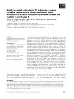
was improved when DRMs were prepared from membranes solubilized in 2.0% CHAPSO. Western blot
analysis showed that PS1-NTF and caveolin-1 (a lipid
raft marker) were localized to a large extent to the interface between 5% and 35% sucrose (fraction 2), while
calnexin (a non-raft marker) was found in the 45%
sucrose fraction (fraction 5) (Fig. 1A). Another lipid
raft marker, flotillin-1, showed poor separation in rat
brain (Fig. 1A). In contrast, CHAPSO DRMs prepared
from SH-SY5Y cells showed a distinct localization of
flotillin-1 to the 5–35% interface (fraction 3, Fig. 1B).
The same pattern was observed for another lipid raft
marker, GM1, which is labeled by the cholera toxin
subunit B (Fig. 1B). The pronounced separation of
lipid raft markers from a non-raft marker in SH-SY5Y
cells indicates that it is easier to prepare DRMs from
SH-SY5Y cells than from brain tissue. The c-secretase
components PS1-NTF and Pen-2 were also found in the
5–35% interface in SH-SY5Y cells. For comparison, we
also prepared DRMs in 1% Triton X-100, a detergent
that is frequently used for isolating DRMs, but no PS1NTF was found in the DRM fraction (Fig. 1C,D).
Thus, 2.0% CHAPSO is suitable for separation of
DRMs containing c-secretase components from soluble material. The 5–35% interface, which contains
DRMs and c-secretase, will be referred to as the DRM
fraction.
Using the protocol described above, we prepared
DRMs from human brain. Six fractions were collected
from the top of the tube and subjected to western blot
analysis using antibodies directed to the c-secretase
1176
components BACE, APP, APP C-terminal fragments
(APP-CTFs) and raft and non-raft markers. Fraction 4
(at the 35–45% interface) and fraction 5 (45% sucrose)
were enriched in the non-lipid raft markers, calnexin
(ER) and adaptin-c (trans-Golgi network). PS1-NTF,
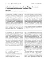
nicastrin, Aph-1aL and Pen-2 were found in the DRM
fraction, while only around 10% of the total protein
was found in this fraction (Fig. 2A,C). Interestingly, the
majority of BACE1, full-length APP and APP-CTFs
were distributed to fractions 4 and 5. The procedure
was repeated using rat brain, and the results were in
line with those obtained for human brain (Fig. 2B,D).
However, in rat brain, the localization of flotillin-1 and
caveolin-1 differed between preparations, and they were
also found in fraction 5 to a varying extent. This could
possibly be due to the more heterogenous and more
lipid-rich starting material as the whole rat brain was
used.
The mature form of nicastrin is found in DRMs
In the active c-secretase complex, nicastrin is glycosylated [32]. To determine the glycosylation status of
nicastrin in DRMs and fractions 4 and 5 from human
and rat brain, endoglycosidase H (Endo H) or N-glycosidase F (peptide-N-glycosidase F, PNGase F) were
applied to deglycosylate nicastrin. Endo H works on a
more limited range of substrates than PNGase F.
Untreated DRMs contained a nicastrin species of
approximately 125 kDa (Fig. 2E). Endo H decreased
the apparent molecular weight of nicastrin from
approximately 125 kDa to approximately 100 kDa,
indicating the presence of high-mannose oligosaccharides. Upon treatment with PNGase F, which also
removes complex oligosaccharides, the deglycosylation
was more pronounced, resulting in a diffuse band at
approximately 80 kDa (Fig. 2E). The deglycosylation
pattern of nicastrin was the same in fractions 4 and 5
as in DRMs, and no differences between human and
rat brain were observed. The above results suggest that
the nicastrin that is present in DRMs (fraction 2) as
well as in fractions 4 and 5 is highly glycosylated,
including high-mannose oligosaccharides and complex
oligosaccharides. The results were confirmed using
another nicastrin antibody (BD Biosciences, San Jose,
CA, USA, data not shown).
DRMs containing c-secretase elute in a
high-molecular-weight SEC fraction
To further purify and investigate the approximate
molecular weight of DRMs containing the c-secretase
complex, we injected the DRM fraction from human
FEBS Journal 275 (2008) 1174–1187 ª 2008 The Authors Journal compilation ª 2008 FEBS
J.-Y. Hur et al.
Human brain c-secretase in DRMs
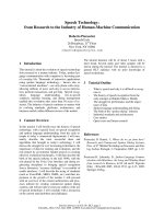
Fig. 1. DRMs prepared in 2% CHAPSO are
enriched in c-secretase components. DRMs
from (A) rat brain and (B) SH-SY5Y cells
were isolated by sucrose gradient
centrifugation after treatment with 2.0%
CHAPSO. In rat brain, six fractions were
collected from the top of the tube: fraction 1, fraction 2 (DRM fraction, interface
between 5% and 35% sucrose), fraction 3,
fraction 4 (interface between 35% and 45%
sucrose), fraction 5 and fraction 6 (pellet). In
SH-SY5Y cells, 12 fractions were collected
from the top of the tube: fraction 1–2, fraction 3 (DRM fraction, interface between 5%
and 35% sucrose), fraction 4–9, fraction 10
(interface between 35% and 45% sucrose),
fraction 11 and fraction 12. The fractions
were subjected to western blot analysis
using flotillin-1 and caveolin-1 (lipid raft
markers), calnexin (a non-raft marker), and
PS1-NTF. In SH-SY5Y cells, the ganglioside
GM1 (a lipid raft marker) was detected by
binding of cholera toxin subunit B using a
dot-blot assay. In (C) and (D), the DRMs
were isolated after treatment with 1% Triton X-100 of (C) rat brain and (D) SH-SY5Y
cells.
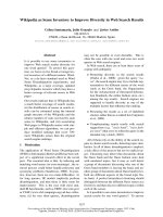
brain onto a Superose 6 SEC column, collected fractions and analyzed them by western blotting using
antibodies directed to all the known c-secretase complex components. When using 0.25% CHAPSO as the
mobile phase, the c-secretase components, APP and
flotillin-1 eluted with the void volume (> 2000 kDa)
(Fig. 3A,B). DRMs from rat brain gave similar results
(Fig. 3C). The relatively narrow peak indicates that
the complex is stable during separation. When using
2% CHAPSO as the mobile phase, most of the nicastrin and PS1-NTF eluted in the void volume, although
a second peak around 230 kDa could be observed
(corresponding to fractions 17–21, Fig. 3F). Thus, the
c-secretase complexes are mainly present in large
DRMs (> 2000 kDa).
The c-secretase complex components can be
co-immunoprecipitated from DRMs
The stability of the complex was further evaluated
by co-immunopreciptation. The starting material for
DRM preparation (P3), fraction 2 (DRMs) and fraction 5 (soluble fraction) from the rat brain DRM
preparation were immunoprecipitated using an antibody against nicastrin (Fig. 4). Western blotting
showed that PS1-CTF, Aph-1aL and Pen-2 co-immunoprecipitated with nicastrin in P3 and the DRM fraction, and, to a lower degree, in the soluble fraction.
Flotillin-1 did not co-immunoprecipitate with nicastrin,
indicating that flotillin-1 and c-secretase are present in
different lipid rafts.
FEBS Journal 275 (2008) 1174–1187 ª 2008 The Authors Journal compilation ª 2008 FEBS
1177
Human brain c-secretase in DRMs
J.-Y. Hur et al.
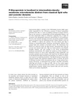
Fig. 2. DRMs containing the c-secretase complex can be isolated from human and rat brain. The protein concentration was analyzed by
BCA assay in (A) human brain and (B) rat brain. (C) Human brain membranes were treated with 2.0% CHAPSO, fractionated on a sucrose
gradient, and subjected to western blot analysis using antibodies directed to the c-secretase complex components’ BACE1, APP, APP-CTFs,
flotillin-1 and caveolin-1 (lipid raft markers) and calnexin and adaptin-c (non-raft markers). (D) The experiment was repeated using rat brain.
The higher-molecular-weight form of nicastrin (> 125 kDa, labeled with an asterisk) was only detected by one antibody, and this was due to
non-specific binding. (E) Fractions 2 (DRMs), 4 and 5 for human brain and rat brain were denatured and incubated overnight at 37 °C with
glycosidases (Endo H and PNGase F). The samples were analyzed by western blot using anti-nicastrin serum. The control was incubated
overnight at 4 °C.
DRMs contain active c-secretase complex
To investigate whether the c-secretase complex present
in DRMs is active, we incubated fractions 2 (DRMs), 4
and 5 from rat brain in the absence or presence of 1 lm
1178
of the c-secretase inhibitor L-685,458 (Fig. 5A), and
found that AICD was produced in DRMs only in the
absence of L-685,458. Although there were higher
amounts of full-length APP and APP-CTFs in fractions 4 and 5, the highest amount of AICD was clearly
FEBS Journal 275 (2008) 1174–1187 ª 2008 The Authors Journal compilation ª 2008 FEBS
J.-Y. Hur et al.
Human brain c-secretase in DRMs
Fig. 3. DRM-associated c-secretase is present in a high-molecular-weight complex, as shown by size-exclusion chromatography (SEC). The
DRM fraction was injected onto a Superose 6HR column and fractions were collected from 10–50 min at a flow rate of 0.5 mLỈmin)1. Solubilization buffer with 0.25% CHAPSO was used as the mobile phase. (A) The absorbance at 254 nm was monitored. The DRM chromatogram was
normalized to the standard chromatogram. (B,C) Every second fraction was analyzed by western blot for (B) human brain and (C) rat brain. The
rat DRM fraction was further analyzed by SEC using solubilization buffer with 0.25% or 2.0% CHAPSO as the mobile phase and the absorbance
at 254 nm was monitored (D). (E,F) Every second fraction was analyzed by western blot for (E) 0.25% CHAPSO and (F) 2.0% CHAPSO.
Fig. 4. The c-secretase complex immunoprecipitates in DRMs. Rat
membranes (P3), the DRM fraction (fraction 2) and fraction 5 were
co-immunoprecipitated with anti-nicastrin serum or control rabbit
IgG. PS1-CTF, Aph-1aL, Pen-2 and flotillin-1 were identified by
western blotting.
generated in DRMs. The immunoreactive band comigrated with a 50-residue synthetic AICD peptide and
was detected by several antibodies (data not shown).
Using an exogenous substrate, C99-FLAG, and a sensitive sandwich ELISA method, we were also able to
detect Ab production, which was inhibited by L-685,458
in the DRM fraction from rat brain (Fig. 5B).
In the next step, we determined whether c-secretase
activity could be detected in a human brain sample
with a postmortem time of 22 h, and were able to
detect AICD production that was inhibited by
L-685,458 (Fig. 5C). Thus, DRMs isolated from postmortem human brain tissue contain active c-secretase
that cleaves endogenous APP-CTFs.
FEBS Journal 275 (2008) 1174–1187 ª 2008 The Authors Journal compilation ª 2008 FEBS
1179
Human brain c-secretase in DRMs
J.-Y. Hur et al.
membranes (postmortem time 0 h), the c-secretase
activity, measured as AICD production, was decreased
by more than 80% at a postmortem time of 6 h. After
this time, the activity continued to decrease but was
still detectable at 24 h postmortem (Fig. 5D) and
remained even after 48 h (data not shown). Thus,
c-secretase activity decreases most rapidly after short
postmortem times, but can be observed in brain tissue
at all time points studied.
c-Secretase co-localizes with lipid rafts in human
brain sections
The presence of a protein in DRMs suggests that it is
associated with lipid rafts. To further investigate
whether the c-secretase components are associated with
lipid rafts, we performed immunofluorescence labeling
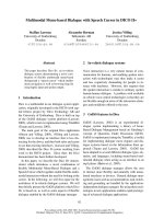
on human brain sections. Multiple fluorescent staining
was used to study co-localization of PS1, nicastrin
and APP with the lipid raft marker GM1. Confocal
microscopy revealed that GM1 immunoreactivity was
most pronounced in the plasma membrane of the cells.
PS1 and nicastrin immunoreactivity overlapped extensively with the lipid raft marker (Fig. 6A,B), but the
overlap of APP and GM1 was limited (Fig. 6C). Thus,
confocal microscopy supports the view that c-secretase
is localized to lipid rafts in human brain.
Discussion
Fig. 5. c-Secretase activity was observed in DRMs by monitoring
AICD and Ab production. (A) The production of AICD was assayed in
fractions 2 (DRMs), 4 and 5 by incubation of 100 lg of protein for
16 h at 37 °C in the absence or presence of the c-secretase inhibitor
L-685,458. The supernatant was subjected to western blot using the
antibody C1 ⁄ 6.1. (B) The DRM fraction (approximately 12 lg protein)
was incubated for 16 h at 37 °C in the absence or presence of the
c-secretase inhibitor L-685,458. Twenty nanograms of C99-FLAG
were added to the samples. Ab40 levels were analyzed by sandwich
ELISA. (C) The production of AICD from human brain was measured
and detected as in (A). (D) Solubilized membranes from rat brain
obtained at various postmortem times were incubated as in (A) and
AICD production was measured as in (A).
To investigate the effect of postmortem time on
c-secretase activity, we collected rat brains after 0, 6,
12 or 24 h postmortem time (see Experimental procedures), prepared P3 and analyzed this fraction for
c-secretase activity. Compared to freshly prepared
1180
Previous studies have shown that APP, BACE and
c-secretase partially localize to lipid rafts, and it has
been suggested that the clustering of these proteins in
lipid rafts increase Ab production [15,21]. These studies were performed in cell lines, which in many cases
overexpressed APP or c-secretase proteins. Recently,
c-secretase was also found to be associated with
DRMs in adult mouse brain [23]. However, the association of c-secretase with lipid rafts in human brain has
not been investigated, and biochemical evidence for
c-secretase activity in DRMs is limited.
Due to their lipid composition, lipid rafts are resistant to certain detergents. Therefore, isolation of
DRMs by treatment with detergents such as Triton
X-100 followed by flotation in a discontinuous sucrose
gradient is frequently used for studying lipid raft components. It should be noted that these preparations are
dependent on the nature and concentration of the
detergent used [20]. Previously, 2% CHAPSO has been
used to isolate DRMs containing an active c-secretase
from SH-SY5Y neuroblastoma cells [22], and 0.5%
Lubrol WX has been used to isolate c-secretase-rich
DRMs from N2a neuroblastoma cells and mouse brain
FEBS Journal 275 (2008) 1174–1187 ª 2008 The Authors Journal compilation ª 2008 FEBS
J.-Y. Hur et al.
Human brain c-secretase in DRMs
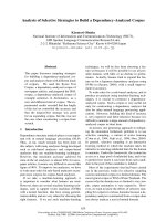
Fig. 6. Confocal microscopy shows partial
co-localization of lipid rafts and c-secretase
in human brain tissue. Immunofluorescence
labeling was performed on human brain
sections. The nucleus was stained with
4’,6-diamidino-2-phenylindole (DAPI). The
cholera toxin subunit B (CT-B) that labels
the lipid rafts is shown by the green fluorescence of the Alexa Fluor 488-coupled goat
anti-rabbit serum. Expression of PS1-CTF,
nicastrin and APP is shown by the red
fluorescence using secondary anti-mouse
Alexa Fluor 594 conjugates. (A) PS1-CTF.
(B) Nicastrin. (C) APP. Scale bar = 1 lm.
[23,33]. We and others have previously studied the
effect of various detergents on the activity of c-secretase prepared from rat brain, and found that 0.4%
CHAPSO resulted in the highest activity [31] but Triton
X-100 abolished the activity [34]. Hence, we used
CHAPSO to prepare DRMs from human and rat
brain. To preserve c-secretase as an active complex, we
started with CHAPSO concentrations in the range
0.25–1.0%. However, it was necessary to increase the
CHAPSO concentration to 2.0% to obtain a good
separation between raft and non-raft markers. We also
noted that the separation was better in SH-SY5Y cells
than in brain samples. Difficulties in obtaining pure
DRM fractions from brain tissue are probably due to
the heterogeneity of the sample and high levels of
myelin.
In the case of human brain, the DRM fraction
resulting from treatment with 2.0% CHAPSO was
FEBS Journal 275 (2008) 1174–1187 ª 2008 The Authors Journal compilation ª 2008 FEBS
1181
Human brain c-secretase in DRMs
J.-Y. Hur et al.
enriched in lipid raft marker proteins and all four
known c-secretase complex components, while APP,
APP-CTFs and BACE were mainly found in other
fractions. The association of c-secretase with DRMs is
in line with previous results from studies in cells or
mouse brain [22,23,33], and suggests that the majority
of c-secretase in human brain is localized to lipid rafts.
Importantly, the localization of PS1 and nicastrin to
lipid rafts was confirmed by immunofluoresence confocal microscopy on human brain sections.
In accordance with our data, previous studies show
that < 25% of BACE is associated with lipid rafts
[21,23,24,26]. Interestingly, it has been suggested that
raft association is necessary for BACE activity [21],
and thus decreasing the amount of raft-associated
BACE could result in lower levels of Ab. Our observation that most of the APP occurs outside the DRM
fraction is in line with previous studies, where the
reported association of APP with DRMs varied from
zero up to 20% [21,23,24,26]. The low levels of BACE,
its substrate and the product (see below) in the DRM
fraction could indicate that the initial step in the amyloidogenic pathway occurs outside lipid rafts. Alternatively, the processing may be initiated by a transient
localization of APP and BACE to lipid rafts.
With regard to APP-CTFs, the published information is limited due to the difficulties in detecting endogenous APP-CTFs in cell lines. Using a c-secretase
inhibitor, APP-CTFs accumulated and were detectable
in both DRMs and the soluble fraction from SHSY5Y neuroblastoma cells or Chinese hamster ovary
(CHO) cells [22,23,25]. In brain tissue, the situation is
different, and endogenous APP-CTFs are readily
detected. In a previous study using adult mouse brain,
the majority of APP-CTFs were found in DRMs,
while full-length APP was found in soluble fractions
[23]. In contrast, we detected APP-CTFs mainly in the
soluble fraction, and it is possible that the choice of
detergent (2% CHAPSO versus 0.5% Lubrol WX)
could explain this discrepancy.
Despite the low substrate levels, AICD production
was easily detected in DRMs from human and rat
brain, while only minor amounts of AICD were generated in the other fractions. To our knowledge, this is
the first study to show c-secretase processing of an
endogenous substrate in DRMs, and the first to show
c-secretase activity in mammalian brain DRMs. Interestingly, we could not detect APP and APP-CTFs in
DRMs after incubating the sample at 37 °C for 16 h.
As shown in Fig. 2C,D, APP and APP-CTFs are present in the DRM fraction before the start of the activity
assay. However, the levels of those fragments were
clearly lower in DRMs than in fraction 4 and 5. We
1182
speculate that the low levels of APP-CTFs might be
degraded by non-specific protease activity during incubation at 37 °C, which also explains why blocking
c-secretase activity using an inhibitor did not lead to
the accumulation of APP-CTFs in DRMs. APP and
APP-CTFs are generally more difficult to detect in
human brain material than in rat brain, probably due
to proteolysis during the long postmortem time. We
were not able to detect production of endogenous Ab,
but, after addition of an exogenous substrate, Ab
could be detected in the DRM fraction from rat brain
using sandwich ELISA. Both AICD and Ab production were inhibited by the c-secretase inhibitor
L-685,458.
In cell studies, only mature nicastrin, which is the
form that associates with the active c-secretase complex [35], is localized to DRMs, while immature nicastrin is detected in other fractions [22,33]. However, in
accordance with our previous results [36], nicastrin was
highly glycosylated in all fractions in the brain study.
Thus, there is a clear difference in the maturation process of nicastrin between cell lines and mammalian
brain.
The predicted molecular weight of the c-secretase
complex at a stoichiometry of 1 : 1 : 1 : 1 (PS : nicastrin : Aph-1 : Pen-2) is approximately 220 kDa [37,38].
Previous studies on soluble c-secretase have estimated
the size of the complex to vary between 200 and
2000 kDa, and the stoichiometry of the c-secretase
complex is not clear [10,36,39–41]. The diverse results
might be due to differences in starting material, preparation procedures and the techniques used (e.g. SEC,
blue native PAGE or gradient centrifugation). By SEC,
the molecular weight of the DRM fraction was estimated to be > 2000 kDa. This high-molecular-weight
fraction contained the raft marker flotillin-1, the c-secretase complex and low amounts of APP and APPCTFs. We suggest that the estimated molecular weight
reflects the size of the DRMs (including other proteins,
lipids and CHAPSO) rather than the size of the c-secretase complex. Elution of the soluble c-secretase complex has been shown to shift from the void volume to a
lower-molecular-weight fraction when the CHAPSO
concentration in the mobile phase is increased [42]. We
detected the majority of the c-secretase components in
the high-molecular-weight fraction from DRMs even
when 2% CHAPSO was used as the mobile phase.
These data show that the c-secretase complex is stably
associated with DRMs. In line with these SEC results,
it was also possible to co-immunoprecipitate PS1,
Aph-1aL and Pen-2 using an anti-nicastrin serum in
2% CHAPSO. Another indication of the stability of
the c-secretase complex is that activity can be observed
FEBS Journal 275 (2008) 1174–1187 ª 2008 The Authors Journal compilation ª 2008 FEBS
J.-Y. Hur et al.
in preparations from human brain with long postmortem times (22 h).
Freshly prepared membranes from rat brain showed
significantly higher c-secretase activity than membranes
prepared at various postmortem times. As access to
human brain material is often limited, and rat
and human brain showed similar results in our
experiments, we suggest that rat brain is a useful substitute for human brain in studies on c-secretase.
The distribution of c-secretase and its substrates
between lipid rafts and disordered domains of the
membrane seems to regulate processing. We speculate
that lowering the levels of c-secretase or APP ⁄ APPCTFs in lipid rafts may be one way to decrease Ab
production. Possibly, c-secretase inhibitors that preferentially distribute to lipid rafts might show increased
selectivity for inhibition of Ab production, and thus be
useful for pharmacological treatment of AD.
In conclusion, c-secretase is present in DRMs prepared from human and rat brain, and confocal microscopy on sections from human brain confirms that
c-secretase is indeed localized to lipid rafts. The
DRM fraction shows high c-secretase activity although
the substrate levels are low, and DRMs prepared
from brain tissue are suitable for studies on active
c-secretase.
Experimental procedures
Human brain material
The cortex from a postmortem human brain (postmortem
time 22 h) of a non-Alzheimer case was obtained from
Huddinge Brain Bank (Huddinge, Sweden) and stored at
)70 °C before use.
Animals
Male Sprague–Dawley rats (200–250 g) were obtained from
B&K Universal (Sollentuna, Sweden). The ethical permit
was granted by the Animal Trial Committee of Southern
Stockholm (no. S60-05). The rats were killed by carbon
dioxide treatment. The brains were dissected to remove
blood vessels and white matter.
Cell culture
The human neuroblastoma cell line, SH-SY5Y, was cultured in Dulbecco’s modified Eagle’s medium supplemented
with 10% fetal bovine serum and 1% penicillin–streptomycin solution (GIBCO ⁄ Invitrogen, Carlsbad, CA, USA).
Cells were grown in 5% CO2 ⁄ 95% air at 37 °C. Nearly
confluent SH-SY5Y cells in three 150 mm dishes were
Human brain c-secretase in DRMs
washed with cold phosphate buffered saline and centrifuged
two times at 1500 g for 5 min at room temperature. The
cell pellet was stored at )20 °C before use.
Preparation of membranes
Membranes were prepared as described previously [36]
with some modifications. Briefly, the brain material was
homogenized by 25 strokes at 1500 r.p.m. using a mechanical pestle homogenizer (RW20; IKALaborteknik, Hattersheim, Germany) in lysis buffer (1 mL buffer ⁄ 0.2 g tissue)
containing 20 mm Hepes (pH 7.5), 50 mm KCl, 2 mm
EGTA and CompleteÔ protease inhibitor mixture (Roche
Applied Science, Indianapolis, IN, USA). All procedures
were carried out on ice. The samples were centrifuged at
1000 g for 10 min to remove nuclei and poorly homogenized material. The pellet was homogenized and then
centrifuged at 1000 g for 10 min, and the post-nuclear
supernatants were pooled and centrifuged once more at
10 000 g for 30 min in order to remove mitochondria. The
supernatant was centrifuged once more, and the final
supernatant was then centrifuged at 100 000 g for 1 h to
yield the final pellet (P3).
Preparation of detergent-resistant membranes
DRMs were prepared as described previously [22] with
some modifications. To isolate DRMs from brain material
or cells, P3 or the cell pellet, respectively, were resuspended in 600 lL of buffer containing 20 mm Tris ⁄ HCl
(pH 7.4), 150 mm NaCl, 1 mm EDTA, 2.0% CHAPSO or
1% Triton X-100, and CompleteÔ protease inhibitor mixture (Roche Applied Science). The samples were incubated
with end-over-end rotation for 20 min at 4 °C. The sample
was adjusted to 45% sucrose and placed at the bottom of
a 14 mL Beckman Ultra-ClearÔ centrifuge tube. Then,
6.9 mL of 35% sucrose followed by 2.3 mL of 5% sucrose
was overlaid. The sample was centrifuged at 100 000 g for
16 h at 4 °C in a SW40Ti rotor (Beckman Coulter, Fullerton, CA, USA). Six fractions were collected from the top
of the tube using a 5 mL syringe (CODAN, Hørsholm,
Denmark). In order to remove sucrose from the six
fractions, PD-10 desalting columns (GE Healthcare, Piscataway, NJ, USA) were used according to the manufacturer’s instructions. A buffer containing 20 mm Hepes
(pH 7.4), 150 mm NaCl, 5 mm EDTA and CompleteÔ
protease inhibitor mixture (Roche Applied Science) was
diluted sevenfold and used to equilibrate the columns. The
samples were applied, eluted and concentrated to 1· buffer
(seven times) using a vacuum centrifuge (Maxi Dry Lyo,
Heto-Holten AIS, Allerød, Denmark). The protein concentration was determined by BCA protein assay according to
the manufacturer’s instructions (Pierce, Rockford, IL,
USA).
FEBS Journal 275 (2008) 1174–1187 ª 2008 The Authors Journal compilation ª 2008 FEBS
1183
Human brain c-secretase in DRMs
J.-Y. Hur et al.
SDS–PAGE and western blotting
Equal amounts of protein (20 lg) were mixed with 4· LDS
sample buffer (Invitrogen) and kept at room temperature for
20 min. The samples were then loaded onto a NuPAGEÔ
4–12% Bis–Tris gel or a 16% Tricine gel (Invitrogen). The
samples were electrophoresed, transferred to nitrocellulose
membranes (Whatman Ltd, Maidstone, UK), and the proteins of interest were detected by specific antibodies.
Antibodies
The following antibodies were used: PS1-NTF (529591; Calbiochem, Darmstadt, Germany), raised against amino acid
residues 1-65 of human PS1; PS1-loop (MAB5232; Chemicon, Billerica, MA, USA), raised against the loop (amino
acid residues 263–378) of human PS1; nicastrin (N1660;
Sigma, St Louis, MO, USA), raised against C-terminal residues 693–709 of human nicastrin; Aph-1aL (PRB-550P;
COVANCE, Berkeley, CA, USA), raised against the
C-terminal region of human Aph-1aL; UD1 (a gift from
J. Naslund, Karolinska Institutet, Sweden), raised against
ă
the N-terminal residues ERVSNEEKLNL of Pen-2; BACE-1
(B0681; Sigma), raised against the N-terminal regions of
human BACE-1 (amino acids 46–62, with C-terminally added
lysine); C1 ⁄ 6.1 (a gift from P. M. Mathews, Nathan Kline
Institute, NY, USA), raised against the C-terminus of b-APP;
calnexin (SPA-860; Stressgen, San Diego, CA, USA), raised
against canine calnexin (residues 575-593); adaptin-c (610385;
BD Biosciences); flotillin-1 (610820; BD Biosciences); caveolin-1 (sc-894; Santa Cruz Biotechnology, Santa Cruz, CA,
USA); cholera toxin, subunit B (C3741; Sigma).
Size-exclusion chromatography
The DRM fraction was injected onto a Superose 6HR column (Amersham Biosciences, Piscataway, NJ, USA), using
a buffer containing 20 mm Hepes, pH 7.0, 150 mm KCl,
2 mm EGTA, CompleteÔ protease inhibitor mixture
(Roche Applied Science) and 0.25% or 2% CHAPSO as
the mobile phase at a flow rate of 0.5 mLỈmin)1. Fractions
(0.5 mL) were collected from 10–50 min, and analyzed by
SDS–PAGE as described above.
Co-immunoprecipitation
Rat membranes (P3) were resuspended in 600 lL of immunoprecipitation buffer containing 20 mm Tris ⁄ HCl
(pH 7.4), 150 mm NaCl, 1 mm EDTA, 2.0% CHAPSO and
CompleteÔ protease inhibitor mixture (Roche Applied
Science). The samples were incubated with end-over-end
rotation for 20 min at 4 °C. P3, fraction 2 and fraction 5
were pre-cleared with a 1 : 1 ratio of protein A ⁄ protein
G Sepharose (GE Healthcare) for 30 min at 4 °C, and incu-
1184
bated with anti-nicastrin or control rabbit IgG overnight at
4 °C. Protein A ⁄ G Sepharose was added for 1 h at 37 °C.
After washing three times with immunoprecipitation buffer,
the beads were eluted in SDS–PAGE sample buffer and
subjected to SDS–PAGE as described above.
c-Secretase activity assay
The production of AICD was assayed by incubating the
samples for 16 h at 37 °C in the absence or presence of the
c-secretase inhibitor L-685,458 (Bachem, Torrance, CA,
USA). After incubation, samples were centrifuged at
100 000 g for 1 h to remove the membranes, and the supernatant was collected, concentrated using a vacuum centrifuge (Maxi Dry Lyo) and analyzed by SDS–PAGE.
Sandwich enzyme-linked immunosorbent assay
Ab40 levels were analyzed by commercial Sandwich
enzyme-linked immunosorbent assay (sandwich ELISA;
Wako Chemicals, Osaka, Japan) according to the manufacturer’s instructions. The DRM fraction was incubated for
16 h at 37 °C in the absence or presence of the c-secretase
inhibitor L-685,458. Twenty nanograms of recombinant
and purified C99-FLAG dissolved in 2,2,2-trifluoroethanol
were added to the samples. The reaction was stopped by
adding RIPA buffer (150 mm NaCl, 1.0% NP-40, 0.5%
sodium deoxycholate, 0.1% SDS, 50 mm Tris ⁄ HCl, pH 8.0)
and boiling for 5 min. The samples were centrifuged at
1000 g for 5 min at room temperature and the supernatants
were dispensed into wells (12 lg protein ⁄ well) coated with
BNT77 antibody (directed to amino acids 11–28 of Ab)
and incubated overnight at 4 °C. Bound Ab40 was detected
by the 3,3¢,5,5¢-tetramethylbenzidine (TMB) reaction using
horseradish peroxidase-conjugated BA27 antibody (directed
to the C-terminus of Ab40). All measurements were performed in duplicate, and Ab40 levels were calculated
from the synthetic Ab(1–40) (Bachem Bioscience, King of
Prussia, PA, USA).
Deglycosylation
The glycosylation status of nicastrin was analyzed as
described previously [36]. Briefly, samples from the DRM
preparation were denatured by heating for 10 min at 100 °C
in the presence of 0.5% v ⁄ v SDS and 1.0% v ⁄ v b-mercaptoethanol, cooled on ice and adjusted to 50 mm sodium
citrate, pH 5.5. For Endo H treatment, 100 milliunits
(as defined by the supplier) of Endo H (Roche Applied
Science) was added. For PNGase F treatment, NP-40 was
added to a final concentration of 1.0%, followed by addition of 15.4 milliunits (as defined by the supplier) of
PNGase F (Roche Applied Science). The samples were
incubated overnight at 37 °C and analyzed by SDS–PAGE.
FEBS Journal 275 (2008) 1174–1187 ª 2008 The Authors Journal compilation ª 2008 FEBS
J.-Y. Hur et al.
Postmortem time study
The rats were killed and kept at room temperature for 2 h.
In order to simulate a slow cooling curve (as in the case of
the human brain), the head was removed, put in plastic
bag, placed in a styrofoam box filled with water (37 °C),
and the box was placed in a cold room (4 °C) [43]. After 6,
12, 24 or 48 h, the brains were removed, and stored at
)70 °C before use. To obtain fresh tissue (postmortem time
0 h), the rat was killed and the brain was immediately
removed and homogenized.
Immunofluorescence labeling and confocal
microscopy
Cryopreserved human brain sections from the frontal cortex
embedded in Tissue-TEK OCT compound (Miles, Elkhart,
IN, USA), were cut in 12 lm thick sections, mounted on
Hypertema Teflon-coated glass slides (Novakemi, Stockholm, Sweden) and air-dried. For staining of lipid rafts, the
brain tissues were labeled using fluorescent cholera toxin
subunit B (CT-B) (Vybrant Alexa Fluor 488 lipid raft labeling kits, Invitrogen Molecular Probes, Carlsbad, CA, USA)
and incubated with anti-CT-B. The brain tissues were fixed
in 4% formaldehyde and 4% sucrose, and permeabilized
with 0.2% Triton X-100. After the blocking step with
blocking buffer (DAKO protein block serum-free; Dako,
Gidstrup, Denmark), primary and secondary antibodies
were diluted in DAKO blocking buffer. For labeling of
PS1, the PS1-loop antibody followed by an Alexa
Fluor 594-conjugated anti-mouse serum (Invitrogen Molecular Probes) was used. The nicastrin antibody (AB5890;
Chemicon International, Temecula, CA, USA) and an
Alexa Fluor 594-conjugated anti-pig serum were used for
labeling of nicastrin. Anti-APP-CT20 C-terminal (171610;
Calbiochem) and Alexa Fluor 488-conjugated anti-rabbit
sera were used for APP. To reduce the background of
staining, we used autofluorescence eliminator reagent
(Chemicon International). All samples were visualized
using an inverted laser scanning microscope (LSM 510
META; Zeiss, Thornwood, NY, USA).
Acknowledgements
We thank Dr Jan Naslund (Karolinska Institutet) for
ă
the UD1 antibody, Dr Paul M. Mathews (The Nathan
S. Kline Institute) for the C1 ⁄ 6.1 antibody, and Dr
Takeshi Nishimura (Dainippon Sumitomo Pharma) for
C99-FLAG. We thank Dr Nenad Bogdanovic and
Inga Volkmann (Karolinska Institutet) for skillful
assistance with human brain tissue preparation. This
work was supported by Dainippon Sumitomo Pharma,
by the Osher Foundation, Gamla Tjanarinnor (H.W.),
ă
Socialstyrelsen (H.W. and L.O.T.), the Foundation for
Human brain c-secretase in DRMs
Alzheimer’s and Dementia Research (SADF) and the
Foundation for Geriatric Diseases at the Karolinska
Institutet (J.F.).
References
1 Selkoe DJ (2001) Presenilin, Notch, and the genesis and
treatment of Alzheimer’s disease. Proc Natl Acad Sci
USA 98, 11039–11041.
2 Selkoe DJ (2001) Alzheimer’s disease: genes, proteins,
and therapy. Physiol Rev 81, 741–766.
3 Iwatsubo T, Odaka A, Suzuki N, Mizusawa H, Nukina
N & Ihara Y (1994) Visualization of Ab42(43) and
Ab40 in senile plaques with end-specific Ab monoclonals: evidence that an initially deposited species is
Ab42(43). Neuron 13, 45–53.
4 Jarrett JT, Berger EP & Lansbury PT Jr (1993) The carboxy terminus of the b amyloid protein is critical for
the seeding of amyloid formation: implications for the
pathogenesis of Alzheimer’s disease. Biochemistry 32,
4693–4697.
5 Naslund J, Haroutunian V, Mohs R, Davis KL, Davies
P, Greengard P & Buxbaum JD (2000) Correlation
between elevated levels of amyloid b-peptide in the
brain and cognitive decline. J Am Med Assoc 283,
1571–1577.
6 Hardy J & Selkoe DJ (2002) The amyloid hypothesis of
Alzheimer’s disease: progress and problems on the road
to therapeutics. Science 297, 353–356.
7 Walsh DM, Klyubin I, Fadeeva JV, Rowan MJ & Selkoe DJ (2002) Amyloid-b oligomers: their production,
toxicity and therapeutic inhibition. Biochem Soc Trans
30, 552–557.
8 Kimberly WT & Wolfe MS (2003) Identity and function
of c-secretase. J Neurosci Res 74, 353–360.
9 Takasugi N, Tomita T, Hayashi I, Tsuruoka M, Niimura M, Takahashi Y, Thinakaran G & Iwatsubo T
(2003) The role of presenilin cofactors in the c-secretase
complex. Nature 422, 438–441.
10 Edbauer D, Winkler E, Regula JT, Pesold B, Steiner H
& Haass C (2003) Reconstitution of c-secretase activity.
Nat Cell Biol 5, 486–488.
11 Chen F, Hasegawa H, Schmitt-Ulms G, Kawarai T,
Bohm C, Katayama T, Gu Y, Sanjo N, Glista M,
Rogaeva E et al. (2006) TMP21 is a presenilin complex
component that modulates c-secretase but not e-secretase activity. Nature 440, 1208–1212.
12 Zhou S, Zhou H, Walian PJ & Jap BK (2005) CD147
is a regulatory subunit of the c-secretase complex in
Alzheimer’s disease amyloid b-peptide production. Proc
Natl Acad Sci US 102, 7499–7504.
13 Wolfe MS (2006) The c-secretase complex: membraneembedded proteolytic ensemble. Biochemistry 45, 7931–
7939.
FEBS Journal 275 (2008) 1174–1187 ª 2008 The Authors Journal compilation ª 2008 FEBS
1185
Human brain c-secretase in DRMs
J.-Y. Hur et al.
14 Siemers ER, Quinn JF, Kaye J, Farlow MR, Porsteinsson A, Tariot P, Zoulnouni P, Galvin JE, Holtzman
DM, Knopman DS et al. (2006) Effects of a c-secretase
inhibitor in a randomized study of patients with Alzheimer disease. Neurology 66, 602–604.
15 Cordy JM, Hooper NM & Turner AJ (2006) The
involvement of lipid rafts in Alzheimer’s disease. Mol
Membr Biol 23, 111–122.
16 Sing CF & Davignon J (1985) Role of the apolipoprotein E polymorphism in determining normal plasma lipid
and lipoprotein variation. Am J Hum Genet 37, 268–285.
17 Simons K & Ikonen E (1997) Functional rafts in cell
membranes. Nature 387, 569–572.
18 Pike LJ (2006) Rafts defined: a report on the Keystone
Symposium on lipid rafts and cell function. J Lipid Res
47, 1597–1598.
19 Sharma P, Varma R, Sarasij RC, Ira, Gousset K,
Krishnamoorthy G, Rao M & Mayor S (2004) Nanoscale organization of multiple GPI-anchored proteins in
living cell membranes. Cell 116, 577–589.
20 Schuck S, Honsho M, Ekroos K, Shevchenko A &
Simons K (2003) Resistance of cell membranes to different detergents. Proc Natl Acad Sci USA 100, 5795–5800.
21 Ehehalt R, Keller P, Haass C, Thiele C & Simons K
(2003) Amyloidogenic processing of the Alzheimer
b-amyloid precursor protein depends on lipid rafts.
J Cell Biol 160, 113–123.
22 Urano Y, Hayashi I, Isoo N, Reid PC, Shibasaki Y,
Noguchi N, Tomita T, Iwatsubo T, Hamakubo T &
Kodama T (2005) Association of active c-secretase complex with lipid rafts. J Lipid Res 46, 904–912.
23 Vetrivel KS, Cheng H, Kim SH, Chen Y, Barnes NY,
Parent AT, Sisodia SS & Thinakaran G (2005) Spatial
segregation of c-secretase and substrates in distinct
membrane domains. J Biol Chem 280, 25892–25900.
24 Hattori C, Asai M, Onishi H, Sasagawa N, Hashimoto
Y, Saido TC, Maruyama K, Mizutani S & Ishiura S
(2006) BACE1 interacts with lipid raft proteins. J Neurosci Res 84, 912–917.
25 Wahrle S, Das P, Nyborg AC, McLendon C, Shoji M,
Kawarabayashi T, Younkin LH, Younkin SG & Golde
TE (2002) Cholesterol-dependent c-secretase activity in
buoyant cholesterol-rich membrane microdomains.
Neurobiol Dis 9, 11–23.
26 Abad-Rodriguez J, Ledesma MD, Craessaerts K, Perga
S, Medina M, Delacourte A, Dingwall C, De Strooper
B & Dotti CG (2004) Neuronal membrane cholesterol
loss enhances amyloid peptide generation. J Cell Biol
167, 953–960.
27 Lee SJ, Liyanage U, Bickel PE, Xia W, Lansbury PT Jr
& Kosik KS (1998) A detergent-insoluble membrane
compartment contains Ab in vivo. Nat Med 4, 730–734.
28 Li YM, Lai MT, Xu M, Huang Q, DiMuzio-Mower J,
Sardana MK, Shi XP, Yin KC, Shafer JA & Gardell SJ
(2000) Presenilin 1 is linked with c-secretase activity in
1186
29
30
31
32
33
34
35
36
37
38
39
40
the detergent solubilized state. Proc Natl Acad Sci USA
97, 6138–6143.
LaVoie MJ, Fraering PC, Ostaszewski BL, Ye W, Kimberly WT, Wolfe MS & Selkoe DJ (2003) Assembly of
the c-secretase complex involves early formation of an
intermediate subcomplex of Aph-1 and nicastrin. J Biol
Chem 278, 37213–37222.
Wrigley JD, Nunn EJ, Nyabi O, Clarke EE, Hunt P,
Nadin A, De Strooper B, Shearman MS & Beher D
(2004) Conserved residues within the putative active site
of c-secretase differentially influence enzyme activity and
inhibitor binding. J Neurochem 90, 1312–1320.
Franberg J, Welander H, Aoki M, Winblad B, Tjernberg LO & Frykman S (2007) Rat brain c-secretase
activity is highly influenced by detergents. Biochemistry
46, 7647–7654.
Shirotani K, Edbauer D, Capell A, Schmitz J, Steiner
H & Haass C (2003) c-secretase activity is associated
with a conformational change of nicastrin. J Biol Chem
278, 16474–16477.
Vetrivel KS, Cheng H, Lin W, Sakurai T, Li T,
Nukina N, Wong PC, Xu H & Thinakaran G (2004)
Association of c-secretase with lipid rafts in post-Golgi
and endosome membranes. J Biol Chem 279, 44945–
44954.
Gu Y, Misonou H, Sato T, Dohmae N, Takio K &
Ihara Y (2001) Distinct intramembrane cleavage of the
b-amyloid precursor protein family resembling c-secretase-like cleavage of Notch. J Boil Chem 276, 35235–
35238.
Edbauer D, Winkler E, Haass C & Steiner H (2002)
Presenilin and nicastrin regulate each other and
determine amyloid b-peptide production via complex
formation. Proc Natl Acad Sci USA 99, 8666–8671.
Farmery MR, Tjernberg LO, Pursglove SE, Bergman A,
Winblad B & Naslund J (2003) Partial purification and
characterization of c-secretase from post-mortem human
brain. J Biol Chem 278, 24277–24284.
De Strooper B (2003) Aph-1, Pen-2, and nicastrin with
presenilin generate an active c-secretase complex.
Neuron 38, 9–12.
Iwatsubo T (2004) The c-secretase complex: machinery
for intramembrane proteolysis. Curr Opin Neurobiol 14,
379–383.
Kimberly WT, LaVoie MJ, Ostaszewski BL, Ye W,
Wolfe MS & Selkoe DJ (2003) c-secretase is a membrane protein complex comprised of presenilin,
nicastrin, Aph-1, and Pen-2. Proc Natl Acad Sci USA
100, 6382–6387.
Gu Y, Sanjo N, Chen F, Hasegawa H, Petit A, Ruan
X, Li W, Shier C, Kawarai T, Schmitt-Ulms G et al.
(2004) The presenilin proteins are components of
multiple membrane-bound complexes that have
different biological activities. J Biol Chem 279, 31329–
31336.
FEBS Journal 275 (2008) 1174–1187 ª 2008 The Authors Journal compilation ª 2008 FEBS
J.-Y. Hur et al.
41 Evin G, Canterford LD, Hoke DE, Sharples RA, Culvenor JG & Masters CL (2005) Transition-state analogue
c-secretase inhibitors stabilize a 900 kDa presenilin ⁄
nicastrin complex. Biochemistry 44, 4332–4341.
42 Hoke DE, Tan JL, Ilaya NT, Culvenor JG, Smith SJ,
White AR, Masters CL & Evin GM (2005) In vitro c-secretase cleavage of the Alzheimer’s amyloid precursor
Human brain c-secretase in DRMs
protein correlates to a subset of presenilin complexes and
is inhibited by zinc. FEBS J 272, 5544–5557.
43 Spokes EG & Koch DJ (1978) Post-mortem stability
of dopamine, glutamate decarboxylase and choline
acetyltransferase in the mouse brain under conditions
simulating the handling of human autopsy material.
J Neurochem 31, 381–383.
FEBS Journal 275 (2008) 1174–1187 ª 2008 The Authors Journal compilation ª 2008 FEBS
1187