Báo cáo khoa học: Cholesterol interaction with the related steroidogenic acute regulatory lipid-transfer (START) domains of StAR (STARD1) and MLN64 (STARD3) ppt
Bạn đang xem bản rút gọn của tài liệu. Xem và tải ngay bản đầy đủ của tài liệu tại đây (725.62 KB, 13 trang )
Cholesterol interaction with the related steroidogenic
acute regulatory lipid-transfer (START) domains of StAR
(STARD1) and MLN64 (STARD3)
Julian Reitz
1
, Katja Gehrig-Burger
1
, Jerome F. Strauss III
2
and Gerald Gimpl
1
1 Institute of Biochemistry, Gutenberg-University Mainz, Germany
2 Department of Obstetrics & Gynecology, Virginia Commonwealth University, Richmond, VA, USA
Cholesterol is an essential multifunctional lipid in most
eukaryotic cells. It exerts a strong influence on the
physical state of the plasma membrane, forms choles-
terol–sphingolipid-rich microdomains such as caveolae
and lipid rafts, is necessary for the activity of several
membrane proteins, and serves as the precursor for
steroid hormones [1–5]. Despite many efforts, the path-
ways and mechanisms of cellular cholesterol trafficking
are currently not well understood. Misfunctions of
cholesterol transport are linked to a variety of diseases
[6,7].
The biosynthesis of steroid hormones requires the
transfer of cholesterol from multiple sources to the
inner mitochondrial membrane, where steroidogenesis
begins with the conversion of cholesterol to pregneno-
lone. The translocation of cholesterol to the inner
mitochondrial membrane, the rate-limiting step in
steroidogenesis, is mediated by steroidogenic acute
regulatory protein (StAR, STARD1) [8–12]. The
mechanism by which STARD1 moves cholesterol to
the inner mitochondrial membrane is currently unclear
[13]. Mutations that inactivate STARD1 in humans
lead to an impaired ability of the adrenal gland to pro-
duce steroid hormones, a potentially lethal disease
known as congenital lipoid adrenal hyperplasia [14].
Ablation of the StarD1 gene in mice also causes
impaired steroidogenesis and adrenal lipid accumula-
tion [15]. STARD1 is synthesized as a 37 kDa phos-
phoprotein with an N-terminal mitochondrial targeting
sequence that is cleaved during mitochondrial entry
(Fig. 1A). Deletion of 62 N-terminal residues (N-62
STARD1), including the leader peptide, resulted in a
Keywords
cholesterol; MLN64; STARD1; STARD3;
START proteins
Correspondence
G. Gimpl, Institute of Biochemistry,
Gutenberg-University Mainz, Becherweg 30,
55128 Mainz, Germany
Fax: +49 6131 3925348
Tel: +49 6131 3923829
E-mail:
(Received 14 January 2008, revised 5 Febru-
ary 2008, accepted 14 February 2008)
doi:10.1111/j.1742-4658.2008.06337.x
The steroidogenic acute regulatory (StAR)-related lipid transfer (START)
domains are found in a wide range of proteins involved in intracellular
trafficking of cholesterol and other lipids. Among the START proteins are
the StAR protein itself (STARD1) and the closely related MLN64 protein
(STARD3), which both function in cholesterol movement. We compared
the cholesterol-binding properties of these two START domain proteins.
Cholesterol stabilized STARD3-START against trypsin-catalyzed degrada-
tion, whereas cholesterol had no protective effect on STARD1-START.
[
3
H]Azocholestanol predominantly labeled a 6.2 kDa fragment of
STARD1-START comprising amino acids 83–140, which contains residues
proposed to interact with cholesterol in a hydrophobic cavity. Photoaffinity
labeling studies suggest that cholesterol preferentially interacts with one
side wall of this cavity. In contrast, [
3
H]azocholestanol was distributed
more or less equally among the polypeptides of STARD3-START. Overall,
our results provide evidence for differential cholesterol binding of the two
most closely related START domain proteins STARD1 and STARD3.
Abbreviations
MLN64 (= STARD3), metastatic lymph node 64; MbCD, methyl-b-cyclodextrin; NBD-cholesterol, 22-[N-(7-nitrobenz-2-oxa-1,3-diazol-4-
yl)amino]-23,24-bisnor-5-cholen-3-ol; SELDI, surface-enhanced laser desorption/ionization; StAR (= STARD1), steroidogenic acute regulatory
protein; START, steroidogenic acute regulatory protein lipid-transfer domain.
1790 FEBS Journal 275 (2008) 1790–1802 ª 2008 The Authors Journal compilation ª 2008 FEBS
cytosolic protein with full activity, as shown in intact
cells and in isolated mitochondria [16–18]. The func-
tionally active C-terminal domain of STARD1 con-
tains the StAR-related lipid-transfer (START) domain.
START domains consist of 200–210 amino acids and
are found in a wide range of proteins involved in
several cellular functions, including lipid transport,
signal transduction, and transcriptional regulation [19].
Among the START proteins are the StAR protein
itself (STARD1) and the closely related metastatic
lymph node 64 (MLN64) protein (STARD3). Both
proteins function as cholesterol-binding proteins
[20,21]. Their START domains share 37% sequence
identity.
STARD3 is overexpressed in certain breast cancers
[22]. The protein contains four transmembrane helices
that target it to the membrane of late endosomes [23]
(Fig. 1A). However, the physiological function of
STARD3 is currently unclear. It may be involved in
steroidogenesis in the human placenta, which lacks
STARD1 [24,25]. The START domain at the C-termi-
nal half of STARD3 is believed to be exposed to the
cytosol. In its isolated form, STARD3-START is able
to promote steroidogenesis even more efficiently than
intact STARD3 [26]. The crystal structure of the unli-
ganded START domain of human STARD3 has been
resolved [20]. This structure shows a hydrophobic tun-
nel that expands throughout the length of the START
domain and is perfectly sized to accommodate a single
cholesterol molecule [20]. A similar structure has been
reported for the cholesterol-regulated START pro-
tein 4 (STARD4) [27]. For another START protein,
the phosphatidylcholine transfer protein (STARD2), it
has been directly shown that the tunnel represents the
binding site of the lipid, in this case phosphatidylcho-
line [28].
To understand the molecular mechanism how cho-
lesterol is transferred by STARD1 and STARD3, the
cholesterol-binding sites of these proteins have to be
identified. As a crystal structure of a cholesterol–
START complex is not yet available, other methods
are required to explore the cholesterol–protein interac-
tion. One approach is molecular modeling based on
the knowledge of the unliganded STARD3 structure.
Two such modeling studies have been recently per-
formed for the START domains of STARD1 and
STARD3 [29,30]. This led to the proposal that
STARD1-START shuttles cholesterol carried in its
hydrophobic cavity between the outer and inner mito-
chondrial membranes [20]. However, spectral and bio-
chemical data supported the view that STARD1
partially unfolds and forms molten globules in the
low-pH environment of the outer mitochondrial
membrane. These intermediates were hypothesized to
facilitate the cholesterol transfer of STARD1 to the
mitochondrial inner membrane through a mechanism
that does not involve sterol shuttling [31,32].
A
START
N
C
START
N
C
B
123
97
66
45
31
21
14
–
–
–
–
–
–
C
m/z
20000 25000 30000 35000
m/z
20000 25000 30000 35000
Intensity
Intensity
0
5
10
15
20
0
10
20
30
40
50
29162.8+H
26167.8+H
STARD1-START
STARD3-START
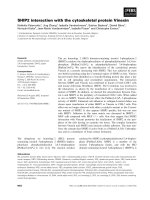
Fig. 1. Expression of the START domains of STARD1 and STARD3.
(A) Domain organization of the START proteins STARD1 (285 amino
acids) and STARD3 (445 amino acids). Both proteins possess a ste-
rol-binding START domain ( 200 amino acids) in their C-terminal
regions. The N-terminal targeting sequence of STARD1 is cleaved
upon entry into the mitochondria, and is nonessential for the activity
of STARD1 [16–18]. The N-terminal part of STARD3 possesses four
transmembrane segments that target the protein to late endosomes.
The START domain in STARD3 is exposed to the cytosol and is func-
tionally active in its isolated form [26]. (B) Purification of the START
domains of STARD1 and STARD3 expressed in Escherichia coli. The
proteins were purified from E. coli, resolved by SDS ⁄ PAGE, and
identified by Coomassie blue staining. Lane 1: marker. Lane 2:
STARD1-START (2 lg of protein). Lane 3: STARD3-START (6 lgof
protein). (C) SELDI-TOF of STARD1-START and STARD3-START.
J. Reitz et al. Cholesterol binding of START proteins
FEBS Journal 275 (2008) 1790–1802 ª 2008 The Authors Journal compilation ª 2008 FEBS 1791
Here, we analyzed the cholesterol-binding character-
istics of the two most related START proteins,
STARD1 and STARD3. Photoaffinity labeling with
radiolabeled 6-azocholestanol as the photoreactive cho-
lesterol probe was employed to characterize and com-
pare the cholesterol binding of the START domains.
This cholesterol analog (previously often termed
photocholesterol) has already been successfully applied
for various proteins [23,33–36]. Overall, this study
addresses the question of whether or not the related
START domains of StARD1 and StARD3 interact
with cholesterol in a similar manner.
Results
Expression of the START domains
The recombinant START proteins each contain a His
6
-
tag at their C-terminus. The proteins were expressed in
BL21 Escherichia coli and purified by affinity chroma-
tography using an Ni
2+
–nitrilotriacetic acid agarose
matrix. Figure 1B shows the Coomassie stains of the
purified proteins. The apparent molecular masses of the
His-tag START proteins in the SDS ⁄ PAGE system were
slightly greater than the calculated molecular masses of
25 769 Da (pI 6.42) and 26 847 Da (pI 8.43) for
STARD1-START and STARD3-START, respectively
(Fig. 1B). This discrepancy has also been observed by
Arakane et al. [17] in the case of STARD1-START. To
explore this issue, we also determined the molecular
masses of both START proteins by surface-enhanced
laser desorption/ionization (SELDI)-TOF MS. Molecu-
lar masses of 26 167 and 29 162 Da were found for
STARD1-START and STARD3-START, respectively
(Fig. 1C). Whereas the molecular mass of STARD1-
START is relatively close (+398 Da) to the calculated
value of 25.7 kDa, the mass of STARD3-START is
about 2.3 kDa higher than that calculated for the
unmodified polypeptide. This could reflect post-transla-
tional protein modification. The expression levels of
STARD1-START and STARD3-START were similar.
Cholesterol binding of the START proteins
In order to verify the cholesterol binding of the
START proteins, we used the fluorescent cholesterol
reporter 22-[N-(7-nitrobenz-2-oxa-1,3-diazol-4-yl)amino]-
23,24-bisnor-5-cholen-3-ol (NBD-cholesterol). This cho-
lesterol analog has successfully been employed to
analyze the cholesterol binding of STARD1-START
[21,31]. A strong increase in the fluorescence intensity
of NBD-cholesterol occurs when the ligand binds to
the hydrophobic environment of the START proteins.
This has recently been studied in detail by Petrescu et al.
[21] in the case of STARD1-START. The binding of
NBD-cholesterol to each of the START proteins shows
a saturating profile (supplementary Fig. S1A,B). The
curves were fitted using a nonlinear regression
algorithm according to one-site models, and yielded K
D
values of 161 ± 45 nm (n = 3) for STARD1-START
and 58 ± 16 nm (n = 3) for STARD3-START. Thus,
STARD3-START bound NBD-cholesterol with a
slightly higher affinity than did STARD1-START.
Two-site models did not result in significantly better
fittings of the binding data.
According to one model of START domain action,
a pH-dependent molten globule transition of STARD1
is required for sterol transfer activity at the level of the
mitochondrial outer membrane [31,32]. Therefore, we
also measured the fluorescence of NBD-cholesterol
(500 nm) bound to STARD1-START (10 nm)atan
acidic pH. At pH 3, the sterol binding of STARD1-
START was about three-fold lower than the sterol
binding measured at pH 7.4 (data not shown).
Analysis of the stabilizing effect of cholesterol
on START proteins
Cholesterol and its analogs are able to stabilize pro-
teins against proteolysis or thermal degradation [37].
To test whether this occurs in the case of the START
proteins, we analyzed the migration behavior of these
proteins in SDS gels under various conditions.
First, the START proteins were incubated (for
20 min at 25 °C) in the presence of cholesterol, photo-
cholesterol, or buffer control. The proteins were irradi-
ated with UV light for 10 min prior to separation by
SDS ⁄ PAGE, western blotting, and immunodetection
with antibody to His (supplementary Fig. S2A). It is
important to note that the His-tag is localized at the
C-terminus of both proteins, so that only molecular
species with an intact C-terminus are visible on the
immunoblots. The immunoblot revealed no significant
differences among treated and untreated START pro-
teins. Faint staining was observed for the putative
dimer forms of the proteins in addition to the predom-
inant monomer ( 30 kDa) bands. We did not find a
slight increase in the molecular size of the START pro-
teins in the photoactivated samples of the photocholes-
terol-containing samples. Most probably, the labeled
species is below the detection limit, due to the low
photoaffinity yield (< 9%).
We next analyzed the resistance of the START pro-
teins to degradation in the presence and absence of
cholesterol. The proteins were pretreated either with
buffer solution or cholesterol–methyl-b-cyclodextrin
Cholesterol binding of START proteins J. Reitz et al.
1792 FEBS Journal 275 (2008) 1790–1802 ª 2008 The Authors Journal compilation ª 2008 FEBS
(MbCD) (0.1 mm) for 20 min at 25 °C. Then, the sam-
ples were incubated for increasing times (6 h, 24 h,
80 h) at 40 °C prior to separation by SDS⁄ PAGE,
western blotting, and immunodetection with antibody
to His (supplementary Fig. S2B). For STARD1-
START, we did not observe any evidence of degrada-
tion during the time course of this experiment. In
contrast, in the case of STARD3-START, an addi-
tional band with a slightly decreased apparent molecu-
lar mass (by 3–4 kDa) appeared after an incubation
period of 24 h or longer. The presence of cholesterol
did not influence the appearance of this additional
band (supplementary Fig. S2B).
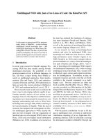
When the samples were treated with trypsin (10 min
or 40 min at 37 °C), additional bands were observed
on the immunoblots for both START proteins
(Fig. 2). Two additional molecular species with slightly
higher electrophoretic mobilities appeared for
STARD1-START. The presence of cholesterol did not
inhibit the appearance of these additional bands, nor
did it affect the protein patterns of the immunoblots.
STARD3-START was more sensitive to trypsinolysis
(Fig. 2). When trypsin was incubated for 40 min, most
of the STARD3-START was either totally degraded
or, more probably, had its C-terminus bearing the His-
tag cleaved. Incubations with trypsin for more than
60 min resulted in immunoblots with no detectable
START proteins (not shown). However, cholesterol
was clearly able to inhibit the trypsinolysis of
STARD3-START (Fig. 2).
Cholesterol labeling of STARD1-START
To determine the cholesterol docking site within the
START domains of STARD1 and STARD3, we per-
formed photoaffinity labeling with [
3
H]photocholester-
ol and subsequent chemical or enzymatic cleavage of
the photoactivated samples. Highly reproducible frag-
mentation patterns were obtained when the protein
was subjected to chemical cleavage by cyanogen
bromide (CNBr), which hydrolyzes peptide bonds
C-terminal to Met residues. The predicted cleavage
products are listed in Table 1 for STARD1-START.
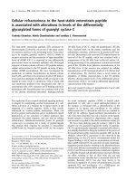
In the case of STARD1-START, the [
3
H]photocholes-
terol radiolabel was incorporated nearly quantitatively
into a single band at about 6.2 kDa (Fig. 3). Even
when we increased the protein amounts from 20 lg
(Fig. 3, filled symbols) to 60 lg (Fig. 3, open symbols),
the label was predominantly incorporated in a
6.2 kDa fragment. A control labeling of STARD1-
START with [
3
H]photocholesterol but without UV
irradiation did not reveal any bands (Fig. 3, dia-
monds). Similarly, when cholesterol was added to the
samples at a ‡ 50-fold molar excess over [
3
H]photo-
cholesterol, the appearance of the 6.2 kDa fragment
++++–++++–Try
+–+––+–+––Cho
40´10´40´10´
STARD3-STARTSTARD1-START
31–
Fig. 2. Stability of the START domains of human STARD1 and
STARD3 in the presence or absence of cholesterol. The START pro-
teins (1 lgÆlL
)1
) were preincubated with buffer solution or choles-
terol-MbCD (Cho) (0.1 m
M) for 20 min at 25 °C. Then, the samples
were incubated in the presence of trypsin (Try) for 10 min or
40 min at 37 °C. The proteins were precipitated with acetone, dis-
solved in water, separated by SDS ⁄ PAGE, and subjected to wes-
tern blotting, using antibody to His and Amersham ECL Plus for
detection.
Table 1. Cleavage and fragmentation of STARD1-START by CNBr. The molecular mass data are calculated average masses [M +H]
+
according to the program PEPTIDE MASS (Expasy).
Molecular mass (Da) Residues Sequence
102.1 1 M
2300.4 2–21 EETLYSDQELAYLQQGEEAM
2885.2 22–47 QKALGILSNQEGWKKESQQDNGDKVM
2294.7 48–68 SKVVPDVGKVFRLEVVVDQPM
1419.6 69–79 ERLYEELVERM
302.3 80–82 EAM
6236.2 83–140 GEWNPNVKEIKVLQKIGKDTFITHELAAEAAGNLVGPRDFVSVRCAKRRGSTCVLAGM
707.7 141–147 DTDFGNM
1705.9 148–163 PEQKGVIRAEHGPTCM
7554.7 164–229 VLHPLAGSPSKTKLTWLLSIDLKGWLPKSIINQVLSQTQVDFANHLRKRLESHPASEARCHHHHHH
J. Reitz et al. Cholesterol binding of START proteins
FEBS Journal 275 (2008) 1790–1802 ª 2008 The Authors Journal compilation ª 2008 FEBS 1793
was suppressed (not shown). A predicted fragment of
this size (6236 Da) corresponds to STARD1-START
residues 83–140, as listed in Table 1. Owing to partial
cleavage, CNBr fragments with sizes similar to the
6236 Da species are possible, such as the combined
fragments with molecular masses of 5185 Da
(= 2300 + 2885 Da), 5179 Da (= 2885 + 2294 Da),
and 6598 Da (= 2885 + 2294 + 1419 Da). To deter-
mine whether partially cleaved fragments are present
within this molecular range, we performed MS (see
inset in Fig. 3). The sample for SELDI-TOF MS was
prepared as described for STARD1-START, except
that unlabeled photocholesterol was used instead of
[
3
H]photocholesterol. In the mass spectrum, two major
peaks are observed within the molecular range 4000–
7000 m ⁄ z, a 5194 Da species and a 6263 Da species.
The 5194 Da species could represent either the com-
bined 5185 Da fragment or the (possibly oxidized) par-
tially uncleaved 5179 Da fragment. The 6263 Da peak
should represent the 6236 Da fragment, perhaps modi-
fied by formylation (+26 Da). Covalent coupling of
one molecule of photocholesterol should add a mass of
about 386 Da to the 6236 Da fragment, resulting in a
6.6 kDa species. A small shoulder area to the right
to the 6263 Da peak (Fig. 3, inset) might include such
a species. However, a partial uncleaved 6598 Da frag-
ment (see above) would overlap with this species and
does not allow us to reach a definite conclusion on this
point. STARD1-START protein labeled with photo-
cholesterol and cleaved by CNBr did not reveal sub-
stantial differences in the mass spectra in comparison
with samples untreated with photocholesterol prior to
cleavage with CNBr, probably because of the low
photoaffinity yield (< 9%), which results in the
labeled species being below the detection limit.
Affinity labeling with [
3
H]photocholesterol and
subsequent CNBR cleavage were carried out for
STARD1-START at neutral and acidic pH. Typical
fragmentation profiles are demonstrated in Fig. 4A (at
neutral pH) and Fig. 4B (at acidic pH). Quantitation
of the results is shown in Table 2. Cholesterol labeling
of the 6.2 kDa fragment was lower at pH 3.0 than at
pH 7.4. Moreover, in gel slices at and close to the gel
front, a markedly higher incorporation of radioactivity
was found at acidic pH than at neutral pH. These gel
slices contain oligopeptide fragments with molecular
masses < 2 kDa, including unbound [
3
H]photocholes-
terol. According to the fragmentation pattern (Table 1),
these could represent peptides with molecular masses of
1705, 751, and 302 Da. Obviously, at pH 3, the choles-
terol labeling of STARD1-START is less specific than
the labeling at pH 7.4.
Cholesterol labeling of STARD3-START
In case of STARD3-START, photoaffinity labeling
with [
3
H]photocholesterol and subsequent CNBr cleav-
age revealed several peaks, which were numbered from
1 to 5 (Fig. 5, circles). The predicted cleavage products
for STARD3-START are listed in Table 3. Peak 1 cor-
responds to molecular mass > 26.6 kDa, and should
represent uncleaved STARD3-START. Peaks 2 and 3
can be assigned to the predicted fragments of
13 262 Da (residues 93–212) and 10 556 Da (resi-
dues 1–92), respectively (Table 3). Peak 4 corresponds
to the fragment of size 2972 Da (residues 213–236).
Peak 5 represents unbound [
3
H]photocholesterol
(Fig. 5, dotted line). SELDI-TOF of CNBr-cleaved
STARD3-START revealed major peaks oat 3187,
11 575, 14 332, and 25 918 Da, and a minor peak at
29 152 Da (not shown). The 25 918 Da species
( 11 575 + 14 332 Da) should be partially cleaved
polypeptide. Thus, each of the masses of the three
Gel slice number
0 102030405060708090100
Radioactivity (dpm)
0
5000
10 000
15 000
20 000
25 000
30 000
35 000
26.6 17.0 14.4
6.5
3.5 1.4
m/z
4000 5000 70006000
Intensity
0
2
4
6
8
6263.1+H
5194.9+H
Fig. 3. Cholesterol labeling and chemical cleavage of STARD1-
START. STARD1-START (20 lg of protein, filled circles and dia-
monds, and 60 lg of protein, open circles) was incubated with
[
3
H]photocholesterol (50 lM) for 20 min at 25 °C. Then, the sam-
ples were either UV-irradiated (circles) or not UV-irradiated (control,
diamonds) for 10 min at 4 °C. The protein was precipitated with
acetone, dissolved in water, and subjected to chemical cleavage
by CNBr for 24 h at 37 °C. The proteins were separated by
SDS ⁄ PAGE. The gel was cut into 1 mm slices and incubated over-
night at room temperature with a scintillation cocktail. The radioac-
tivity of each slice was counted. The molecular mass (in kDa) was
estimated from a control lane loaded with molecular size markers,
and is given at the top of each panel. The reference line (dotted)
corresponds to unbound [
3
H]photocholesterol. The inset shows a
SELDI-TOF mass spectrum of STARD1-START cleaved by CNBr in
(and calibrated for) the mass range 4000–7000 m ⁄ z. The sample
for MS was prepared as described, except that unlabeled photo-
cholesterol was used instead of [
3
H]photocholesterol.
Cholesterol binding of START proteins J. Reitz et al.
1794 FEBS Journal 275 (2008) 1790–1802 ª 2008 The Authors Journal compilation ª 2008 FEBS
fragments is higher (215–1070 Da) than calculated for
the corresponding unmodified polypeptide. This sug-
gests that unknown post-translational protein modifi-
cations are more or less equally distributed along the
length of the protein. In control experiments in the
presence of an excess of unlabeled cholesterol, low
amounts of radioactivity were detected in the gel slices
over the whole length of the gel (except at peak 5, cor-
responding to unbound photocholesterol) (Fig. 5, dia-
monds). Similar low amounts of radioactivity were
observed when the START protein was denaturated
by heat (5 min at 95 °C) (not shown).
Discussion
We have explored the cholesterol binding of the
START domains of the two most related START pro-
teins, STARD1 and STARD3. Both proteins bound
the fluorescent cholesterol reporter NBD-cholesterol
with high affinity. With respect to the sterol binding of
STARD1-START, our results were within the range
previously reported [21]. Cholesterol is able to stabilize
proteins, e.g. by protecting them from thermal dena-
turation or proteolytic degradation, as shown for the
oxytocin receptor [37], the Torpedo californica acetyl-
choline receptor [38], and rhodopsin [39]. When
STARD3-START was incubated for many hours (24–
80 h) at 40 °C, an additional band (truncated by
3 kD in apparent molecular mass) appeared in
immunoblots. This additional molecular species could
represent either a denaturated form of the protein with
higher electrophoretic mobility or an N-terminal trun-
cated fragment of STARD3-START resulting from
cleavage by a protease still present in our preparation.
In each case, the presence of cholesterol was not able
to suppress the appearance of this additional molecular
species. However, cholesterol had a protective effect
against the trypsinolysis of STARD3-START, whereas
the cleavage of STARD1-START was not affected.
Both START proteins possess several cleavage sites
Table 2. Efficiency of labeling of the 6.2 kDa fragment with [
3
H]photocholesterol in STARD1-START. Labeling was performed with
[
3
H]photocholesterol (50 lM) and STARD1-START (5 lM). The samples were UV-irradiated for 10 min at 4 °C at the indicated pH in a volume
of 100 lL. The protein was precipitated with acetone, dissolved in water, and subjected to chemical cleavage by CNBr for 24 h at 37 °C.
The proteins were separated by SDS ⁄ PAGE. The gel was cut into 1 mm slices. The slices were incubated with scintillation cocktail, and the
radioactivity of each slice was counted. To calculate the labeling efficiency, the radioactivity in the peak area ( 15 slices) corresponding to a
molecular mass of 6.2 kDa was integrated. Control samples were treated under the same conditions except for the UV crosslinking step.
These control values (integrated radioactivity of 15 slices corresponding to a molecular mass of 6.2 kDa) were subtracted from the sample
data. Labeling efficiency is the amount of [
3
H]photocholesterol incorporated into the 6.2 kDa fragment of STARD1-START (0.5 nmol), with
100% being equal to 0.5 nmol of the photolabel. The data are means ± SD (n = 3). To obtain the relative labeling efficiencies, the data were
normalized to 100%.
Membranes Labeling efficiency (%) Relative efficiency (%)
STARD1-START, pH 7.4 8.8 ± 1.9 100.0 ± 21.5
STARD1-START, pH 3.0 5.6 ± 2.2 63.6 ± 25.0
Gel slice number
Radioactivity (dpm)
Radioactivity (dpm)
0
15 000
A
Gel slice number
0 102030405060708090100 0 102030405060708090100
0
15 000
B
pH 7.4 pH 3.0
*
*
Fig. 4. Cholesterol labeling and CNBr cleavage of STARD1-START at different pH values. The START proteins (each 20 lg of protein) were
incubated with [
3
H]photocholesterol (50 lM) for 20 min at 25 °C at pH 7.4 (A) or pH 3.0 (B). Then, the samples were UV-irradiated for
10 min at 4 °C. The protein was cleaved by CNBr and further processed as described in the legend for Fig. 3. The asterisks mark the
position of the 6.2 kDa band. The reference lines (dotted) correspond to the gel front line containing unbound [
3
H]photocholesterol and
fragments of less than 1 kDa.
J. Reitz et al. Cholesterol binding of START proteins
FEBS Journal 275 (2008) 1790–1802 ª 2008 The Authors Journal compilation ª 2008 FEBS 1795
(Arg and Lys residues) for trypsin within their N-ter-
minal sequence, which could lead to the observed frag-
mentation pattern. One simple explanation of the data
is that the N-terminal region of STARD3-START
directly interacts with cholesterol, thus impeding the
access of trypsin. Alternatively, cholesterol could stabi-
lize a conformation of the protein that is more resis-
tant to trypsinolysis.
What is known about the cholesterol-binding site of
the START domains of STARD1 and STARD3? The
crystal structure of human STARD3-START revealed
an a ⁄ b-fold consisting of a nine-stranded twisted
b-sheet and four a-helices [20]. The START domains
of STARD3 [20], STARD4 [27], phosphatidylcholine
transfer protein [28,40], and related bacterial proteins
share this basic structure [41,42]. A STARD1-START
model based on the structure of STARD3-START is
shown in Fig. 6A,B in two views. The view in Fig. 6B
is related to that in Fig. 6A by a 90° rotation about
the y-axis. The b-strands in the order b
1
–b
2
–b
3
–b
9
–b
8
–
b
7
–b
6
–b
5
–b
4
form a U-shaped unclosed b-barrel with a
predominant hydrophobic cavity that is optimally sized
to bind a single cholesterol molecule (Fig. 6A). The
roof of the cavity is mainly formed by the C-terminal
a
4
-helix. The access of cholesterol to this cavity may
be enabled by conformational changes of the a
4
-helix
and the adjacent loops. In the case of STARD1-
START, we have identified a 6.2 kDa fragment
comprising amino acids 83–140 as a major cholesterol-
binding site (Fig. 7, residues 83–140, highlighted in
gray). The corresponding structures, colored yellow in
Fig. 6A,B, are the b-strands b
7
–b
6
–b
5
–b
4
including
the W
3
-loop (connecting b
5
and b
6
) and part of the
a
3
-helix. This suggests that cholesterol bound in the
cavity is preferentially in contact with one side wall of
this cavity. The geometry of the cavity in STARD1-
START is well suited for a ligand with the size and
shape of cholesterol [29,30]. Critical residues proposed
to interact with cholesterol are localized within the
fragment containing amino acids 83–140. These resi-
dues are in magenta in Fig. 6B. For example, the
acidic side chain of Glu107 in STARD1-START
(Glu169 in STARD1) (corresponding to Asp117 in
STARD3-START) was proposed to be involved in
specific cholesterol binding, most likely with the
3b-hydroxyl group of cholesterol [20]. Cholesterol
might also interact with the conserved and buried Arg
residue at position 126 in STARD1-START (Arg136
in STARD3-START) [20]. The charged residues
Glu107 and Arg126 in human STARD1-START,
which are equivalent to Glu168 and Arg187 in the
hamster STARD1 model, were found to form a salt
bridge at the bottom of the hydrophobic pocket of the
START domain [29,30]. In STARD3-START, these
residues may interact with the 3b-hydroxyl group of
cholesterol via hydrogen bonding to an included
water molecule [30], as was concluded from molecular
Gel slice number
0 10203040506070
Radioactivity (dpm)
0
1000
2000
3000
4000
5000
6000
7000
26.6 17.0 14.4 6.5 3.5 1.4
1
2
3
4
5
Fig. 5. Cholesterol labeling and chemical cleavage of STARD3-
START. The protein (20 lg) was incubated with [
3
H]photocholes-
terol (50 l
M) for 20 min at 25 °C. As a control, STARD3-START
(20 lg) was incubated with [
3
H]photocholesterol (50 lM) in the
presence of a 50-fold molar excess of cholesterol (diamonds).
Then, the samples were UV-irradiated, cleaved by CNBr, and
further processed as described in the legend for Fig. 3. The
molecular mass (in kDa) was estimated from a control lane
loaded with molecular size markers, and is given at the top of
panel. The reference line (dotted) corresponds to unbound
[
3
H]photocholesterol.
Table 3. Cleavage and fragmentation of STARD3-START by CNBr. The molecular mass data are calculated average masses [M +H]
+
according to the program PEPTIDE MASS (Expasy).
Molecular mass
(Da) Residues Sequence
10 555.7 1–92 GSDNESDEEVAGKKSFSAQEREYIRQGKEATAVVDQILAQEENWKFEKNNEYGD
TVYTIEVPFHGKTFILKTFLPCPAELVYQEVILQPERM
13 262.2 93–212 VLWNKTVTACQILQRVEDNTLISYDVSAGAAGGVVSPRDFVNVRRIERRRDRY
LSSGIATSHSAKPPTHKYVRGENGPGGFIVLKSASNPRVCTFVWILNTDLKGRLPRYLIHQSLAATM
2972.3 213–236 FEFAFHLRQRISELGARAHHHHHH
Cholesterol binding of START proteins J. Reitz et al.
1796 FEBS Journal 275 (2008) 1790–1802 ª 2008 The Authors Journal compilation ª 2008 FEBS
modeling and structure-based thermodynamics [29,30].
Water molecules were in fact discovered inside the
STARD3 crystal [20]. The replacement of the two
charged residues Glu107 and Arg126 in STARD1-
START by hydrophobic residues of similar volume
resulted in the total loss of STARD1 activity [30].
According to molecular modeling, another residue
located within the 6.2 kDa fragment could be involved
in cholesterol interaction: Leu137 (Leu199) in
STARD1-START (STARD1), and the corresponding
Ser147 (Ser362) in STARD3-START (STARD3)
[29,30]. In STARD1-START, cholesterol might contact
Leu137 indirectly, mediated by at least one water mol-
ecule, whereas in STARD3-START cholesterol was
suggested to form a direct hydrogen bond with Ser147
[29,30]. Nevertheless, the major contributions to the
C
N
β
4
α
1
α
4Ω
3
Ω
2
Ω
1
β
5
β
6
β
7
β
1
β
2
β
3
α
2
α
3
β
8
β
9
N
C
E
L
R
A
B
Fig. 6. Model of STARD1-START. The model was build after sequence alignment of STARD1-START with STARD3-START, for which a crys-
tal structure is known [20]. For a better depiction of the elongated hydrophobic pocket, the same ribbon diagram is displayed from two
different views [(A) and (B)] using the program
CHIMERA [51]. The view in (B) is related to that in (A) by a 90° rotation about the y-axis. The
photocholesterol docking region is shown in yellow, and comprises half of the a
3
-helix and the strands b
3
–b
7
, including their connecting
loops. The residues Glu107 (E), Arg126 (R) and Leu137 (L) (all marked in magenta) are located within this region and have been proposed to
interact with cholesterol (see Discussion). Otherwise, the model is colored according to the secondary structure, with helices in red,
b-strands in green, and loops in gray.
Fig. 7. Alignment of the START domains of human STARD1 and STARD3. Sequence identities are marked by a star, and residues contribut-
ing to the tunnel in STARD3 are marked in bold. STARD1 missense mutations causing congenital adrenal hyperplasia are underlined. The
numbering of residues within the whole sequences of STARD3 and STARD1, respectively, is in parentheses. STARD1-START and STARD3-
START share 37% sequence identity and 60% amino acid similarity. Residues 83–140, corresponding to the photocholesterol-interacting
fragment in STARD1-START, are marked in bold and highlighted in gray.
J. Reitz et al. Cholesterol binding of START proteins
FEBS Journal 275 (2008) 1790–1802 ª 2008 The Authors Journal compilation ª 2008 FEBS 1797
energy of cholesterol binding are most likely provided
by nonpolar contacts with side chains lining the hydro-
phobic cavity of STARD1-START [29].
In contrast to STARD1-START, STARD3-START
did not show preferential incorporation of photocho-
lesterol into a single polypeptide. If one assumes the
same cholesterol-binding site as in STARD1-START,
one should expect that photocholesterol is primarily
incorporated into the CNBr fragment 93–212. How-
ever, this was clearly not the case. Instead, cholesterol
labeling of STARD3-START was distributed more or
less equally among the three fragments. This could
indicate that the cholesterol molecule localized within
the binding pocket of STARD1-START possesses a
lower degree of freedom than the cholesterol molecule
inside the tunnel of STARD3-START. Although both
START domains show high structural similarity, a
recent modeling approach provided evidence for slight
differences in the orientation of the cholesterol ring
within their cavities that may result in distinct contact
sites for photocholesterol [29].
How is the nearly solvent-inaccessible cavity opened
or closed in response to cholesterol loading and
release? Access into the cavity is mainly occluded by
the C-terminal a
4
-helix and the adjacent loops
(Fig. 6A). Conformational changes of the amphipathic
a
4
-helix allow opening of the cavity. This scenario is
supported by spectroscopic measurements demonstrat-
ing a loss of helical structure in STARD1 after binding
of the cholesterol reporter NBD-cholesterol [21]. The
a
4
-helix is believed to contact the phospholipid bilayer
of the outer mitochondrial membrane [43]. According
to one hypothesis, STARD1 thereby undergoes an
acid-inducible structural change to a molten globule
state [44]. Biophysical data provided evidence for a
stronger association of STARD1 with the mitochon-
drial outer membrane (e.g. with the protonated phos-
pholipid head groups) at an acidic pH ( 3.5) [45].
We show here that under acidic pH conditions, the
efficiency in photocholesterol labeling of STARD1-
START was significantly but not dramatically
decreased. Thus, a putative molten globule state of
STARD1-START might be slightly more capable
of releasing its bound cholesterol. However, the
STARD1-mediated translocation of cholesterol into
the mitochondria is not well understood. Probably,
STARD1 acts in concert with other proteins, such as
STARD4 and the peripheral benzodiazepine receptor,
to transfer cholesterol from the outer to the inner
membrane of the mitochondrion [43,46].
Taken together, our observations provide evidence
for differential cholesterol interactions with the two
most closely related START proteins. The importance
of the cholesterol-binding site in STARD1-START is
underlined by the fact that several disease-related
mutations or truncations in human STARD1 appear
to correspond to residues lining the interior of the
hydrophobic cavity, or in the C-terminal a-helix,
when mapped onto the STARD3-START structure
[14,18,47].
However, it is important to mention that any con-
clusions drawn from studies employing cholesterol
analogs such as NBD-cholesterol or photocholesterol
have to be judged with caution [35]. For example,
photocholesterol is structurally different from choles-
terol, having, associated with the B-ring, an additional
ring structure consisting of two nitrogen atoms, and
could be involved in significantly different interactions
(e.g. hydrogen bonding) with certain amino acid side
chains. Thus, it cannot be excluded that the difference
in photocholesterol binding does not truly reflect a dif-
ference in binding of native cholesterol. An ultimate
understanding of the interaction of cholesterol with
START proteins requires the structure(s) of choles-
terol-occupied START proteins.
Experimental procedures
Expression of the START domains
The recombinant START proteins were produced in BL21
E. coli expressing human STARD3-START (amino
acids 216–445) [26], or N-62-STARD1 (STARD1-START),
as previously described [17]. Each of the expressed proteins
contained a His
6
-tag at the C-terminus. The bacteria were
cultivated in LB medium containing 25 lgÆmL
)1
kanamycin
for STARD1-START or 25 lgÆmL
)1
ampicillin for
STARD3-START. For expression of the proteins, 400 mL
of medium (with antibiotic) was inoculated with 1 mL of
overnight culture. The medium was shaken at 37 °C until an
attenuance of 0.5–1.0 at 600 nm was achieved. Expression
was induced by the addition of 0.5 m isopropyl-b-d-thio-
galactopyranoside. After 4.5 h, the bacteria were pelleted.
The pellet was resuspended on ice in 10 mL of the fol-
lowing buffer: 300 mm NaCl, 50 mm NaH
2
PO
4
,20mm
Tris ⁄ HCl (pH 7.4), and 10 mm b-mercaptoethanol. The
bacteria were sonicated on ice (3 · 15 pulses of 1 s, output
level 7), using a Branson Sonifier 250 (Branson, Danbury,
CT, USA). The suspension was centrifuged at 4 °C for
30 min at 20 000 g (J2-21-centrifuge; Beckman, Munich,
Germany). The supernatant was incubated with 500 lLof
Ni
2+
–nitrilotriacetic acid–agarose matrix (Qiagen, Hilden,
Germany). The mixture was rotated at 4 °C overnight. The
matrix was placed in a column and washed with 20 mL
of the following buffer: 300 mm NaCl, 50 mm NaH
2
PO
4
(pH 8.0), and 20 mm imidazole. STARD1-START was
eluted with 2 mL of the following buffer: 300 mm NaCl,
Cholesterol binding of START proteins J. Reitz et al.
1798 FEBS Journal 275 (2008) 1790–1802 ª 2008 The Authors Journal compilation ª 2008 FEBS
50 mm NaH
2
PO
4
(pH 8.0), and 250 mm imidazole. To
avoid aggregation of STARD3-START, the STARD3 elu-
tion buffer contained 40% (w ⁄ v) glycerol. The eluted pro-
teins were dialyzed (molecular mass cutoff 12 kDa; Sigma,
Schnelldorf, Germany) against the following buffer: 50 mm
KCl, 50 mm Hepes (pH 7.4), and 1 mm dithiothreitol. For
dialysis of STARD3-START, the following buffer was
used: 150 mm NaCl, 50 mm KCl, 50 mm Tris (pH 7.4),
10 mm dithiothreitol, and 40% (w ⁄ v) glycerol.
Immunoblotting
Proteins were separated by SDS ⁄ PAGE and were trans-
fered onto a nitrocellulose membrane using a tank blot sys-
tem. Immunodetection was performed with appropriate
antibodies: mouse anti-His serum (1 : 2000) and mouse
anti-peroxidase Ig (1 : 1000). The proteins were detected
with Amersham ECL Plus (GE Healthcare Life Sciences,
Munich, Germany). The results were displayed and docu-
mented using a VersaDoc 3000 imaging system (Bio-Rad,
Munich, Germany).
Photoaffinity labeling
Photoaffinity labeling of the START proteins was performed
using the photoreactive cholesterol analog [
3
H]6,6-azocho-
lestanol (termed [
3
H]photocholesterol). [
3
H]Photocholesterol
was synthesized according to an established protocol
[48]. Twenty micrograms of protein in a final volume of
200 lL were incubated with [
3
H]photocholesterol (50 lm,
30–185 GBqÆmmol
)1
) for 20 min at room temperature. The
sterol was complexed with MbCD (0.6 mgÆmL
)1
). For UV
irradiation, either a 200 W Hg-lamp (k 330 nm; Leitz,
Wetzlar, Germany) or a Transilluminator 4000 (Stratagene,
Heidelberg, Germany) was used. The distance between the
lamp of the Transilluminator and the samples was about
5 cm. During the irradiation, the samples were incubated on
ice in 1.5 mL reaction tubes. The samples were irradiated for
10 min. When the 200 W Hg-lamp was used, the samples
were irradiated in a cooled quartz cuvette with a magnetic
stir-bar. The crosslinking efficiency obtained with the Trans-
illuminator was found to be similar to that obtained with the
200 W Hg-lamp. The proteins were precipitated with 1 mL
of cold acetone ()20 °C). The sample was stored at )20 °C
for at least 1 h. The proteins were pelleted by centrifugation
at 20 000 g for 10 min at 4 °C. The supernatant was
removed. The pellet was dried with gaseous N
2
. The protein
pellets were subjected to SDS ⁄ PAGE or to chemical or
enzymatic cleavage.
Cleavage of proteins
For chemical cleavage, CNBr (Fluka, Germany) was used.
The pellet (20 lg of protein) was resuspended in 30 lLof
H
2
O. Seventy microliters of formic acid containing 100 lg
of CNBr were added. The sample was incubated for 24 h
at 37 °C in the dark. The solvent was evaporated with
gaseous N
2
. For enzymatic cleavage, the protease LysC
(Roche, Germany) was used. The pellet (20 lg of protein)
was resuspended in 20 lL of the following buffer: 100 mm
NH
4
HCO
3
(pH 8.5). One microgram of LysC in 1 lLof
the same buffer was added, and the sample was incubated
at 37 °C for 24 h in the dark in a gaseous N
2
atmosphere.
SDS
⁄
PAGE
To determine the molecular masses of the proteins, the
Laemmli protocol was employed. For the separation of
small protein fragments, the method described by Schaegger
and von Jagow [49] was used.
Scintillation counting
The fragments of the labeled and cleaved proteins were sep-
arated by tube gels (100 mm in length, 4 mm in diameter)
or slab gels (50 mm in length, 1.5 mm in thickness). The
gels were cut into 1 mm slices. Each slice was incubated
overnight at room temperature in a scintillation vial (Canb-
erra Packard, Dreieich, Germany) with 4 mL of the follow-
ing scintillation cocktail: 90% (v ⁄ v) Lipoluma; 9% (v ⁄ v)
Lumasolve; and 1% (v ⁄ v) H
2
O (Lumac-LSC; Perkin-Elmer,
Groningen, the Netherlands). For scintillation counting, a
Tri-Carb 2100 TR-counter (Packard, Dreieich) was used.
Fluorescence spectroscopy
The fluorescent cholesterol reporter NBD-cholesterol was
used to verify the cholesterol binding of STARD1-START
and STARD3-START. The measurements were performed
with a Photon Technologies International (Birmingham, NJ,
USA) spectrofluorometer (Quantamaster). The proteins were
diluted with 25 mm potassium phosphate buffer (pH 7.4)
including 0.0002% Tween-20 to a final concentration of
10 nm. The sample was transferred in a quartz cuvette that
was placed in a cuvette holder equipped with a magnetic stir-
bar. The sterol was added from ethanolic stock solutions.
The samples were incubated for 10 min at 37 ° C before the
fluorescence was recorded at constant temperature (37 °C).
NBD-cholesterol was excited at 473 nm. Fluorescence emis-
sion was monitored at 530 nm. Excitation and emission
bandpasses were set to 4 nm. To reduce light scatter, a cutoff
filter (495 nm) was placed in the emission path. The binding
data were calculated using sigmaplot (version 8.0).
MS
A SELDI-TOF mass spectrometer (Ciphergen Biosystems,
Go
¨
ttingen, Germany) was used to measure the molecular
J. Reitz et al. Cholesterol binding of START proteins
FEBS Journal 275 (2008) 1790–1802 ª 2008 The Authors Journal compilation ª 2008 FEBS 1799
masses of polypeptides. Typically, 1 lg of native protein
(corresponding to 0.26 nmol of STARD1-START or
0.29 nmol of STARD3-START) or cleaved protein was
added to one spot of H4-protein chips (reversed phase).
Sinapinic acid or a-cyano-4-hydroxycinnamic acid (Cipher-
gen) were used as energy-absorbing matrices according
to the manufacturer’s protocol. Proteins for calibrations
were cyctochrome c (12 230 Da), superoxide dismutase
(15 591 Da), myoglobin (16 951 Da), b-lactoglobulin
(18 363 Da), and horseradish peroxidase (43 240 Da).
Protein quantification
To determine the protein content of the samples, the
method described by Bradford [50] was used.
Acknowledgements
We thank Professor Falk Fahrenholz for his interest in
and support for this study. We thank Christa Wolpert
for technical assistance and Annette Roth for help
with MS. This study was supported by a Boehringer-
Ingelheim Stipendium to Julian Reitz.
References
1 Yeagle PL (1985) Cholesterol and the cell membrane.
Biochim Biophys Acta 822, 267–287.
2 Burger K, Gimpl G & Fahrenholz F (2000) Regulation
of receptor function by cholesterol. Cell Mol Life Sci
57, 1577–1592.
3 Pucadyil TJ & Chattopadhyay A (2006) Role of choles-
terol in the function and organization of G-protein cou-
pled receptors. Prog Lipid Res 45, 295–333.
4 Simons K & Ikonen E (2000) How cells handle choles-
terol. Science 290, 1721–1726.
5 Ramstedt B & Slotte JP (2006) Sphingolipids and the
formation of sterol-enriched ordered membrane
domains. Biochim Biophys Acta 1758, 1945–1956.
6 Ikonen E (2006) Mechanisms for cellular cholesterol
transport: defects and human disease. Physiol Rev 86 ,
1237–1261.
7 Maxfield FR & Tabas I (2005) Role of cholesterol and
lipid organization in disease. Nature 438, 612–621.
8 Stocco DM (2000) Intramitochondrial cholesterol trans-
fer. Biochim Biophys Acta 1486, 184–197.
9 Kallen CB, Billheimer JT, Summers SA, Stayrook SE,
Lewis M & Strauss JF III (1998) Steroidogenic acute
regulatory protein (StAR) is a sterol transfer protein.
J Biol Chem 273, 26285–26288.
10 Clark BJ, Wells J, King SR & Stocco DM (1994) The
purification, cloning, and expression of a novel luteiniz-
ing hormone-induced mitochondrial protein in MA-10
mouse Leydig tumor cells. Characterization of the
steroidogenic acute regulatory protein (StAR). J Biol
Chem 269, 28314–28322.
11 Sugawara T, Holt JA, Driscoll D, Strauss JF III, Lin
D, Miller WL, Patterson D, Clancy KP, Hart IM,
Clark BJ et al. (1995) Human steroidogenic acute regu-
latory protein: functional activity in COS-1 cells, tissue-
specific expression, and mapping of the structural gene
to 8p11.2 and a pseudogene to chromosome 13. Proc
Natl Acad Sci USA 92, 4778–4782.
12 Lin D, Sugawara T, Strauss JF III, Clark BJ, Stocco
DM, Saenger P, Rogol A & Miller WL (1995) Role of
steroidogenic acute regulatory protein in adrenal and
gonadal steroidogenesis. Science 267, 1828–1831.
13 Strauss JF III, Kishida T, Christenson LK, Fujimoto T
& Hiroi H (2003) START domain proteins and the
intracellular trafficking of cholesterol in steroidogenic
cells. Mol Cell Endocrinol 202, 59–65.
14 Bose HS, Sugawara T, Strauss JF III & Miller WL
(1996) The pathophysiology and genetics of congenital
lipoid adrenal hyperplasia. International Congenital
Lipoid Adrenal Hyperplasia Consortium. N Engl J Med
335, 1870–1878.
15 Caron KM, Soo SC, Wetsel WC, Stocco DM, Clark BJ
& Parker KL (1997) Targeted disruption of the mouse
gene encoding steroidogenic acute regulatory protein
provides insights into congenital lipoid adrenal hyper-
plasia. Proc Natl Acad Sci USA 94, 11540–11545.
16 Arakane F, Sugawara T, Nishino H, Liu Z, Holt JA,
Pain D, Stocco DM, Miller WL & Strauss JF III (1996)
Steroidogenic acute regulatory protein (StAR) retains
activity in the absence of its mitochondrial import
sequence: implications for the mechanism of StAR
action. Proc Natl Acad Sci USA 93, 13731–13736.
17 Arakane F, Kallen CB, Watari H, Foster JA, Sepuri
NB, Pain D, Stayrook SE, Lewis M, Gerton GL &
Strauss JF III (1998) The mechanism of action of ste-
roidogenic acute regulatory protein (StAR). StAR acts
on the outside of mitochondria to stimulate steroido-
genesis. J Biol Chem 273, 16339–16345.
18 Wang X, Liu Z, Eimerl S, Timberg R, Weiss AM, Orly J
& Stocco DM (1998) Effect of truncated forms of the ste-
roidogenic acute regulatory protein on intramitochondri-
al cholesterol transfer. Endocrinology 139, 3903–3912.
19 Ponting CP & Aravind L (1999) START: a lipid-bind-
ing domain in StAR, HD-ZIP and signalling proteins.
Trends Biochem Sci 24
, 130–132.
20 Tsujishita Y & Hurley JH (2000) Structure and lipid
transport mechanism of a StAR-related domain. Nat
Struct Biol 7, 408–414.
21 Petrescu AD, Gallegos AM, Okamura Y, Strauss JF III
& Schroeder F (2001) Steroidogenic acute regulatory
protein binds cholesterol and modulates mitochondrial
membrane sterol domain dynamics. J Biol Chem 276,
36970–36982.
Cholesterol binding of START proteins J. Reitz et al.
1800 FEBS Journal 275 (2008) 1790–1802 ª 2008 The Authors Journal compilation ª 2008 FEBS
22 Tomasetto C, Regnier C, Moog-Lutz C, Mattei MG,
Chenard MP, Lidereau R, Basset P & Rio MC (1995)
Identification of four novel human genes amplified and
overexpressed in breast carcinoma and localized to the
q11–q21.3 region of chromosome 17. Genomics 28 , 367–376.
23 Alpy F, Latchumanan VK, Kedinger V, Janoshazi A,
Thiele C, Wendling C, Rio MC & Tomasetto C (2005)
Functional characterization of the MENTAL domain.
J Biol Chem 280, 17945–17952.
24 Tuckey RC, Bose HS, Czerwionka I & Miller WL
(2004) Molten globule structure and steroidogenic activ-
ity of N-218 MLN64 in human placental mitochondria.
Endocrinology 145, 1700–1707.
25 Holtta-Vuori M, Alpy F, Tanhuanpaa K, Jokitalo E,
Mutka AL & Ikonen E (2005) MLN64 is involved in
actin-mediated dynamics of late endocytic organelles.
Mol Biol Cell 16, 3873–3886.
26 Watari H, Arakane F, Moog-Lutz C, Kallen CB, Tom-
asetto C, Gerton GL, Rio MC, Baker ME & Strauss JF
III (1997) MLN64 contains a domain with homology to
the steroidogenic acute regulatory protein (StAR) that
stimulates steroidogenesis. Proc Natl Acad Sci USA 94,
8462–8467.
27 Romanowski MJ, Soccio RE, Breslow JL & Burley SK
(2002) Crystal structure of the Mus musculus choles-
terol-regulated START protein 4 (StarD4) containing a
StAR-related lipid transfer domain. Proc Natl Acad Sci
USA 99, 6949–6954.
28 Roderick SL, Chan WW, Agate DS, Olsen LR, Vetting
MW, Rajashankar KR & Cohen DE (2002) Structure
of human phosphatidylcholine transfer protein in com-
plex with its ligand. Nat Struct Biol 9, 507–511.
29 Murcia M, Faraldo-Gomez JD, Maxfield FR & Roux
B (2006) Modeling the structure of the StART domains
of MLN64 and StAR proteins in complex with choles-
terol. J Lipid Res 47, 2614–2630.
30 Mathieu AP, Fleury A, Ducharme L, Lavigne P &
LeHoux JG (2002) Insights into steroidogenic acute
regulatory protein (StAR)-dependent cholesterol
transfer in mitochondria: evidence from molecular
modeling and structure-based thermodynamics
supporting the existence of partially unfolded states of
StAR. J Mol Endocrinol 29, 327–345.
31 Baker BY, Yaworsky DC & Miller WL (2005) A pH-
dependent molten globule transition is required for
activity of the steroidogenic acute regulatory protein,
StAR. J Biol Chem 280, 41753–41760.
32 Bose HS, Whittal RM, Baldwin MA & Miller WL
(1999) The active form of the steroidogenic acute regu-
latory protein, StAR, appears to be a molten globule.
Proc Natl Acad Sci USA 96, 7250–7255.
33 Thiele C, Hannah MJ, Fahrenholz F & Huttner WB
(2000) Cholesterol binds to synaptophysin and is
required for biogenesis of synaptic vesicles. Nat Cell
Biol 2, 42–49.
34 Kramer-Albers EM, Gehrig-Burger K, Thiele C, Trotter
J & Nave KA (2006) Perturbed interactions of mutant
proteolipid protein ⁄ DM20 with cholesterol and lipid
rafts in oligodendroglia: implications for dysmyelination
in spastic paraplegia. J Neurosci 26, 11743–11752.
35 Gimpl G & Gehrig-Burger K (2007) Cholesterol repor-
ter molecules. Biosci Rep 27, 335–358.
36 Mintzer EA, Waarts BL, Wilschut J & Bittman R
(2002) Behavior of a photoactivatable analog of choles-
terol, 6-photocholesterol, in model membranes. FEBS
Lett 510, 181–184.
37 Gimpl G & Fahrenholz F (2002) Cholesterol as stabi-
lizer of the oxytocin receptor. Biochim Biophys Acta
1564, 384–392.
38 Perez-Ramirez B (1994) Thermal stability of Torpedo
californica acetylcholine receptor in a cholesterol lipid
environment. Mol Cell Biochem 132, 91–99.
39 Albert AD, Boesze-Battaglia K, Paw Z, Watts A &
Epand RM (1996) Effect of cholesterol on rhodopsin
stability in disk membranes. Biochim Biophys Acta
1297, 77–82.
40 Vordtriede PB, Doan CN, Tremblay JM, Helmkamp
GM Jr & Yoder MD (2005) Structure of PITPbeta in
complex with phosphatidylcholine: comparison of
structure and lipid transfer to other PITP isoforms.
Biochemistry 44, 14760–14771.
41 Shen Y, Goldsmith-Fischman S, Atreya HS, Acton T,
Ma L, Xiao R, Honig B, Montelione GT & Szyperski
T (2005) NMR structure of the 18 kDa protein CC1736
from Caulobacter crescentus identifies a member of the
START domain superfamily and suggests residues
mediating substrate specificity. Proteins 58, 747–750.
42 Nakabayashi M, Shibata N, Komori H, Ueda Y,
Iino H, Ebihara A, Kuramitsu S & Higuchi Y (2005)
Structure of a conserved hypothetical protein,
TTHA0849 from Thermus thermophilus HB8, at 2.4 A
resolution: a putative member of the StAR-related
lipid-transfer (START) domain superfamily. Acta
Crystallogr F Struct Biol Cryst Commun 61, 1027–1031.
43 Miller WL (2007) Steroidogenic acute regulatory
protein (StAR), a novel mitochondrial cholesterol
transporter. Biochim Biophys Acta 1771, 663–676.
44 Bose HS, Lingappa VR & Miller WL (2002) The steroi-
dogenic acute regulatory protein, StAR, works only at the
outer mitochondrial membrane. Endocr Res 28, 295–308.
45 Yaworsky DC, Baker BY, Bose HS, Best KB, Jensen
LB, Bell JD, Baldwin MA & Miller WL (2005) pH-
Dependent interactions of the carboxyl-terminal helix of
steroidogenic acute regulatory protein with synthetic
membranes. J Biol Chem 280, 2045–2054.
46 Alpy F & Tomasetto C (2005) Give lipids a START:
the StAR-related lipid transfer (START) domain in
mammals. J Cell Sci 118, 2791–2801.
47 Bose HS, Baldwin MA & Miller WL (1998) Incorrect
folding of steroidogenic acute regulatory protein (StAR)
J. Reitz et al. Cholesterol binding of START proteins
FEBS Journal 275 (2008) 1790–1802 ª 2008 The Authors Journal compilation ª 2008 FEBS 1801
in congenital lipoid adrenal hyperplasia. Biochemistry
37, 9768–9775.
48 Burger K (2000) Cholesterin und progesteron – modula-
toren G-protein gekoppelter Signaltransduktionswege.
Dissertation, Institute of Biochemistry, Mainz.
49 Schaegger H & von Jagow G (1987) Tricine-sodium
dodecyl sulfate-polyacrylamide gel electrophoresis for
the separation of proteins in the range from 1 to
100 kDa. Anal Biochem 166, 368–379.
50 Bradford MM (1976) A rapid and sensitive method
for the quantitation of microgram quantities of protein
utilizing the principle of protein-dye binding. Anal
Biochem 72, 248–254.
51 Pettersen EF, Goddard TD, Huang CC, Couch GS,
Greenblatt DM, Meng EC & Ferrin TE (2004) UCSF
Chimera – a visualization system for exploratory
research and analysis. J Comput Chem 25, 1605–1612.
Supplementary material
The following supplementary material is available
online:
Fig. S1. Cholesterol binding of the START proteins.
Fig. S2. Stability of the START domains of human
STARD1 and STARD3 in the presence of photo-
cholesterol or cholesterol.
This material is available as part of the online article
from
Please note: Blackwell Publishing are not responsible
for the content or functionality of any supplementary
materials supplied by the authors. Any queries (other
than missing material) should be directed to the corre-
sponding author for the article.
Cholesterol binding of START proteins J. Reitz et al.
1802 FEBS Journal 275 (2008) 1790–1802 ª 2008 The Authors Journal compilation ª 2008 FEBS