Báo cáo khoa học: In vivo degradation of nitric oxide synthase (NOS) and heat shock protein 90 (HSP90) by calpain is modulated by the formation of a NOS–HSP90 heterocomplex pot
Bạn đang xem bản rút gọn của tài liệu. Xem và tải ngay bản đầy đủ của tài liệu tại đây (498.36 KB, 11 trang )
In vivo degradation of nitric oxide synthase (NOS) and heat
shock protein 90 (HSP90) by calpain is modulated by the
formation of a NOS–HSP90 heterocomplex
Monica Averna, Roberto Stifanese, Roberta De Tullio, Franca Salamino, Sandro Pontremoli
and Edon Melloni
Department of Experimental Medicine (DIMES)-Biochemistry Section, and Centre of Excellence for Biomedical Research (CEBR),
University of Genoa, Italy
The interaction of nitric oxide synthase (NOS) with a
variety of proteins plays an important role in the regu-
lation of NO production [1–4]. Of these interacting
proteins, heat shock protein 90 (HSP90) has been pro-
posed to exert a relevant role for both NOS function
and stability [1,5–7]. Thus, HSP90 may serve as an
allosteric positive modulator of NOS isozymes by
inducing the acquisition of the active conformation or
by enhancing the affinity of NOS for the Ca
2+
sensor
calmodulin [8]. It has also been proposed that the
association of NOS with HSP90 favours the correct
insertion of the haem group into apo-NOS and the
formation of stable NOS dimers [9,10]. As the haem-
deficient monomeric NOS form following treatment
with HSP90 inhibitors is rapidly polyubiquitinated and
degraded by the proteasome pathway, HSP90 has been
considered to be indirectly involved in the selective
proteolytic degradation of NOS [11–16]. In addition to
proteasome degradation, several reports have indicated
that, in extreme cytotoxic conditions, calpain becomes
uncontrollably activated, producing extensive degrada-
tion of NOS and HSP90 [17–26].
Keywords
Ca
2+
homeostasis; calpain; calpastatin; heat
shock protein 90; nitric oxide synthase
Correspondence
S. Pontremoli, Department of Experimental
Medicine (DIMES)-Biochemistry Section,
University of Genoa, Viale Benedetto XV,
1-16132 Genoa, Italy
Fax: +39 010 518343
Tel: +39 010 3538128
E-mail:
(Received 8 October 2007, revised
19 February 2008, accepted 11 March 2008)
doi:10.1111/j.1742-4658.2008.06394.x
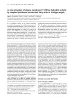
We have shown previously that isolated heat shock protein 90 (HSP90)
and nitric oxide synthase (NOS), once associated in a heterocomplex,
become completely resistant to calpain digestion. In this study, it is shown
that, in vivo, under conditions of calpain activation, the protection of NOS
degradation occurs. In addition, the extent of NOS degradation is a func-
tion of the level of HSP90 expression. Thus, in rat brain, which contains a
large excess of HSP90, almost all neuronal NOS is associated with the
chaperone protein. In this condition, neuronal NOS retains its full catalytic
activity, although limited proteolytic conversion to still active low-molecu-
lar-mass (130 kDa) products takes place. In contrast, in aorta, which con-
tains much smaller amounts of HSP90, endothelial NOS is not completely
associated with the chaperone, and undergoes extensive degradation with a
loss of protein and catalytic activity. On the basis of these findings, we pro-
pose a novel role of the HSP90–NOS heterocomplex in protecting in vivo
NOS from proteolytic degradation by calpain. The efficiency of this effect
is directly related to the level of intracellular HSP90 expression, generating
a high HSP90 to NOS ratio, which favours both the formation and stabil-
ization of the HSP90–NOS heterocomplex. This condition seems to occur
in rat brain, but not in aorta, thus explaining the higher vulnerability to
proteolytic degradation of endothelial NOS relative to neuronal NOS.
Abbreviations
[Ca
2+
]
i
, intracellular Ca
2+
concentration; C.I.1, synthetic calpain inhibitor-1; eNOS, endothelial nitric oxide synthase; HMS, hypertensive Milan
strain; HSD, high-sodium diet; HSP90, heat shock protein 90; iNOS, inducible NOS; NMS, normotensive Milan strain; nNOS, neuronal nitric
oxide synthase; NOS, nitric oxide synthase.
FEBS Journal 275 (2008) 2501–2511 ª 2008 The Authors Journal compilation ª 2008 FEBS 2501
We have recently demonstrated that the suscep-
tibility to calpain degradation of purified endothelial
NOS (eNOS) and neuronal NOS (nNOS) is signifi-
cantly reduced in the presence of equimolar amounts
of HSP90 [27]. Using immunoprecipitation studies, it
has also been established that the protective effect is
caused by HSP90-specific recruitment by active calpain
molecules. In this associated form, HSP90 becomes
resistant to digestion, although the protease still retains
50% of its proteolytic activity against external sub-
strates. Furthermore, when NOS isozymes are associ-
ated with this binary complex, they also become
resistant to proteolytic degradation. These observa-
tions imply a correlation between the vulnerability of
NOS isozymes and the availability of HSP90 to gener-
ate stable ternary complexes. This relationship is
strongly supported by the different digestibility of
NOS in Jurkat and BAE-1 cells, expressing high and
low levels of HSP90, respectively.
To verify the occurrence of such a protective effect
in vivo, we used normotensive Milan strain (NMS)
rats as a model. Thus, we induced a mild elevation of
intracellular Ca
2+
concentration ([Ca
2+
]
i
) by the
administration of a high-sodium diet (HSD) [28], and
studied calpain degradation of NOS and HSP90 in
brain and aorta. To amplify the range of fluctuations
in [Ca
2+
]
i
, hypertensive Milan strain (HMS) rats were
also used, as they are characterized by a constitutive
elevation in [Ca
2+
]
i
and a higher responsiveness to
HSD.
We report here that, in the brain and aorta of HSD-
treated rats, the extent and patterns of proteolytic deg-
radation of NOS isozymes and HSP90 are similar to
those previously detected in Jurkat and BAE-1 cells
loaded with Ca
2+
[27]. As the differences in expression
of HSP90 in the two rat tissues are similar to those
present in these cell models [27], we propose that the
occurrence of conditions which favour the formation
and stabilization of proteolytically resistant complexes
of NOS with HSP90 are crucial in determining the
in vivo resistance of NOS and HSP90 to calpain degra-
dation.
Results
Levels of HSP90 and NOS isozymes in rat brain
and aorta
The level of HSP90 and the type of NOS isoform pres-
ent in rat brain and aorta were determined by immu-
noblotting (Fig. 1). In brain, nNOS was the most
preferentially expressed isoform, together with traces
of eNOS (Fig. 1A). In aorta, only eNOS isozyme was
detectable (Fig. 1B). In both tissues, no expression of
inducible NOS (iNOS) was found (Fig. 1A,B). HSP90
was present in rat brain in amounts six- to sevenfold
A
B
C
Fig. 1. NOS isozymes and HSP90 expressed in brain and aorta of
NMS rats. (A) Aliquots (50 lg protein) of NMS rat brain soluble
material, obtained as described in Experimental procedures, were
submitted to 6% SDS-PAGE and blotted as described previously.
NOS isozymes were detected with the specific mAbs. (B) Aliquots
(50 lg protein) of NMS rat thoracic aorta total lysate, obtained as
described in Experimental procedures, were submitted to 6% SDS-
PAGE and blotted as described previously. NOS isozymes were
detected with specific mAbs. (C) HSP90 levels were detected from
the same samples as reported in (A) and (B) using the specific
mAb. The immunoreactive bands detected in (A–C) were quantified
(see bars) as described in Experimental procedures. The values
reported are the arithmetical means ± standard deviation of five dif-
ferent experiments carried out on five different animals of each
strain.
In vivo degradation of NOS and HSP90 by calpain M. Averna et al.
2502 FEBS Journal 275 (2008) 2501–2511 ª 2008 The Authors Journal compilation ª 2008 FEBS
higher than in aorta, resulting in a much higher
HSP90 to NOS ratio in brain (Fig. 1C).
Calpain activation in rat brain and aorta
following HSD treatment
To promote in vivo calpain activation, NMS rats were
treated with HSD, which has been established previ-
ously to induce a mild elevation in [Ca
2+
]
i
, slightly
higher in aorta than in brain [28]. To amplify the
range of elevation in [Ca
2+
]
i
, HMS rats were also
used, as a limited increase in [Ca
2+
]
i
in both aorta and
brain has been found to be constitutively present in
these animals.
To assess the in vivo activation of calpain, we relied
on the following well-established methods: (a) the
occurrence of calpain consumption [26,29–31]; (b) a
specific pattern of calpastatin digestion, resulting in an
imbalance within the proteolytic system [32]; and (c)
the degradation of calpain target proteins [26,30]. As
shown in Fig. 2A, following HSD treatment, the levels
of both l- and milli-calpain isoforms were reduced to
a limited extent in brain, whereas, in aorta (Fig. 2B),
the decrease in the two protease isoforms was more
pronounced.
Moreover, in brain, the natural inhibitor of calpain,
calpastatin, was preferentially converted into still
active 15 kDa fragments (Table 1), whereas, in aorta,
the inhibitor was predominantly inactivated. As both
the inactivation and fragmentation of calpastatin are
known to be produced by active calpain [32], these
observations further indicate that calpain is activated
in both tissues, although at a higher rate in aorta. Fur-
ther direct evidence in support of calpain activation in
aorta was provided by the degradation of talin and
desmin in HSD-treated rats (Fig. 3A). Indeed, this
process was completely prevented (Fig. 3B) by the
administration to the animals of synthetic calpain
inhibitor-1 (C.I.1) [33,34].
Digestion of HSP90 and NOS in brain and aorta
of normotensive and hypertensive rats treated
with HSD
Following HSD treatment, no appreciable changes in
NOS activity occurred in the brain of NMS rats,
although a small fraction of the native 160 kDa syn-
thase was converted into the still active 130 kDa form
(Fig. 4A). The level of HSP90 remained unchanged
during the period of treatment (Fig. 4A). By contrast,
in aorta, more than 50% of native eNOS progressively
disappeared (Fig. 4B,D), together with a significant
degradation of HSP90, which was only partially
replaced by an 84 kDa form (Fig. 4B,D). The involve-
ment of calpain in these digestion processes was dem-
onstrated by the protective effect on both HSP90
and NOS degradation of the administration to the
HSD-treated NMS animals of C.I.1 (Fig. 5).
In the brain of hypertensive rats, in spite of a pre-
existing condition of altered Ca
2+
homeostasis, no
appreciable changes in total nNOS activity or the level
of HSP90 were observed (Fig. 6A,C). By contrast with
NMS rats, a small fraction of a still active 130 kDa
form was already present in the brain of untreated
HMS rats and increased following HSD treatment
(Fig. 6A). However, in aorta, the digestion of eNOS
and HSP90 appeared to be more extensive (Fig. 6B).
Indeed, approximately 80–90% of eNOS protein and
A
B
Fig. 2. Levels of calpain isoforms and calpain substrates in the aorta
of NMS and HMS rats treated with HSD. Aliquots (100 lg protein) of
brain soluble material (A) and aorta total lysate (B), prepared as
described in Experimental procedures, from untreated or 4-week
HSD-treated NMS and HMS rats, were submitted to 8% SDS-PAGE,
followed by immunoblotting revealed with serum-l-calpain mAb 56.3
[36] and monoclonal IgG milli-calpain. The immunoreactive material
was detected and quantified as described in Experimental proce-
dures. The values reported are the arithmetical means ± standard
deviation of five different experiments carried out on five different
animals of each strain.
M. Averna et al. In vivo degradation of NOS and HSP90 by calpain
FEBS Journal 275 (2008) 2501–2511 ª 2008 The Authors Journal compilation ª 2008 FEBS 2503
activity, together with 60–70% of HSP90, were lost
(Fig. 6B,D).
The degradation pattern of nNOS in the brain of
HSD-treated rats, resulting in the accumulation of the
still active 130 kDa form, can be reproduced in in vitro
conditions if nNOS digestion by calpain is carried out
in the presence of HSP90 [27]. This finding can also
explain the large extent of digestion of eNOS in aorta,
in which, in association with a higher degree of calpain
activation, a lower level of HSP90 is also present.
Identification of HSP90–NOS heterocomplexes in
aorta and brain lysates
In order to explore the relationship between the
HSP90 to NOS ratio and the formation of calpain-
resistant heterocomplexes, we first studied, by immu-
noprecipitation analysis, the association of the two
proteins in brain and aorta. As shown in Fig. 7A, fol-
lowing the addition of IgG1-HSP90 antibody to crude
extracts of brain or aorta, NOS was immunoprecipitat-
ed, indicating a specific association of the two proteins.
We then determined the amount of each enzyme
involved in such complexes by submitting samples of
crude extracts of rat brain and aorta to gel filtration
chromatography. As shown in Fig. 7B, in brain, nNOS
eluted entirely as a single peak at a volume corre-
sponding to a molecular mass higher than that of the
free native enzyme. HSP90 was eluted in two peaks:
the first coincident with that of nNOS, and the second
containing more than 60% of total chaperone protein,
with an elution volume identical to that of free HSP90.
Thus, all nNOS appeared to be engaged in a complex
with HSP90, whereas the major fraction of the chaper-
one was present in the free form.
In aorta (Fig. 7C), approximately 85–90% of eNOS
was recovered in association with HSP90 and the
remaining 10–15% was found in the free form; how-
ever, the amount of HSP90 recovered as free protein
was much lower than that engaged in the complex.
The large difference in the amount of free chaperone
observed in the two tissues is indicative of the existence
Table 1. Levels of native and 15 kDa calpastatin species in brain
and aorta of NMS and HMS rats treated with HSD for 4 weeks.
The data reported are the arithmetical means ± standard deviation
of five different experiments carried out on five different animals of
each strain.
Animal Treatment
a
Total
calpastatin
activity (%)
b
15 kDa
fragment
activity (%)
c
Loss of total
calpastatin
activity (%)
d
Brain
NMS None 97 ± 5 3 ± 0.3 0
NMS HSD 40 ± 2 40 ± 3 20
HMS None 90 ± 6 10 ± 2 0
HMS HSD 5 ± 1 49 ± 2 46
Aorta
NMS None 99 ± 5 1 ± 1 0
NMS HSD 16 ± 2 25 ± 2 59
HMS None 64 ± 4 16 ± 2 20
HMS HSD 2 ± 0.5 14 ± 1 84
a
NMS and HMS rats were fed for 4 weeks with HSD as described
in Experimental procedures.
b
Total calpastatin activity was mea-
sured as described in Experimental procedures and [28,43].
c
Aliqu-
ots (1.5 mg protein) of the soluble material, obtained as described
previously from untreated or 4-week HSD-treated NMS and HMS
rat brain and thoracic aorta homogenates, were submitted to 12%
SDS-PAGE divided into 10 lanes [28]. The 15 kDa calpastatin spe-
cies were identified on the basis of their electrophoretic mobility,
and quantified following extraction from the gel by measuring their
inhibitory activity as described previously [28,42].
d
The loss of
calpastatin activity was calculated by subtracting the sum of the
percentage of the active calpastatin species from 100.
A
B
Desmin
Desmin
Talin
Talin
NMS HMS
NMS
+HSD
HMS
+HSD
+ HSD
+ C.I. 1
+ HSDControl
Fig. 3. Levels of calpain substrates in aorta of NMS and HMS rats
treated with HSD and C.I.1. Aliquots (50 lg protein) of aorta total
lysate (A), prepared as described in Experimental procedures, from
untreated and HMS rats, were submitted to 8% SDS-PAGE fol-
lowed by immunoblotting. Samples (50 lg protein) of aorta total
lysate (B) from untreated or 4-week HSD-treated NMS rats, in the
absence (+HSD) or presence (+HSD+C.I.1) of 25 l
M C.I.1, were
submitted to 8% SDS-PAGE followed by immunoblotting. Desmin
and talin were detected with specific mAbs.
In vivo degradation of NOS and HSP90 by calpain M. Averna et al.
2504 FEBS Journal 275 (2008) 2501–2511 ª 2008 The Authors Journal compilation ª 2008 FEBS
of conditions favouring and stabilizing the heterocom-
plex much more efficiently in brain than in aorta. This
could explain the higher susceptibility of eNOS to
calpain digestion.
Discussion
Although several reports [11–26] have indicated that
calpain and the proteasome pathway are the two
major systems responsible for the proteolytic degrada-
tion of NOS, some pertinent questions still remain
unsolved. Indeed, although it has been established,
especially by the use of NOS and HSP90 inhibitors,
that proteasome-promoted degradation selectively
removes inactive structurally damaged NOS forms, or
monomeric haem-deficient isozyme species [11–16],
the precise molecular events that trigger the proteo-
lytic degradation of NOS in vivo still remain to be
defined. One of these molecular signals could be
altered or decreased HSP90 function, favouring the
accumulation of abnormal or monomeric NOS mole-
cules and their degradation by the proteasome system
[1,12]. Furthermore, proteolytic degradation of NOS
by calpain has been described in conditions of
extreme cytotoxicity [17,19,21,23,26]. In these experi-
ments, as a result of high Ca
2+
overload, several
calpain targets, including NOS, can undergo proteo-
lytic digestion. For this reason, the degradation of
NOS can be attributed to an overactivation of cal-
pain rather than to a selective regulated proteolytic
mechanism.
In previous studies, we have observed that HSP90 is
five- to tenfold less susceptible than nNOS and eNOS
to calpain degradation [27] as a result of the formation
of a calpain–HSP90 complex in which the protease can
no longer degrade the bound chaperone. NOS iso-
zymes, once recruited into the HSP90–calpain binary
complex, also become resistant to calpain digestion.
This protective effect may be of physiological rele-
vance, as conditions promoting NO production also
induce calpain activation. Thus, the formation of
NOS–HSP90 complexes may provide a new insight
into the understanding of the mechanisms involved in
modulating NO production. In such a case, the avail-
ability of adequate amounts of HSP90 becomes the
limiting factor.
Our study poses new important questions that need
to be addressed. The first question concerns the vul-
nerability of different NOS isoforms to proteolysis
in vivo under conditions of small changes to [Ca
2+
]
i
.
The second question concerns the capacity of HSP90
to protect NOS in vivo against proteolytic degrada-
A
B
CD
Fig. 4. In vivo digestion of NOS and HSP90 in NMS rats during HSD treatment. Aliquots (20 lg protein) of rat brain soluble material (A) and
aliquots (50 lg protein) of rat aorta total lysate (B), obtained as described in Experimental procedures, from untreated or HSD-treated NMS
rats, were submitted to 6% SDS-PAGE and blotted as described previously. nNOS, eNOS and HSP90 were detected with specific mAbs.
(C) nNOS (open circles) and HSP90 (filled circles) immunoreactive materials detected in (A) and the corresponding nNOS activity (open
squares) were quantified as described in Experimental procedures. (D) eNOS (open circles) and HSP90 (filled circles) immunoreactive materi-
als detected in (B) and the corresponding eNOS activity (open squares) were quantified as described in Experimental procedures. The values
reported are the arithmetical means ± standard deviation of five different experiments carried out on five different animals of each strain.
M. Averna et al. In vivo degradation of NOS and HSP90 by calpain
FEBS Journal 275 (2008) 2501–2511 ª 2008 The Authors Journal compilation ª 2008 FEBS 2505
tion. Finally, a third question involves the possi-
ble relationship between such protection and the
well-known different expression of HSP90 in various
tissues.
To answer these questions, we have used animals
treated with HSD, which has been shown previously
to induce an increase in the level of [Ca
2+
]
i
and a
correlated calpain activation [28]. This increase in
[Ca
2+
]
i
is more intense in aorta than in brain.
Under these conditions, in brain, no change in the
level of HSP90 was observed, although a limited and
conservative degradation of nNOS occurred without
a loss of catalytic activity. In contrast, in aorta, both
eNOS and HSP90 were highly degraded. The differ-
ent vulnerability of the two NOS isoforms to proteo-
lytic degradation is strictly related to the availability
of HSP90, which is expressed in higher concentra-
tions in the brain than in the aorta. Furthermore,
the patterns of digestion of eNOS and nNOS
observed in HSD-treated animals are identical to
those previously obtained in reconstructed systems
containing the synthases together with different levels
of HSP90.
Our data suggest that a large reservoir of HSP90
maintains all NOS engaged in a calpain-resistant het-
erocomplex, which is protected from proteolysis, even
under conditions of prolonged protease activation.
This conclusion is further supported by the finding
reported here that, in brain, the nNOS–HSP90 com-
plex is in equilibrium with a large amount of stabiliz-
ing free chaperone, a condition that does not occur in
aorta. The reduced availability of HSP90 in aorta can
thus explain the increased vulnerability of eNOS rela-
tive to nNOS to proteolysis. On the basis of these find-
ings, we propose a novel mechanism in which HSP90
can provide functional stability of NOS isozymes
under conditions characterized by an alteration in
intracellular Ca
2+
homeostasis.
Experimental procedures
Materials
Leupeptin C.I.1, aprotinin, phosphatase inhibitor cocktail I
and II, NADPH, calmodulin, FAD, FMN, tetrahydrobiop-
terin, l-arginine and aldolase were purchased from Sigma
Aldrich, Milan, Italy. l-[
14
C]arginine (925 Bq; specific activ-
ity, 1Æ14 · 10
11
BqÆmol
)1
), Sephacryl S-300, Sephadex G-
200 resins, Superose
Ò
12 10 ⁄ 300 GL column and protein
G-Sepharose were obtained from GE Healthcare, Milan,
Italy. Ferritin was purchased from Boehringer Mannheim,
Mannheim, Germany. Dowex 50W8 resin (Na
+
form) was
obtained from Bio-Rad Laboratories, Milan, Italy. 4-(2-
Aminoethyl)benzenesulfonylfluoride (AEBSF) was obtained
from Calbiochem (Missiagua, Canada). The ECL
Ò
Detec-
tion System was obtained from GE Healthcare.
Monoclonal antibodies (mAbs)
nNOS, eNOS, iNOS and HSP90 antibodies were purchased
from BD Transduction Laboratories, Milan, Italy. b-Actin
and milli-calpain antibodies were obtained from Sigma
Aldrich, Milan, Italy. Desmin and talin antibodies were
purchased from Novus Biologicals, Littleton, CO, USA.
IgG1-calpastatin (mAb 35.23) and serum l-calpain (mAb
A
B
Fig. 5. Levels of NOS isozymes and HSP90 in brain and aorta of
NMS rats treated with HSD and C.I.1. Aliquots of brain soluble
material (20 lg protein) and of aorta total lysate (50 lg protein),
obtained as described in Experimental procedures, from untreated
or 4-week HSD-treated NMS rats, in the absence (+HSD) or pres-
ence (+HSD+C.I.1) of 25 l
M C.I.1, were submitted to 6% SDS-
PAGE followed by immunoblotting, revealed with IgG1-eNOS or
IgG1-nNOS mAbs (A) or IgG1-HSP90 mAb (B). The immunoreactive
material of eNOS, nNOS and HSP90 was detected and quantified
as described in Experimental procedures. The values reported are
the arithmetical means ± standard deviation of five different experi-
ments carried out on five different animals of each strain.
In vivo degradation of NOS and HSP90 by calpain M. Averna et al.
2506 FEBS Journal 275 (2008) 2501–2511 ª 2008 The Authors Journal compilation ª 2008 FEBS
56.3) mAbs were produced as indicated in [35] and [36],
respectively.
Animals
NMS and HMS rats [37] were housed in controlled condi-
tions (22 ± 1 °C; humidity, 50 ± 5%; lighting, 8–20 h).
Systolic blood pressure was measured by tail-cuff plethys-
mography [W&W Electronic, BP recorder 8005 (Huntsinlle,
AL, USA)] on prewarmed (37 °C) rats, following the
procedure originally described by Byrom and Wilson [38].
Normotensive and hypertensive rats showed mean arterial
blood pressures of 100 ± 5 and 145 ± 10 mmHg, respec-
tively.
Experimental hypertension
Experimental hypertension was induced in 60-day-old rats
by feeding ad libitum with a standard rat chow and provid-
ing NaCl dissolved in tap water at a concentration of
10 gÆL
)1
for a period of time ranging from 15 to 30 days.
Each animal received approximately 0.7 gÆday
)1
of NaCl.
Where indicated, 25 lm C.I.1 was dissolved in tap water in
the presence of 10 gÆL
)1
NaCl, and administered to NMS
and HMS rats for 4 weeks [28]. Each rat received 0.5–
0.7 mgÆday
)1
of C.I.1. Experiments were carried out follow-
ing the institution’s ethical guidelines. During the course of
the experiments, no appreciable changes were observed in
food consumption and body weight.
Preparation of tissue homogenates
NMS and HMS rats were sacrificed by decapitation; the
brain was immediately removed, minced, homogenized in a
Potter–Elvehjem homogenizer and sonicated in three vol-
umes of 50 mm sodium borate buffer, pH 7.5, containing
1mm EDTA, 0.5 mm 2-mercaptoethanol, 0.1 mgÆmL
)1
leupeptin and 2 mm AEBSF (buffer A). The particulate
material was discarded by centrifugation (100 000 g for
10 min). Thoracic aorta was rapidly excised from the same
animals. After the removal of the adhering connective
tissue, the tissue was cut into several segments (approxi-
mately 2 mm each), homogenized in a Potter–Elvehjem
homogenizer and lysed by sonication in three volumes of
buffer A. The protein concentration was determined follow-
ing the procedure of Bradford [39].
Immunoblot
Rat brain and aorta lysates (20–50 lg) were diluted in a
final volume of 100 lL of the SDS-PAGE loading buffer
and submitted to 6% SDS-PAGE [40]. The protein bands
AB
C
D
Fig. 6. In vivo digestion of NOS and HSP90 in HMS rats during HSD treatment. Aliquots (20 lg protein) of rat brain soluble material (A) and
aliquots (50 lg protein) of rat aorta total lysate (B), obtained as described in Experimental procedures, from untreated or HSD-treated HMS
rats, were submitted to 6% SDS-PAGE and blotted as described previously. nNOS, eNOS and HSP90 were detected with specific mAbs.
(C) nNOS (open circles) and HSP90 (filled circles) protein detected in (A) and the corresponding nNOS activity (open squares) were quantified
as described in Experimental procedures. (D) eNOS (open circles) and HSP90 (filled circles) immunoreactive materials detected in (B) and
the corresponding eNOS activity (open squares) were quantified as described in Experimental procedures. The values reported are the arith-
metical means ± standard deviation of five different experiments carried out on five different animals of each strain.
M. Averna et al. In vivo degradation of NOS and HSP90 by calpain
FEBS Journal 275 (2008) 2501–2511 ª 2008 The Authors Journal compilation ª 2008 FEBS 2507
were then blotted onto a nitrocellulose membrane and
saturated with a NaCi/P
i
solution, pH 7.5, containing 5%
powered milk. The blots were probed with specific antibod-
ies, followed by a peroxidase-conjugated secondary anti-
body as described previously, and then developed with the
ECL Detection System [41]. The immunoreactive material
was detected with a Bio-Rad Chemi Doc XRS apparatus
and quantified using quantity one 4.6.1 software (Bio-
Rad Laboratories). The procedure was made quantitative
by the use of known amounts of proteins submitted to
SDS-PAGE and staining with the appropriate antibody.
The bands were then scanned, and the areas of the peaks
obtained were used to create a calibration curve.
Immunoprecipitation
Brain and thoracic aorta, excised from NMS rats, were
lysed in ice-cold 20 mm Tris ⁄ HCl, 2.5 mm EDTA, 2.5 mm
EGTA, 0.14 m NaCl, pH 7.4 (immunoprecipitation buffer),
containing 1% Triton X-100, 10 lgÆ mL
)1
aprotinin,
20 lgÆmL
)1
leupeptin, 10 lgÆmL
)1
AEBSF and phosphatase
inhibitor cocktail I and II (10 lgÆmL
)1
), followed by brief
sonication. Cell lysates were centrifuged at 12 000 g for
15 min at 4 °C, and protein quantification of the superna-
tants was performed using the Lowry assay. For the immu-
noprecipitations, 500 lg of detergent-soluble protein
(crude extract) was previously precleared with protein
G-Sepharose, and then incubated in the presence of 2 lgof
IgG1-HSP90 mAb at 4 °C overnight. Protein G-Sepharose
was then added and incubated for an additional hour. The
immunocomplexes were washed three times with immuno-
precipitation buffer, heated in SDS-PAGE loading buffer
for 5 min [40] and submitted to 6% SDS-PAGE. Proteins
were then transferred by electroblotting onto a nitro-
cellulose membrane, and immunoblotting analysis was
performed as described above.
Identification of NOS–HSP90 association by gel
filtration
Aliquots (0.5 mg protein) of the soluble material of brain
homogenate and thoracic aorta total lysate, obtained from
NMS rats as described previously, were submitted to gel
filtration chromatography on a Superose
Ò
12 10 ⁄ 300 GL
column (total volume, 24 mL) equilibrated in buffer A con-
taining 50 mm NaCl using an FPLC system. The flow rate
was 100 lLÆmin
)1
and the eluted proteins were collected in
500 lL fractions. The molecular weights of the eluted pro-
teins were calculated from the elution volumes of ferritin
(M
r
= 450 kDa) and aldolase (M
r
= 160 kDa), utilized as
standard proteins.
A
B
C
Fig. 7. Identification of NOS–HSP90 association in rat brain and
aorta. (A) Aliquots (500 lg protein) of brain and aorta crude extract,
prepared as described in Experimental procedures, were incubated
overnight at 4 °C with IgG1-HSP90 antibody (see Experimental
procedures), as reported also in [7,44,45]. The mixtures were then
incubated for 1 h at room temperature with 50 lL of protein
G-Sepharose. The particles were collected and washed three times
with immunoprecipitation buffer. The particles were then suspended
in SDS-PAGE loading solution, heated for 5 min at 90 °C and submit-
ted to 6% SDS-PAGE. NOS isozymes and HSP90 were identified
with specific mAbs (see Experimental procedures). The values
reported are the arithmetical means ± standard deviation of five dif-
ferent experiments carried out on five different animals of each
strain. (B, C) Aliquots (500 lg protein) of the soluble material of brain
homogenate and thoracic aorta total lysate, obtained from NMS rats
as described previously, were submitted to gel filtration chromatog-
raphy (see Experimental procedures). Aliquots (30 lL) of each eluted
fraction were suspended in SDS-PAGE loading solution [40] and sub-
mitted to 6% SDS-PAGE, followed by immunoblotting. HSP90 (filled
circles) and NOS isoforms (open circles) were probed with the appro-
priate antibody. The immunoreactive material was quantified as
described in Experimental procedures.
In vivo degradation of NOS and HSP90 by calpain M. Averna et al.
2508 FEBS Journal 275 (2008) 2501–2511 ª 2008 The Authors Journal compilation ª 2008 FEBS
Aliquots (30 lL) of each eluted fraction were suspended
in SDS-PAGE loading buffer [40] and submitted to 6%
SDS-PAGE. Proteins were then transferred to a nitrocellu-
lose membrane by electroblotting, and immunoblotting
analysis was performed as described above. The immunore-
active material was detected and quantified as described
above.
Assay of NOS activity
NOS activity was assayed by detecting the production of
citrulline from l-[
14
C]arginine, as reported previously [23]
with the following modifications. Aliquots (100 lg protein)
of the crude homogenate were incubated in a total volume
of 250 lL in buffer A containing 1 mm NADPH, 200 mm
calmodulin, 20 lm tetrahydrobiopterin, 1 lm FAD, 1 lm
FMN, 5 lml-arginine and 925 Bq of l-[
14
C]arginine (spe-
cific radioactivity, 1Æ14 · 10
11
BqÆmol
)1
)at37°C. After
30 min, 2 mL of ice-cold stop buffer (50 mm Hepes,
pH 5.5, containing 5 mm EDTA) was added. These incuba-
tions were then submitted to anion exchange chromatogra-
phy using 2 mL of packed Dowex 50W8 Na
+
form resin
pre-equilibrated with stop buffer. l-Citrulline was eluted by
washing the resin with 3 mL of stop buffer, and the radio-
activity present was counted in a liquid scintillation coun-
ter. One unit of NOS activity was defined as the amount of
enzyme producing 1 pmol citrullineÆmin
)1
in the specified
conditions.
Separation and quantification of calpastatin
species in rat brain and aorta
Aliquots of the soluble material (10 lanes with 100 lg pro-
tein each), prepared as described above from untreated or
treated NMS and HMS rat brain and thoracic aorta homo-
genates, were submitted to 12% SDS-PAGE [28]. Calpasta-
tin species were identified following protein extraction from
the gel, as described previously [42]. Calpastatin activity
was measured as described in [43].
Acknowledgements
This work was supported in part by grants from Min-
istero Haliano per I’Universita
`
e la Ricerca, Fondo per
gli Investimenti della Ricerca di Base and Progetti di
Ricerca di Interesse Nazionale projects, and from the
University of Genoa.
References
1 Kone BC, Kuncewicz T, Zhang W & Yu Z (2003) Pro-
tein interaction with nitric oxide synthases: controlling
the right time, the right place, and the right amount of
nitric oxide. Am J Physiol Renal Physiol 285, 178–190.
2 Kone BC (2000) Protein–protein interactions controlling
nitric oxide synthases. Acta Physiol Scand 168, 27–31.
3 Gratton J, Fontana J, O’Connor D, Garcia-Cardena
G, McCabe T & Sessa C (2000) Reconstitution of an
endothelial nitric-oxide synthase (eNOS), hsp90, and
caveolin-1 complex in vitro. J Biol Chem 275, 22268–
22272.
4 Garcia-Cardena G, Martasek P, Masters BS, Skidd
PM, Conet J, Lisanti MP & Sessa WC (1997) Dissecting
the interaction between nitric oxide synthase (NOS) and
caveolin. Functional significance of the NOS caveolin
binding domain in vivo. J Biol Chem 272, 25437–
25440.
5 Piech A, Dessy C, Havaux X, Feron O & Balligand J
(2003) Differential regulation of nitric oxide synthases
and their allosteric regulators in heart and vessels of
hypertensive rats. Cardiovasc Res 57, 456–467.
6 Bender AT, Silverstein AM, Demady DR, Kanelakis
KC, Noguchi S, Pratt WB & Osawa Y (1999) Neuro-
nal nitric-oxide synthase is regulated by the HSP90-
based chaperone system in vivo. J Biol Chem 274 ,
1472–1478.
7 Papapetropoulos A, Fulton D, Lin MI, Fontana J,
McCabe TJ, Zoellner S, Garcia-Cardena G, Zhou Z,
Gratton J & Sessa WC (2004) Vanadate is a potent acti-
vator of endothelial nitric-oxide synthase: evidence for
the role of the serine ⁄ threonine kinase akt and the
90 kDa heat shock protein. Mol Pharmacol 65, 407–
415.
8 Song Y, Zweier JL & Xia Y (2001) Heat-shock protein
augments neuronal nitric oxide synthase activity by
enhancing Ca
2+
⁄ calmodulin binding. Biochem J 355,
357–360.
9 Minami Y, Kimura Y, Kawasaki H, Suzuki K & Yaha-
ra I (1994) The carboxy-terminal region of mammalian
HSP90 is required for its dimerization and function
in vivo. Mol Cell Biol 14, 1459–1464.
10 Billecke SS, Bender AT, Kanelakis KC, Murphy PJM,
Lowe ER, Kamada Y, Pratt WB & Osawa Y (2002)
HSP90 is required for heme binding and activation of
apo-neuronal nitric-oxide synthase. J Biol Chem 277,
20504–20509.
11 Dunbar AY, Kamada Y, Jenkins GJ, Lowe ER, Bille-
cke SS & Osawa Y (2004) Ubiquitination and degrada-
tion of neuronal nitric-oxide synthase in vitro: dimer
stabilization protects the enzyme from proteolysis. Mol
Pharmacol 66, 964–969.
12 Osawa Y, Lowe ER, Everett AC, Dunbar AY & Bille-
cke SS (2003) Proteolytic degradation of nitric oxide
synthase: effect of inhibitors and role of HSP90-based
chaperones. J Pharmacol Exp Ther 304, 493–497.
13 Govers R, de Bree P & Rabelink TJ (2003) Involvement
of the proteasome in activation of endothelial nitric
oxide synthase. Life Sci 73, 2225–2236.
M. Averna et al. In vivo degradation of NOS and HSP90 by calpain
FEBS Journal 275 (2008) 2501–2511 ª 2008 The Authors Journal compilation ª 2008 FEBS 2509
14 Kolodziejski PJ, Musial A, Koo JS & Eissa NT (2002)
Ubiquitination of inducible nitric oxide synthase is
required for its degradation. Proc Natl Acad Sci USA
99, 12315–12320.
15 Musial A & Eissa T (2001) Inducible nitric-oxide
synthase is regulated by the proteasome degradation
pathway. J Biol Chem 276, 24268–24273.
16 Bender A, Demady DR & Osawa Y (2000) Ubiquitina-
tion of neuronal nitric-oxide synthase in vitro and
in vivo. J Biol Chem 275, 17407–17411.
17 Gamerdinger M, Manthey D & Behl C (2006) Oestro-
gen receptor subtype-specific repression of calpain
expression and calpain enzymatic activity in neuronal
cells – implications for neuroprotection against Ca-med-
iated excitotoxicity. J Neurochem 97, 57–68.
18 Araujo IM & Carvalho CM (2005) Role of nitric oxide
and calpain activation in neuronal death and survival.
Curr Drug Targets CNS Neurol Disord 4, 319–324.
19 Stalker TJ, Gong Y & Scalia R (2005) The calcium-
dependent protease calpain causes endothelial dysfunc-
tion in type 2 diabetes. Diabetes 54, 1132–1140.
20 Stalker TJ, Skvarka CB & Scalia R (2003) A novel role
for calpains in the endothelial dysfunction of hypergly-
cemia. FASEB J 17, 1511–1513.
21 Araujo IM, Ambrosio AF, Leal EC, Santos PF, Carv-
alho AP & Carvalho CM (2003) Neuronal nitric oxide
synthase proteolysis limits the involvement of nitric
oxide in kainate-induced neurotoxicity in hippocampal
neurons. J Neurochem 85, 791–800.
22 Walker G, Pfeilschifter J, Otten U & Kunz D (2001)
Proteolytic cleavage of inducible nitric oxide synthase
(iNOS) by calpain I. Biochim Biophys Acta 1568, 216–
224.
23 Su Y & Block ER (2000) Role of calpain in hypoxic
inhibition of nitric oxide synthase activity in pulmonary
endothelial cells. Am J Physiol Lung Cell Mol Physiol
278, 1204–1212.
24 Bellocq A, Doublier S, Suberville S, Perez J, Escoubet
B, Fouqueray B, Puyol DR & Baud L (1999) Somato-
statin increases glucocorticoid binding and signalling in
macrophages by blocking the calpain-specific cleavage
of HSP90. J Biol Chem 274, 36891–36896.
25 Laine
´
R & Ortiz de Montellano PR (1998) Neuronal
nitric oxide synthase isoforms a and l are closely
related calpain sensitive proteins. Mol Pharmacol 54,
305–312.
26 Hajimohammadreza I, Raser KJ, Nath R, Nadimpalli
R, Scott M & Wang KKW (1997) Neuronal nitric oxide
synthase and calmodulin-dependent protein kinase IIa
undergo neurotoxin-induced proteolysis. J Neurochem
69, 1006–1013.
27 Averna M, Stifanese R, DeTullio R, Salamino F, Bertuc-
cio M, Pontremoli S & Melloni E (2007) Proteolytic deg-
radation of NOS isoforms by calpain is modulated by the
expression levels of HSP90. FEBS J 274, 6116–6127.
28 Averna M, Stifanese R, DeTullio R, Passalacqua M,
Defranchi E, Salamino F, Melloni E & Pontremoli S
(2007) Regulation of calpain activity in rat brain with
altered Ca
2+
homeostasis. J Biol Chem 282, 2656–2665.
29 Stifanese R, Averna M, Salamino F, Cantoni C, Min-
gari MC, Prato C, Pontremoli S & Melloni E (2006)
Characterization of the calpain ⁄ calpastatin system in
human hemopoietic cell lines. Arch Biochem Biophys
456, 48–57.
30 Goll DE, Thompson VF, Li H, Wei W & Cong J
(2003) The calpain system. J Physiol Rev 83, 731–801.
31 Melloni E, Pontremoli S, Salamino F, Sparatore B,
Michetti M & Horecker BL (1984) Two cytosolic
Ca
2+
-dependent, neutral proteinases from rabbit liver:
purification and properties of the proenzyme. Arch
Biochem Biophys 232, 505–512.
32 De Tullio R, Averna M, Salamino F, Pontremoli S &
Melloni E (2000) Differential degradation of calpastatin
by l and m-calpain in Ca
2+
enriched human neuroblas-
toma LAN-5 cells. FEBS Lett 475, 17–21.
33 Sasaki T, Kishi M, Saito M, Tanaka T, Higuchi N,
Kominami E, Katunuma N & Murachi T (1990) Inhibi-
tory effect of di- and tripeptidyl aldehydes on calpains
and cathepsins. J Enzym Inhib 3, 195–201.
34 Lu Q & Mellgren RL (1996) Calpain inhibitors and ser-
ine protease inhibitors can produce apoptosis in HL-60
cells. Arch Biochem Biophys 334, 175–181.
35 Melloni E, De Tullio R, Averna M, Tedesco I, Salami-
no F, Sparatore B & Pontremoli S (1998) Properties of
calpastatin forms in rat brain. FEBS Lett 431, 55–58.
36 Pontremoli S, Melloni E, Damiani G, Salamino F,
Sparatore B, Michetti M & Horecker BL (1988) Effects
of a monoclonal anti-calpain antibody on responses of
stimulated human neutrophils. Evidence for a role for
proteolytically modified protein kinase C. J Biol Chem
263, 1915–1919.
37 Bianchi G, Ferrari P & Berber BR (1984) The Milan
hypertensive strain. In: Handbook of Hypertension,
Vol. 4 (de Jong W, ed.), pp. 328–349. Elsevier Science
Publisher.
38 Byrom FB & Wilson CA (1938) A plethysmographic
method for measuring systolic blood pressure in the
intact rat. J Physiol 93, 301–304.
39 Bradford MM (1976) A rapid and sensitive method for
the quantitation of microgram quantities of proteins
utilizing the principle of protein–dye binding. Anal
Biochem 72, 248–254.
40 Laemmli UK (1970) Cleavage of structural proteins
during the assembly of the head of bacteriophage T4.
Nature 227, 680–685.
41 Palejwala S & Goldsmith LT (1992) Ovarian expression
of cellular Ki-ras p21 varies with physiological status.
Proc Natl Acad Sci USA 89, 4202–4206.
42 Averna M, De Tullio R, Salamino F, Minafra R,
Pontremoli S & Melloni E (2001) Age-dependent
In vivo degradation of NOS and HSP90 by calpain M. Averna et al.
2510 FEBS Journal 275 (2008) 2501–2511 ª 2008 The Authors Journal compilation ª 2008 FEBS
degradation of calpastatin in kidney of hypertensive
rats. J Biol Chem 276, 38426–38432.
43 Salamino F, Sparatore B, De Tullio R, Pontremoli R,
Melloni E & Pontremoli S (1991) The calpastatin defect
in hypertension is possibly due to a specific degradation
by calpain. Biochim Biophys Acta 1096, 265–269.
44 Thomas SR, Chen K & Keaney JF Jr (2002) Hydrogen
peroxide activates endothelial nitric-oxide synthase
through coordinated phosphorylation and dephosphory-
lation via a phosphoinositide 3-kinase-dependent signal-
ling pathway. J Biol Chem 277, 6017–6024.
45 Garcia-Cardena G, Fan R, Stern DF, Liu J & Sessa
WC (1996) Endothelial nitric oxide synthase is regulated
by tyrosine phosphorylation and interacts with caveo-
lin-1. J Biol Chem 271, 27237–27240.
M. Averna et al. In vivo degradation of NOS and HSP90 by calpain
FEBS Journal 275 (2008) 2501–2511 ª 2008 The Authors Journal compilation ª 2008 FEBS 2511