Báo cáo khoa học: A novel phosphorylated glycoprotein in the shell matrix of the oyster Crassostrea nippona pptx
Bạn đang xem bản rút gọn của tài liệu. Xem và tải ngay bản đầy đủ của tài liệu tại đây (2.27 MB, 13 trang )
A novel phosphorylated glycoprotein in the shell matrix of
the oyster Crassostrea nippona
Tetsuro Samata, Daisuke Ikeda, Aya Kajikawa, Hideyoshi Sato, Chihiro Nogawa, Daishi Yamada,
Ryo Yamazaki and Takahiro Akiyama
Laboratory of Cell Biology, Faculty of Environmental Health, Azabu University, Sagamihara, Japan
Subsequent to the pioneering work of Miyamoto et al.
[1], Sudo et al. [2] and Shen et al. [3], more than 20
genes encoding the organic matrix (OM) components
of molluscan shells have been determined and their
deduced amino acid sequences clarified [4–12]. How-
ever, the information available to date has been
restricted to the nacreous and prismatic layers of pearl
oysters, leaving the other shell layers poorly investi-
gated at the molecular level. One exception is the find-
ing of acidic glycoprotein MSP-1 in the foliated layer
of Patinopecten yessoensis [4].
Through their control of nucleation, growth, mor-
phology and polymorphism of CaCO
3
crystals, these
OMs are commonly assumed to be intimately associ-
ated with every phase of molluscan biomineralization,
and thus with the overall regulation of the shell micro-
structure. More recent investigations have primarily
involved in vitro measurement of OM activities related
to crystal formation [13–18]. Although these studies
have clearly shown that OM modulates molluscan bio-
mineralization, the results nevertheless demonstrate
marked methodology-dependent variation. The func-
tion of OM thus remains unclear, even in vitro, and is
a topic of future research.
Molluscan shells are composed of either aragonite
or calcite. By contrast to the widespread occurrence
of aragonite, calcite is limited to several taxa
with species-dependent microstructures composed of
Keywords
domain structure; foliated layer; oyster shell;
phosphorylated matrix protein; poly-Asp
sequences
Correspondence
T. Samata, Laboratory of Cell Biology,
Faculty of Environmental Health, Azabu
University, 1-17-71 Fuchinobe, Sagamihara,
Kanagawa 229-0006, Japan
Fax: +81 42 769 2560
Tel: +81 42 769 2560
E-mail:
Database
The nucleotide sequences have been sub-
mitted to DDBJ with the accession number
AB207821–AB207826
(Received 11 January 2008, revised 31
March 2008, accepted 7 April 2008)
doi:10.1111/j.1742-4658.2008.06453.x
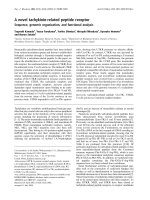
We found a novel 52 kDa matrix glycoprotein MPP1 in the shell of Cras-
sostrea nippona that was unusually acidic and heavily phosphorylated.
Deduced from the nucleotide sequence of 1.9 kb cDNA, which is likely to
encode MPP1 with high probability, the primary structure of this protein
shows a modular structure characterized by repeat sequences rich in Asp,
Ser and Gly. The most remarkable of these is the DE-rich sequence, in
which continuous repeats of Asp are interrupted by a single Cys residue.
Disulfide-dependent MPP1 polymers occurring in the form of multimeric
insoluble gels are estimated to contain repetitive locations of the anionic
molecules of phosphates and acidic amino acids, particularly Asp. Thus,
MPP1 and its polymers possess characteristic features of a charged mole-
cule for oyster biomineralization, namely accumulation and trapping of
Ca
2+
. In addition, MPP1 is the first organic matrix component considered
to be expressed in both the foliated and prismatic layers of the molluscan
shell microstructure. In vitro crystallization assays demonstrate the induc-
tion of tabular crystals with a completely different morphology from those
formed spontaneously, indicating that MPP1 and its polymers are poten-
tially the agent that controls crystal growth and shell microstructure.
Abbreviations
CBB, Coomassie brilliant blue; GISM, translucent gelatinous insoluble organic matrix; ISM, insoluble organic matrix; MPP1, molluscan
phosphorylated protein 1; ntp, nucleotide position; OM, organic matrix; SM, soluble organic matrix.
FEBS Journal 275 (2008) 2977–2989 ª 2008 The Authors Journal compilation ª 2008 FEBS 2977
prismatic, foliated, chalky and granular structures. In
particular, the shells of oyster species are composed of
a highly complex microstructure consisting of the
chalky layer in addition to the foliated and prismatic
layers. The foliated layer is formed by the aggregation
of units termed lath, each with a width of 2 lm and
length of 10 lm [19], whereas the chalky layer has a
homogeneous morphology composed of tiny calcite
granules [19]. A variety of studies, mostly based on
amino acid analysis of bulk soluble matrix (SM) and
insoluble matrix (ISM) [20–24], have shown the pres-
ence of OM in oyster species with particularly highly
acidic properties. This high acidity is due to Asp and
phosphoserin (p-Ser) [25]. Much of the accumulating
data on oyster shell biomineralization were obtained by
Wheeler et al., who have provided summaries of their
extensive studies [26,27]. Their investigation of the frac-
tionation and functional analysis of the OM compo-
nents highlighted the inhibitory activity of the OM
against crystal formation in vitro. Immunocytochemical
studies of the OM in the prismatic layer of C. virginica,
as reported by Kawaguchi and Watabe [28], revealed
that the ISM constituted the framework of the OM
and SM, which comprised several phosphorylated pro-
teins and might be distributed on the surface of the
ISM and surrounded calcite crystals. Atomic force
microscope and scanning electron microscope observa-
tions of foliar chips after pyrolysis and their subse-
quent crystallization revealed that crystal formation
occurred on the surface of the laths under the regula-
tion of the OM, which showed pulsed secretion [19].
As noted above, the primary structure of oyster OM
has yet to be precisely determined. In the present
study, we aimed to elucidate the overall picture of the
OM components of Crassostrea nippona by a combina-
tion of biochemical and genetical analyses. For gene
analysis, given the close similarity of the shell structure
and amino acid composition of the OM of the oyster
and scallop, we started with the isolation of cDNA
clones homologous with MSP-1 gene. Additional
in vitro crystallization assays were then performed to
investigate the function of the OM components.
Results
Biochemical characterization of the OM
components extracted from oyster shell
Fractionation of the bulk OM separated two fractions,
namely the SM at approximately 20 mg per 50 g of
shell and the ISM, which was further sub-divided into
two components: a predominant translucent gelatinous
insoluble organic matrix (GISM) pellet at approxi-
mately 120 mg per 50 g of shell and a small quantity
of fibrous precipitate at approximately 5 mg per 50 g
of shell.
After SDS ⁄ PAGE of GISM, which was largely-
solubilized in a sample buffer containing 2-mercaptoeth-
anol after boiling, and subsequent staining procedures
with negative staining, Stains-all and Methyl green visu-
alized an exclusive band of approximately 52 kDa,
which showed a negative reaction with Coomassie
brilliant blue (CBB) (Fig. 1).
SDS ⁄ PAGE of the 52 kDa component after enzy-
matic deglycosylation and dephosphorylation showed
apparent downward shifts in molecular masses of
2.5 and 3.5 kDa, respectively (Fig. 2).
Table 1 shows that the 52 kDa component in GISM
exhibits an amino acid composition, strikingly domi-
nated by Asx (aspartic acid plus asparagine), which,
together with Ser and Gly, accounted for more than
80% of the total residue. By contrast, the bulk SM
showed a different amino acid composition, which
comprised large amounts of Asx, Glx (glutamic acid
plus glutamine) and Gly, and a much smaller amount
of Ser than that of GISM.
Amino acid sequence analysis using a peptide
sequencer failed to determine the N-terminal sequence
of the 52 kDa component. Likewise, LC⁄ MS ⁄ MS anal-
ysis of the V8 protease digests of the 52 kDa compo-
nent did not reveal any peptide with sequences
corresponding to those of the 44 kDa deduced protein
MAB C D
66.2
45
(kDa)
Fig. 1. SDS ⁄ PAGE electrophoretogram of GISM in the OM of
C. nippona. The same amount of sample was applied to each lane.
Lane M, molecular mass standards; lane A, CBB staining; lane B,
Stains-all staining; lane C, negative staining; lane D, Methyl green
staining. Arrows on the right side of the lanes indicate the position
of the 52 kDa component. A weakly stained band in lane A does
not correspond to the 52 kDa component but a minor component
with a molecular mass of 45 kDa.
A novel acidic glycoprotein from the oyster shells T. Samata et al.
2978 FEBS Journal 275 (2008) 2977–2989 ª 2008 The Authors Journal compilation ª 2008 FEBS
encoded by the 1.9 kb cDNA and those reported so
far. On the other hand, the deduced 44 kDa protein
was identified as a top protein score of 55 (probability
based mowse score) using the Mascot search engine
for the fragments digested with endoproteinase Asp-N
of the 52 kDa component, whereas scores of the other
proteins in the database were lower than 20. Among
each peptide sequence with high peptide scores, a short
but specific sequence of DCGVDCGYYEPV (score of
19) at the N-terminal region of the deduced 44 kDa
protein and an additional sequence of DNNGDGNG
(score of 16) in the NG repeat sequence at the C-ter-
minal region were characteristic. The same result was
obtained using the Sequest search engine. The most
appropriate condition for Asp-N digestion was the
addition of 75 ng of enzyme to 15 lg of protein.
FTIR analysis of GISM showed the most intensive
absorption peaks at 1654 cm
)1
and 1561 cm
)1
, corre-
sponding to amides I and II, respectively, characteristic
in protein moiety (Fig. 3) [29]. The small peak at
1243 cm
)1
may represent amide III, sulfates or phos-
phates and that at 1408 cm
)1
may be associated with
carboxylate [29,30]. An additional large absorption
peak occurred at around 1097 cm
)1
, which was consid-
ered to be associated with carbohydrates [29,30].
Cloning and sequencing of cDNA encoding the
OM component in the foliated layer
A nucleotide fragment of approximately 320 bp was
amplified using the primer pairs of F1 and R1. Nucle-
otide sequences of the primer positions in this frag-
ment (fragment A) completely matched that of MSP-1
gene corresponding to F1 and mismatched at five
nucleotide positions corresponding to R1, and the
deduced amino acid sequences at the N-terminal and
C-terminal regions were SGSSSSS and GGDGGDG.
3¢-Rapid amplification of cDNA ends (3¢-RACE)
using the set of primers of the adaptor primer and the
AB
M1 2 M1 2
66.2
45
(kDa)
66.2
45
(kDa)
Fig. 2. SDS ⁄ PAGE electrophoretogram of (A) dephosphorylated
GISM and (B) deglycosylated GISM. Lane M, molecular mass stan-
dards; lane 1, dephosphorylated GISM (A) and deglycosylated
GISM (B); lane 2, native GISM. The same amount of the sample
was applied to each lane. Arrows on the right side of the lanes indi-
cate the position of the native and the (A) dephosphorylated and
(B) deglycosylated 52 kDa components.
Fig. 3. FTIR spectrum of the GISM fraction. The vertical scale
shows the intensity exhibited by %T.
Table 1. Amino acid compositions of the 52 kDa component and
the SM of C. nippona together with that of the deduced 44 kDa
protein. Values were calculated by mole percentage. Asx,
Asp + Asn; Glx, Glu + Gln; Ser, Ser + p-Ser. Any amount of amino
acids lower than approximately 1% in the 52 kDa component and
the SM may not be accurate because of contamination from the
poly(vinylidene difluoride) membrane.
44 kDa
deduced
protein
52 kDa
component SM
Asx 26.77 28.49 32.87
Thr 0.40 0.63 1.31
Ser 33.80 30.29 4.72
Glx 3.42 2.83 11.51
Pro 0.60 1.16 0.69
Gly 28.97 25.93 32.40
Ala 0.40 0.98 1.38
Val 0.60 1.11 1.24
Met 0.00 0.73 1.12
Cys 2.62 1.92 0.86
Ile 0.00 0.41 0.33
Leu 0.00 0.67 0.98
Tyr 1.81 2.27 3.88
Phe 0.00 0.45 0.61
Trp 0.00 0.00 0.73
Lys 0.40 1.08 2.75
His 0.00 0.00 0.00
Arg 0.20 0.85 2.62
T. Samata et al. A novel acidic glycoprotein from the oyster shells
FEBS Journal 275 (2008) 2977–2989 ª 2008 The Authors Journal compilation ª 2008 FEBS 2979
gene-specific primer F2 based on the nucleotide
sequence of fragment A amplified a fragment of
approximately 1040 bp (fragment B), in which the F1
primer annealed with the same sequence, located
345 bp upstream of the primer position. The 5¢-region
of the nucleotide sequence completely matched that of
fragment A.
Next, 5¢-rapid amplification of cDNA ends (5¢-
RACE) revealed the presence of one positive clone
(fragment C) with a length of approximately 1050 bp.
The 3¢-region of the nucleotide sequence completely
matched that of fragment B.
Using PCR employing two gene-specific primers of
F3 and R3 to obtain the full-length cDNA, a fragment
of approximately 1.7 kb was amplified, which included
sequences consistent with those of the above-men-
tioned fragments, A, B and C. After addition of the
remaining sequences, namely approximately 100 bp of
the 5¢-region and 110 bp of the 3¢-region, the full
length of the obtained clone was determined to be
approximately 1.9 kb. An additional two clones that
lacked nucleotide sequences between nucleotide posi-
tion (ntp) 913–1011 and ntp 754–1083 of the 1.9 kb
clone were amplified. The full lengths of these two
clones were approximately 1.8 and 1.56 kb, respec-
tively. The nucleotide sequences reported here have
been submitted to the GenBank TM ⁄ EBI Data Bank
with accession numbers AB207821–AB207826.
The cDNA preserved the fundamental structure
necessary for an ORF such as the start and stop
codons, poly A signal and polyA tail. An in-frame
stop codon TAG was located at ntp 1696–1698 with
a putative polyadenylation signal (AATAAA) located
at ntp 1866–1871 of the 1.9 kb cDNA. The relevant
Fig. 4. Nucleotide sequence of the 1.9 kb
cDNA and deduced amino acid sequence.
Numbers on the left indicate the nucleotide
positions in the 1.9 kb cDNA sequence
(upper) and positions of the amino acid resi-
dues in the deduced protein (lower). The
putative signal peptide is underlined. The
start codon (ATG), stop codon (TAG) and
putative polyadenylation signal (AATAAA)
are boxed.
A novel acidic glycoprotein from the oyster shells T. Samata et al.
2980 FEBS Journal 275 (2008) 2977–2989 ª 2008 The Authors Journal compilation ª 2008 FEBS
nucleotide and deduced amino acid sequences are
shown in Fig. 4.
Deduced protein structure encoded by the 1.9 kb
cDNA
The deduced protein encoded by the 1.9 kb cDNA
fragment encompassed 516 amino acid residues and
had a calculated molecular mass before post-transla-
tional modification of 46561.41 Da. Following the typ-
ical sequence for signal peptide, comprising 19 amino
acids, the N-terminal amino acid of the mature protein
was expected to be Ala based on the prediction using
neutral networks and hidden Markov models. Eventu-
ally, the molecular mass of the mature protein was
estimated to be 44490.85 Da, containing 497 amino
acid residues.
The amino acid composition of the deduced protein
was characterized by a high proportions of Ser
(33.80%), Gly (28.97%) and Asp (26.77%), which
together accounted for more than 80% of the total
amino acid residues (Table 1). By contrast, the occur-
rence of basic amino acids was markedly low, with
only two Lys residues, resulting in a much higher pro-
portion of acidic to basic amino acids than in MSP-1.
The deduced 44 kDa protein revealed a modular
structure with a domain characterized by repeat
sequences rich in Ser and Gly, named the SG domain.
This was segmented eight times by comparatively short
repeats of a DE-rich sequence (Fig. 5). The sequence
of N-terminal region was followed by an NGD
domain rich in Asn, Gly and Asp, which formed nine
segments of NGD. Another NGD domain, containing
seven segments of NGD, was characterized by five sets
of GDYNGN ⁄ A occurring at the C-terminal region.
Similar short sequences of GGDGGDGDN occurred
twice at the C-terminal side. The NGD domain at the
N-terminal region was connected by an SDG-rich
sequence comprised mainly of an irregular arrange-
ment of Ser, Gly and Asp. A similar sequence repeated
twice at the C-terminal region with nine repeats of SD.
The subsequent SG domain was dominated by
sequences of (Ser)
n
–(Gly), where n = 1–4. The DE-
rich sequence predominantly contained the acidic
amino acids, which appeared in a characteristic
manner as (DEDCED), (DDGDEDCEDE), (DED-
CDDDD), (DDDDCEDDDD) and (DDDDDCD-
DDD). In the sequence, Asp was contained preferably
over Glu, and a single Cys residue was located at its
center.
A search of the nonredundant GenBank CDS data-
base using blast (protein–protein blast and Search
for short, nearly exact matches) showed a similarity of
34.4% between the sequence throughout the molecules
of the deduced 44 kDa protein and MSP-1, with only
exceptional high similarity between the SG domain of
them (Fig. 6). Partially high correspondence with phos-
phophorin, a dentin Ca-binding phosphoprotein [31],
and Lustrin A [3], a molluscan OM protein from a
gastropod Haliotis rufescens, was observed over the 50
amino acids comprising the SG domain of this protein.
No clear homology with any other protein occurring
in the database.
Motif analyses by scanprosite (provided by Swiss
Institute of Bioinformatics, SIB, Geneva, Switzerland)
and netphosk (provided by Center for Biological
Sequence Analysis BioCentrum-DTU Technical Uni-
versity of Denmark, Lyngby, Denmark) suggested that
35 and 45 casein kinase II phosphorylation sites were
present, respectively. A motif of an N-glycosylation
site was detected at two positions of the molecule. An
additional motif of GAGs (glucose aminoglycans)-
binding indicated as DGSD was confirmed at two
positions of the C-terminal region.
With consideration of the phosphorylation sites
and excluding the putative signal peptide, use of the
scansite tools of the ExPASy server showed that the
deduced 44 kDa protein had a very low theoretical pI
of 1.21 considering the 35 casein kinase II phosphory-
lation sites.
The additional two proteins encoded by the 1.8 and
1.56 kb cDNAs lacked amino acid residues between
256 and 288 corresponding to the SG domain in
the second unit of the deduced 44 kDa protein and
Fig. 5. Schematic representation of the domain structures of
MPP1 and MSP-1. The SG domain, DE-rich sequence and NGD
domain of MPP1 are arranged to constitute the unit structure
twice, named unit-1 and unit-2. The sequence between these two
units was completely conserved.
T. Samata et al. A novel acidic glycoprotein from the oyster shells
FEBS Journal 275 (2008) 2977–2989 ª 2008 The Authors Journal compilation ª 2008 FEBS 2981
residues 203 to 316 corresponding to the whole second
unit of the protein, respectively.
Tissue specific expression of a transcript of the
1.9 kb cDNA
As shown in Fig. 7, northern blot hybridization
showed that a transcript of approximately 1.5–2.0 kb
was detected solely in the RNA from mantle pallial,
where it contributed to the formation of the foliated
layer. A band slightly smaller in size, and which had
a much weaker intensity of the chemiluminescence
reaction than the former band, was detected in the
mRNA from the mantle edge, where it contributed to
the formation of the prismatic layer. By contrast,
they were not expressed in gill or adductor muscle.
In vitro assay of OM activity
In the systems used for the ‘CaCO
3
crystal growth
assay’, characteristic inhibitory efficiency against crystal
formation was recognized after addition of the SM,
GISM and the 52 kDa component to the crystallizing
solution. Inhibition was observed as a change in crystal
morphology, from a characteristic rhombohedral shape
to a poor crystalline habit with rounded edges for calcite
crystals, and from a spherical shape with needle-like
structure to a spherulite shape with smooth surfaces for
aragonite crystals, and the complete loss of crystal shape
for both in an additive volume-dependent manner. One
interesting result obtained by contrast interference
microscopy was the induction of tabular crystals of oval
to quadrangular shape with rough edges and very fine
parallel stria along the bottom face when the three
above mentioned components were added to the arago-
nitic crystallizing solution with the underlying GISM-
derived membrane. These crystals were observed to be
tightly adhered to the membrane in a manner com-
pletely different from those inorganically formed or
those formed without fixative (Fig. 8A-1, 2). Scanning
electron microscopy of the edge of the crystals revealed
the presence of rod-like rectangular structures with
a striking morphological appearance and dimensions
closely comparable to those of the folia (Fig. 8B).
Consistent with the findings of Wheeler et al. [15], an
instantaneous decrease in pH was seen in the ‘CaCO
3
precipitation assay’ when CaCl
2
was added to the bicar-
bonate solution, followed by an additional downward
trend intercalated by relatively stable periods. The dura-
tion of the stable periods was increased and the rate of
pH decrease was attenuated in a volume-dependent
Fig. 6. Alignment of the amino acid
sequences of MPP1 and MSP-1. Asterisks
show identical amino acids, and dashes
correspond to deletions. Numbers on the
right and left indicate the number of the
amino acid residues in the MPP1 (upper)
and MSP-1 (lower) sequences.
Fig. 7. Electrophoretogram of a transcript of the 1.9 kb cDNA by
northern hybridization. Samples of total RNA were isolated from
different oyster tissues: lane A, mantle edge, responsible for pris-
matic layer formation; lane B, mantle pallial, responsible for foliated
layer formation; lane C, adductor muscle; lane D, gill; lane M,
molecular weight standard of RNA. The arrow indicates the 2.0 kb
RNA marker.
A novel acidic glycoprotein from the oyster shells T. Samata et al.
2982 FEBS Journal 275 (2008) 2977–2989 ª 2008 The Authors Journal compilation ª 2008 FEBS
manner with respect to the additive (Fig. 9). Notably,
this tendency toward the inhibition of crystal nucleation
was more intensive with the 52 kDa component than
with the same amount of phosphovitin used for refer-
ence (Fig. 9).
Discussion
A common feature of the oyster OM as reported in a
number of studies is the overall similarity of amino
acid composition among the bulk SM, ISM and even
several purified components that comprise the SM, as
described above [20–24]. Because no other component
exhibits the same composition or is stained with both
negative staining and Methyl green, we assume that
the 52 kDa component, which accounts for a consider-
able part of the gelatinous material in the foliated
layer, is the main phosphorylated glycoprotein. A sec-
ond key component in oyster biomineralization might
be the polyanionic components contained in the SM,
although their primary structures are still unclear.
The predicted amino acid composition of the
deduced 44 kDa protein agrees well with that of the
52 kDa component in the foliated layer of C. nippona
(Table 1) and the 54 kDa phospholylated component
(RP-1) in the same layer of C. virginica [26], as well as
those of the bulk OMs reported from several oyster
species described to date [20–24]. In addition,
LC ⁄ MS ⁄ MS analysis of the endoproteinase Asp-N
digest of the 52 kDa component revealed the presence
of several peptides with amino acid sequences corre-
sponding to those in the sequence of the genetically
determined 44 kDa protein, although amino acid
sequence analyses using the peptide sequencer failed to
determine the N-terminal sequence of the 52 kDa
component, strongly suggestive of the presence of
N-terminal block. As noted in the present study,
FTIR, amino acid composition and motif analyses all
suggest that the size discrepancy between the deduced
A1
B
A2
Fig. 8. Surface views of crystals induced by ‘CaCO
3
crystal
growth assay’. (A1) Spontaneously formed aragonite crystals,
Scale bar = 100 lm. (A2) Tabular crystals of oval to quadrangular
shape with rough edges induced on the GISM-derived membrane
after addition of the 52 kDa component at 5 lg. Scale
bar = 100 lm. (B) Scanning electron microscopy of the edge of
the tabular crystals. Scale bar = 100 lm. The presence of residual
undissolved CaCO
3
crystals was carefully checked by scanning
electron microscopy, an energy dispersive X-ray spectrometer,
FTIR and an X-ray diffractometer. Experiments were repeated at
least 10 times for each batch.
A
B
C
D
E
0:00
2:00
4:00
6:00
8:00
10:00
12:00
14:00
(min)
(pH)
8.8
8.6
8.4
8.2
8.0
7.8
7.6
7.4
7.2
7.0
Fig. 9. Recordings of CaCO
3
precipitation by ‘CaCO
3
precipitation
assay’. (A) Reference experiment performed by addition of distilled
water (DW) to the crystallizing solution. (B, D, E) Addition of the
52 kDa component to the crystallizing solution at 2.5 lg (B), 10 lg
(D) and 50 lg (E). (C) Addition of phosphovitin to the crystallizing
solution at 50 lg.
T. Samata et al. A novel acidic glycoprotein from the oyster shells
FEBS Journal 275 (2008) 2977–2989 ª 2008 The Authors Journal compilation ª 2008 FEBS 2983
44 kDa protein and the 52 kDa component may be
attributed to post-translational phosphorylation and
glycosylation. This assumption was supported by the
results obtained for the enzymatic dephosphorylation
and deglycosylation experiments of the 52 kDa com-
ponent. These data indicate with high probability that
the 1.9 kb cDNA is the gene encoding the 52 kDa
protein. Finally, we conclude that the 52 kDa compo-
nent is a main novel phosphorylated glycoprotein that
is intimately involved in shell formation of C. nippona
and thus can be designated: MPP1 (
molluscan phosph-
orylated
protein 1). Although MPP1 shares high
homology with MSP-1 as a whole, the differences
between them are obvious with respect to the presence
of a DE-rich sequence and the lack of a K domain,
together with the relatively high amount of Cys in
MPP1 (Fig.5), their respective molecular masses
(52 kDa for MPP1 versus 74.5 kDa for MSP-1), the
number of potential phosphorylation sites (35–45 sites
in MPP1 versus 9–10 sites in MSP-1) and their respec-
tive pI (1.21 for MPP1 versus 3.15 for MSP-1, consid-
ering ten casein kinase II phosphorylation sites).
The complete primary structures of two highly acidic
OM proteins from the prismatic layer and one from
the foliated layer have been reported, namely Aspein,
with a GS(D)
5
repeat [9]; Asprich, whose D block has
a maximum 10 Asp repeat [11]; and MSP-1 in the foli-
ated layer, which lacks the poly-D sequences [4]. In
addition, a 17 kDa protein, caspartin, isolated from
the prismatic layer of Pinna nobilis [32], had Asp as
the first of 75 N-terminal amino acid residues; how-
ever, its complete primary structure has not been
revealed. Among these three genetically determined
proteins, only MSP-1 has been confirmed as being dis-
tributed in the shell, as demonstrated by the N-termi-
nal amino acid sequence of the OM component
matching that deduced from the nucleotide sequence
of the MSP-1 gene, although a band with a compara-
ble molecular size as that of MSP-1 could not be vali-
dated by SDS ⁄ PAGE.
Regarding the modular structure of MPP1, the
remarkable DE-rich sequence appears to be anoma-
lous, in that the continuous repeats of Asp are inter-
rupted by a single Cys residue, which is conserved in
all DE-rich sequences except one. This sequence con-
servation of Cys hints at its functional significance,
namely that it is incorporated in the formation of
intra- or inter-molecular disulfide bonds. In the latter
case, MPP1 monomer may be self-assembled to a poly-
mer, converting them to an insoluble form, although
the mechanism of this insolubility is unknown.
The secondary structure of MPP1 estimated by the
method of Chau and Fasman [33] consists predomi-
nantly of a loop structure, which mainly corresponds
to the repeated arrangement of the SG domain with
densely distributed phosphorylation sites inserted by
the DE-rich sequence. In turn, this gives rise to the
regular arrangement of the anionic molecules of
phosphates and acidic amino acids. Given this
assumption, disulfide-dependent MPP1 polymers
occurring in the form of multimeric insoluble gels
can be estimated to contain a massively repeating
acidic region. MPP1 polymers may thus participate in
oyster shell formation by accumulating Ca
2+
through
an ionotropic effect of phosphates, analogous to that
with sulfates [34], which extend from the peptide
chain. Further binding of Ca
2+
to carboxyl groups
of Asp or Glu arranged in the DE-rich sequence
occurs, followed by the subsequent reaction of the
Ca
2+
with CO
3
2)
, which may be concentrated by the
specific function of nacrein whose presence in oyster
shells has been genetically determined [35]. In this
way, subsequent sequential reaction of the anionic
and cationic ions may result in the nucleation of
CaCO
3
crystals. With regard to the biochemistry of
the reactions between the OM and Ca
2+
, Weiner and
Hood [22] and Weiner and Traub [36] proposed that
the regular spacing of the carboxyl side chains of
Asp is a close reflection of that of Ca
2+
in CaCO
3
crystal lattices, and thus controls crystal polymor-
phism. However, it should be noted that highly acidic
proteins have been associated with calcitic shell lay-
ers, indicating the potential involvement of the Asp-
and ⁄ or p-Ser rich components in calcite formation
not only in the prismatic layers, but also in the foli-
ated layers. This notion is supported by the results of
the present study.
By contrast to this notion, however, our in vitro
crystallization assay showed that the OM compo-
nents had an inhibitory effect against CaCO
3
crystal
formation. This does not necessari ly imply a negative
role for the OM components in oyster shell biomin-
eralization because, although the soluble and the
additive components inhibited crystal formation when
present in the isolated state, the same molecules
induced tabular crystals with a completely different
morphology from spontaneously formed crystals
when pre-mixed with underlying GISM-derived mem-
brane. Unfortunately, X-ray diffractional analysis of
the tabular crystals failed to determine their mineral-
ogy due to their small quantities, which were far less
than the minimum detectable quantity. The basement
membrane is an artificial material, which is prepared
from the gelatinous pellet by clumping together on
drying. The surface area of the membrane may
be hydrated again and returned to the form of a
A novel acidic glycoprotein from the oyster shells T. Samata et al.
2984 FEBS Journal 275 (2008) 2977–2989 ª 2008 The Authors Journal compilation ª 2008 FEBS
concentrated gel in the crystallizing solution, imply-
ing that oyster shell formation may occur in a gelati-
nous environment containing a multimeric complex
of the MPP1 molecule. A similar environment was
envisaged in the case of the formation of the nacre-
ous layer, to which jelly component comprising
MSI60 might be related [37,38]. For formation of
the multimeric complex, GAGs that were estimated
to be in close contact with GISM [28] might be
responsible because the potential binding sites of
GAG were found in the deduced 44 kDa protein.
As an additional but decisive contributor to calcite
induction, we emphasize the role of phosphate, which
has been specifically identified as the accessory mole-
cule of p-Ser in the OM of the foliated layer. This
notion is supported by the fact that phosphate content
in the foliated layer far exceeds that in the aragonitic
shell layers [20]. One study identified phosphate as
favorably controlling calcite formation when added to
the calcium carbonate solution in trace amounts [39].
The precise effect of phosphate in polymorphism con-
trol awaits future study.
Additional identification of the MPP1-related com-
ponent in the prismatic layer of C. nippona, as well
as in vitro crystallization assays using recombinant
proteins or synthesized peptides, will initiate a new
phase in the elucidation of oyster shell formation,
and highlight the control of CaCO
3
polymorphism
and shell microstructure in molluscs. In further trials
to obtain a whole figure of molluscan shell biominer-
alization, several additional factors must be taken
into consideration; namely, the behaviour of cells, the
composition of extrapallial fluids, functions of the
signal molecules regulating expression of the OM
component, as well as environmental factors, as
described by Kuboki et al. [40]. Genetical research
combined with an analyses of these factors may com-
prise a potential tool for the elucidation of molluscan
biomineralization in the future.
Experimental procedures
Molluscan materials
We used live individuals of C. nippona cultured at the hatch-
ery of Shimane Technology Center for Fisheries, Japan.
Extraction and purification of the organic matrix
proteins
Shell surfaces were cleaned with an electric rotary grinder
(JOY-ROBO, Cannock, UK) to roughly remove perios-
tracum and adherent hard tissues. Pieces of folia were
carefully separated from the powder of chalky material
and then immersed in 5% NaClO for 30 min to remove
organic contaminants. After rinsing with distilled water
(DW) and air-drying, folia were ground into powder with
a ball mill (ITO Manufacturing, Nagano, Japan). The
powdered folia was decalcified with 5% acetic acid for
3 days at 4 °C under constant stirring and with pH regu-
lated at over 4.5, followed by dialysis against DW. The
dialyzed solution was centrifuged at 15 000 g for 30 min
to obtain separation of the supernatant SM and precipi-
tated GISM. These two fractions were boiled in a sample
buffer containing 5% 2-mercaptoethanol for 1 min and
then subjected to SDS ⁄ PAGE using Pagel (gradient gel
of 5–20%; ATTO, Tokyo, Japan) under reducing condi-
tions in a Dual Mini Slab Chamber (ATTO). After elec-
trophoresis, bands were stained with CBB (Sigma-Aldrich
Chemie, Steinheim, Germany), Stains-all (BDH, Dorset,
UK) [41], Methyl green (CHROMA, Rockingham, VT,
USA) [42] and negative staining [43], all as previously
described.
Amino acid composition and N-terminal
sequence analysis
Following separation with SDS ⁄ PAGE, OM components
were electro-blotted onto a poly(vinylidene difluoride)
membrane (Immobilon Transfer Membranes; Millipore,
Bedford, MA, USA) by a semi-dry blotting system (Nihon
Eidou, Tokyo, Japan) and then stained with CBB. To
determine the amino terminal sequence, the target protein
bands were cut from the membrane and subjected directly
to an automated amino acid sequence analyzer LF3000
(Beckman Coulter, Fullerton, CA, USA). To determine
amino acid composition, membrane pieces corresponding
to the protein bands were hydrolyzed in 5.7 m HCl at
110 °C for 24 h. Hydrolyzed samples were analyzed with
an L-8500 automated amino acid analyzer (Hitachi,
Tokyo, Japan) using ion-exchange, post-column Ninhydrin
detection.
Enzymatic digestion and LC ⁄ MS ⁄ MS analysis
V8 protease (Pierce, Rockford, IL, USA) and endoprotein-
ase Asp-N protease (Roche, Basel, Switzerland) were added
to the gel pieces, which contained the 52 kDa component
dissolved in 50 mm sodium phosphate buffer (pH 7.8). The
amounts of the enzymes and proteins were changed at a ratio
between 1 : 50 and 1 : 200. After incubation at 37 °C for
18 h, the protease digests were dried and dissolved in 10 lL
of trifluoroacetic acid, and then cleaned up by Zip-tip (Milli-
pore). Purified digests were subjected to LC ⁄ MS ⁄ MS anal-
ysis on a Paradigm MS4 LC System coupled to a model
LCQ ion trap mass spectrometer (Thermo Fisher Scientific,
Waltham, MA, USA) equipped with an electrospray inter-
T. Samata et al. A novel acidic glycoprotein from the oyster shells
FEBS Journal 275 (2008) 2977–2989 ª 2008 The Authors Journal compilation ª 2008 FEBS 2985
face utilizing a C18 column (Michrom Bioresources, Auburn,
CA, USA).
Deglycosylation and dephosphorylation
experiments
PNgase F (Roche) digestion of GISM was carried out as
described below. After addition of 100 lL of incubation
buffer [50 mm sodium phosphate buffer (pH 7.8), 10 mm
EDTA (pH 8.0), 0.5% (v ⁄ v) Nonidet P40, 0.2% (w ⁄ v)
SDS, 1% (v ⁄ v) 2-mercaptoethanol] to an equivalent volume
of GISM, the mixture was incubated for 18 h at 37 °C with
2 units of PNgase F.
Alkaline phosphatase (Roche) digestion of GISM was
carried out according to the manufacturer’s instructions.
After addition of 5 l Lof10· phosphatase buffer to 45 lL
of GISM, the reaction mixture was incubated for 1.5 h at
37 °C with 4 units of alkaline phosphatase.
FTIR analysis
Samples were mixed with KBR and analyzed by FTIR
(Magna-IR 750, Thermo Fisher Scientific).
cDNA cloning
Tissue collection for RNA extraction
The outer mantle epithelial tissue responsible for secretion
of the foliated layer was carefully separated from that part
of the mantle edge responsible for secretion of the prismatic
layer and immediately frozen in liquid nitrogen.
Total RNA extraction
Total RNA was extracted from 300 mg of mantle epithelial
tissue using Isogen (Nippongene, Tokyo, Japan) and purified
with a SV RNA Isolation System (Promega, Madison, WI,
USA). The total amount of RNA was calculated with a
spectrophotometer (GeneQuant; GE Healthcare Bioscience,
Quebec, Canada).
PCR amplification
Single-stranded cDNA was synthesized with SuperScript III
RNase H
)
Reverse Transcriptase (Invitrogen, Carlsbad,
CA, USA), and purified after transcription using a Wizard
SV Gel and PCR Clean-Up System (Promega). A cDNA
fragment encoding the oyster OM protein was amplified
using a set of gene-specific primers of F1 (forward 953,
3¢-end corresponding to ntp 953 of the MSP-1 gene) (5¢-
TCC GGC TCA AGC TCT AGC TCT-3¢) and R1 (reverse
1369 of the MSP-1 gene) (5¢-TCC ATC ACC TCC ATT
GCC TCC-3¢), corresponding to the amino acid sequences
of the SGSSSSS and GGNGGDG of the MSP-1 gene,
respectively. Primers were supplied by Texas Genomics
Japan (Tokyo, Japan). PCR amplification was performed
using KOD-Plus as an enzyme for extensive reaction with a
thermal cycler (Bio-Rad Laboratories, Hercules, CA, USA).
3¢-RACE was carried out using a set of primers of an
adaptor primer (TCG AAT TCG GAT CCG AGC TCT)
and the gene-specific primer of F2 (forward 918) (5¢-TGC
GAT GAT GAT GAC AGC GGA-3¢), based on the nucle-
otide sequence of the cDNA fragment obtained from the
first PCR.
5¢-RACE was primed using a Smart Race Kit (Clontech,
Mountain View, CA, USA) using a set of an adaptor UPM
and the gene-specific primer of R2 (reverse 1056) (5¢-TGC
GAG GAT GGT GGT GAT GGA-3¢), designed from the
nucleotide sequence of the cDNA fragment amplified by 3¢-
RACE.
The full length of the cDNA encoding the oyster OM
protein was amplified using a set of the gene-specific prim-
ers of F3 (forward 136) (5¢-CCT AGA AGA ATA CAT
CGG GGT-3¢), and R3 (reverse 1827) (5¢-TCT GGC ATG
AAA CAC GAC AAC-3¢), based on the nucleotide
sequences of the 5¢ and 3¢ terminal regions, respectively.
TA cloning
After purification and A-tailing, the PCR products were
used for ligation with pGEM-T Easy Vectors (Promega),
and catalyzed with T4 DNA ligase at 4 °C for 16 h. The
ligation products were supplied for transformation of
JM109 high-efficiency competent cells (Promega). Positive
clones were selected by blue ⁄ white colour screening and
standard ampicillin selection, followed by purification using
a Qiaprep Spin Miniprep Kit (Qiagen, Tokyo, Japan).
Sequencing
The purified clones were labelled with a Thermo Sequence
Primer Cycle Sequencing Kit (GE Healthcare Bioscience)
and sequenced with an automated DNA sequence analyzer
DSQ-1000L (Shimadzu, Kyoto, Japan).
Northern blot hybridization
Total RNA was extracted with Isogen (Nippongene) from
each tissue (mantle edge, mantle pallial, gill and adductor
muscle) of C. nippona and purified using a SV RNA Isola-
tion System (Promega). RNA samples were segregated by
electrophoresis on a 1% (w ⁄ v) formaldehyde agarose gel
and transferred to a positively-charged nylon membrane
(GE Healthcare Bioscience). Hybridization was performed
at 58 °C using an Alkali Phos Direct Labelling and Detec-
tion Kit (GE Healthcare Bioscience). Probes for analysis
were designed in correspondence to ntp 4–124 of the
1.9 kb cDNA. CDP-star was used for detection and
A novel acidic glycoprotein from the oyster shells T. Samata et al.
2986 FEBS Journal 275 (2008) 2977–2989 ª 2008 The Authors Journal compilation ª 2008 FEBS
chemiluminescence was confirmed on a HyperfilmÔ ECL
(GE Healthcare Bioscience).
In vitro crystallization assay
We used two kinds of in vitro assay systems to elucidate
OM activities related to crystal formation, and each dif-
fered in the method that they used to monitor crystal for-
mation and in the composition of the crystallizing solution.
In the first system, named the ‘CaCO
3
crystal growth
assay’, crystals were induced by incubation of an additive
SM, GISM or the 52 kDa component with or without
basement GISM-derived membrane as a fixative in a satu-
rated solution of CaCO
3
. Several kinds of experiments have
been attempted to use different solutions modulated for
crystal induction. We used the system described by Sekigu-
chi and Samata [44], which was an improvement of a sys-
tem originally developed by Kitano [45,46], in which super-
saturation for CaCO
3
could be maintained only in the ori-
ginal solution by bubbling CO
2
gas, followed by a gradual
decline in saturation by CO
2
removal. This experiment used
two types of crystallizing solutions, namely calcitic crystal-
lizing solution (10 mm CaCl
2
) to ensure 100% calcite
formation and aragonitic crystallizing solution (10 mm
CaCl
2
containing 12 mm MgCl
2)
to ensure 100% aragonite
formation.
The second system, called the ‘CaCO
3
precipitation
assay’, was developed by Wheeler et al. [15] to elucidate the
effect of the OM on the rate of precipitation of CaCO
3
in a
crystallizing solution with 20 mm CaCl
2
and 20 mm NaH-
CO
3
(pH 8.7). The precipitation rate was determined by
recording the decrease in pH of the crystallizing solution at
the time that nucleation occurs.
For these two systems, bulk SM, GISM and the 52 kDa
component extracted from SDS ⁄ PAGE gel and subsequent
purification with Microcon-3 (Millipore) were added at an
amount in the range 0.5–50 lgÆmL
)1
. As a control, we used
DW and 50 lgÆmL
)1
phosphovitin. The morphology of the
induced crystals was determined with a differential interfer-
ence contrast microscope (BH2; Olympus, Tokyo, Japan),
and a scanning electron microscope (JSM-5400LV; JEOL,
Tokyo, Japan). Crystal type was determined using an X-ray
diffractometer (JDX 8010; JEOL).
Acknowledgements
We thank Dr T. Yamane for assistance and advice
on sample collection, Dr N. Wada for discussion of
in vitro crystallization assays, Dr R. Mineki for advice
on calculation of amino acid composition and Dr
D. Higo for analysis of LC ⁄ MS data. This work was
supported in part by the Promotion and Mutual Aid
Corporation for Private Schools of Japan, Grant-in-Aid
for Matching Fund Subsidy for Private Universities.
References
1 Miyamoto H, Miyashita T, Okushima M, Nakano S,
Morita T & Matsushiro A (1996) A carbonic anhydrase
from the nacreous layer in oyster pearls. Proc Natl Acad
Sci USA 93, 9657–9660.
2 Sudo S, Fujikawa T, Nagakura T, Ohkubo T, Sakagu-
chi K, Tanaka M, Nakashima K & Takahashi T (1997)
Structures of mollusc shell framework proteins. Nature
387, 563–564.
3 Shen X, Belcher AM, Hansma PK, Stucky GD & Morse
DE (1997) Molecular cloning and characterization of
lustrin A, a matrix protein from shell and pearl nacre of
Haliotis rufescens. J Biol Chem 272, 32472–32481.
4 Sarashina I & Endo K (2001) The complete primary
structure of molluscan shell protein 1 (MSP-1), an
acidic glycoprotein in the shell matrix of the scallop
Patinopecten yessoensis. Mar Biotechnol 3, 362–369.
5 Samata T, Hayashi N, Kono M, Hasegawa K, Horita
C & Akera S (1999) A new matrix protein family
related to the nacreous layer formation of Pinctada
fucata. FEBS Lett 462, 225–229.
6 Kono M, Hayashi N & Samata T (2000) Molecular
mechanism of the nacreous layer formation in Pinctada
maxima. Biochem Biophys Res Commun 269, 213–218.
7 Marin F, Corstjens P, de Gaulejac B, de Vrind-De Jong
E & Westbroek P (2000) Mucins and molluscan calcifi-
cation. Molecular characterization of mucoperlin, a
novel mucin-like protein from the nacreous shell layer
of the fan mussel Pinna nobilis (Bivalvia, pteriomor-
phia). J Biol Chem 275, 20667–20675.
8 Weiss IM, Gohring W, Fritz M & Mann K (2001) Per-
lustrin, a Haliotis laevigata (abalone) nacre protein, is
homologous to the insulin-like growth factor binding
protein N-terminal module of vertebrates. Biochem
Biophys Res Commun 285, 244–249.
9 Tsukamoto D, Sarashina I & Endo K (2004) Structure
and expression of an unusually acidic matrix protein of
pearl oyster shells. Biochem Biophys Res Commun 320,
1175–1180.
10 Suzuki M, Murayama E, Inoue H, Ozaki N, Tohse H,
Kogure T & Nagasawa H (2004) Characterization of
Prismalin-14, a novel matrix protein from the prismatic
layer of the Japanese pearl oyster (Pinctada fucata).
Biochem J 382, 205–213.
11 Gotliv BA, Kessler N, Sumerel JL, Morse DE, Tuross
N, Addadi L & Weiner S (2005) Asprich: a novel aspar-
tic acid-rich protein family from the prismatic shell
matrix of the bivalve Atrina rigida. Chembiochem 6,
304–314.
12 Yano M, Nagai K, Morimoto K & Miyamoto H
(2006) Shematrin: a family of glycine-rich structural
proteins in the shell of the pearl oyster Pinctada
fucata. Comp Biochem Physiol B 144, 254–262.
T. Samata et al. A novel acidic glycoprotein from the oyster shells
FEBS Journal 275 (2008) 2977–2989 ª 2008 The Authors Journal compilation ª 2008 FEBS 2987
13 Watabe N & Wilbur KM (1960) Influence of the
organic matrix on crystal type in mollusks. Nature 188,
334.
14 Wilbur KM & Watabe N (1963) Experimental studies
on calcification in mollusc and the alga Coccoliths
huxleyi. Ann NY Acad Sci 109, 82–112.
15 Wheeler AP, George JW & Evans CA (1981) Control
of calcium carbonate nucleation and crystal growth by
soluble matrix of oyster shell. Science 212, 1397–1398.
16 Belcher AM, Wu XH, Christensen RJ, Hansma PK &
Morse DE (1996) Control of crystal phase switching
and orientation by soluble mollusc-shell proteins.
Nature 381, 56–58.
17 Falini G, Albeck S, Weiner S & Addadi L (1996) Con-
trol of aragonite or calcite polymorphism by mollusk
shell macromolecules. Science 271, 67–69.
18 Heinemann F, Treccani L & Fritz M (2006) Abalone
nacre insoluble matrix induces growth of flat and ori-
ented aragonite crystals. Biochem Biophys Res Commun
344, 45–49.
19 Sikes CS, Wheeler AP, Wierzbicki A, Mount AS &
Dillaman RM (2000) Nucleation and growth of calcite
on native versus pyrolyzed oyster shell folia. Biol Bull
198, 50–66.
20 Wheeler AP, Rusenko KW & Sikes CS (1988) Regula-
tion of in vivo CaCO
3
crystallization by fractions of oys-
ter soluble matrix. Mar Biol 98, 71–80.
21 Weiner S & Hood L (1975) Soluble protein of the
organic matrix of mollusk shells: a potential template
for shell formation. Science 190, 987–989.
22 Kasai H & Ohta N (1980) Relationship between the
amino acid composition and shell structure. In Study of
Molluscan Paleobiology, Professor M. Omori Memorial
(Habe T & Omori M, eds), pp. 101–106. Kokusai Insa-
tsu, Tokyo, Japan.
23 Samata T (1988) Studies on the organic matrix in mol-
luscan shells-I. Amino acid composition of the organic
matrix in the nacreous and prismatic layers. Venus 47,
127–140.
24 Samata T (1990) Ca-binding glycoproteins in molluscan
shell with different types of ultrastructure. Veliger 33,
191–201.
25 Borbas JE, Wheeler AP & Sikes CS (1991) Molluscan
shell matrix phosphoproteins: correlation of degree of
phosphorylation to shell mineral microstructure and to
in vitro regulation of mineralization. J Exp Zool 258,
1–13.
26 Wheeler AP & Sikes CS (1989) Matrix-crystal interac-
tions in CaCO
3
biomineralization. In Biominaraliza-
tion, Chemical and biochemical perspectives (Mann S,
Webb J & Williams RJP eds), pp 95–131. VCH,
Weinheim.
27 Wheeler AP (1992) Phosphoprotein of oyster (Crassos-
trea verginica) shell organic matrix. In Hard Tissue Min-
eralization and Demineralization (Suga S & Watabe N,
eds), pp. 171–187. Springer-Verlag, Berlin Hederberg,
Tokyo.
28 Kawaguchi T & Watabe N (1993) The organic matrices
of the shell of the American oyster Crassostrea virginica
Gmelin. J Exp Mar Biol Ecol 170, 11–28.
29 Dauphin Y (2003) Soluble organic matrices of the cal-
citic prismatic shell layers of two Pteriomorphid bival-
ves. Pinna nobilis and Pinctada margaritifera. J Biol
Chem 278, 15168–15177.
30 Pouchert CJ (1970) The Aldrich Library of Infrared
Spectra. Aldrich Chemical Co. Inc., Milwaukee, WI.
31 Gu K, Chang SR, Slaven MS, Clarkson BH, Ruther-
ford RB & Ritchie HH (1999) Human dentin phospho-
phoryn nucleotide and amino acid sequence. Eur J Oral
Sci 106, 1043–1047.
32 Marin F, Amons R, Guichard N, Stigter M, Hecker A,
Luquet G, Layrolle P, Alcaraz G, Riondet C & Westb-
roek P (2005) Caspartin and calprismin, two proteins of
the shell calcitic prisms of the Mediterranean fan mussel
Pinna nobilis. J Biol Chem 280, 33895–33908.
33 Chou PY & Fasman GD (1978) Prediction of the sec-
ondary structure of proteins from their amino acid
sequence. Adv Enzymol 47, 145–148.
34 Greenfield GEM, Wilson DC & Crenshaw MA (1984)
Ionotropic nucleation of calcium carbonate by mollus-
can matrix. Amer Zool 24, 925–932.
35 Norizuki M & Samata T (2008) Distribution and func-
tion of the nacrein-related proteins inferred from struc-
tural analysis. Mar Biotechnol 10–13, 234–241.
36 Weiner S & Traub W (1984) Macromolecules in mollusc
shells and their functions in biomineralization. Phil
Trans R Soc Lond B 304, 421–438.
37 Levi-Kalisman Y, Falini G, Addadi L & Weiner S
(2001) Structure of the nacreous organic matrix of a
bivalve mollusk shell examined in the hydrated state
using cryo-TEM. J Struct Biol 135, 8–17.
38 Addadi L, Joester D, Nudelman F & Weiner S (2006)
Mollusk shell formation: a source of new concepts for
understanding biomineralization processes. Chemistry
12, 980–987.
39 Oomori T, Kyan A & Kitano Y (1988) Magnesian cal-
cite synthesis from calcium bicarbonate solution con-
taining magnesium ions in the presence of fluoride and
phosphate ions. Geochem J 22, 275.
40 Kuboki Y, Fujisawa R, Mizuno M, Dong L & Takita
H (2003) From biomineralization to hard tissue enge-
neering. In Biomineralization (BIOM2001) – Formation,
Diversity, Evolution and Application (Kobayashi I &
Ozawa H, eds), pp. 22–29. Tokai University Press,
Kanagawa.
41 Campbell KP, MacLennan DH & Jorgensen AO (1983)
Staining of the Ca
2+
-binding proteins, calsequestrin,
calmodulin, troponin C, and S-100, with the cationic
carbocyanine dye ‘Stains-all’. J Biol Chem 258, 11267–
11273.
A novel acidic glycoprotein from the oyster shells T. Samata et al.
2988 FEBS Journal 275 (2008) 2977–2989 ª 2008 The Authors Journal compilation ª 2008 FEBS
42 Cutting JA & Roth TF (1973) Staining of phosphor-
proteins on acrylamide gel electrophoregrams. Anal
Biochem 54, 386–394.
43 Fernandez-Patron C, Calero M, Collazo PR, Garcia
JR, Madrazo J, Musacchio A, Soriano F, Estrada R,
Frank R & Castellanos-Serra LR (1995) Protein reverse
staining: high-efficiency microanalysis of unmodified
proteins detected on electrophoresis gels. Anal Biochem
224, 203–211.
44 Sekiguchi N & Samata T (2003) Functionrelated to
crystallization of the organic matrix isolated from
molluscan and larger foraminifera shells. J Fossil Res
36, 12–15.
45 Kitano Y (1968) Factors controlling the crystal type
and minor element content of carbonates formed in
marine biological systems – the geochemical significance
of carbonate fossil. Chem Ind 21, 1017–1028.
46 Kitano Y (1986) Geochemical study on the formation
of carbonate sediment. J Oceanogr Soc Japan 42, 402–
420.
T. Samata et al. A novel acidic glycoprotein from the oyster shells
FEBS Journal 275 (2008) 2977–2989 ª 2008 The Authors Journal compilation ª 2008 FEBS 2989