Báo cáo khoa học: Nop53p interacts with 5.8S rRNA co-transcriptionally, and regulates processing of pre-rRNA by the exosome ppt
Bạn đang xem bản rút gọn của tài liệu. Xem và tải ngay bản đầy đủ của tài liệu tại đây (858.95 KB, 15 trang )
Nop53p interacts with 5.8S rRNA co-transcriptionally, and
regulates processing of pre-rRNA by the exosome
Daniela C. Granato1, Glaucia M. Machado-Santelli2 and Carla C. Oliveira1
1 Department of Biochemistry, Institute of Chemistry, University of Sao Paulo, Brazil
˜
2 Department of Cellular and Development Biology, Institute of Biomedical Sciences, University of Sao Paulo, Brazil
˜
Keywords
exosome activation; pre-60S; pre-rRNA
processing; protein–RNA interaction;
ribosome biogenesis
Correspondence
C. C. Oliveira, Department of Biochemistry,
Institute of Chemistry, University of Sa
˜o
Paulo, Av. Prof. Lineu Prestes 748, Sao
˜
Paulo, CEP 05508-900, Brazil
Fax: +55 11 38155579
Tel: +55 11 30913810 (ext. 208)
E-mail:
(Received 2 April 2008, revised 22 May
2008, accepted 20 June 2008)
doi:10.1111/j.1742-4658.2008.06565.x
In eukaryotes, pre-rRNA processing depends on a large number of nonribosomal trans-acting factors that form intriguingly organized complexes.
One of the early stages of pre-rRNA processing includes formation of the
two intermediate complexes pre-40S and pre-60S, which then form the
mature ribosome subunits. Each of these complexes contains specific prerRNAs, ribosomal proteins and processing factors. The yeast nucleolar
protein Nop53p has previously been identified in the pre-60S complex and
shown to affect pre-rRNA processing by directly binding to 5.8S rRNA,
and to interact with Nop17p and Nip7p, which are also involved in this
process. Here we show that Nop53p binds 5.8S rRNA co-transcriptionally
through its N-terminal region, and that this protein portion can also partially complement growth of the conditional mutant strain Dnop53 ⁄ GAL::NOP53. Nop53p interacts with Rrp6p and activates the exosome in vitro.
These results indicate that Nop53p may recruit the exosome to 7S
pre-rRNA for processing. Consistent with this observation and similar to
the observed in exosome mutants, depletion of Nop53p leads to accumulation of polyadenylated pre-rRNAs.
Synthesis of mature ribosomal subunits in yeast involves
many steps of rRNA processing, directed by at least 180
factors that include proteins and snoRNP complexes.
The protein factors include rRNA-modifying enzymes,
endonucleases, exonucleases, RNA helicases, GTPases
and snoRNA-associated proteins [1,2]. Three of the
rRNAs (18S, 5.8S and 25S) are transcribed as a 35S precursor, which undergoes a series of processing reactions,
including endo- and exonucleolytic cleavage and nucleotide modifications. Some of the processing factors and
ribosomal proteins assemble into the complex early
during transcription [3–6], leading to formation of various pre-ribosomal particles, the first of which is the 90S
complex [7,8]. Most of the factors forming the 90S complex are involved in processing of 18S rRNA, or are part
of the 40S ribosome subunits [7,8].
Co-purification of proteins and mass spectrometry
studies have identified many of the factors involved in
rRNA processing, such as the small ribosomal subunit
(SSU) complex processome and Dim2p [9,10]. The processing factors of the large ribosomal subunit bind later
during transcription of the 35S pre-rRNA, or after the
early cleavages at sites A0, A1 and A2 that separate the
pre-40S and pre-60S complexes [8,11,12], and include
some of the large ribosomal subunit proteins, as well as
27S processing factors [11]. As some ribosomal proteins
bind early during rRNA transcription, they also play
an important role in rRNA processing. Rpl3p and the
IPI complex have recently been shown to be involved in
cleavages at ITS2, and their depletion leads to accumulation of the pre-rRNAs 35S and 27S, and a decrease in
mature 25S levels [2,9].
Abbreviations
ETS, external transcribed spacer; IPI, involved in processing of ITS2; ITS2, internal transcribed spacer 2; LSU, large ribosomal subunit;
snoRNP, small nucleolar ribonucleoprotein; SSU, small ribosomal subunit; TAP, tandem affinity purification; TEV, tobacco etch virus protein;
YNB, yeast minimal synthetic medium.
4164
FEBS Journal 275 (2008) 4164–4178 ª 2008 The Authors Journal compilation ª 2008 FEBS
D. C. Granato et al.
The exosome is a complex of exoribonucleases that
is involved in the late steps of pre-rRNA processing,
and is directly responsible for the 3¢ fi 5¢ exonucleolytic digestion of the 3¢ extension of the 7S pre-rRNA
and formation of the mature 5.8S rRNA [13]. Interestingly, despite being directly involved in the late
steps of processing, depletion of essential subunits of
the exosome leads to accumulation of the pre-rRNAs
35S, 27S and 7S [13–16]. The exosome is also
involved in processing of snoRNAs and degradation
of defective rRNAs and cytoplasmic mRNAs [17,18].
These results indicate that the exosome has two types
of substrates, one type that requires maturation
through removal of 3¢ extensions, and another type
that has not been correctly processed and is going to
be subjected to rapid and complete degradation. For
the exosome to differentiate between these two kinds
of substrates, it requires either RNA signals or association with other proteins [19]. One of the exosomeinteracting proteins is Rrp47p, which also participates
in 3¢ fi 5¢ processing of nuclear stable RNAs [20]. The
exosome also associates with the TRAMP complex
(composed of the factors Trf4p ⁄ Trf5p–Air1p ⁄ Air2p–
Mtr4p) that is responsible for the polyadenylation of
aberrant RNAs, thereby stimulating exosome activity
in vitro and in vivo [21–24].
Many other exosome-interacting proteins have been
identified in yeast. Rrp43p has been reported to interact with Nip7p and Nop17p [25,26]. The nuclear Lsm
complex has been shown to be a necessary cofactor for
5¢ and 3¢ exoribonucleases involved in the processing
of 7S pre-rRNA [27]. The Rex complex, formed by the
RNase D class of RNases, is also required for 5.8S
rRNA maturation [28]. In addition, the Ski complex,
formed by proteins Ski2p, Skip3p and Ski8p, is an
exosome cofactor involved in 3¢ fi 5¢ cytoplasmic
mRNA degradation [29,30].
Nop53p has been shown to bind 5.8S rRNA, and its
depletion leads to accumulation of 7S, a phenotype
similar to that caused by the depletion of core exosome subunits [31]. Nop53p interacts with the nucleolar proteins Nop17p and Nip7p [31], both of which
interact with the exosome and are involved in
pre-rRNA processing [25,26]. In this study, we demonstrate that Nop53p binds 5.8S rRNA through its
N-terminal region, and that Nop53p is recruited to
pre-rRNA early during transcription. We also show
that Nop53p interacts directly with the exosome subunit Rrp6p and with the TRAMP subunit Trf4p, and
demonstrate that Nop53p activates the exosome in
in vitro RNA degradation assays. These results indicate
that Nop53p is an exosome regulatory factor.
Nop53p activates the exosome in vitro
Results
Nop53p is recruited co-transcriptionally to
pre-rRNA
Nop53p is a nucleolar protein that has previously been
shown to be involved in pre-rRNA processing and to
co-immunoprecipitate the 27S and 7S pre-rRNAs and
the mature 5.8S rRNA [31–33], and to bind 5.8S
rRNA in vitro [31]. In order to determine whether
Nop53p interacts with the pre-rRNA early during
transcription, chromatin immunoprecipitation (ChIP)
experiments were performed, using the fusion protein
protein A–Nop53p, and protein A as a negative control. Immunoprecipitated chromatin was analyzed by
PCR reactions using primers complementary to various regions of the rDNA, or to the snR37 (box
H ⁄ ACA) and snR74 (box C ⁄ D) snoRNA genes, with
the latter being used as controls. The results show that
protein A–Nop53p immunoprecipitates 5.8S and 25S
chromatin and, to a lesser extent, 18S chromatin
(Fig. 1). In order to verify whether protein A–Nop53p
chromatin binding was dependent on active transcription, ChIP was also performed in the presence of
RNases A ⁄ T1. The results show that, in the presence
of RNases A ⁄ T1, protein A–Nop53p chromatin immunoprecipitation is reduced to the same levels as the
control protein A (Fig. 1). Further evidence for the
Nop53p co-transcriptional interaction with pre-rRNA
was obtained by observation of direct interaction
between Nop53p and RNA polymerase I transcription
factor Rrn3p [34] by protein pull-down (Fig. 1E). In
these experiments, recombinant GST–Rrn3p pulled
down His–Nop53p, whereas GST did not (Fig. 1E).
These results indicate that Nop53p binds 5.8S rRNA
co-transcriptionally, which is in accordance with its
nucleolar localization.
Analysis of Nop53p regions involved in RNA
interaction
Although Nop53p binds RNA [31], no RNA recognition motif could be identified in its sequence. In
order to determine the region of Nop53p that is
responsible for the interaction with RNA in the
pre-60S complex, truncated Nop53p mutants were
obtained, which correspond to the breakdown fragments of Nop53p visualized on SDS–PAGE gels
(Fig. 1) [31], and may contain stable structural
domains of the protein. Co-immunoprecipitation
experiments were then performed using the truncated
mutants fused to protein A (A–N-Nop53p and A–C-
FEBS Journal 275 (2008) 4164–4178 ª 2008 The Authors Journal compilation ª 2008 FEBS
4165
Nop53p activates the exosome in vitro
D. C. Granato et al.
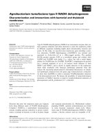
Fig. 1. Nop53p immunoprecipitates 5.8S chromatin and interacts with RNA polymerase I. A ChIP assay with A–Nop53p or protein was performed, followed by PCR reactions with primers for amplification of various regions of the rDNA and snoRNAs. (A) PCR for amplification of
18S and 25S chromatin regions. (B) Amplification of 5.8S region using samples from ChIP in the absence (upper panel) or presence (lower
panel) of RNases A ⁄ T1. (C) Amplification of snoRNA chromatin. For (A)–(C), ‘Int’ represents the intergenic region of chromosome V, used as
an internal control; I, input; S, sheared; E, eluted. (D) Quantification of the bands obtained in the PCR reactions. Values represent the ratio of
the rDNA bound to column to the input. Bars represent standard deviation. (E) Western blot for detection of proteins after pull-down assay.
Total extracts from cells expressing either GST or GST–Rrn3p (TE1) were incubated with glutathione–Sepharose, the flow-through fraction
was collected (FT1), and, after washing, total extracts of cells expressing His–Nop53p (TE2) were loaded. The flow-through fraction was
collected again (FT2), the resin was washed, and the bound fraction was obtained (B). His–Nop53p is pulled down by GST–Rrn3p, but not by
GST. His–Nop53p was detected using monoclonal antibody against His. GST and GST–Rrn3p were detected using anti-GST serum. Bands
corresponding to full-length and breakdown products of His–Nop53p are indicated on the right. The asterisks indicate a protein present in
Escherichia coli extract that runs close to His–Nop53p.
Nop53p). In these experiments, various salt concentrations were used to analyze the strength of the
interaction between truncated Nop53p mutants and
the pre-60S complex. The protein A–Nop53p fusion
efficiently precipitated 27S, 7S and 5.8S rRNAs, even
in the presence of 500 mm potassium acetate (Fig. 2A),
indicating that Nop53p binds stably to the pre-60S
complex. Although Nop53p precipitates 25S, lower
relative amounts of this rRNA were co-precipitated at
higher salt concentrations, indicating that Nop53p
4166
binds less efficiently to mature 60S subunits, which is
also consistent with its nucleolar localization.
The truncated Nop53p mutant fusion A–N-Nop53p
(N-terminal portion of Nop53p) also precipitates
pre-60S rRNAs, but much less efficiently than the
full-length protein, and only in the presence of up to
300 mm potassium acetate (Fig. 2A). Interestingly, the
A–C-Nop53p fusion (C-terminal portion of Nop53p)
co-purifies 27S, 25S, 7S and 5.8S rRNAs more efficiently than the N-terminal portion of Nop53p
FEBS Journal 275 (2008) 4164–4178 ª 2008 The Authors Journal compilation ª 2008 FEBS
D. C. Granato et al.
Nop53p activates the exosome in vitro
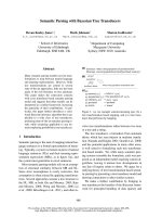
Fig. 2. Truncated mutants of Nop53p still
associate with pre-60S. (A) Co-immunoprecipitation of RNA with full-length or truncated Nop53. Northern blot hybridization of
RNA co-immunoprecipitated with protein A–Nop53p, protein A–N-Nop53p or
protein A–C-Nop53p. Probes used were
specific against rRNAs or scR1 (internal
control). (B) Western blot of the proteins
obtained from the same experiments,
detected using anti-mouse IgG.
(Fig. 2A). A–C-Nop53p co-precipitates 27S pre-rRNA
even in the presence of 500 mm potassium acetate,
indicating that the C-terminal portion of Nop53p is
stably bound to pre-rRNP complexes. A western blot
of bound fractions from the same experiments showed
that protein A fusions bound efficiently to the columns
under all conditions used (Fig. 2B), showing that the
differences in efficiency of rRNA precipitation between
truncated Nop53p mutants are due to different stabilities of interaction with the pre-60S complex and not
inefficient binding to the column.
In order to determine whether the Nop53p truncated mutants also bind RNA directly, in vitro RNA
binding assays were performed. In these experiments,
full-length Nop53p bound RNAs corresponding to
various fragments of pre-rRNA (Fig. 3A). Although
Nop53p specifically co-immunprecipitates pre-60S
chromatin (Fig. 1) and rRNAs (Fig. 2) [31], Nop53p
did not show a clear sequence specificity for binding
in these in vitro RNA binding assays. Interestingly,
however, all the rRNAs regions tested were AU-rich
and predicted to form secondary structures.
N-Nop53p also bound RNA, although not as efficiently as the full-length protein (Fig. 3A). C-Nop53p,
on the other hand, did not bind RNA in vitro, showing the same result as the negative control GST
(Fig. 3A). We therefore conclude that Nop53p binds
RNA through its N-terminal region and has affinity
for AU-rich and structured RNAs. In the pre-60S
complex, Nop53p binding to rRNA might be more
specific and stabilized by protein–protein interactions
with its C-terminal portion.
In order to analyze the affinity of Nop53p for
AU-rich RNA sequences in more detail, in vitro RNA
binding assays were performed using RNA oligonucleotides. Full-length Nop53p bound poly-rU and
poly-rAU oligomers, but not poly-rC (Fig. 3B), corroborating the results described above. In these experiments, 5.8S rRNA was used as a positive control for
Nop53p interaction. In summary, although no sequence
specificity was detected in these in vitro assays, Nop53p
showed higher affinity for U- and AU-rich sequences.
Truncated Nop53p mutants still localize to the
nucleolus
Although Nop53p is a nucleolar protein, no nuclear
localization signal could be detected in its sequence. In
FEBS Journal 275 (2008) 4164–4178 ª 2008 The Authors Journal compilation ª 2008 FEBS
4167
Nop53p activates the exosome in vitro
D. C. Granato et al.
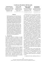
Fig. 3. RNA binding assay with truncated
Nop53p mutants. (A) Radioactively labeled
in vitro transcribed fragments of rRNA were
incubated with 10 pmol of full-length
Nop53p, or the truncated forms GST–NNop53p or GST–C-Nop53p, or with GST.
RNA–protein complexes were analyzed by
native gel electrophoresis and visualized by
phosphorimaging. Lanes 1, 6, 11 and 16,
RNAs incubated with full-length Nop53p;
lanes 2, 7, 12 and 17, RNAs incubated with
GST–C-Nop53p; lanes 3, 8, 13 and 18, RNAs
incubated with GST–N-Nop53p; lanes 4, 9,
14 and 19, RNAs incubated with GST; lanes
5, 10, 15 and 20, free RNA. (B) Nop53p
shows a preference for U-rich sequences.
Increasing amounts of Nop53p were incubated with various RNA oligonucleotides.
Free RNA and RNA–protein complexes
(RNP) are indicated on the right. No protein
was added in lanes 1, 5, 9 and 13.
order to identify the portion of Nop53p that is responsible for its subcellular localization, the truncated
mutants of the protein were fused to a GFP tag
(GFP–N-Nop53p and GFP–C-Nop53p). Confocal
images of split fluorescence channels showed the same
pattern of localization for RFP–Nop1p and GFP–NNop53p and GFP–C-Nop53p. Interestingly, although
GFP–C-Nop53p is concentrated in the nucleolus, it
can also be visualized throughout the nucleus. The
co-localization of GFP–Nop53p truncated mutants
and RFP–Nop1p was confirmed by fluorescence
profiles in several cell images (Fig. 4A,B). These results
indicate that protein interactions might be responsible
for directing Nop53p to the nucleus and for its concentration in the nucleolus.
4168
The N-terminal half of Nop53p complements
a conditional mutant strain
As shown above, the N-terminal portion of Nop53p
binds 5.8S rRNA directly and is concentrated in the
nucleolus, whereas the C-terminal portion of Nop53p
might interact with the proteins in the pre-60S complex,
but is less concentrated in the nucleolus. These results
raised the question of whether any of the truncated
mutants of Nop53p, when under control of a constitutive promoter, could complement the growth of the conditional strain Dnop53 ⁄ GAL::A-NOP53 in glucose
medium. Interestingly, N-Nop53p partially complements growth of the conditional strain (Fig. 5A).
When pre-rRNA processing was analyzed in these
FEBS Journal 275 (2008) 4164–4178 ª 2008 The Authors Journal compilation ª 2008 FEBS
D. C. Granato et al.
Nop53p activates the exosome in vitro
Fig. 4. Subcellular localization and protein interaction of the Nop53p N- and C-terminal fragments. Yeast strain NOP53 expressing GFP–NNop53p and RFP–Nop1p (A) or GFP–C-Nop53p and RFP–Nop1p (B) was analyzed by laser scanning confocal microscopy. Each channel
labeling is shown separately and merged in the lower right panels. The upper panels show representative profiles of green and red fluorescence, indicating RFP–Nop1p (red line) and GFP–N-Nop53p (green line) or GFP–C-Nop53p (green line) co-localization.
transformants, it was possible to see that, although
27S pre-rRNA and 25S rRNA levels in the strains
Dnop53 ⁄ GAL::A-NOP53 expressing either N-Nop53p
or C-Nop53p were very similar to those of the control
strain Dnop53 ⁄ GAL::A-NOP53 ⁄ pGAD, expression of
N-Nop53p led to lower accumulation of the 7S
pre-rRNA intermediate (Fig. 5B). The strain expressing
C-Nop53p showed higher levels of 7S pre-rRNA and
lower levels of the mature 5.8S rRNA (Fig. 5B). Quantification of 7S ⁄ 5.8S ratio in these strains showed that
N-Nop53p partially complements the function of the
Dnop53 ⁄ GAL::A-NOP53 strain (Fig. 5C). These results
indicate that direct binding to 5.8S rRNA is important
for Nop53p function.
Dnop53::GAL-NOP53 accumulates polyadenylated
forms of pre-rRNA
Nop53p affects pre-rRNA processing, and its depletion
leads to the accumulation of 27S and 7S pre-rRNAs,
which are degraded from the 5¢ end [31]. Therefore,
cells depleted of Nop53p show similar phenotypes to
exosome mutants, indicating that unprocessed rRNA
intermediates may accumulate in a polyadenylated
form in the Dnop53::GAL-NOP53 strain, as demonstrated for Drrp6 mutants [35,36]. In order to analyze
the polyadenylation of rRNA processing intermediates
in the Dnop53::GAL-NOP53 strain, total RNA was
extracted after 12 h of Nop53p depletion, and poly-A
RNA was isolated using oligo(dT) cellulose columns.
Analysis of the purified poly-A RNA demonstrated
that 27S and 7S pre-rRNAs accumulated in the polyadenylated form in Dnop53::GAL-NOP53 (Fig. 6A),
indicating that these RNAs are not efficiently
processed or degraded by the exosome in the absence
of Nop53p.
In order to determine whether the effect of Nop53p
depletion on 5.8S processing by the exosome was indirect, or whether it involved direct exosome binding,
protein interaction experiments were performed. We
FEBS Journal 275 (2008) 4164–4178 ª 2008 The Authors Journal compilation ª 2008 FEBS
4169
Nop53p activates the exosome in vitro
D. C. Granato et al.
Fig. 5. Analysis of complementation of
Dnop53 ⁄ GAL::A-NOP53 by truncated
mutants of Nop53p. (A) Analysis of complementation of strain Dnop53 ⁄ GAL::A-NOP53
by truncated Nop53p mutants, under the
control of a constitutive promoter, by
growth in glucose medium. N-Nop53p partially complements growth on glucose
plates. (B) rRNA processing was analyzed in
yeast strains Dnop53 ⁄ GAL::A-NOP53
expressing either Nop53p, N-Nop53p or
C-Nop53p by Northern blot hybridization
with probes against rRNAs, indicated on the
right. (C) Quantification of the 7S ⁄ 5.8S
rRNA ratio, showing the efficiency of 5.8S
rRNA maturation in cells expressing the
truncated forms of Nop53p.
have previously tried to identify interactions between
Nop53p and the exosome subunits using the twohybrid system, but no positive interaction was detected
[31]. Therefore, we tested the interaction between
Nop53p and some of the exosome subunits using GST
pull-down assays. In these experiments, we detected a
specific interaction between the recombinant proteins
His–Nop53p and GST–Rrp6p (Fig. 6B). Control
experiments with GST–Mtr3p showed that His–
Nop53p does not interact with this other exosome subunit, nor does it interact with GST, which was used as
a negative control (Fig. 6B). Despite the higher level of
GST expression compared to GST–Rrp6p or to GST–
Mtr3p, His–Nop53p was only pulled down by GST–
Rrp6p, confirming the specificity of this interaction.
These results led to the conclusion that, by binding to
the 5.8S rRNA and through its interaction with
Rrp6p, Nop53p may direct the exosome to the 7S
intermediate for processing.
The TRAMP complex has been shown to be responsible for polyadenylation of the RNAs that are substrates for degradation by the exosome [22]. As
depletion of Nop53p leads to the accumulation of
polyadenylated pre-rRNAs, this raises the question of
whether Nop53p also interacts with TRAMP subunits.
4170
The TRAMP subunit Trf4p was therefore fused to
GST, expressed in Escherichia coli, and its interaction
with Nop53p tested through GST pull-down. The
results show that GST–Trf4p pulls down His–Nop53p,
but the negative control GST does not (Fig. 6C).
These results indicate that Nop53p not only interacts
with the exosome, but also with the TRAMP complex,
corroborating the view that it is a regulatory factor for
processing of 7S pre-rRNA.
Nop53p activates the exosome RNase activity
in vitro
In order to test whether the Nop53p–Rrp6p interaction
is important for control of exosome function, in vitro
RNA degradation assays were performed. Yeast
exosome was isolated by tandem affinity purification
(TAP)–Rrp43p purification and was incubated with a
substrate RNA for in vitro RNA degradation, in the
presence of Nop53p or BSA, the latter being used as a
negative control (Fig. 7A). The results show that
TAP–Rrp43p exosome degrades an in vitro transcribed
RNA corresponding to a region of ITS2, a natural
exosome substrate during rRNA maturation (Fig. 7A;
lane 2). Upon incubation of the substrate RNA with
FEBS Journal 275 (2008) 4164–4178 ª 2008 The Authors Journal compilation ª 2008 FEBS
D. C. Granato et al.
Nop53p activates the exosome in vitro
Fig. 6. Analysis of rRNA polyadenylation in
the strain Dnop53 ⁄ GAL::A-NOP53 and interaction with the exosome. (A) Total RNA
was isolated from strains NOP53 and
Dnop53 ⁄ GAL::A-NOP53, and run through oligo(dT)–Sepharose columns. Polyadenylated
RNAs were analyzed by Northern blot
hybridization against probes specific for
rRNAs. I, input; FT, flow-through; EL, eluted
polyadenylated RNA. (B, C) Western blot for
detection of proteins after pull-down assay.
(B) Total extract from Escherichia coli cells
expressing either GST, GST–Rrp6p or GST–
Mtr3p (TE1) was incubated with glutathione–Sepharose resin, the flow-through fraction was collected (FT1), and, after washing,
the total extract of cells expressing His–
Nop53p (TE2) was loaded. The flow-through
fraction was collected again (FT2), the resin
was washed (not shown), and bound fraction was obtained (B). His–Nop53p is pulled
down by GST–Rrp6p. His–Nop53p was
detected using antibody against His. GST,
GST–Rrp6p and GST–Mtr3p were detected
using anti-GST serum. Bands corresponding
to full-length and breakdown products of
fusion proteins are indicated on the right.
(C) Same procedure as in (B), but with total
extract from E. coli cells expressing either
GST or GST–Trf4p (TE1), or expressing His–
Nop53p (TE2). His–Nop53p is pulled down
by GST–Trf4p.
the TAP–Rrp43p complex, there is a 20% decrease in
the intensity of the substrate band and a corresponding
increase in the intensity of faster-migrating bands that
correspond to degradation products (Fig. 7A; lane 2).
Although Nop53p does not degrade the RNA by itself
(Fig. 7A; lane 10), addition of 10 pmol Nop53p to the
reaction containing the TAP–Rrp43p complex
increases the RNase activity of the exosome by 16%
(Fig. 7A; lane 7). Addition of 20 and 30 pmol Nop53p
further increased the RNase activity of the exosome by
27% and 42%, respectively (Fig. 7A; lanes 8 and 9).
Addition of BSA to the reaction had no effect
(Fig. 7A; lanes 2–6). Control experiments with TAP–
Nop58p-purified box C ⁄ D snoRNP complex showed
no degradation of the RNA, as expected (Fig. 7A;
lanes 11–16).
FEBS Journal 275 (2008) 4164–4178 ª 2008 The Authors Journal compilation ª 2008 FEBS
4171
Nop53p activates the exosome in vitro
D. C. Granato et al.
with the exosome (Fig. 7B). His–Nop53p was also
co-purified with TAP–Nop58p, although in much
lower levels, probably due to the interaction between
Nop53p and the box C ⁄ D assembly factor Nop17p
[31]. These results strongly indicate that Nop53p is an
exosome cofactor, stimulating the RNase activity of
the complex.
Discussion
Fig. 7. Effect of Nop53p on RNA degradation by the exosome.
In vitro RNA degradation assay to test the effect of Nop53p on
exosome RNase activity. (A) A radioactively labeled RNA oligo corresponding to the 5¢ region of the rRNA spacer ITS2 was incubated
with 100 ng of the exosome complex isolated using TAP–Rrp43p,
or with 100 ng of box C ⁄ D snoRNP isolated using TAP–Nop58p,
and 10, 20 or 30 pmol of His–Nop53p or BSA. Reaction mixtures
were incubated for 1 h at 30 °C, and the products were analyzed
by denaturing acrylamide gel electrophoresis. The main degradation
products generated by the exosome complex are indicated. (B)
Analysis of protein complexes recovered through TAP purification.
TAP–Nop58p co-purified Nop1p and TAP–Rrp43p co-purified Mtr3p,
indicating that the box C ⁄ D snoRNP and exosome complexes,
respectively, were intact. Both complexes co-purified His–Nop53p
in the pull-down assay, although Nop53p interaction with the
exosome was much stronger.
The recovery of TAP-purified complexes was
analyzed by the detection of other subunits of the
exosome and box C ⁄ D snoRNP by western blotting
(Fig. 7B). TAP–Nop58p co-purified endogenous
Nop1p and TAP–Rrp43p co-purified Mtr3p, indicating
that the box C ⁄ D snoRNP and exosome complexes,
respectively, were recovered. In addition, incubation of
TAP complexes with His–Nop53p from E. coli extracts
showed that His–Nop53p is recovered with TAP–
Rrp43p, further confirming the interaction of Nop53p
4172
We used in vivo and in vitro approaches to characterize
the role that Nop53p plays in rRNA processing in
yeast. It had previously been demonstrated that
Nop53p binds 5.8S rRNA and participates in the late
steps of maturation of the large ribosomal subunit
RNAs [31–33], and here we show that the role played
by Nop53p involves protein–protein and protein–RNA
interactions. Nop53p co-precipitates 5.8S and 25S
chromatin and, to a lower extent, 18S chromatin,
which indicates that it binds pre-rRNA co-transcriptionally. Nop53p recruitment to rDNA chromatin is
dependent on active transcription, as no precipitation
of chromatin above background level was obtained
with A-Nop53p after treatment with RNases. We also
show here that Nop53p interacts directly with RNA
polymerase I transcription factor Rrn3p [34]. These
data indicate that, although Nop53p is present in the
pre-60S complex [1,11] and affects 7S pre-rRNA
processing by the exosome [31], it binds 5.8S rRNA
co-transcriptionally. Other protein complexes have
been shown to interact with transcription factors and
also influence pre-rRNA processing, including the
CURI complex formed by CK2, Utp21, Rrp7p and
Ifh1p, which is proposed to couple rRNA and ribosomal protein transcription [37]. Some of the U3
snoRNP protein subunits (Utp) have also been shown
to bind rRNA early during transcription and to participate in rRNA processing [4,5,7]. SSU processome
factors, mainly involved in processing of the 18S
rRNA, bind the precursor rRNA co-transcriptionally
[4]. Later during processing, factors involved in the
maturation of 27S pre-rRNA assemble onto the RNA,
forming the large ribosomal subunit (LSU) complex
[8,38]. Nop53p may participate in formation of the
LSU knob, and as it is present in the gradient fractions that contain LSU pre-rRNAs, it may remain
bound to the 5.8S rRNA during its processing [32].
The nucleolar localization of Nop53p seems to be
the result of protein–protein interactions, as no nuclear
localization signal could be identified in the Nop53p
sequence and truncated versions of this protein still
localize to the nucleolus. We have shown that Nop53p
interacts with various nuclear proteins – Nop17p,
FEBS Journal 275 (2008) 4164–4178 ª 2008 The Authors Journal compilation ª 2008 FEBS
D. C. Granato et al.
Nip7p, Rrn3p, Rrp6p and Trf4p (Figs 1 and 6) [31].
Identification of these protein interactions indicates
that one of these factors, or the whole complex, might
be responsible for directing Nop53p to the nucleolus.
A recent example of an rRNA processing factor that is
dependent on protein interaction for its subcellular
localization is human hRrp47p, an exosome cofactor,
which was shown to depend on its interaction with the
exosome subunit PM ⁄ Scl_100 (an Rrp6p ortholog) for
direction to the nucleus [39]. Interestingly, the Nop53p
C-terminal region co-immunoprecipitates the pre-60S
complex more efficiently than the N-terminal portion
of the protein. The Nop53p N-terminal region, on the
other hand, is involved in RNA interaction and can
partially complement the conditional strain Dnop53 ⁄
GAL::NOP53 in glucose medium. These results indicate that interaction with RNA is responsible for
Nop53p molecular function in 27S and 7S pre-rRNA
processing, and that this interaction may be stabilized
in the pre-60S complex through protein interaction
with the C-terminal portion of Nop53p. Similarly, the
ribosomal protein Rpl25p affects processing of 27S
pre-rRNA and has three functional domains for
nuclear import, RNA binding and 60S subunit assembly [40]. Mutations of each of these domains result in
defective ITS2 processing and accumulation of
pre-rRNA 27S, indicating that assembly of Rpl25p is
necessary but not sufficient for processing [40]. Despite
not having a canonical RNA-binding motif, Nop53p
binds RNA, but does not show strict RNA sequence
specificity in in vitro RNA binding experiments. Similarly, Nop9p, another example of an RNA-binding
protein involved in pre-rRNA processing, associates
with 20S pre-rRNA but does not show sequence specificity for in vitro binding [41].
Depletion of Nop53p leads to accumulation of the
7S pre-rRNA and polyadenylated RNAs, a phenotype very similar to that resulting from depletion of
exosome subunits. The results shown here indicate
that Nop53p function is directly related to the interaction with 5.8S rRNA and the exosome in the
pre-60S complex. In this context, Nop53p could
be responsible for directing the exosome to 7S
pre-rRNA, thereby regulating the function of the
complex. In the absence of Nop53p, the exosome is
not efficiently directed to the 7S pre-rRNA for processing, leading to the accumulation of its polyadenylated form. As RNAs polyadenylated by the TRAMP
complex are targeted for degradation by the exosome
[22], polyadenylated 7S was expected to be degraded
in strain Dnop53 ⁄ GAL::A-NOP53. However, polyadenylated 7S pre-RNA accumulates in this strain
and appears to be degraded in the 5¢ fi 3¢ direction
Nop53p activates the exosome in vitro
[31], leading to the conclusion that Nop53p is an
exosome cofactor.
Accordingly, in vitro RNA degradation assays with
the exosome complex isolated using TAP–Rrp43p
showed that, although Nop53p does not degrade RNA
by itself, its presence stimulates the RNase activity of
the exosome. It is possible that the stimulation of the
exosome activity is due to recruitment of the complex
to the substrate via Nop53p–Rrp6p interaction, or
through TRAMP recruitment. Interestingly, Nop53p
has also been identified as interacting with components
of the TRAMP complex [42], corroborating the results
shown here. A similar role is seen for another RNAbinding protein, of the Nrd1 complex, which can direct
the exosome to specific RNA substrates and stimulate
exosome degradation of substrates [43]. We can
conclude that Nop53p must play an important role in
exosome activity.
In summary, we show here that Nop53p binds 5.8S
rRNA co-transcriptionally through its N-terminal portion and may interact with other pre-60S processing
factors through its C-terminal portion. As depletion of
Nop53p leads to accumulation of polyadenylated 7S
pre-rRNA, and as Nop53p interacts with the exosome
subunit Rrp6p and activates the RNase activity of the
complex in vitro, Nop53p may be involved in recruitment of the exosome to the 7S pre-rRNA for processing and formation of the mature 5.8S rRNA.
Experimental procedures
Plasmid construction
The plasmids used in this study are listed in Table 1 and
cloning procedures are summarized below. DNA fragments
of NOP53 coding for the N-terminal (amino acids 1–210)
and C-terminal (amino acids 210–456) portions of the protein were PCR-amplified from Saccharomyces cerevisiae
genomic DNA and cloned into vectors pBTM or pGADC2
for two-hybrid analyses and into YCplac33GAL-A, fused
to the protein A tag, under the control of the GAL1
promoter [31]. Subsequently, the fragments coding for the
N- and C-terminal regions of NOP53 were subcloned into
pGEX (GE Healthcare, Piscataway, NJ, USA), using the
restriction sites BamHI ⁄ SalI and EcoRI ⁄ PstI, respectively,
generating vectors pGEX-N-NOP53 and pGEX-C-NOP53.
Plasmids pGAD-N-NOP53 and pGAD-C-NOP53, containing NOP53 truncation mutants under the control of the
constitutive ADH1 promoter, were also used for complementation analysis of conditional strain Dnop53 ⁄ GAL::
NOP53. The plasmids pYCplac33GAL-A-NOP53, pET28NOP53, pGADC2-NOP53 and pGEM-5.8S have been
described previously [31].
FEBS Journal 275 (2008) 4164–4178 ª 2008 The Authors Journal compilation ª 2008 FEBS
4173
Nop53p activates the exosome in vitro
D. C. Granato et al.
Table 1. Plasmids used in this study.
Plasmids
Relevant characteristics
Reference
pGADC2
GAL4 activation domain,
LEU2 2 lm
GAL4::NOP53, LEU2 2 lm
GAL4::N-NOP53, LEU2 2 lm
GAL4::C-NOP53, LEU2 2 lm
GAL1::ProtA, URA3, CEN4
GAL1::ProtA-NOP53,
URA3,CEN4
GAL1::ProtA-N-NOP53,
URA3, CEN4
GAL1::ProtA-C-NOP53,
URA3, CEN4
MET25::GFP, URA3, CEN6
MET25::GFP-N-NOP53,
URA3, CEN6
MET25::GFP-C-NOP53,
URA3, CEN6
ADH1::RFP-NOP1, LEU2 2 lm
His::NOP53, KanR
GST::N-NOP53, AmpR
GST::C-NOP53, AmpR
GST::RRN3, AmpR
GST::RRP6, AmpR
GST::MTR3, AmpR
GST::TRF4, AmpR
[44]
pGAD-NOP53
pGAD-N-NOP53
pGAD-C-NOP53
YCplac33GAL-A
YCp33GAL-ANOP53
YCp33GAL-A-NNOP53
YCp33GAL-A-CNOP53
pGFP-N-FUS
pGFP-N-NOP53
pGFP-C-NOP53
pRFP-NOP1
pET-NOP53
pGEX-N-NOP53
pGEX-C-NOP53
pGEX-RRN3
pGEX-RRP6
pGEX-MTR3
pGEX-TRF4
[31]
This study
This study
[31]
[31]
This study
This study
[45]
This study
This study
[26]
[31]
This
This
This
This
[46]
This
study
study
study
study
study
Maintenance and handling of E. coli and yeast
strain
Escherichia coli strains DH5a and BL21(DE3) were maintained in LB medium and manipulated according to
standard techniques [47]. The yeast strains used in this
work, with a brief description of the relevant genetic markers, are shown in Table 2. Growth and handling of S. cere-
visiae strains were performed as previously described [49].
Carbon source-conditional strains were incubated in YP
medium containing 2% galactose, and transferred to 2%
glucose for the indicated periods of time. Yeast strains were
transformed using the lithium acetate method [49].
Protein pull-down and immunoblot assays
Protein A pull-down assays were performed using extracts
from 500 mL yeast cultures grown to an attenuance at
600 nm of 1.3–1.5 at 30 °C in yeast minimal synthetic medium (YNB)-Gal and the required supplements. Yeast
whole-cell extracts were prepared by suspending cells in
1 mL of ice-cold buffer A (20 mm Tris ⁄ HCl pH 8.0, 5 mm
magnesium acetate, 150 mm potassium acetate, 0.2% v ⁄ v
Triton X-100, 1 mm dithiothreitol, 1 mm phenylmethanesulfonyl fluoride). Cells were disrupted by vortexing with
1 volume of glass beads and the extracts cleared by centrifugation at 16 000 g for 30 min at 4 °C [31]. Extracts containing protein A fusion proteins were incubated with
200 lL of IgG–Sepharose beads (GE Healthcare) for 2 h at
4 °C. The IgG–Sepharose beads were extensively washed
with ice-cold buffer A, and bound proteins were suspended
in 80 lL of SDS–PAGE sample buffer. A similar procedure
was used for protein A–N-Nop53p and protein A–CNop53p domain co-immunoprecipitation assays, except that
the potassium acetate concentration was raised from 150 to
500 mm during incubation with IgG–Sepharose beads and
washing of the beads,. Samples were fractionated by SDS–
PAGE followed by immunoblot analyses with anti-mouse
IgG (GE Healthcare). Pull-down of His–Nop53p was
assayed as follows: whole-cell extracts from E. coli cells
expressing either GST, GST–Rrn3p or GST–Trf4p were
generated in low-salt buffer (20 mm Tris ⁄ HCl pH 8.0,
5 mm magnesium acetate, 50 mm potassium acetate, 0.1%
v ⁄ v Triton X-100, 1 mm dithiothreitol, 1 mm phenyl-
Table 2. Yeast and bacteria strains used in this study.
Strain
Relevant characteristics
Reference
NOP53
Nop53 ⁄ Dnop53 2n
2n MATa, his3D1 leu2D0, lys2D0 ura3D0 met15D0
MATa ⁄ a, his3D1 ⁄ his3D1 leu2D0 ⁄ leu2D0 lys2D0 ⁄ LYS2 ura3D0 ⁄ ura3D0 MET15 ⁄
met15D0 NOP53 ⁄ NOP53::KANR
MET15 his3D1 leu2D0 ura3D0 NOP53::KANR ⁄ YCpGAL-A-NOP53
NOP53, YCp33GAL-A
NOP53, YCp33GAL-A-NOP53
NOP53, YCp33GAL-A-N-NOP53
NOP53, YCp33GAL-A-C-NOP53
Dnop53, YCp33GAL-A-NOP53,pGAD-NOP53
Dnop53,YCp33GAL-A-NOP53,pGAD-N NOP53
Dnop53,YCp33GAL-A-NOP53,pGAD-C-NOP53
Dnop53, YCp33GAL-A-NOP53,pGADC2
NOP53, pGFP-N-NOP53,pRFP-NOP1
NOP53, pGFP-C-NOP53,pRFP-NOP1
supE44 DlacU169 (/80 lacZDM15) hsdR17 recA1 endA1 gyrA96 thi1 relA1
E. coli B F– ompT hsdS(rB– mB–) dcm+ Tetr gal l (DE3) endA Hte [argU ileY leuW Camr]*
Euroscarf
Euroscarf
Dnop53 ⁄ GAL:: NOP53 (YDG151)
YDG152
YDG153
YDG154
YDG155
YDG156
YDG157
YDG158
YDG159
YDG160
YDG161
DH5a
BL21 Codon Plus (DE3) RIL
4174
[31]
[31]
[31]
This study
This study
This study
This study
This study
This study
This study
This study
[48]
Stratagene
FEBS Journal 275 (2008) 4164–4178 ª 2008 The Authors Journal compilation ª 2008 FEBS
D. C. Granato et al.
methanesulfonyl fluoride) and mixed with 500 lL of glutathione–Sepharose beads (GE Healthcare). After washing
bound material with the same buffer, whole-cell extracts
from E. coli cells expressing His–Nop53p were added to the
glutathione–Sepharose beads and incubated at 4 °C for 2 h.
The glutathione–Sepharose beads were precipitated and
washed again with the low-salt buffer, and bound proteins
were eluted and resolved on SDS–PAGE, and transferred
to poly(vinylidene) difluoride (PVDF) membranes (Bio-Rad
Laboratories, Hercules, CA, USA), which were incubated
with anti-(poly histidine) serum (GE Healthcare) or antiGST serum (Sigma, St Louis, MO, USA). The immunoblots were developed using the enhanced chemiluminescence
system (GE Healthcare).
The purification of complexes using TAP–Rrp43p and
TAP–Nop58p was performed as described previously [50],
with some modifications. Yeast cells expressing TAP–
Rrp43p or TAP–Nop58p were grown in 4 L of yeast
complete medium containing glucose. Isolation of the complexes was performed by incubating the total yeast extracts
for 2 h at 4 °C with IgG–Sepharose beads (GE Healthcare),
followed by extensive washing with TMN buffer [10 mm
Tris pH 7.6, 100 mm NaCl, 5 mm MgCl2, 0.1% Nonidet
P40 (Sigma-Aldrich), 1 mm dithiothreitol, 1 mm phenylmethanesulfonyl fluoride]. The exosome and box C ⁄ D
snoRNP complexes were eluted from the beads by incubating the resin with 20 U of tobacco etch virus (TEV) protease for 18 h.
Pull-down of His–Nop53p using TAP complexes was performed by incubating the TAP–Rrp43p or TAP–Nop58p
total yeast extracts with IgG–Sepharose beads as described
above. The resin was then washed and total extract of
E. coli cells expressing His–Nop53p was added and incubated for 2 h at 4 °C, followed by extensive washing with
low-salt buffer. The bound proteins were eluted with 20 U
of TEV protease for 18 h.
Recombinant His–Nop53p, GST–N-Nop53p and
GST–C-Nop53p expression and purification
Extracts for GST pull-down assays were prepared as
follows. E. coli BL21(DE3) cells harboring plasmids encoding either test proteins or control proteins were incubated
in 500 mL LB medium containing 100 lgỈmL)1 ampicillin.
At an attenuance at 600 nm of approximately 0.8, 0.5 mm
isopropyl thio-b-d-galactoside (IPTG) was added and the
cultures were transferred to 37 °C for 2 h (pGEX-NNOP53) or 4 h (pGEX-C-NOP53). Cells were harvested by
centrifugation at 3700 g for 20 min at 4°C, and suspended
in Tris ⁄ NaCl buffer (50 mm Tris ⁄ HCl pH 8.0, 150 mm
NaCl, 1 mm dithiothreitol, 1 mm phenylmethanesulfonyl
fluoride, 0.5% v ⁄ v Nonidet P40). The cell suspension was
lysed in French press, and the extracts were cleared by centrifugation at 16 000 g for 60 min at 4 °C. The supernatant
was incubated with 100 lL glutathione–Sepharose beads
Nop53p activates the exosome in vitro
(GE Healthcare) for 1 h at 4 °C. After extensive washing
with the binding buffer, proteins were eluted from the glutathione–Sepharose beads using Tris ⁄ NaCl buffer containing 20 mm glutathione. E. coli BL21(DE3) harboring
plasmid pET28-NOP53 was incubated in LB medium containing 100 lgỈmL)1 kanamycin at 37 °C. At an attenuance
at 600 nm of approximately 0.8, 0.5 mm IPTG was added
and the culture was transferred to 30 °C for 2 h. Cells were
harvested by centrifugation at 3700 g for 20 min at 4 °C
and suspended in buffer containing 50 mm Tris ⁄ HCl
pH 8.0, 50 mm NaCl, 1 mm phenylmethanesulfonyl fluoride. Cell extracts were prepared as described above for
GST-fused proteins, and soluble material was incubated
using Q–Sepharose beads (GE Healthcare). His–Nop53p
was further purified by incubating the flow through from
the Q–Sepharose with Ni-NTA beads (Qiagen, Valencia,
CA, USA) for 2 h at 4 °C, followed by chromatography on
a heparin–Sepharose column (GE Healthcare), using the
same buffer as above for binding and a KCl gradient
(50 mm to 1 m) for elution.
Subcellular localization of Nop53p
The subcellular localization of Nop53p truncation mutants
was analyzed by monitoring the fluorescence signal produced by a GFP fusion to the N- and C-terminal portions
of Nop53p. The subcellular localization of Nop1p was
analyzed by monitoring fluorescence of RFP fused to the
N-terminus of Nop1p. GFP–N-Nop53p, GFP–C-Nop53p
and RFP–Nop1p proteins were expressed in strain NOP53,
transformed with plasmid vector pGFP-C-FUS [45]
containing the respective genes, and pRFP-NOP1, as previously described [31]. Cells were mounted on polylysinecoated slides and observed using a laser scanning confocal
microscope (LSM510; Zeiss, Jena, Germany). The fluorescent images were obtained by confocal laser scanning using
argon (458, 488 and 514 nm), helium–neon 1 (543 nm) and
helium–neon 2 (633 nm) lasers connected to an inverted
fluorescence microscope (Zeiss Axiovert 100M). The profile
module of the LSM510 software was used to analyze the
green and red fluorescence co-localization.
Co-immunoprecipitation of RNAs
Whole-cell extracts of yeast strains W303 ⁄ YCp33-GAL::A,
DNOP53 ⁄ YCp33-GAL::A-NOP53, NOP53 ⁄ YCp33-GAL::
A-NOP53-N,
NOP53 ⁄ YCp33-GAL::A-NOP53-C
were
prepared as described above, and protein A-tagged Nop53p
(A–Nop53p) was isolated by incubation with IgG–Sepharose beads (GE Healthcare) for 2 h at 4 °C [26]. Following
extensive washing with buffer A, RNA was isolated from
bound fractions by directly extracting the bead suspension
with phenol. Subsequently, the RNA was precipitated,
suspended in diethylpyrocarbonate-treated water and analyzed by electrophoresis in 1.5% agarose gels and by
FEBS Journal 275 (2008) 4164–4178 ª 2008 The Authors Journal compilation ª 2008 FEBS
4175
Nop53p activates the exosome in vitro
D. C. Granato et al.
Table 3. Oligonucleotides used for Northern blot hybridization or
PCR.
Oligo
Sequence
Reference
P2
P4
P5
CATGGCTTAATCTTTGAGAC
CGTATCGCATTTCGCTGCGTTC
CTCACTACCAAACAGAATGTTT
GAGAAGG
GCCGCTTCACTCGCCGTTACTA
AGGC
GTTTGACCTCAAATCAGGTAGG
CTCTTCGAAGGCACTTTACA
TATCTGGTTGATCCTGCCAG
AATTCAGGGAGGTAGTGACA
CCGATTGGCAAAAAC
TGTTGGAGCACAAGCAAG
GCTGCAGAAGATGAAACAA
GCATCAGACACTAATTGC
GGAAATGCGTAGGGAAGACCA
ATTTCATGACG
GATGCCTCTTTAGAACAAGGTT
ACAAATCCTG
CTTTCAACAACGGATCTCTTGG
GGTCACCCACTACACTACTCGG
TCTAGCCGCGAGGAAGGA
GTTCGCCTAGACGCTCTCTTC
CCTTCTCAAACATTCTGTTTGG
[51]
[52]
[16]
P7
25SFor3252
25SRev3501
18SFor701
18SRevPE
SnR37For
SnR37Rev
SnR74For
SnR74Rev
Cr.V interg. For
Cr.V interg. Rev
5.8SFor2865
5S
scR1Rev
UC1
ITS2 For 3020
[26]
This
This
This
[26]
This
This
This
This
This
study
study
study
study
study
study
study
study
This study
[31]
[31]
[53]
[52]
This study
Northern blot as described previously [26,31], using probes
specific to pre-rRNA and rRNAs. For comparison, 1% of
RNA recovered from total extract was loaded on gels, and
normalized for loading to endogenous scR1 RNA.
RNA extraction and analysis
Exponentially growing cultures of yeast strains NOP53,
DNOP53 ⁄ GAL-A-NOP53, DNOP53 ⁄ GAL-A-NOP53 ⁄ ADNOP53,
DNOP53 ⁄ GAL-A-NOP53 ⁄ AD-C-NOP53
and
DNOP53 ⁄ GAL-A-NOP53 ⁄ C2 strains were transferred from
YNB-Gal to YNB-Glu medium. Cells were collected at the
time of the shift (t0) and after 24 h of incubation in glucose
medium. RNA extraction was performed using a modified
hot phenol method [31]. Oligo probes used in the Northern
hybridization analyses are listed in Table 3. For poly-A
RNA purification, total RNA (400 lg) from NOP53 and
DNOP53 ⁄ GAL-A-NOP53 strains grown for 12 h in glucose
medium was purified using 50 lL of oligo(dT)–Sepharose
(Gibco, Grand Island, NY, USA), following the manufacturer’s protocol.
RNA binding and degradation assays
RNA fragments corresponding to the rRNA regions
5.8S+29, 25S, 5¢ ETS and ITS2 were transcribed in vitro
using linearized vectors pGEM-5.8S, pGEM-25S, pGEM5¢ETS and pGEM-ITS2 as templates in the presence of
4176
10 lCi of a-32P-UTP, as described previously [31]. One
pmol of radiolabeled RNA was incubated with 10 pmol of
purified His–Nop53p, GST–N-Nop53p, GST–C-Nop53p or
GST in buffer A for 20 min at 25 °C. For the band-shift
analysis, the RNA–protein complexes were analyzed in 6%
polyacrylamide gel. For RNA degradation assays, 100 ng
of the TAP–Rrp43p- or TAP–Nop58p-purified complexes,
or 0.5 pmol of GST–Rrp6p, was incubated with 0.5 pmol
of radioactively labeled oligo RNA, in the presence of 10,
20 or 30 pmol of His–Nop53p or BSA.
Chromatin immunoprecipitation
Strain DNOP53 ⁄ GAL::A-NOP53 grown at 30 °C in
YP-GAL to an attenuance at 600 nm of 0.2–0.6 was pelleted
by centrifugation at 3700 g for 20 min at 4°C and processed
as described previously [4,54]. Chromatin solution was incubated for 2 h with IgG–Sepharose beads pre-washed with
lysis buffer (50 mm Hepes-KOH pH 7.5, 150 mm NaCl,
1 mm EDTA, 1% Triton, 0.1% deoxycholate, 1 mm phenylmethanesulfonyl fluoride). The immunoprecipitated material
was washed three times with 1 mL low-salt and high-salt
buffers for 5 min, and the chromatin obtained, as well as the
input, was submitted to reverse crosslinking and analyzed by
PCR. Several regions of various genes were amplified after
ChIP. [a-32P] dATP was added to the PCR (0.5 lCi per
25 lL). The results of ChIP were quantified [55] using a
phosphorimager (Molecular Dynamics, Sunnyvale, CA,
USA), and normalized against the input. An intergenic
region from chromosome V was used as an internal control.
The values on the histogram correspond to the mean of
three PCR reactions from three immunoprecipitated chromatin preparations. For treatment with RNase, RNase mix
(RNase A ⁄ RNase T1; Fermentas, Glenburnie, MD, USA)
was added to the total cell extract at a final concentration of
10 lgỈlL)1. The total extract was incubated for 1 h at 25 °C
and subjected to immunoprecipitation at 4 °C for 2 h.
Acknowledgements
We would like to thank Nilson I. T. Zanchin (LNLS,
Campinas, Brazil) and Sandro R. Valentini (UNESP,
Araraquara, Brazil) for anti-Nip7p and anti-Rpl5p antisera, respectively. We also thank Beatriz A. Castilho
(UNESP, Sao Paulo, Brazil) and Daniel C. Pimenta
˜
(Butantan Institute, Sao Paulo, Brazil) for experimental
˜
help. We thank Juliana S. Luz for anti-Nop1p and antiMtr3p antisera, and Tereza C. Lima Silva (LNLS,
Campinas, Brazil) and Zildene G. Correa (LNLS, Campinas, Brazil) for DNA sequencing. D.C.G. was the
`
recipient of a Fundacao de Amparo a Pesquisa do
¸ ˜
Estado de Sao Paulo (FAPESP) fellowship. This work
˜
was supported by FAPESP grants 03 ⁄ 06031-3 and
05 ⁄ 56493-9 to C.C.O.
FEBS Journal 275 (2008) 4164–4178 ª 2008 The Authors Journal compilation ª 2008 FEBS
D. C. Granato et al.
References
1 Babler J, Grandi P, Gadal O, Lessmann T, Petfalski E,
Tollervey D, Lechner J & Hurt E (2001) Identification
of a 60S preribosomal particle that is closely linked to
nuclear export. Mol Cell 8, 517–529.
2 Rosado VI, Kressler D & de la Cruz J (2007) Functional analysis of Saccharomyces cerevisiae ribosomal
protein Rpl3p in ribosome synthesis. Nucleic Acids Res
35, 4203–4213.
3 Ferreira-Cerca S, Poll G, Gleizes PE, Tschochner H
& Milkereit P (2005) Roles of eukaryotic ribosomal
proteins in maturation and transport of pre-18S rRNA
and ribosome function. Mol Cell 20, 263–275.
4 Gallagher JE, Dunbar DA, Grannemann S, Mitchell
BM, Osheim Y, Beyer AL & Baserga SJ (2004) RNA
polymerase I transcription and pre-rRNA processing
are linked by specific SSU processome components.
Genes Dev 18, 2506–2517.
5 Dez C, Dlakic M & Tollervey D (2007) Roles of the
HEAT repeat proteins Utp10 and Utp20 in 40S ribosome maturation. RNA 13, 1516–1527.
6 Perez-Fernandez J, Roman A, Rivas J, Bustelo XR &
Dosil M (2007) The 90S pre-ribosome is a multimodular
structure that is assembled through a hierarchical mechanism. Mol Cell Biol 27, 5414–5429.
7 Dragon F, Gallagher JE, Compagnone-Post PA, Mitchell BM, Porwancher KA, Wehner KA, Wormsley S,
Settlage R, Shabanowitz J, Osheim Y et al. (2002)
A large nucleolar U3 ribonucleoprotein required for
18S ribosomal RNA biogenesis. Nature 417, 967–
970.
8 Grandi P, Rybin V, Bassler J, Petfalski E, Strauss D,
Marzioch M, Schafer T, Kuster B, Tschochner H, Tollervey D et al. (2002) 90S pre-ribosomes include the 35S
pre-rRNA, the U3 snoRNP, and 40S subunit processing
factors but predominantly lack 60S synthesis factors.
Mol Cell 10, 105–115.
9 Krogan NJ, Peng WT, Cagney G, Robinson MD, Haw
R, Zhong G, Guo X, Zhang X, Canadien V, Richards
DP et al. (2004) High-definition macromolecular composition of yeast RNA-processing complexes. Mol Cell
13, 225–239.
10 Vanrobays E, Gelugne JP, Caizergues-Ferrer M &
Lafontaine DL (2004) Dim2p, a KH-domain protein
required for small ribosomal subunit synthesis. RNA
10, 645–656.
11 Nissan TA, Baßler J, Petfalski E, Tollervey D & Hurt
E (2002) 60S pre-ribosome formation viewed from
assembly in the nucleolus until export to the cytoplasm.
EMBO J 21, 5539–5547.
12 Wehner KA, Gallagher JE & Baserga SJ (2002) Components of an interdependent unit within the SSU
processome regulate and mediate its activity. Mol Cell
Biol 22, 7258–7267.
Nop53p activates the exosome in vitro
13 Mitchell P, Petfalski E, Shevchenko A, Mann M & Tollervey D (1997) The exosome: a conserved eukaryotic
RNA processing complex containing multiple 3¢ fi 5¢
exoribonucleases. Cell 91, 457–466.
14 Zanchin NIT & Goldfarb DS (1999) The exosome subunit Rrp43p is required for the efficient maturation of
5.8S, 18S and 25S rRNA. Nucleic Acids Res 27, 1283–
1288.
15 Allmang C, Mitchell P, Petfalski E & Tollervey D
(2000) Degradation of ribosomal RNA precursors by
the exosome. Nucleic Acids Res 28, 1684–1691.
16 Oliveira CC, Gonzales FA & Zanchin NIT (2002) Temperature-sensitive mutants of the exosome subunit
Rrp43p show a deficiency in mRNA degradation and
no longer interact with the exosome. Nucleic Acids Res
30, 4186–4198.
17 Allmang C, Kufel J, Chanfreau G, Mitchell P, Petfalski
E & Tollervey D (1999) Functions of the exosome in
rRNA, snoRNA and snRNA synthesis. EMBO J 18,
5399–5410.
18 van Hoof A, Staples RR, Baker RE & Parker R (2000)
Function of the Ski4p (Csl4p) and Ski7p proteins in
3¢-to-5¢ degradation of mRNA. Mol Cell Biol 20, 8230–
8243.
19 Houseley J, La Cava J & Tollervey D (2006) RNAquality control by the exosome. Nat Rev Mol Cell Biol
7, 529–539.
20 Mitchell P, Petfalski E, Houalla R, Podtelejnikov A,
Mann M & Tollervey D (2003) Rrp47p is an exosomeassociated protein required for the 3¢ processing of
stable RNAs. Mol Cell Biol 23, 6982–6992.
21 Kadaba S, Krueger A, Trice T, Krecic AM, Hinnebusch AG & Anderson J (2004) Nuclear surveillance
and degradation of hypomodified initiator tRNAMet in
S. cerevisiae. Genes Dev 18, 1227–1240.
22 LaCava J, Houseley J, Saveanu C, Petfalsky E, Thompson E, Jacquier A & Tollervey D (2005) RNA degradation by the exosome is promoted by a nuclear
polyadenylation complex. Cell 121, 713–724.
23 Wyers F, Rougemaille M, Badis G, Rousselle JC,
Dufour ME, Boulay J, Regnault B, Devaux F, Namane
A, Seraphin B et al. (2005) Cryptic pol II transcripts
are degraded by a nuclear quality control pathway
involving a new poly(A) polymerase. Cell 121, 725–737.
24 Vanacova S, Wolf J, Martin G, Blank D, Dettwiller S,
Friedlein A, Langen H, Keith G & Keller W (2005) A
new yeast poly(A) polymerase complex involved in
RNA quality control. PLoS Biol 3(6), e189.
25 Zanchin NIT & Goldfarb DS (1999) Nip7p interacts
with Nop8p, an essential nucleolar protein required for
60S ribosome biogenesis, and the exosome subunit
Rrp43p. Mol Cell Biol 19, 1518–1525.
26 Gonzales FA, Zanchin NIT, Luz JS & Oliveira CC
(2005) Characterization of Saccharomyces cerevisiae
Nop17p, a novel Nop58p-interacting protein that is
FEBS Journal 275 (2008) 4164–4178 ª 2008 The Authors Journal compilation ª 2008 FEBS
4177
Nop53p activates the exosome in vitro
27
28
29
30
31
32
33
34
35
36
37
38
39
40
D. C. Granato et al.
involved in pre-rRNA processing. J Mol Biol 346, 437–
455.
Kufel J, Allmang C, Verdone L, Beggs J & Tollervey D
(2003) A complex pathway for 3¢ processing of the yeast
U3 snoRNA. Nucleic Acids Res 31, 6788–6797.
van Hoof A, Lennertz P & Parker R (2000) Three conserved members of the RNase D family have unique
and overlapping functions in the processing of 5S, 5.8S,
U4, U5, RNase MRP and RNase P RNAs in yeast.
EMBO J 15, 1357–1365.
Araki Y, Takahashi S, Kobayashi T, Kajiho H, Hoshino S & Katada T (2001) Ski7p G protein interacts with
the exosome and the Ski complex for 3¢-to-5¢ mRNA
decay in yeast. EMBO J 20, 4684–4693.
Wang L, Lewis MS & Johnson AW (2005) Domain
interactions within the Ski2 ⁄ 3 ⁄ 8 complex and between
the Ski complex and Ski7p. RNA 11, 1291–1302.
Granato DC, Gonzales FA, Luz JS, Cassiola F, Machado-Santelli GM & Oliveira CC (2005) Nop53p, an
essential nucleolar protein that interacts with Nop17p
and Nip7p, is required for pre-rRNA processing in
Saccharomyces cerevisiae. FEBS J 272, 4450–4463.
Thomson E & Tollervey D (2005) Nop53p is required
for late 60S ribosome subunit maturation and nuclear
export in yeast. RNA 11, 1215–1224.
Sydorskyy Y, Dilworth DJ, Halloran B, Yi EC, Makhnevych T, Wozniak R & Aitchison JD (2005) Nop53p
is a novel nucleolar 60S ribosomal subunit biogenesis
protein. Biochem J 388, 819–826.
Claypool JA, French SL, Johzuka K, Eliason K, Vu L,
Dodd JA, Beyer AL & Nomura M (2004) Tor pathway
regulates Rrn3p-dependent recruitment of yeast RNA
polymerase I to the promoter but does not participate
in alteration of the number of active genes. Mol Biol
Cell 15, 946–956.
Kuai L, Fang F, Butler JS & Sherman F (2004) Polyadenylation of rRNA in Saccharomyces cerevisiae. Proc
Natl Acad Sci USA 101, 8581–8586.
Carneiro T, Carvalho C, Braga J, Rino J, Milligan L,
Tollervey D & Carmo-Fonseca M (2007) Depletion of
the yeast nuclear exosome subunit Rrp6 results in accumulation of polyadenylated RNAs in a discrete domain
within the nucleolus. Mol Cell Biol 27, 4157–4165.
Rudra D, Mallick J, Zhao Y & Warner JR (2007) Potential interface between ribosomal protein production and
pre-rRNA processing. Mol Cell Biol 27, 4815–4824.
Venema J & Tollerevey D (1999) Ribosome synthesis in
Saccharomyces cerevisiae. Annu Rev Genet 33, 261–311.
Schilders G, van Dijk E & Pruijn GJ (2007) C1D and
hMtr4p associate with the human exosome subunit
PM ⁄ Scl-100 and are involved in pre-rRNA processing.
Nucleic Acids Res 35, 2564–2572.
van Beekvelt CA, Graaff-Vincent M, Faber AW, van’t
´
Riet J, Venema J & Raue HA (2001) All three functional domains of the large ribosomal subunit protein
4178
41
42
43
44
45
46
47
48
49
50
51
52
53
54
55
L25 are required for both early and late pre-rRNA processing steps in Saccharomyces cerevisiae. Nucleic Acids
Res 29, 5001–5008.
Thomson E, Rappsilber J & Tollervey D (2007) Nop9
is an RNA binding protein present in pre-40S ribosomes and required for 18S rRNA synthesis in yeast.
RNA 13, 2165–2174.
Krogan NJ, Cagney G, Yu H, Zhong G, Guo X, Ignatchenko A, Li J, Pu S, Datta N, Tikuisis AP et al.
(2006) Global landscape of protein complexes in the
yeast Saccharomyces cerevisiae. Nature 440, 637–643.
Vasiljeva L & Buratowski S (2006) Nrd1 interacts with
the nuclear exosome for 3¢ processing of RNA polymerase II transcripts. Mol Cell 21, 239–248.
James P, Halladay J & Craig EA (1996) Genomic
libraries and a host strain designed for highly efficient
two-hybrid selection in yeast. Genetics 144, 1425–1436.
Niedenthal RK, Riles L, Johnston M, Hegemann JH
(1996) Green fluorescent protein as a marker for gene
expression and subcellular localization in budding yeast.
Yeast 12, 773–786.
Luz JS, Tavares JR, Gonzales FA, Santos MC & Oliveira CC (2007) Analysis of the Saccharomyces cerevisiae
exosome architecture and of the RNA binding activity
of Rrp40p. Biochimie 89, 686–691.
Sambrook J, Maniatis T & Fritsch EF (1989) Molecular
Cloning: A Laboratory Manual, 2nd edn. Cold Spring
Harbor Laboratory Press, Cold Spring Harbor, NY.
Hanahan D (1983) Studies on transformation of Escherichia coli with plasmids. J Mol Biol 166, 557–580.
Sherman F, Fink GR & Hicks JB (1986) Laboratory
Course Manual for Methods in Yeast Genetics. Cold
Spring Harbor Laboratory Press, Cold Spring Harbor,
NY.
Mitchell P (2001) Purification of yeast exosome.
Methods Enzymol 342, 356–364.
Fatica A, Dlakic A & Tollervey D (2002) Naf1p is a box
H ⁄ ACA snoRNP assembly factor. RNA 8, 1502–1514.
Zanchin NIT, Roberts P, DeSilva A, Sherman F &
Goldfarb DS (1997) Saccharomyces cerevisiae Nip7p is
required for efficient 60S ribosome subunit biogenesis.
Mol Cell Biol 17, 5001–5015.
Baker KE & Parker R (2006) Conventional 3¢ end formation is not required for NMD substrate recognition
in Saccharomyces cerevisiae. RNA 12, 1441–1445.
Keogh MC & Buratowski S (2004) Using chromatin
immunoprecipitation to map co-transcriptional mRNA
processing in Saccharomyces cerevisiae. Methods Mol
Biol 257, 1–16.
Nedea E, He X, Kim M, Pootoolal J, Zhong G, Canadien V, Hughes T, Buratowski Moore CL & Greenblatt
J (2003) Organization and function of APT, a sub-complex of the yeast cleavage and polyadenylation factor
involved in the formation of mRNA and snoRNA 3¢
ends. J Biol Chem 278, 33000–33010.
FEBS Journal 275 (2008) 4164–4178 ª 2008 The Authors Journal compilation ª 2008 FEBS