Báo cáo khoa học: Towards understanding the functional role of the glycosyltransferases involved in the biosynthesis of Moraxella catarrhalis lipooligosaccharide ppt
Bạn đang xem bản rút gọn của tài liệu. Xem và tải ngay bản đầy đủ của tài liệu tại đây (988.5 KB, 14 trang )
Towards understanding the functional role of the
glycosyltransferases involved in the biosynthesis of
Moraxella catarrhalis lipooligosaccharide
Ian R. Peak
1
, I. D. Grice
1
, Isabelle Faglin
1
, Zoran Klipic
1
, Patrick M. Collins
1
,
Lucien van Schendel
1
, Paul G. Hitchen
2
, Howard R. Morris
3
, Anne Dell
2
and Jennifer C. Wilson
1
1 Institute for Glycomics, Griffith University, Gold Coast Campus, Queensland, Australia
2 Division of Molecular Biosciences, Faculty of Natural Sciences, Imperial College London, UK
3 M-SCAN Mass Spectrometry Research and Training Centre, Silwood Park, Ascot, UK
Glycosyltransferases are enzymes that synthesize the
carbohydrate structures of the lipooligosaccharides
(LOSs) that are abundant on the surface of Gram-
negative bacteria. These carbohydrate-rich structures
have been implicated in the pathogenic mechanisms of
many bacteria. Generally, each glycosyltransferase is
exquisitely unique, in that it has its own donor, accep-
tor and linkage specificity, and a vast array of these
enzymes are required to assemble these complex struc-
tures [1–3].
Recently, there has been some progress towards
identifying the genes expressing the glycosyltrans-
ferase enzymes involved in LOS biosynthesis in
Moraxella catarrhalis, a human upper respiratory
Keywords
glycosyltransferase; lipooligosaccharide
biosynthesis; Moraxella catarrhalis; MS;
NMR spectroscopy
Correspondence
J. C. Wilson, Institute for Glycomics, Griffith
University, Gold Coast Campus, PMB 50,
QLD 4215, Australia
Fax: +61 7 555 28908
Tel: +61 7 555 28077
E-mail: Jennifer.wilson@griffith.edu.au
Database
Sequences have been deposited under the
accession numbers DQ071425 (2951 locus),
DQ071426 (3292 locus) and DQ071427–
DQ071431 (lgt2 alleles)
(Received 24 August 2006, revised 29
January 2007, accepted 16 February 2007)
doi:10.1111/j.1742-4658.2007.05746.x
The glycosyltransferase enzymes (Lgts) responsible for the biosynthesis of
the lipooligosaccharide-derived oligosaccharide structures from Moraxella
catarrhalis have been investigated. This upper respiratory tract pathogen is
responsible for a spectrum of illnesses, including otitis media (middle ear
infection) in children, and contributes to exacerbations of chronic obstruct-
ive pulmonary disease in elderly patients. To investigate the function of the
glycosyltransferase enzymes involved in the biosynthesis of lipooligosaccha-
ride of M. catarrhalis and to gain some insight into the mechanism of sero-
type specificity for this microorganism, mutant strains of M. catarrhalis
were produced. Examination by NMR and MS of the oligosaccharide
structures produced by double-mutant strains (2951lgt1 ⁄ 4D and
2951lgt5 ⁄ 4D) and a single-mutant strain (2951lgt2D) of the bacterium has
allowed us to propose a model for the serotype-specific expression of lipo-
oligosaccharide in M. catarrhalis. According to this model, the presence ⁄
absence of Lgt4 and the Lgt2 allele determines the lipooligosaccharide
structure produced by a strain. Furthermore, it is concluded that Lgt4
functions as an N-acetylglucosylamine transferase responsible for the addi-
tion of an a-d-GlcNAc (1 fi 2) glycosidic linkage to the (1 fi 4) branch,
and also that there is competition between the glycosyltransferases Lgt1
and Lgt4. That is, in the presence of an active Lgt4, GlcNAc is preferen-
tially added to the (1 fi 4) chain of the growing oligosaccharide, instead of
Glc. In serotype B strains, which lack Lgt4, Lgt1 adds a Glc at this posi-
tion. This implies that active Lgt4 has a much higher affinity ⁄ specificity for
the b-(1 fi 4)-linked Glc on the (1 fi 4) branch than does Lgt1.
Abbreviations
APT, attached proton test; BHI, Brain Heart Infusion; DSS, 2,2-dimethylsilapentane-S-sulphonic acid; LOS, lipooligosaccharide;
OS, oligosaccharide; TMS, trimethylsilyl.
2024 FEBS Journal 274 (2007) 2024–2037 ª 2007 The Authors Journal compilation ª 2007 FEBS
tract pathogen [4–6]. Along with Haemophilus influenzae
and Streptococcus pneumoniae, this microorganism
is responsible for acute otitis media (middle ear
infection) in infants [7]. M. catarrhalis also contributes
to a spectrum of respiratory tract conditions occurring
in adult patients and causing or exacerbating sinus-
itis, pneumonia and chronic obstructive pulmonary
disease [7–9].
There are three major LOS serotypes of M. catarr-
halis, A, B and C, which differ in the carbohydrate
content of the oligosaccharide component of their
LOS. These serotypes represent 61%, 28.8% and
5.3% of clinical isolates in one study [10]. Structural
analysis has revealed the oligosaccharide structure of
each of the serotypes [11–14]. Several glycosyltrans-
ferase-encoding genes have been identified for sero-
types A and B, but the exact function of some of the
glycosyltransferase enzymes remain unclear [4–6]. Fur-
thermore, the mechanism of LOS biosynthesis with
regard to serotype specificity remains to be elucidated
for this microorganism. Herein are described two
double-mutant strains of M. catarrhalis 2951, namely,
2951lgt1 ⁄ 4D and 2951lgt5 ⁄ 4D. In addition, a single-
mutant strain 2951lgt2D is also described. The oligo-
saccharide structures produced by these mutant
strains have provided significant insights into the
sequential addition of carbohydrate moieties to the
final LOS structure. Furthermore, examination of
these oligosaccharide structures has revealed the fol-
lowing: (a) lgt4 encodes an N-acetylglucosamine
transferase; and (b) removal of the lgt4 gene leads to
replacement of a GlcNAc with a Glc. These findings
have prompted us to conclude that there is competi-
tion between glycosyltransferases Lgt1 and Lgt4.
Results
In order to investigate the function of the glycosyl-
transferase enzymes involved in the biosynthesis of
LOS of M. catarrhalis, and to gain some insights into
the mechanism of serotype specificity for this micro-
organism, mutant strains of M. catarrhalis were
produced. Mutation of genes encoding the glycosyl-
transferase enzymes that assemble the LOS of
M. catarrhalis leads to mutant bacteria that produce
truncated oligosaccharide structures. Examination of
the truncated oligosaccharide structures has allowed
us to infer a function for the role of the glycosyl-
transferase enzymes in LOS biosynthesis. The oligo-
saccharide structures isolated from these mutant
bacteria are designated 2951lgt2D, 2951lgt1 ⁄ 4D, and
2951lgt5 ⁄ 4D.
Analysis of the genes encoding the
glycosyltransferase enzymes responsible for LOS
biosynthesis for serotype A M. catarrhalis
Table 1 gives the strains and plasmids utilized in this
study. Table 2 summarizes the oligonucleotide primers
used to amplify or sequence the glycosyltransferase
genes.
lgt2
Sequence analysis of lgt2 from CCUG 3292 (serotype
B) revealed that it is 765 bp in length, and is identical
to that of the reported serotype B strain 7169 (des-
cribed as lgt2
B ⁄ C
by Edwards et al. [5]). The corres-
ponding gene from serotype A strain 2951 (lgt2
A
)is
also 765 bp long, but differs significantly from lgt2
B ⁄ C
:
these alleles differ by only one nucleotide in the first
287 nucleotides, whereas the remainder of the gene is
only 52% identical, giving an overall 70% identity.
The lgt2 gene was amplified and sequenced from sev-
eral strains of different serotypes (described in
Table 1), using primers UORF2:2205 (within the con-
served 5¢ region of lgt2) and DORF3:3434 (within
lgt1). Our results confirm a previous report [5] that all
serotype B and C strains contain the lgt2
B ⁄ C
allele,
whereas all serotype A strains contain the lgt2
A
allele.
The function of the lgt2
A
allele has not been previously
described.
lgt1, lgt4, and lgt5
Amplification and sequencing of lgt1 from CCUG
3292 (using primers DORF3:2768 and UORF3:4093)
revealed an identical sequence to that of lgt1 of
strains 7169 (serotype B [4]), and ATCC 43617 (sero-
type B [5]). Amplification using these primers from
strain 2951 produced a molecule approximately 1 kbp
larger than that from the serotype B strain CCUG
3292. Sequence analysis of this larger fragment
revealed that it contains an lgt1 allele (984 bp, 95%
identical to lgt1 of ATCC 43617 and 7169), and an
additional ORF of 996 bp with similarity to glycosyl-
transferase-encoding genes. The presence of this addi-
tional gene, lgt4, was previously reported in strains of
serotypes A and C [5], and our PCR and sequence
analysis results confirm that this gene is restricted to
strains of serotypes A and C, although its function
has not been described to date. We have previously
described an additional gene, lgt5, present in all sero-
types, that encodes an a-(1 fi 4)-galactosyltrans-
ferase [6].
I. R. Peak et al. Moraxella catarrhalis LOS biosynthesis
FEBS Journal 274 (2007) 2024–2037 ª 2007 The Authors Journal compilation ª 2007 FEBS 2025
Table 1. Strains used in this study, and plasmids used for mutagenesis.
Strain Genotype, relevant phenotype, or comment Source ⁄ Reference
Moraxella catarrhalis
2951 Wild type, serotype A, LOS structure determined [27]
2951galE UDP-glucose-4-epimerase deficient [27]
CCUG 3292 Wild type, serotype B, LOS structure determined CCUG
ATCC 25238 Wild type, serotype A, LOS structure determined ATCC
ATCC 25239 Wild type, serotype A ATCC
CCUG 26394 Wild type, serotype A, strain D6 CCUG [10]
CCUG 26397 Wild type, serotype B, strain F17 CCUG [10]
CCUG 26400 Wild type, serotype B, strain J9 CCUG [10]
CCUG 26404 Wild type, serotype C, strain W3 CCUG [10]
CCUG 26391 Wild type, serotype C, strain B3 CCUG [10]
2951lgt2D lgt2::kan
r
, 2951 transformed with p2Kan This study
2951lgt1 ⁄ 4D lgt1::kan
r
, Dlgt4, 2951 transformed with pK8 This study
2951lgt5 ⁄ 4D lgt5::kan
r
, Dlgt4, 2951 transformed with p4:3292K This study
Escherichia coli
DH5a/80 dLacZDM15 recA1 endA1 gyrA96 thi-1 hsdR17 (r
k
–
,m
k
+
) supE44 relA1
deoRD(lacZYA-argF)U169
Invitrogen
Plasmid ⁄ vector
pBluescriptSK F1(+) ColE1 ori lacZa Amp
r
Stratagene
pGemT-Easy F1 ori lacZ Amp
r
Promega
pUC4kan pBR322 ori lacZ Amp
r
Kan
r
Pharmacia
For mutation of lgt2
p2 : 3292 lgt2
3292
UORF2:2040 and DORF2:3120 in pGemT-Easy This study
pRA1 lgt2
3292
in pBluescript (PstI site deleted from vector) This study
p2Kan As pRA1, kan
r
in PstI site of lgt2 This study
For mutation of lgt1 ⁄ 4
pIF1.4 lgt1
3292
DORF3:2768 and UORF3:4093 in pGemT-Easy This study
pIF1.4
AN
As pIF1.4, with StyI site removed from vector This study
pK8 As pIF1.4
AN
, with kan
r
in StyI site of lgt1 This study
For mutation of lgt4 ⁄ 5
p4:3292 UORF4:3684 and DORF4:5047 in pGemT-Easy This study
p4:3292K As p4:3292, with kan
r
in XbaI site of lgt5 This study
Table 2. Oligonucleotide sequence used to amplify and ⁄ or sequence the glycosyltransferase genes.
Oligonucleotide sequence
(5¢–to3¢) Comment
Forward or reverse with respect to
orientation of submitted sequences
Primers used to amplify and ⁄ or sequence lgt2
UORF2:2040 CATGGCATCGATGGGCTATAC Within lgt3 R
UORF2:2205 GTAACACCCACTGATATTAGC Within lgt2 R
DORF2:3120 AGTGGGGCTTTTGTCAGACAG Within lgt1 F
Primers used to amplify and ⁄ or sequence lgt1
UORF3:4093 ATCACCAATCCATAATGCATG Within lgt5 F
DORF3:2768 ATGTAATCAGCATCGAAGACG Within lgt2 R
DORF3:3434 GCGTATTAAGAACTTACAAGG Within lgt1 R
2951:DORF3A AACTCAACAAGATAGTCAAAC Within lgt2
2951
F
2951:UORF3A ATGATAAAGTACTCAATGGTG Within lgt4 R
Primers used to amplify and ⁄ or sequence lgt4 + lgt5
UORF4:3684 TCAATTTGCTCATGTAATGGC Within lgt1 F
2951:UORF4A ACAGGACAGCCCAAATATAAG Within lgt4 F
2951:UOrf4B AAAAGGTGTCGTAATCTCACC Within lgt4 F
2951:DOrf4B GTGAGATTACGACACCTTTTG Within lgt4 R
DORF4:4515 TTTCTAGATTTATACCATGGTG Within lgt5 R
DORF4:4132 AAAAGAAGACAAACAAGCAGC Within lgt5 R
DORF4:5047 TTATCGGTACATATTGATTGG Downstream of lgt5 R
Moraxella catarrhalis LOS biosynthesis I. R. Peak et al.
2026 FEBS Journal 274 (2007) 2024–2037 ª 2007 The Authors Journal compilation ª 2007 FEBS
Structural analysis of the oligosaccharide
derived from single-mutant (2951lgt2D) and
double-mutant (2951lgt1
⁄
4D and 2951lgt5
⁄
4D)
strains of serotype A M. catarrhalis
The genes lgt1, lgt2 and lgt5 were cloned from strain
CCUG 3292, and disrupted by insertion of Kan
r
into
convenient restriction sites within each ORF (Fig. 1).
Linearized plasmid containing the mutant allele was
transferred into strain 2951 by natural transformation.
Allelic replacement was confirmed by PCR.
2951lgt2D
MS analysis of the LOS oligosaccharide showed that
mutation of the gene encoding Lgt2 results in a trun-
cated oligosaccharide as compared to the oligosaccha-
ride produced by the wild-type bacteria (Fig. 2).
Negative-ion ESI-MS spectra for the 2951lgt2D oligo-
saccharide gave a molecular ion signal at m ⁄ z 1251,
consistent with the calculated molecular mass for an
oligosaccharide of composition Hex
5
ÆHexÆNAcÆKdo
(1252 atomic mass unit). MALDI-MS analysis of the
methylated oligosaccharide (75% acetonitrile fraction
from Sep-pak C18 purification) yielded a molecular
ion signal at 1611 m ⁄ z [M + Na]
+
, supporting this
assignment. GC-MS sugar analysis [trimethylsilyl
(TMS) derivative] confirmed the presence of Glc, Glc-
NAc and Kdo, whereas GC-MS linkage analysis of the
permethylated sample identified terminal Glc, terminal
GlcNAc, 2-linked Glc, and 3,4,6-linked Glc (Table 3).
For each of the oligosaccharides studied, NMR
spectral assignment was aided by a combination of
one-dimensional and two-dimensional experiments,
including
1
H,
13
C-attached proton test (APT), COSY,
1
H-
13
C-HSQC and
1
H-
13
C-HSQC-TOCSY and edited
versions of these experiments. Chemical shift assign-
ments for 2951lgt2D are given in Table 4. The
sequence of the sugar residues was confirmed by exam-
ination of 400 ms NOESY and
1
H-
13
C-HSQC-NOESY
experimental results. For the 2951lgt2D oligosaccha-
ride, complete
1
H chemical shift assignment of the
highly branched a-d-glucose residue (residue C) was
made possible by examination of the COSY spectra, as
many of the ring protons for this residue lie outside
the crowded 3.2–4.1 p.p.m. region of the spectra. For
the other hexose residues, the anomeric and H2 pro-
tons could also be assigned using the COSY spectra.
1
H and
13
C chemical shift assignments for the other
ring protons and carbons were possible using the
1
H-
13
C-HSQC-TOCSY spectra in combination with
the
13
C-APT and
1
H-
13
C-HSQC spectra. The anomeric
configuration for three of the six hexoses (residues A,
B, and C) could be confirmed by
3
J
1,2
coupling con-
stants from a
1
H-NMR spectrum. However,
3
J
1,2
coupling constants could not be determined for the
anomeric protons of residues D and E, due to overlap,
or for the anomeric proton of residue G, which over-
laps with the signal from H5 of residue C. Complete
assignment of the Kdo residue was possible by exam-
ination of TOCSY correlations with the well-dispersed
H3 and H8 methylene protons of Kdo. Glycosidic
linkages for each sugar residue in the oligosaccharide
were confirmed by examination of 400 ms NOESY
and
1
H-
13
C-HSQC-NOESY spectra.
2951lgt1 ⁄ 4D
An lgt1⁄ 4 double mutant was constructed by trans-
forming the 2951 strain with the lgt1
3292
::KAN con-
struct. As 3292 does not contain lgt4, this construct
recombined in lgt1 and lgt5 (confirmed by PCR and
sequencing), inactivating lgt1 as a result of the Kan
r
insertion, and also deleting lgt4 (Fig. 1B): it was con-
firmed that the construct had not illegitimately recom-
bined in lgt2,aslgt2 was amplified and sequenced
from strain 2951lgt1⁄ 4D using primers UORF2:2040
and DORF2:3120 and found to be identical to lgt2
2951
.
Mutation of the genes encoding Lgt1 and Lgt4
results in a very truncated oligosaccharide (Fig. 2) as
compared to the oligosaccharide produced by the wild-
type bacteria. Negative-ion ESI-MS spectra for the
2951lgt1 ⁄ 4D oligosaccharide gave a molecular ion at
m ⁄ z 886 consistent with the composition Hex
4
ÆKdo.
MALDI-MS analysis of the methylated oligosac-
charide (75% acetonitrile fraction from Sep-pak C18
purification) gave a molecular ion at m ⁄ z 1161
[M + Na]
+
, supporting this assignment. GC-MS
sugar analysis confirmed the presence of Glc and Kdo.
The sample failed to give GC-MS linkage data; how-
ever, the NMR data described below are unequivocal.
1
H and
13
C assignments for the 2951lgt1 ⁄ 4D oligosac-
charide are given in Table 5. Chemical shift assignment
for this oligosaccharide was relatively straightforward,
due to the excellent signal dispersion and the reduced
number of sugar residues, as shown in Fig. 3. The
chemical shift for each of the anomeric signals was sig-
nificantly different from those chemical shifts for the
same residue in the less truncated oligosaccharides
2951lgt2D and 2951lgt5 ⁄ 4D. This has previously been
noted for the synthetically prepared analog of the
2951lgt1 ⁄ 4D oligosaccharide [15].
Complete assignment of residue C, the central
a-d-Glc residue linked to the Kdo and three b-d-Glc
residues via (1 fi 6), (1 fi 4) and (1 fi 3) glycosidic
linkages, was achieved by examination of a COSY
I. R. Peak et al. Moraxella catarrhalis LOS biosynthesis
FEBS Journal 274 (2007) 2024–2037 ª 2007 The Authors Journal compilation ª 2007 FEBS 2027
spectrum, as shown in Fig. 4. The anomeric configur-
ation of residue C was confirmed by the
3
J
1,2
coupling
constant of 3.95 Hz. The sequential assignment of resi-
dues D, B and G, the three b-d-Glc residues (each with
a
3
J
1,2
coupling constant of 8 Hz), was significantly
aided by
1
H,
13
C-HSQC-TOCSY and one-dimensional
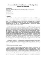
Fig. 1. Schematic representation of LOS biosynthetic locus of strains 3292 (serotype B) and 2951 (serotype A), including constructs for mut-
agenesis. Large open arrows represent ORFs that are highly similar between strains. Filled regions of arrows represent sequence diversity
between strains. Small arrows represent oligonucleotide primers used for amplification. (A) Construction of plasmid for mutation of lgt2:
lgt2
B ⁄ C
and flanking sequence were amplified from strain 3292 using primers UORF2:2040 and DORF2:3120, and disrupted by insertion of
kan
r
into the PstI site. Transformation of strain 2951 resulted in allelic replacement. (B) Construction of plasmid for mutation of lgt1 and dele-
tion of lgt4: lgt1 and flanking sequence were amplified from strain 3292 using primers UORF3:4093 and DORF3:2768, and disrupted by
insertion of kan
r
into the StyI site. Transformation of strain 2951 resulted in recombination within lgt5 and lgt1, resulting in deletion of lgt4
and disruption of lgt1. (C) Construction of plasmid for mutation of lgt5 and deletion of lgt4: lgt5 and flanking sequence were amplified from
strain 3292 using primers DORF4:5047 and UORF4:3684, and disrupted by insertion of kan
r
into the NsiI site. Transformation of strain 2951
resulted in recombination within lgt5 and lgt1, resulting in deletion of lgt4 and disruption of lgt5.
Moraxella catarrhalis LOS biosynthesis I. R. Peak et al.
2028 FEBS Journal 274 (2007) 2024–2037 ª 2007 The Authors Journal compilation ª 2007 FEBS
selective TOCSY experiments. Although the anomeric
signals of each of the b-d-Glc residues were well
resolved, it was necessary to resort to one-dimensional
selective TOCSY experiments to assign the remaining
ring protons of these residues (D, B and G), which are
overlapped in both the
1
H and
13
C dimensions. Com-
plete assignment of the Kdo residue was possible from
examination of TOCSY correlations with the well-
dispersed H3 and H8 methylene protons of Kdo. An
APT experiment was used to obtain the
13
C shift of
the C1 and C2 resonances of Kdo. Examination of the
400 ms NOESY spectrum confirmed each of the gly-
cosidic linkages for the 2951lgt1 ⁄ 4D oligosaccharide
and the terminal location of each of the b-d-Glc resi-
dues D, B, and G. As lgt1 has previously been shown
to encode an a-(1 fi 2)-glucosyltransferase that adds
residue A [4], we concluded that lgt4 encodes the
a-(1 fi 2)- N-acetylglycosyltransferase.
Fig. 2. Structures of wild-type serotype A
oligosaccharide (strain 2951) and 2951lgt2D,
2951lgt1 ⁄ 4D and 2951lgt5 ⁄ 4D mutant oligo-
saccharides. Letters refer to designated
sugar residues.
I. R. Peak et al. Moraxella catarrhalis LOS biosynthesis
FEBS Journal 274 (2007) 2024–2037 ª 2007 The Authors Journal compilation ª 2007 FEBS 2029
2951lgt5 ⁄ 4D
In order to further investigate the function of lgt4,an
lgt4 ⁄ 5 double mutant was also produced. To delete
lgt4 from strain 2951, lgt1 and lgt5 were amplified
from strain 3292, and Kan
r
was inserted into lgt5.As
strain 3292 does not contain lgt4, this construct recom-
bined in lgt1 and lgt5, inactivating lgt5 as a result of
the Kan
r
insertion, and also deleting lgt4 (Fig. 1C).
Mutation of the genes encoding Lgt4 and Lgt5
results in a truncated oligosaccharide, as shown in
Fig. 2. Compared to the wild-type 2951 oligosac-
charide, or the recently published 2951lgt5D oligosac-
charide [6], it was immediately obvious from the
2951lgt5 ⁄ 4D oligosaccharide NMR spectra that the
a-d-GlcNAc residue on the (1 fi 4) branch was missing,
due to the lack of peaks indicative of an N-acetamido
peak at 2.19 p.p.m. (
1
H) and 25.4 ⁄ 177.0 p.p.m. (
13
C).
MS sugar analysis supported this, indicating the pres-
ence of Glc, Gal and Kdo (in the approximate ratio
6 : 1 : 1). As expected, mutation of the lgt5 gene resul-
ted in a truncation of the (1 fi 6) branch, and conse-
quently the b-d-Gal was found in the terminal position
because the a-d-Gal found in the wild-type 2951 oligo-
saccharide was missing. What was most surprising,
however, was that mutation of the lgt4 and lgt5
genes resulted in an oligosaccharide in which the
a-d-GlcNAc residue on the (1 fi 4) branch was
replaced by an a-d-Glc residue.
MALDI-MS analysis of the methylated oligosaccha-
ride gave a molecular ion at m ⁄ z 1773.8 [M + Na]
+
(75% acetonitrile fraction following Sep-pak C18 puri-
fication), consistent with the composition Hex
7
ÆKdo.
GC-MS sugar analysis (TMS derivative) indicated
an oligosaccharide composed of Glc, Gal, and Kdo.
GC-MS linkage analysis of the permethylated sample
Table 3. GC-MS analysis of partially methylated alditol acetates
obtained from 2951lgt2D oligosaccharide (OS) and 2951lgt5 ⁄ 4D OS
from serotype A M. catarrhalis following Sep-pak C18 purification
(75% acetonitrile fractions).
Sample
Elution
time
(min)
Characteristic
fragment ions Assignment
2951lgt2D OS 18.52 118, 129, 145, 205 Terminal Glcp
19.68 129, 130, 161, 190 2-linked Glcp
21.98 118, 333 3,4,6-linked Glcp
22.37 117, 159, 205 Terminal GlcpÆNAc
2951lgt5 ⁄ 4D OS 18.52 118, 129, 145, 205 Terminal Glcp
18.82 118, 129, 145, 205 Terminal Galp
19.68 129, 130, 161, 190 2-linked Glcp
19.88 113, 118, 162, 233 4-linked Glcp
21.98 118, 333 3,4,6-linked Glcp
Table 4.
1
H and
13
C chemical shifts (p.p.m.) for oligosaccharides isolated from the M. catarrhalis 2951lgt2D mutant in D
2
O referenced to 2,2-dimethylsilapentane-S-sulphonic acid (DSS)
(0.0 p.p.m.), at 298 K, on a Bruker Avance spectrometer operating at 600 and 150 MHz, respectively.
Sugar residue
1
H Chemical shift (d, p.p.m.)
13
C Chemical shift (d, p.p.m.)
H1 H2 H3 H4 H5 H6a H6b NH(C ¼ O)CH
3
3
J
1,2
(Hz) C1 C2 C3 C4 C5 C6 NH(C ¼ O)CH
3
(A) a-D-Glcp-(1 fi 5.38 3.35 3.73 3.39 3.97 3.6 99.3 74.7 75.6 72.0 74.7 63.5
(B) fi 2)-b-
D-Glcp-(1 fi 5.16 3.36 3.57 3.43 3.47 7.2 100.1 83.7 77.8 72.2 78.1 63.2
(C) fi 3,4,6)-a-
D-Glcp-(1 fi 5.11 3.89 4.49 3.91 4.566 4.10 4.00 4.2 102.4 76.4 77.7 76.5 72.5 70.4
(D) b-
D-Glcp-(1 fi 5.04 3.34 3.51 3.32 3.54 3.96 3.71 ND
a
104.9 76.4 78.9 72.7 78.9 63.9
(E) a-
D-GlcNAcp-(1 fi 5.05 3.99 3.71 3.55 3.80 2.16 ND
a
101.6 56.4 74.8 72.4 74.8 62.9 25.4 ⁄ 177.0
(G) fi 2)-b-
D-Glcp-(1 fi 4.58 3.46 3.55 3.37 3.41 3.82 3.90 ND
b
105.3 78.0 78.0 72.3 78.1 63.5
H3ax H3eq H4 H5 H6 H7 H8a H8b C1 C2 C3 C4 C5 C6 C7 C8
fi 5)-a-Kdop 2.00 1.88 4.14 4.06 3.84 4.04 3.78 3.61 178.4 98.9 37.1 68.4 78.0 74.1 71.1 65.8
a
The anomeric protons of residues D and E overlap, and so
3
J
1,2
coupling constants were not determined for these residues.
b
The anomeric protons of residues G and H5 of residue C
overlap, and so a a
3
J
1,2
coupling constant was not determined for residue G.
Moraxella catarrhalis LOS biosynthesis I. R. Peak et al.
2030 FEBS Journal 274 (2007) 2024–2037 ª 2007 The Authors Journal compilation ª 2007 FEBS
identified terminal Glc, terminal Gal, 2-linked Glc,
4-linked Glc, and 3,4,6-linked Glc (Table 3).
1
H and
13
C assignments for the 2951lgt5⁄ 4D oligo-
saccharide are given in Table 6. The anomeric confi-
guration for each sugar residue was obtained from
the
3
J
1,2
coupling constants, as shown in Table 6.
Chemical shift assignment for this oligosaccharide
was more challenging than for the other oligosaccha-
rides, due to the poor dispersion of signals in the
3.2–4.1 p.p.m. region of the spectra. Although the
1
H
signals of the anomeric protons of the 2951lgt5⁄ 4D
oligosaccharide were well dispersed, the
13
C signals
were not, as can be seen in the anomeric region of
the
1
H-
13
C-HSQC spectrum shown in Fig. 5. In fact,
the
13
C anomeric signal for residues D, G and H
overlapped, as did those of E and B. For this rea-
son, it was necessary to perform a series of selective
one-dimensional TOCSY experiments, irradiating
each of the anomeric signals in turn to obtain the
1
H chemicals shifts of the corresponding remaining
ring protons. Again, assignment of the Kdo residue
was possible from examination of TOCSY correla-
tions in the 120 ms
1
H-
13
C-HSQC-TOCSY spectrum
from the dispersed H3 and H8 methylene protons of
Kdo, and an APT experiment was used to elucidate
the
13
C shift of the C1 and C2 resonance of Kdo.
For the highly branched, central a-d-Glc residue C,
the locations of the H3 ⁄ C3 and H5 ⁄ C5 correlations
were conspicuous in the
1
H-
13
C-HSQC spectrum.
Examination of the anomeric
13
C line of residue C
in the 120 ms
1
H-
13
C-HSQC-TOCSY spectrum
revealed the location of the remaining ring protons.
CD B G
Fig. 3. Anomeric region of the
1
H-NMR spectrum (600 MHz,
298 K, D
2
O) 2951lgt1 ⁄ 4D mutant OS. Letters refer to designated
sugar residues as shown for 2951lgt1 ⁄ 4D in Fig. 2.
C1-C2
C2-C3
C3-C4
C4-C5
H5-H6R/S
H6R-H6S
C1
Fig. 4.
1
H,
1
H-COSY NMR spectrum (600 MHz, 298 K, D
2
O)
2951lgt1 ⁄ 4D mutant oligosaccharide.
Table 5.
1
H and
13
C chemical shifts (p.p.m.) for oligosaccharides isolated from the M. catarrhalis 2951lgt1 ⁄ 4D mutant in D
2
O referenced to
DSS (0.0 p.p.m.), at 298 K, on a Bruker Avance spectrometer operating at 600 and 150 MHz, respectively.
Sugar residue
1
H Chemical shift (d, p.p.m.)
13
C Chemical shift (d, p.p.m.)
H1 H2 H3 H4 H5 H6a H6b
3
J
1,2
(Hz) C1 C2 C3 C4 C5 C6
(B) b-
D-Glcp-(1 fi 4.68 3.33 3.49 3.38 3.44 3.90 3.71 7.9 103.8 75.8 78.4 72.2 78.4 63.5
(C) fi 3,4,6)-a-
D-Glcp- 5.13 3.80 4.26 3.95 4.41 4.17 4.02 3.9 102.2 75.0 79.2 75.6 72.6 70.0
(D) b-
D-Glcp-(1 fi 4.93 3.36 3.49 3.38 3.43 3.90 3.71 8.0 104.1 76.0 78.6 72.2 78.6 63.5
(G) b-
D-Glcp-(1 fi 4.48 3.30 3.49 3.38 3.43 3.90 3.71 8.0 105.0 75.8 78.5 72.4 78.5 63.5
H3ax H3eq H4 H5 H6 H7 H8a H8b C1 C2 C3 C4 C5 C6 C7 C8
fi 5)-a-Kdop 2.03 1.86 4.13 4.08 3.83 4.03 3.78 3.61 179.3 99.2 37.0 68.7 78.0 74.0 71.3 65.8
I. R. Peak et al. Moraxella catarrhalis LOS biosynthesis
FEBS Journal 274 (2007) 2024–2037 ª 2007 The Authors Journal compilation ª 2007 FEBS 2031
For each sugar residue, the
1
H anomeric chemical
shift gave TOCSY correlations in the 120 ms
1
H-
13
C-
HSQC-TOCSY spectrum with the
13
C positions of
each of the ring protons, including C6. This informa-
tion, coupled with the selective one-dimensional
TOCSY experiments, was sufficient to structurally
assign the 2951lgt5 ⁄ 4D oligosaccharide. The only residue
for which assignments (H5 ⁄ C5 and H6 ⁄ C6) remained
outstanding was the terminal b-d-gal, H. Even from
the selective one-dimensional TOCSY experiment, it
was not possible to ascertain their assignment. All
other assignments for the 2951lgt5 ⁄ 4D oligosaccharide
were in agreement with literature values (where appro-
priate) for the wild-type 2951 oligosaccharide [11–14].
Discussion
Functional analysis of glycosyltransferase
enzymes
In serotype B strains (3292) of M. catarrhalis, Lgt1
catalyzes the addition of the a-d-Glc-(1 fi 2) glyco-
sidic linkage to both the (1 fi 6) and (1 fi 4) branches
of the growing oligosaccharide chain [4]. This serotype
lacks the lgt4 glycosyltransferase gene present in sero-
type A and C strains. The presence of the lgt4 gene
on the lgt locus of serotype A and C strains of
M. catarrhalis has been noted [5], however, its function
has not been determined. Serotype A and C strains
have the lgt4 gene and express GlcNAc on their LOS
structures. Serotype B strains lack this gene, and do
not have GlcNAc as part of their LOS. It was there-
fore of interest to determine whether the lgt4 gene
encoded an N-acetylglucosamine transferase. In order
to ascribe a function to the product of this gene, we
endeavored to produce mutant strains of M. catarr-
halis 2951 lacking the lgt4 gene, in order to ascertain
from the degree of truncation the function of the Lgt4
glycosyltransferase. Unfortunately, all attempts to pro-
duce bacteria expressing lgt4D mutant oligosaccharide
were unsuccessful.
In an alternative approach to studying the function
of the Lgt4 glycosyltransferase, 2951lgt1⁄ 4D double-
mutant bacteria were produced. The mutational strat-
egy for this double mutation took advantage of the
absence of lgt4 in serotype B strains, in that the mutant
alleles were constructed using 3292 (serotype B)-derived
alleles that would delete lgt4 when introduced into a
serotype A strain (Fig. 1B). This strategy was also suc-
cessfully employed to make another double mutation,
2951lgt5 ⁄ 4D (Fig. 1C, and see below). The LOS from
mutant bacteria was harvested, and the truncated oligo-
saccharide examined by NMR and MS analysis. The
Table 6.
1
H and
13
C chemical shifts (p.p.m.) for oligosaccharides isolated from the M. catarrhalis 2951lgt5 ⁄ 4D mutant in D
2
O referenced to
DSS (0 p.p.m.), at 298 K, on a Bruker Avance spectrometer operating at 600 and 150 MHz, respectively. ND, not determined.
Sugar residue
1
H Chemical shift (d, p.p.m.)
13
C Chemical shift (d, p.p.m.)
H1 H2 H3 H4 H5 H6a H6b
3
J
1,2
(Hz) C1 C2 C3 C4 C5 C6
(A) fi 4)-a-
D-Glcp-(1 fi 5.42 3.63 3.87 3.66 4.10 3.76 3.99 4.2 99.7 74.0 75.7 81.0 73.2 62.9
(B) fi 2)-b-
D-Glcp-(1 fi 5.06 3.44 3.57 3.41 3.60 3.70 3.82 7.2 101.3 81.8 77.9 72.5 77.9 63.2
(C) fi 3,4,6)-a-
D-Glcp- 5.11 3.87 4.38 4.01 4.56 4.05 4.16 < 1 102.4 75.5 78.4 76.2 72.8 70.6
(D) b-
D-Glcp-(1 fi 4.90 3.39 3.50 3.38 3.51 3.73 3.94 7.5 105.3 76.1 78.6 72.3 78.6 63.7
(E) a-
D-Glcp-(1 fi 5.27 3.47 3.67 3.44 3.98 3.77 4.4 101.1 74.8 76.2 72.0 74.8 63.1
(G) fi 2)-b-
D-Glcp-(1 fi 4.60 3.46 3.54 3.41 3.50 3.72 3.92 7.7 105.7 79.1 77.4 72.5 79.1 63.6
(H) b-
D-Galp-(1 fi 4.44 3.51 3.64 3.89 ND ND 7.7 105.8 73.9 75.5 71.5 ND ND
H3ax H3eq H4 H5 H6 H7 H8a H8b C1 C2 C3 C4 C5 C6 C7 C8
fi 5)-a-Kdop 2.03 1.89 4.13 4.06 3.82 4.02 3.62 3.78 178.4 98.9 37.1 68.4 78.0 74.2 71.0 65.7
A
E
B
C
D
GH
Fig. 5. Anomeric region of the
1
H,
13
C-HSQC NMR spectrum
(600 MHz, 298 K, D
2
O) 2951lgt5 ⁄ 4D mutant oligosaccharide. Refer
to Fig. 2 for letter designations.
Moraxella catarrhalis LOS biosynthesis I. R. Peak et al.
2032 FEBS Journal 274 (2007) 2024–2037 ª 2007 The Authors Journal compilation ª 2007 FEBS
2951lgt1 ⁄ 4D double-mutant oligosaccharide was com-
posed of a central a-d-Glc residue (1 fi 6)-, (1 fi 4)-
and (1 fi 3)-linked to three b-d-Glc residues, as shown
in Fig. 2. As mentioned previously, the presence of the
glucosyltransferase Lgt1 could account for the addition
of an a-d-Glc (1 fi 2) glycosidic linkage to the (1 fi 6)
branch; however, in serotype A and C strains, there is
an a-d-GlcNAc residue with a (1 fi 2) glycosidic link-
age on the (1 fi 4) chain. To further investigate the
function of Lgt4 and to explore the interrelationship
between the activity of the Lgt1 and Lgt4 glycosyl-
transferase enzymes, a 2951Lgt5 ⁄ 4D serotype A mutant
was produced. Lgt5 is the galactosyltransferase respon-
sible for the addition of a terminal a-d-Galp (1 fi 4) to
the (1 fi 6) branch of serotype A strain 2951 [6]. Dis-
ruption of the genes that encode Lgt4 and Lgt5 would
ensure retention of Lgt1 glycosyltransferase activity,
and potentially produce truncated oligosaccharides
lacking the terminal a-d-Galp (1 fi 4) on the (1 fi 6)
branch and the a-d-GlcNAc (1 fi 2) glycosidic linkage
on the (1 fi 4) branch i.e., an oligosaccharide compri-
sing six sugar units (not including Kdo). Fascinatingly,
NMR and MS examination of the oligosaccharide iso-
lated from the mutant M. catarrhalis 2951lgt5 ⁄ 4D
revealed that an oligosaccharide containing seven
hexose sugar units was produced by these mutant bac-
teria. This is clearly evident from the anomeric region
of the
1
H,
13
C-HSQC spectrum, as shown in Fig. 5, and
the MALDI-MS methylation data. Moreover, this
1
H,
13
C-HSQC spectrum differed from that of the oligo-
saccharide produced by Lgt5D mutant M. catarrhalis,
because the spectrum lacked an N-acetamido methyl
peak at 2 p.p.m. that would have been indicative of a
GlcNAc being retained at the terminal position of the
(1 fi 4) chain. Additionally, the MS sugar analysis
clearly indicated the absence of the GlcNAc residue.
Instead, in the absence of Lgt4 and Lgt5, a glucose resi-
due was added to the terminal position of the (1 fi 4)
chain by Lgt1. This finding demonstrates that, in the
absence of a functional Lgt4, Lgt1 is able to add an
a-d-Glc (1 fi 2) glycosidic linkage to the (1 fi 4) branch.
Serotype B strains lack lgt4, and therefore have a
Glc at this position. We and others [4–6] have
observed the presence of two different alleles of lgt2.
In serotype A strains of M. catarrhalis, Lgt2 (Lgt2
A
)
adds a b-d-Gal (1 fi 4) to the a-d-Glc (1 fi 2) glycosi-
dic linkage added by Lgt1 (see Fig. 2) to the (1 fi 6)
branch. In serotype B strains, however, Lgt2 (Lgt2
B ⁄ C
)
adds a b-d-Gal (1 fi 4) to both the (1 fi 4) and
(1 fi 6) branches. This allele (Lgt2
B ⁄ C
)oflgt2, present
in serotype B and C strains of M. catarrhalis, corre-
lates with the extension to the (1 fi 4) branch regard-
less of whether the acceptor molecule is a terminal
glucose or N-acetylgalactosamine. Our results suggest
that the serotype A allele (Lgt2
A
) is unable to extend
either the terminal glucose or N-acetylgalactosamine
onto the (1 fi 4) branch. This observation implies that
the serotype A Lgt2 has a higher acceptor specificity
than that found in serotype B and C strains.
Furthermore, 2951lgt1D M. catarrhalis bacteria pro-
duced LOS-derived oligosaccharide with the same
degree of truncation as the 2951lgt1 ⁄ 4D double-mutant
bacteria as determined by tricine SDS ⁄ PAGE (data
not shown). A possible explanation for this observa-
tion is that it is necessary for Lgt1 to catalyze the
addition of the a-d-Glc (1 fi 2) glycosidic linkage to
the (1 fi 6) branch before Lgt4 can act by adding the
a-d-GlcNAc (1 fi 2) glycosidic linkage to the (1 fi 4)
branch. Such a requirement for addition of a hexose
to one chain before an enzyme can add to another
has been reported for biosynthesis of LOS ⁄ lipopoly-
saccharide in other organisms; for example, Lic2C
catalyzes the addition of glucose to the core HepII of
H. influenzae, but requires that LgtF has added glucose
to HepI first [16].
Accordingly, we propose that Lgt4 is a N-acetyl-
glucosylamine transferase responsible for the addition
of an a-d-GlcNAc (1 fi 2) glycosidic linkage to the
(1 fi 4) branch. In the presence of an active Lgt4 (lgt4
is present only in serotype A and C strains), GlcNAc
is preferentially added to the (1 fi 4) chain. This
implies that active Lgt4 has a much higher affin-
ity ⁄ specificity for the b-(1 fi 4)-linked Glc than does
Lgt1. Competitive addition of hexoses has previously
been reported in LOS ⁄ lipopolysaccharide biosynthesis;
for example, in pathogenic Neisseria strains, lgtA and
lgtC encode an N-acetylglucosylamine transferase and
a galactosyltransferase, respectively. In the presence of
active LgtA and LgtC, GlcNAc is added by LgtA.
Only in the absence of LgtA can LgtC add Gal [17].
From our experimental data and those of others, the
role of Lgt1–5 in serotypes A and B has been con-
firmed (Fig. 6): the presence of Lgt3 is required for the
addition of three glucosyl residues to the core
GlcÆKdo, as mutation of lgt3 results in production of
Kdo with only a single Glc residue [4]. Lgt1 then adds
a-(1 fi 2)-Glc to the (1 fi 4) chain and also to the
(1 fi 6) chain in serotype B strains. Lgt4 (present only
in serotypes A and C), then adds an a-(1
fi 2)-
GlcNAc to the (1 fi 4) chain instead of Glc. Lgt2
A
(in
serotype A strains) and Lgt2
B ⁄ C
(prototypic isoform,
in serotype B and C strains) then adds b-(1 fi 4)-Gal
to the (1 fi 6) chain. In serotype B strains, the proto-
typic Lgt2 also adds b-(1 fi 4)-Gal to the (1 fi 4)
chain. Finally, Lgt5 adds an a-(1 fi 4)-Gal to terminal
Gal residues, when present. This model is consistent
I. R. Peak et al. Moraxella catarrhalis LOS biosynthesis
FEBS Journal 274 (2007) 2024–2037 ª 2007 The Authors Journal compilation ª 2007 FEBS 2033
with genetic and experimental data. According to this
model, the presence ⁄ absence of lgt4 and the lgt2 allele
determine the LOS structure produced by a strain (as
proposed by Edwards et al. [5]). The elucidation of the
biosynthetic pathway of LOS structures of known
serotypes will allow further examination of the role of
LOS in pathogenesis. In addition, it will provide
answers to some fundamental questions regarding glyc-
osyltransferase structure and acceptor specificity.
Experimental procedures
Bacteria and growth
Bacterial strains are described in Table 1. Escherichia coli
was grown at 37 °C in LB agar (Oxoid Ltd, Basingstoke,
Hampshire, UK) with shaking at 200 r.p.m., or on LB agar,
with ampicillin (Sigma-Aldrich, St Louis, MO, USA)
at 100 lgÆmL
)1
, kanamycin (Gibco, Invitrogen, Mt Waverly,
Victoria, Australia) at 50 lgÆmL
)1
, and X-Gal (Fermentas
International, Burlington, Ontario, Canada) at 40 lgÆmL
)1
,as
appropriate. M. catarrhalis was grown at 37 °C on Brain
Heart Infusion (BHI) (Oxoid) agar, with kanamycin added at
15 lgÆmL
)1
where appropriate. For structural analysis,
M. catarrhalis was grown at 37 °C in BHI broth supplemented
with 0.5% yeast extract (Oxoid), with shaking at 220 r.p.m.
Transformation of M. catarrhalis
After overnight growth, bacteria were suspended in 500 lL
of sterile NaCl ⁄ P
i
. A bacterial suspension (20 lL) was
placed on a BHI plate and left to dry. Approximately 1 lg
of restriction-digested plasmid dissolved in 30 lLof
NaCl ⁄ P
i
was added to the bacteria and incubated at 37 °C
for 3–4 h, before transfer of bacteria to BHI ⁄ kanamycin
plates and overnight incubation at 37 °C. Sterile NaCl ⁄ P
i
(30 lL) was added instead of the DNA as control in each
transformation.
Standard recombinant DNA techniques
Standard DNA manipulation techniques were employed,
essentially as previously described [18,19]. Chromosomal
DNA was purified as previously described, and plasmid
DNA was purified from E. coli by the alkali-lysis method,
or using purification kits obtained from Qiagen (Qiagen,
Hilden, Germany). Enzymes were obtained from New
England Biolabs (Beverly, MA, USA), and used as recom-
mended. DNA was purified from agarose gel using kits
obtained from Eppendorf (Eppendorf AG, Hamburg
Germany). Primers were synthesized by Geneworks
(Geneworks, Hindmarsh, SA, Australia).
Mutation
Mutant alleles were constructed as described below before
transformation into M. catarrhalis as described above.
Mutation of lgt2
lgt2 and flanking regions were amplified from strain 3292
using primers UORF2:2040 and DORF2:3120, and cloned
into pGEMTeasy (Promega Corporation, Madison, WI,
USA) to generate p2:3292. lgt2 was excised from p2:3292
with SpeI and EcoRI, and cloned into SpeI ⁄ Eco RI-cut
pBluescriptSKII (Stratagene, La Jolla, CA, USA) (removes
Fig. 6. Proposed model for the serotype-
specific expression of the LOS-derived
oligosaccharide in M. catarrhalis. Data for
serotype A [6, this study] and for serotype
B [4,6].
Moraxella catarrhalis LOS biosynthesis I. R. Peak et al.
2034 FEBS Journal 274 (2007) 2024–2037 ª 2007 The Authors Journal compilation ª 2007 FEBS
PstI site from pBluescript) to generate pRA1. Kan
r
, excised
from pUC4Kan (GE Healthcare, Little Chalfont, UK) with
PstI, was cloned into the unique PstI site of lgt2. The
resulting plasmid, p2Kan, has Kan
r
in the opposite orienta-
tion relative to lgt2.
Mutation of lgt1 (3292) and double mutation of
lgt1 and lgt5 (2951)
lgt1 (3292) was amplified using primers DORF3:2768 and
UORF3:4093, and cloned into pGEM-Teasy to generate
pIF1.4. pIF1.4 was digested with ApaI and NcoI to remove
the StyI site of the vector, blunted with Klenow (New Eng-
land Biolabs), and religated, to generate pIF1.4
AN
. Kan
r
,
excised from pUC4Kan with HincII, was ligated into the
unique StyI site (blunted with Klenow) of pIF1.4
AN
. The
resulting plasmid, pK8, has Kan
r
in the same orientation
relative to lgt1.
Double mutation of lgt4 and lgt5 (2951)
The region encompassing lgt1 and lgt5 was amplified from
strain 3292 using primers (UROF4:3684 and DORF4:5047),
and the resulting amplimer was cloned into pGEMTeasy to
generate p3-5(3292). This plasmid was linearized by partial
digestion with NsiI (one site in pGEMTeasy, one site
located in lgt5), and ligated with Kan
r
, which had been
excised from pUC4Kan with PstI. Recombinants were
screened by restriction mapping for plasmids in which the
Kan
r
had ligated into the Nsi I site in lgt5. One such recom-
binant plasmid was named p3-5K, and was used to trans-
form M. catarrhalis strain 2951.
Electrophoretic separation and visualization
of LOS by T-SDS ⁄ PAGE and silver stain
Bacteria from overnight growth were resuspended in
NaCl ⁄ P
i
, and treated with proteinase K (ICN Biomedicals,
Aurora, OH, USA) and SDS (Sigma-Aldrich) (0.05% w ⁄ v
final), before electrophoretic separation on precast 10–20%
Tris ⁄ Tricine acrylamide minigels (Bio-Rad Laboratories,
Hercules, CA, USA), using Tris ⁄ tricine buffer [20]. LOS
was visualized using an ammoniacal silver stain method
previously described [21].
Extraction and hydrolysis of LOS
Bacteria harvested from 6–12 L of culture were dried
by successive washes with ethanol (Merck, Darmstadt,
Germany), acetone (Biolab Australia, Clayton, Victoria,
Australia), and petroleum ether (boiling point 40–60 °C)
(Merck). LOS was extracted from the dry cell mass using
the phenol ⁄ chloroform ⁄ petroleum ether extraction method
[22] with modifications by Qureshi et al. [23]. The water-
soluble oligosaccharide component was isolated from the
LOS by acid hydrolysis as outlined by Phillips et al. [24].
All solvents were distilled prior to use or were of HPLC
grade.
Purification of oligosaccharides
The oligosaccharides were then dissolved in milli-Q water
(Millipore Corporation, Bedford, MA, USA) and passed
through conditioned Alltech Maxi-Clean C18 (Alltech
Associates Inc., Deerfield, IL, USA) 300 mg cartridges to
remove lipophilic components. The eluant was then centri-
fuge-filtered with Micro-spin centrifuge filters (Alltech
Associates Inc.) (0.45 lm positive charged Nylon-66) at
2000 g. The filtrate was lyophilized before size exclusion
chromatography using a Bio-Rad Bio-Gel P2 extra-fine
column 15 (500 mm) and eluted with 0.05 m pyridinium
acetate buffer (pH 5.4) at a flow rate of 16 mLÆh
)1
. Frac-
tions (1 mL) were assessed for carbohydrate content by
charring on TLC plates. Fractions containing carbohydrate
were lyophilized, and then analyzed by NMR spectroscopy
and MS.
Structural analysis by NMR
Purified oligosaccharides were dissolved in D
2
O (99.998%;
Cambridge Isotope Laboratories Inc., Andover, MA, USA)
and cycled through three steps of lyophilization ⁄ dissolution
to remove exchangeable protons.
1
H-NMR and
13
C-NMR
experiments were performed at 600 MHz and 150 MHz,
respectively, at 298 K or 278 K, in D
2
O using a Bruker,
(Karlsruhe, Germany) Avance spectrometer. Chemical
shifts are reported in p.p.m. referenced to DSS. Spectral
assignment was aided by the recording of
1
H one-dimen-
sional gradient COSY, TOCSY (60 and 120 ms mixing
time),
13
C-APT,
1
H-
13
C-HSQC and edited
1
H-
13
C-HSQC
(CH and CH
2
correlations opposite sign),
1
H-
13
C-HSQC-
TOCSY and edited
1
H-
13
C-HSQC-TOCSY (60 and 120 ms
mixing time) (one bond C–H correlations opposite sign),
and
1
H-
13
C-HSQC-NOESY and NOESY (400 ms) spectra.
In addition, for the 2951lgt5 ⁄ 4D mutant oligosaccharide,
one-dimensional selective TOCSY experiments were used to
assist with the assignment process. All spectra were
acquired using unmodified pulse sequences from the Bruker
pulse sequence library.
Structural analysis by MS
ESI-MS
Lyophilized oligosaccharide samples were resuspended in
acetonitrile ⁄ water, 70 : 30 v ⁄ v, to a concentration of
1mgÆmL
)1
, and injected directly into a Bruker Esquire
3000 ion-trap mass spectrometer in negative ion mode at a
flow rate of 550 lLÆh
)1
.
I. R. Peak et al. Moraxella catarrhalis LOS biosynthesis
FEBS Journal 274 (2007) 2024–2037 ª 2007 The Authors Journal compilation ª 2007 FEBS 2035
Sugar analysis ⁄ TMS derivatives
Samples were hydrolyzed in 1 m methanolic hydrogen chlor-
ide at 80 °C for 16 h, and the reagent was removed under a
stream of nitrogen. Hexosamines were re-N-acetylated in
500 lL of methanol ⁄ pyridine ⁄ acetic anhydride (500 : 1 : 5,
v ⁄ v) for 15 min at room temperature, and then dried under
a nitrogen atmosphere. TMS derivatization was performed
in 100 lL of Tri-Sil ‘Z’ (Pierce, Chicago, IL, USA) at room
temperature for 30 min, after which the reagent was
removed under nitrogen. Derivatized monosaccharides were
resuspended in 1 mL of hexane, and centrifuged at 1800 g
for 10 min with an Eppendorf (Hamburg, Germany) 5810R
centrifuge, and the supernatant was transferred and dried
under nitrogen for analysis by GC-MS. Samples were ana-
lyzed by GC-MS using temperature program A.
MALDI-MS permethylation analysis
Permethylation using NaOH ⁄ CH
3
I (Sigma-Aldrich) was
performed as previously described [25]. After derivatization,
the reaction products were purified on a Sep-pak C18
(Waters Corporation, Milford, MA, USA) as previously
described [25]. MALDI-MS was performed on the 75%
acetonitrile fraction using a Perceptive Biosystems Voyager
DE STR mass spectrometer (Foster City, CA, USA) in the
reflectron mode with delayed extraction. Permethylated
samples were dissolved in 1% trifluoroacetic acid (Sigma-
Aldrich) in methanol, and 1 lL aliquots were premixed
with 1 lL of matrix (2,5-dihydrobenzoic acid) before load-
ing onto a metal plate.
MS linkage analysis (partially methylated
alditol acetates)
Purified (75% acetonitrile Sep-pak C18) permethylated
samples were converted to their partially methylated aldi-
tol acetates as previously described [26]. Permethylated
samples were hydrolyzed with 2 m trifluoroaetic acid (aq.)
at 121 °C for 2 h, and this was followed by reduction with
sodium borodeuteride (Sigma-Aldrich) in 2 m NH
3
(aq.)
(10 mgÆmL
)1
). The reaction was terminated with glacial
acetic acid (Sigma-Aldrich), and excess borates were
removed by repeated additions (· 4) of 10% acetic acid in
methanol. Samples were acetylated with Ac
2
O at 100 °C for
1 h. Samples were then analyzed by GC-MS using tempera-
ture program B.
GC-MS analysis
This was carried out using a Perkin Elmer (Waltham, MA,
USA) Clarus 500. Samples were dissolved in hexanes prior
to on-column injection on an RTX-5 (30 m · 0.32 mm
internal diameter; Restek Corp., Bellefonte, PA, USA).
Temperature program A: The temperature of the oven was
held at 65 °C for 1 min before being increased to 140 °Cat
a rate of 25 C°Æmin
)1
, and then to 200 °C at a rate of
5C°Æmin
)1
, and finally to 300 °C at a rate of 10 C°Æmin
)1
.
Temperature program B: The temperature of the oven was
held at 60 ° C for 1 min, before being increased to 300 °C
at a rate of 8 C°Æmin
)1
.
Acknowledgements
This work was supported by grants from the Institute
for Glycomics, Griffith University. A. Dell and H. R.
Morris are supported by funding from the Biotechno-
logy and Biological Sciences Research Council and
Wellcome Trust. A. Dell is a Biotechnology and Bio-
logical Sciences Research Council Professorial Fellow.
References
1 Breton C, Snajdrova L, Jeanneau C, Koca J & Imberty
A (2006) Structures and mechanisms of glycosyltrans-
ferases. Glycobiology 16, 29R–37R.
2 Breton C, Heissigerova H, Jeanneau C, Moravcova J &
Imberty A (2002) Comparative aspects of glycosyltrans-
ferases. Biochem Soc Symp 69, 23–32.
3 Breton C, Mucha J & Jeanneau C (2001) Structural and
functional features of glycosyltransferases. Biochimie 83,
713–718.
4 Edwards KJ, Allen S, Gibson BW & Campagnari AA
(2005) Characterization of a cluster of three glycosyl-
transferase enzymes essential for Moraxella catarrhalis
lipooligosaccharide assembly. J Bacteriol 187, 2939–
2947.
5 Edwards KJ, Schwingel JM, Datta AK & Campagnari
AA (2005) Multiplex PCR assay that identifies the
major lipooligosaccharide serotype expressed by Morax-
ella catarrhalis clinical isolates. J Clin Microbiol 43,
6139–6143.
6 Wilson JC, Collins PM, Klipic Z, Grice ID & Peak IR
(2006) Identification of a novel glycosyltransferase
involved in LOS biosynthesis of Moraxella catarrhalis.
Carbohydr Res 341, 2600–2606.
7 Verduin CM, Hol C, Fleer A, van Dijk H & van
Belkum A (2002) Moraxella catarrhalis: from emerging
to established pathogen. Clin Microbiol Rev 15,
125–144.
8 Karalus R & Campagnari A (2000) Moraxella catarr-
halis: a review of an important human mucosal patho-
gen. Microbes Infection 2, 547–559.
9 Sethi S & Murphy TF (2001) Bacterial infection in
chronic obstructive pulmonary disease in 2000: a state-
of-the-art review. Clin Microbiol Rev 14, 336–363.
10 Vaneechoutte M, Verschraegen G, Claeys G & Abeele
A-MVD (1990) Serological typing of Branhamella
Moraxella catarrhalis LOS biosynthesis I. R. Peak et al.
2036 FEBS Journal 274 (2007) 2024–2037 ª 2007 The Authors Journal compilation ª 2007 FEBS
catarrhalis strains on the basis of lipopolysaccharide
antigens. J Clin Microbiol 28, 182–187.
11 Edebrink P, Jansson P-E, Mahbubur Rahman M,
Widmalm G, Holme T & Rahman M (1995) Structural
studies of the O-antigen oligosaccharides from two
strains of Moraxella catarrhalis serotype C. Carbohydr
Res 266, 237–261.
12 Edebrink P, Jansson P-E, Mahbubur Rahman M,
Widmalm G, Holme T, Rahman M & Weintraub A
(1994) Structural studies of the O-polysaccharide from
the lipopolysaccharide of Moraxella (Branhamella)
catarrhalis serotype A (strain ATCC 25238). Carbohydr
Res 257, 269–284.
13 Edebrink P, Jansson P-E, Widmalm G, Holme T &
Rahman M (1996) The structures of oligosaccharides
isolated from the lipopolysaccharide of Moraxella
catarrhalis serotype B, strain CCUG 3292. Carbohydr
Res 295, 127–146.
14 Masoud H, Perry MB, Brisson J-R, Uhrin D &
Richards JC (1994) Structural elucidation of the back-
bone oligosaccharide from the lipooligosaccharide of
Moraxella catarrhalis serotype A. Can J Chem 72,
1466–1477.
15 Lycknert K, Edebrink P & Widmalm G (2004) A con-
formational carbohydrate scaffold is present in the
short-chain lipopolysaccharides of Moraxella catarrhalis.
Angew Chem Int Ed Engl 43, 2288–2290.
16 Hood DW, Deadman ME, Cox AD, Makepeace K,
Martin A, Richards JC & Moxon ER (2004) Three
genes, lgtF, lic2C and lpsA, have a primary role in
determining the pattern of oligosaccharide extension
from the inner core of Haemophilus influenzae LPS.
Microbiology 150, 2089–2097.
17 Gotschlich EC (1994) Genetic locus for the biosynthesis
of the variable portion of Neisseria gonorrhoeae lipoo-
ligosaccharide. J Exp Med 180, 2181–2190.
18 Ausubel FM, Brent R, Kingston RE, Moore DD,
Seidman JG, Smith JA & Struhl K (1987) Current
Protocols in Molecular Biology. Greene Publishing
Associates Inc., John Wiley & Sons Inc., Hoboken, NJ.
19 Sambrook J, Fritsch EF & Maniatis T (1989) Mole-
cular Cloning; a Laboratory Manual. Cold Spring
Harbor Laboratory Press, Cold Spring Harbor, NY.
20 Lesse AJ, Campagnari AA, Bittner WE & Apicella MA
(1990) Increased resolution of lipopolysaccharides and
lipooligosaccharides utilizing tricine-sodium dodecyl
sulfate-polyacrylamide gel electrophoresis. J Immunol
Methods 126, 109–117.
21 Apicella MA, Griffiss JM & Schneider H (1994) Isolation
and characterization of lipopolysaccharides, lipooligo-
saccharides, and lipid A. Methods Enzymol 235, 242–252.
22 Galanos C, Luderitz O & Westphal O (1969) A new
method for the extraction of R lipopolysaccharides. Eur
J Biochem 9, 245–249.
23 Qureshi N, Takayama K & Ribi E (1982) Purification
and structural determination of nontoxic lipid A
obtained from the lipopolysaccharide of Salmonella
typhimurium
. J Biol Chem 257, 11808–11815.
24 Phillips NJ, Apicella MA, Griffiss JM & Gibson BW
(1992) Structural characterization of the cell surface
lipooligosaccharides from a non-typable strain of Hae-
mophilus influenzae. Biochem 31, 4515–4526.
25 Dell A, Reason AJ, Khoo KH, Panico M, McDowell
RA & Morris HR (1994) Mass spectrometry of carbo-
hydrate-containing biopolymers. Methods Enzymol 230,
108–132.
26 Albersheim P, Nevins DJ, English P & Karr A (1967) A
method for the analysis of sugars in plant cell-wall poly-
saccharides by gas-liquid chromatography. Carbohydr
Res 5, 340–345.
27 Zaleski A, Scheffler NK, Densen P, Lee FKN,
Campagnari AA, Gibson BW & Apicella MA (2000)
Lipooligosaccharide P
k
(Gala1–4Galb1–4Glc) epitope of
Moraxella catarrhalis is a factor in resistance to bacteri-
cidal activity mediated by normal human serum. Infect
Immun 68, 5261–5268.
I. R. Peak et al. Moraxella catarrhalis LOS biosynthesis
FEBS Journal 274 (2007) 2024–2037 ª 2007 The Authors Journal compilation ª 2007 FEBS 2037