Báo cáo khoa học: Regulation of the muscle-specific AMP-activated protein kinase a2b2c3 complexes by AMP and implications of the mutations in the c3-subunit for the AMP dependence of the enzyme docx
Bạn đang xem bản rút gọn của tài liệu. Xem và tải ngay bản đầy đủ của tài liệu tại đây (731.12 KB, 10 trang )
Regulation of the muscle-specific AMP-activated protein
kinase a2b2c3 complexes by AMP and implications of the
mutations in the c3-subunit for the AMP dependence of
the enzyme
Kerstin Lindgren*, Mattias Ormestad*, Ma
˚
rten Persson, Sofia Martinsson, L. Thomas Svensson
and Margit Mahlapuu
Discovery Research, Biovitrum AB, Go
¨
teborg, Sweden
The AMP-activated protein kinase (AMPK) is an evo-
lutionarily conserved enzyme that is important for
metabolic sensing both within individual cells and at a
whole body level [1–3]. AMPK activation has been
shown to increase glucose uptake and fatty acid oxida-
tion in skeletal muscle, suppress glucose output from
the liver, and diminish adiposity [4–7], and as a result
AMPK is considered a promising novel drug target
for the treatment of diabetes and related metabolic
disorders.
AMPK is a heterotrimeric complex consisting of a
catalytic a-subunit and regulatory b- and c-subunits,
Keywords
allosteric regulation by AMP; AMP-activated
protein kinase; c3 isoform
Correspondence
M. Mahlapuu, Discovery Research,
Biovitrum AB, Biotech Center, Arvid
Wallgrens Backe 20, SE-413 46 Go
¨
teborg,
Sweden
Fax: +46 31 749 1101
Tel: +46 31 749 1126
E-mail:
*These authors contributed equally to this
study
(Received 9 February 2007, revised 20
March 2007, accepted 4 April 2007)
doi:10.1111/j.1742-4658.2007.05821.x
The AMP-activated protein kinase is an evolutionarily conserved hetero-
trimer that is important for metabolic sensing in all eukaryotes. The muscle-
specific isoform of the regulatory c -subunit of the kinase, AMP-activated
protein kinase c3, has a key role in glucose and fat metabolism in skeletal
muscle, as suggested by metabolic characterization of humans, pigs and
mice harboring substitutions in the AMP-binding Bateman domains of c3.
We demonstrate that AMP-activated protein kinase a2b2c3 trimers are
allosterically activated approximately three-fold by AMP with a half-
maximal stimulation (A
0.5
) at 1.9 ± 0.5 or 2.6 ± 0.3 lm, as measured for
complexes expressed in Escherichia coli or mammalian cells, respectively.
We show that mutations in the N-terminal Bateman domain of c3 (R225Q,
H306R and R307G) increased the A
0.5
values for AMP, whereas the fold
activation of the enzyme by 200 lm AMP remained unchanged in compari-
son to the wild-type complex. The mutations in the C-terminal Bateman
domain of c3 (H453R and R454G), on the other hand, substantially
reduced the fold stimulation of the complex by 200 lm AMP, and resulted
in AMP dependence curves similar to those of the double mutant,
R225Q ⁄ R454G. A V224I mutation in c3, known to result in a reduced gly-
cogen content in pigs, did not affect the fold activation or the A
0.5
values
for AMP. Importantly, we did not detect any increase in phosphorylation
of Thr172 of a2 by the upstream kinases in the presence of increasing con-
centrations of AMP. Taken together, the data show that different muta-
tions in c3 exert different effects on the allosteric regulation of the a2b2c3
complex by AMP, whereas we find no evidence for their role in regulating
the level of phosphorylation of a2 by upstream kinases.
Abbreviations
AICAR, 5-aminoimidazole-4-carboxamide-1-b-
D-ribonucleoside; AMPK, AMP-activated protein kinase; AMPKK, AMP-activated protein kinase
kinase; CAMKKb,Ca
2+
⁄ calmodulin-dependent protein kinase b; CBS, cystathionine-b-synthase; TAK1, transforming growth factor b-activating
kinase 1; ZMP, 5-aminoimidazole-4-carboxamide riboside monophosphate.
FEBS Journal 274 (2007) 2887–2896 ª 2007 Biovitrum AB. Journal compilation ª 2007 FEBS 2887
all of which are required for its activity [8,9]. In the
mammalian genome, two isoforms of the a-subunit (a1
and a2) and the b-subunit (b1 and b2), and three iso-
forms of the c-subunit (c1, c2 and c3), are present,
with 12 possible heterotrimeric combinations. The cat-
alytic a-subunit contains a conventional serine ⁄ threo-
nine protein kinase domain [10], and phosphorylation
of Thr172 within the activation loop of the a-subunit
is essential for the activity of the enzyme. Several
upstream kinases have been shown to phosphorylate
AMPK at Thr172, including tumor suppressor LKB1
[11–13], Ca
2+
⁄ calmodulin-dependent protein kinase b
(CAMKKb) [14,15] and transforming growth fac-
tor b-activating kinase (TAK1) [16]. The three AMPK
c-subunit isoforms differ in their N-terminal sequences
but share the four tandem repeats of a structural
module known as the cystathionine-b-synthase (CBS)
domain, located in the C-terminal region [17]. First
recognized by Bateman [18], CBS motifs contain about
60 residues and are found in a range of diverse pro-
teins. The basic functional unit is believed to contain
two CBS motifs, which associate closely together [19],
binding ligands containing adenosine [20], and the
term ‘Bateman domain’ has been suggested to refer to
the structure formed by two tandem repeats [21]. In
mammalian AMPK, the CBS domains have been
shown to bind the allosteric activator of the kinase,
AMP, whereas the two pairs of CBS domains both
bind one molecule of AMP [20]. In terms of the struc-
ture of AMPK, the b-subunit has been reported to act
as a scaffold for binding of the a-subunit and c-sub-
unit [22].
Previously, we have shown that AMPK c3 is selec-
tively expressed in glycolytic (white, fast-twitch type II)
skeletal muscle [23], and is thus the only AMPK sub-
unit isoform exhibiting strictly restricted tissue-specific
expression. Furthermore, in both human and rodent
skeletal muscle, c3 primarily forms complexes with the
a2 and b2 isoforms [23,24]. Two naturally occurring
missense mutations have been identified in the first
CBS domain of the pig c3 gene, resulting in increased
(R225Q) or decreased (V224I) skeletal muscle glycogen
content [25–27]. R225Q carriers are also characterized
by a higher oxidative capacity in white skeletal muscle
fibers [28,29]. Transgenic mice with skeletal muscle-
specific expression of the R225Q substituted form of
mouse c3 replicate the phenotype observed in pigs,
with elevated glycogen levels and increased fat oxida-
tion in muscle tissue [30,31]. Recently, R225W and
R307C substitutions in the human c3 gene have been
reported to result in increased muscle glycogen con-
tent, indicating that the function of AMPK c3 is con-
served across the mammalian species [32]. The R225Q
mutation in c3 is equivalent to the R302Q substitution
in c2, the dominant isoform of the c-subunit in the
heart. The R302Q mutation, together with several
other substitutions identified in CBS domains of the
c2 gene, cause Wolff–Parkinson–White syndrome in
humans, which is characterized by electrophysiologic
abnormalities and hypertrophy of the heart, and also
by an increase in cardiac glycogen content [33–36].
Recent studies using transgenic mice overexpressing c2
harboring these mutations showed a disease phenotype
that closely mimicked the human condition [37–40].
Direct binding, modeling and mutagenesis studies on
AMPK c1 and c2 suggest that the mutations in CBS
domains directly interfere with AMP binding, as the
mutations are predicted to lie around the mouth of the
AMP-binding cleft [20,41]. However, the mechanism
by which the mutations lead to the observed pheno-
types remains controversial.
In this study, we have utilized AMPK a2b2c3 hetero-
trimers expressed in Escherichia coli to characterize the
enzyme kinetic parameters and regulation mechanisms
for these physiologically relevant complexes. We have
also evaluated the impact of CBS domain mutations on
a-subunit phosphorylation and AMP dependence of
a2b2c3, to improve our understanding of the functional
consequences of the substitutions.
Results
In this work, we used AMPK a2b2c3 heterotrimers
expressed in E. coli to characterize the regulation of
these complexes by AMP as well as to evaluate the
effect of substitutions within the AMPK c3 gene on
the activity of the enzyme. Neumann et al. [42] des-
cribed gaining milligram amounts of the AMPK
a1b1c1inE. coli using a tricistronic vector with T7
RNA polymerase controlling transcription of a single
a1b1c1 messenger, with each subunit carrying its indi-
vidual ribosome-binding site. In our hands, the expres-
sion of AMPK a2b2c3 in bacteria by a similar
approach gave a lower yield of the trimer, due to solu-
bility problems. However, AMPK a2b2c3 complex
could be purified from the soluble protein fraction by
using a polyhistidine tag fused to a2, and the forma-
tion of a stable trimer was demonstrated by SDS ⁄
PAGE and western blot analysis using subunit-specific
antibodies (Fig. 1A,B). As E. coli is not capable of
phosphorylating Thr172 of the a-subunit, the bacteri-
ally expressed trimers were initially inactive, but could
be activated through phosphorylation by the upstream
kinase LKB1 (Fig. 1B). After incubation with LKB1,
the enzymatic activity of the bacterially expressed
AMPK a2b2c3 trimers was similar to that of the
Regulation of AMPK a2b2c3 complexes by AMP K. Lindgren et al.
2888 FEBS Journal 274 (2007) 2887–2896 ª 2007 Biovitrum AB. Journal compilation ª 2007 FEBS
a2b2c3 complexes purified from transiently transfected
mammalian cells, with respect to both the total activity
and the AMP dependence (Fig. 1C). We measured the
activity of the recombinant AMPK a2b2c3 complexes
over a range of AMP concentrations, and calculated
the concentration of AMP giving half-maximal stimu-
lation (A
0.5
). The bacterially expressed phosphorylated
AMPK a2b2c3 trimers had an A
0.5
value of 1.9 ±
0.5 lm (Table 1), which is very similar to the A
0.5
for
a2b2c3 complexes expressed in COS7 cells
(2.6 ± 0.3 lm).
In order to study the effect of the mutations within
AMPK c3 on the activity of the enzyme, we intro-
duced several missense mutations into the c3 gene
(Fig. 2), and investigated the effect of these substitu-
tions on the allosteric activation of the kinase by
AMP. Our main focus was to study the effects of
mutations in positions V224, R225 and R307 in the
first Bateman domain, given that substitutions in these
positions occur in vivo in the pig and ⁄ or human
AMPK c3 genes [25,26,32]. Also, the physiologic con-
sequence of the R225Q mutation in the AMPK c3
A
C
D
B
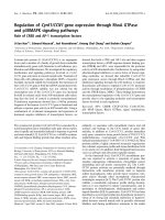
Fig. 1. Characterization of the AMPK a2b2c3 complexes expressed
in E. coli. (A) SDS ⁄ PAGE of bacterially expressed AMPK a2b2c3
complexes after purification by nickel–ion and gel filtration chroma-
tography, stained with silver stain. (B) Representative western blot
of AMPK a2b2c3 complexes expressed in E. coli and COS7 cells
using subunit-specific antibodies raised to either a2, b2, c3or
phosphorylated Thr172 in the a-subunit (apThr172). Bacterially
expressed complexes were phosphorylated by incubation with an
upstream kinase, LKB1. (C) The kinase activity of the AMPK
a2b2c3 trimers expressed as pmoles of phosphate transferred to
the SAMS peptide in the absence (open bars) or presence (black
bars) of AMP (160 l
M). Data are presented as mean ± SEM of
n ¼ 4. In the activity assay, the equivalent amounts of hetero-
trimers expressed in bacteria or mammalian cells were used, as
estimated by western blot (B). (D) Western blot analysis of AMPK
a2b2c3 complexes expressed in E. coli using antibodies to a2or
apThr172. The trimers carry either a wild-type (WT) or R225Q ⁄
R454G version of c3. AMPK complexes were phosphorylated by
LKB1 or CAMKKb in the absence or presence of AMP (0, 50, 200
or 500 l
M AMP).
Table 1. Effect of the mutations in the AMPK c3 gene on the AMP
dependence of the enzyme. Fold stimulation reflects the activation
of the corresponding AMPK complexes by 200 l
M AMP relative to
the basal activity in the absence of added AMP (calculated from
the data presented in Fig. 3B). Significant difference in the AMP-sti-
mulated versus basal activity for each complex was determined by
two-sided Student’s t-test. A
0.5
(concentration of AMP giving half-
maximal stimulation) was calculated from the curves presented in
Fig. 3C (curve fitting by
KALEIDAGRAPH 4.03 with the Michaelis–Men-
ten equation). Values are means ± SEM. NS, not significant; ND,
not determined.
Mutations in c3 Fold activation by AMP P-value A
0.5
(lM)
Wild-type 2.8 ± 0.1 < 0.01 1.9 ± 0.5
V224I 2.6 ± 0.1 < 0.001 2.3 ± 0.7
R225Q 3.5 ± 0.3 < 0.005 131 ± 68
H306R 3.8 ± 0.2 < 0.05 29 ± 6
R307G 3.0 ± 0.2 < 0.001 70 ± 20
H453R 1.6 ± 0.1 < 0.05 ND
R454G 1.5 ± 0.1 < 0.05 ND
R225Q ⁄ R454G 1.3 ± 0.1 NS ND
K. Lindgren et al. Regulation of AMPK a2b2c3 complexes by AMP
FEBS Journal 274 (2007) 2887–2896 ª 2007 Biovitrum AB. Journal compilation ª 2007 FEBS 2889
gene has been extensively studied using transgenic
mouse models [30,31]. Additionally, we generated
H306R, H453R, R454G and R225Q ⁄ R454G substitu-
tions in c3, which occur at the positions corresponding
to R225Q and R307G, in the CBS2 and CBS4
domains (Fig. 2). All complexes were expressed in
E. coli and purified from the soluble fraction by
nickel–ion chromatography in quantities similar
to those of the trimers containing a wild-type c3
(Fig. 3A), suggesting that there was no difference in
the assembly of the different mutants into a hetero-
trimeric complex. Western blot analysis showed that,
in the conditions tested, the phosphorylation level of
Thr172 of a2 by the upstream kinase LKB1 did not
differ for any of the mutants (Fig. 3A). The V224I,
R225Q, H306R and R307G substitutions in AMPK c3
did not affect the fold activation of the enzyme as
compared to the wild-type trimers, when measured in
the presence of 200 lm AMP (Fig. 3B, Table 1). How-
ever, the H453R and R454G substitutions substantially
reduced the AMP dependence of the enzyme down to
approximately the same level as that of the double
mutant R225Q ⁄ R454G (Fig. 3B, Table 1). We calcula-
ted the A
0.5
values for the mutant complexes, which
showed robust activation by AMP at a concentration
of 200 lm (V224I, R225Q, H306R and R307G;
Fig. 3C) using the hyperbolic curve model (Hill coeffi-
cient ¼ 1). All the substitutions tested, except V224I,
increased the A
0.5
value for AMP as compared to the
wild-type trimers (Fig. 3C, Table 1; R
2
‡ 0.97).
Pharmacologic activation of AMPK can be achie-
ved using 5-aminoimidazole-4-carboxamide-1-b-d-ribo-
nucleoside (AICAR). Once taken up by cells, AICAR is
phosphorylated to 5-aminoimidazole-4-carboxamide
riboside monophosphate (ZMP), which mimics the
effects of AMP on AMPK. In our hands, 160 lm ZMP
caused a two-fold activation of the bacterially expressed
wild-type AMPK a2b2c3 complexes, with half-maximal
stimulation (A
0.5
)at61±19lm ZMP. ZMP failed to
significantly increase the activity of the AMPK a2b2c3
complexes with an R225Q mutation in c3, when meas-
ured at the concentration of 160 lm (Fig. 3C).
The presence of AMP (0–500 lm) during the
phosphorylation of the bacterially expressed AMPK
a2b2c3 trimers with LKB1 or CAMKKb did not
increase the phosphorylation level of Thr172 of a2
as shown by western blot analysis (Fig. 1D). Simi-
larly, the phosphorylation of AMPK trimers con-
taining R225Q ⁄ R454G mutations in c3 by LKB1 or
CAMKKb did not differ at varying AMP concentra-
tions (Fig. 1D). In line with this, we did not detect any
increase in the activity of the AMPK a2b2c3 com-
plexes in the presence of AMP in the phosphorylation
reaction by LKB1, as measured through their ability
to incorporate phosphate into the SAMS substrate, as
compared to the complexes phosphorylated in the
absence of AMP (data not shown).
Discussion
We have previously shown that AMPK c3 is the pre-
dominant c isoform expressed in glycolytic skeletal
muscle, where it primarily forms heterotrimers with
the a2 and b2 isoforms [23]. The present study des-
cribes bacterial expression of AMPK a2b2c3 trimers,
and provides the first characterization of the enzyme
kinetic parameters and regulation mechanisms for
these physiologically relevant complexes.
Fig. 2. An alignment of the amino acids in
the four CBS domains in human AMPK c1,
c2 and c3. Sequences were aligned using
CLUSTALX (1.83). A versatile coloring scheme
was incorporated to highlight conserved fea-
tures in the alignment, using a default color
code of
CLUSTALX. Above the alignment is
the conservation score generated by the
program. The enlargement shows details of
the mutations, with red borders indicating
the mutations that have been described in
vivo in pigs (V224I and R225Q in c3) or
humans (mutations in c2, R225W and
R307C in c3), and red letters indicating the
mutations analyzed in the present study.
Regulation of AMPK a2b2c3 complexes by AMP K. Lindgren et al.
2890 FEBS Journal 274 (2007) 2887–2896 ª 2007 Biovitrum AB. Journal compilation ª 2007 FEBS
The particular subunit isoforms present in the
AMPK complex determine the concentration of AMP
causing a half-maximal increase in the total activity as
well as the fold stimulation by AMP [42–46]. Our
experiments show that AMPK a2b2c3 trimers are acti-
vated by AMP with a half-maximal stimulation (A
0.5
)
at 1.9 ± 0.5 or 2.6 ± 0.3 lm, as measured for com-
plexes expressed in E. coli or mammalian cells, respect-
ively (Fig. 3C, Table 1). Typically, 200 lm AMP
caused an activation of 2–4-fold of the AMPK a2b2c3
trimers expressed in bacteria or COS7 cells, relative to
the activity in the absence of AMP. Previously, allos-
teric regulation of c3-containing AMPK complexes by
AMP has been questioned, on the basis of activity
measurements on rat brain protein immunoprecipitates
using antibodies to c3 [17]. One plausible reason for
these discrepancies may be a poor specificity of the
antibodies applied, in combination with the fact that
protein lysate from brain was used, as we and others
have not been able to detect any c3 mRNA or protein
in this tissue [23,25]. In addition, it is possible that the
antibody binding itself interferes with the AMP-bind-
ing properties of c3. Additionally, the low A
0.5
value
characterizing the activation of a2b2c3 trimer by AMP
makes the system highly sensitive to the possible pres-
ence of any traces of AMP, either endogenous or as a
result of the presence of AMP-generating proteins as
contaminants [47].
Several upstream kinases (AMPKKs) have been
reported to activate mammalian AMPK through phos-
phorylation of Thr172 in the a-subunit, i.e. LKB1
[11–13], CAMKKb [15,48] and TAK1 [16]. LKB1 is
believed to be the major upstream kinase for AMPK
in skeletal muscle, as knocking out LKB1 almost com-
pletely prevents both AICAR- and contraction-induced
a2-AMPK signaling [49]. Initially, AMP was thought
to increase phosphorylation of AMPK by AMPKK
both by direct activation of the upstream kinase and
by making the AMPK a better substrate for AMPKK,
through binding to the c-subunits [50]. However, sev-
eral recent studies have challenged this notion [15,48],
and the role of AMP in phosphorylation of AMPK
has remained controversial. While this manuscript was
A
B
C
Fig. 3. Effect of the substitutions in the AMPK c3 gene on the
activity of the enzyme. (A) AMPK a2b2c3 complexes with either
wild-type c3 (WT) or mutant c3 harboring the indicated substitu-
tions were expressed in E. coli, and equal amounts of the trimers
were phosphorylated in vitro by LKB1. Representative western
blots probed with antibodies to a2, b2, c3orapThr172 are shown.
(B) Activity of the AMPK a2b2c3 complexes was measured by a
radioactive filter paper assay using SAMS substrate in the absence
(open bars) or presence (black bars) of AMP (200 l
M). The data pre-
sented are the mean ± SEM from three to four independent phos-
phorylation experiments (n ¼ 2 in the activity assay), expressed
relative to the activity of the wild-type AMPK complexes measured
in the absence of AMP (the activity of the wild-type complex with-
out AMP is set to 1). (C) The activity of the wild-type and mutated
AMPK a2b2c3 complexes was measured over a range of AMP
(from 0 to 160 l
M) or ZMP (from 0 to 160 lM) concentrations. The
data are expressed relative to the basal activity in the absence of
added AMP for each complex (n ¼ 3 in the activity assay; basal
activity is set to 0). The graph is plotted in
KALEIDAGRAPH 4.03 (Syn-
ergy Software), using curve fitting against the one-site Michaelis–
Menten equation; V ¼ V
max
· [S] ⁄ (A
0.5
+ [S]), where V
max
is the
maximal activation, A
0.5
¼ [AMP] at 50% AMPK activation, and
V ¼ AMPK activity. Given the very low level or lack of activation by
AMP, we were unable to measure A
0.5
values for H453R, R454G
and R225Q ⁄ R454G. All the AMPK complexes were expressed in
E. coli except for the wild-type trimers denoted by asterisks, which
were expressed in COS7 cells. In (B) and (C), equivalent amounts
of AMPK were used, as estimated by western blot analysis (A).
K. Lindgren et al. Regulation of AMPK a2b2c3 complexes by AMP
FEBS Journal 274 (2007) 2887–2896 ª 2007 Biovitrum AB. Journal compilation ª 2007 FEBS 2891
in preparation, two studies reported that AMP failed
to promote phosphorylation of c1-containing AMPK
trimers, when recombinant upstream kinase and
AMPK preparations were used [44,47]. In line with
these studies, we did not detect any increase in phos-
phorylation of a2ina2b2c3 complexes by LKB1 or
CAMKKb in the presence of increasing concentrations
of AMP (Fig. 1D).
Despite the considerable effort invested in this area,
the nature of the mutations in the CBS domains of the
AMPK c3 gene has remained controversial. A previous
report provided evidence that the activity of AMPK
was reduced in skeletal muscle of c3 R225Q mutant
pigs [25]. In resting muscle from c3 R225Q mutant
mice, AMPK activity was reported to be unaltered [30]
or reduced [31]. However, in vivo activity measurements
are complicated by the potential inhibitory effects of
glycogen overload, which characterizes skeletal muscle
of mice and pigs carrying the c3 R225Q substitution,
on AMPK activation. Therefore, the effect of the
mutation has been addressed in mammalian cells. In
COS7 cells transfected with plasmids encoding a2b2c3
R225Q or V224I mutants, both substitutions resulted
in diminished AMP dependence of AMPK, as com-
pared to the wild-type trimers. In addition, AMPK
total activity and phosphorylation of a2 were shown to
be markedly elevated in cells expressing a2b2c3
R225Q, as compared to the wild-type or V224I-con-
taining trimers [30]. However, in our laboratory we
have failed to detect any increase in the basal activity
for R225Q complexes assayed from material purified
from transiently transfected COS7 cells (data not
shown). It has to be noted that the activity of the
AMPK is highly sensitive to the lysis protocol used
(hypoxia, glucose deprivation and mechanical stress
during the cell harvest activate AMPK), which poten-
tially complicates the comparisons of the results from
different research groups and may partly explain the
discrepancies observed. In an effort to mimic the
R225Q mutation in c3, the equivalent position has
been mutated in AMPK c1 (R70Q) as well as in c2
(R302Q) [41,44,45,51]. In the present study, we evalu-
ated the impact of CBS domain mutations (locations of
the substitutions are shown in Fig. 2) on the AMP
dependence of the AMPK a2b2c3 using the enzyme
expressed in E. coli, activated in vitro by an LKB1 pre-
paration. Importantly, we did not detect any difference
in phosphorylation of Thr172 of a2 by LKB1 when we
compared the different mutants to the wild-type trimers
(Fig. 3A), although we have to acknowledge that the
semiquantitative nature of the western blot technique
makes the exact measurement difficult. It is noteworthy
that for the mutations in the N-terminal Bateman
domain formed by CBS1–2 (V224I, R225Q, H306R
and R307G), the fold activation of the complex by
200 lm AMP was similar to that of wild-type com-
plexes (Fig. 3B, Table 1). Mutations in the C-terminal
Bateman domain formed by CBS3–4 (H453R and
R454G), on the other hand, significantly reduced AMP
stimulation and resulted in AMP dependence curves
highly similar to that of the double mutant R225Q ⁄
R454G (Fig. 3B, Table 1). The R225Q, H306R and
R307G mutations substantially increased the A
0.5
value
for AMP as compared to the wild-type trimers
(Fig. 3C, Table 1). The V224I mutation, on the other
hand, resulted in AMP dependence curves that were
very similar to those of wild-type complexes (Fig. 3C,
Table 1). According to the current model of the allos-
teric control of mammalian AMPK, the N-terminal
and C-terminal pairs of CBS domains both bind one
molecule of AMP [20,41]. However, the different effects
on AMP activation observed in this study of substitu-
tions in corresponding positions of the two Bateman
domains (H306R and R307G in CBS2 are equivalent
to H453R and R454G in CBS4, respectively, Fig. 2)
indicate that the two suggested AMP-binding sites in
c3 are not functionally equivalent. Notably, the AMP
dependence curves of the AMPK a2b2c3 complexes
appear hyperbolic rather than sigmoid (Fig. 3C). While
this manuscript was in preparation, a crystal struc-
ture of the Schizosaccharomyces pombe AMPK was
reported, demonstrating that one AMP molecule
bound to a single site formed by the CBS domains of
the c-subunit [52]. Nevertheless, the absence of regula-
tion of Saccharomyces cerevisiae AMPK activity by
AMP [53,54] raises the possibility that nucleotide bind-
ing in Sc. pombe AMPK may differ from that in the
human enzyme. The present study does not explain the
mechanisms by which the mutations in CBS domains
of AMPK c3 interfere with activation by AMP.
However, our data would be consistent with a single
AMP-binding site being present in c3.
The A
0.5
value for ZMP was increased for the
R225Q AMPK complexes as compared to the wild-
type heterotrimers, which was expected, as ZMP is
thought to bind to the same site as AMP and activate
AMPK in a similar manner. This result may explain
the impairment of AICAR-stimulated muscle glucose
uptake described in R225Q transgenic mice, as com-
pared to the wild-type littermates [30].
The present study does not answer the question of
how the reduced AMP dependence of the R225Q vari-
ants of c3 would lead to the increased glycogen content
described in the skeletal muscle of the carriers of this
mutation [25,30]. Also, we did not detect any difference
in the regulation of AMPK complexes containing a
Regulation of AMPK a2b2c3 complexes by AMP K. Lindgren et al.
2892 FEBS Journal 274 (2007) 2887–2896 ª 2007 Biovitrum AB. Journal compilation ª 2007 FEBS
V224I form of c3 by AMP, whereas this mutation is
known to lead to reduced skeletal muscle glycogen
stores [26]. Clearly, an important challenge is to deter-
mine the effect of the mutations in the CBS domains
on AMPK activity in the presence of physiologic AMP
and ATP concentrations, which is technically difficult.
The in vitro AMPK activity assay is commonly per-
formed at 200 lm ATP, utilizing radiolabeled ATP,
which causes technical problems when the concentra-
tion is increased to close to physiologic levels [55,56].
The possibility of altered functionality of the AMPK
a2b2c3 trimers expressed in E. coli, as compared to the
in vivo complexes, also has to be acknowledged. Native
AMPK is known to be post-translationally modified at
multiple sites, other than Thr172, in the a-subunit
[13,46,57], and the possibility that the absence of these
modifications has an impact on the activity of the
mutant complexes cannot be excluded. Another possi-
bility would be that interactions other than with
AMP ⁄ ATP, occurring in eukaryotic cells only, have an
impact on the activity of the enzyme. Further studies
in relevant cell systems as well as in animal models are
required to investigate these issues.
Experimental procedures
Expression and purification of recombinant
AMPK a2b2c3inE. coli
The tricistronic plasmids containing the three AMPK sub-
units a2 (mouse, accession number NP_835279) with N-ter-
minal polyhistidine tag, b2 (human, accession number
O43741) and c3 (human, accession number Q9UGI9) in
pET vector (Novagen, Madison, WI), were transformed
into competent host cells (E. coli BL21 DE3 pLysS; Nov-
agen). Single colonies were used to inoculate 5 mL of LB
medium containing 100 lgÆ mL
)1
carbenicillin, 50 lgÆmL
)1
chloramphenicol and 0.5% glucose. Following incubation
in a shaker incubator (250 r.p.m.) overnight at 37 °C, the
starter cultures were used to inoculate 0.5 L of the above
medium. Protein expression was induced with 1 mm isopro-
pyl thio-b-d-galactoside (final concentration) at an A
600
of
0.5–0.7, and cultures were grown for an additional 3 h at
37 °C. Cells were harvested (4000 r.p.m. for 15 min at
4 °C; Allegra 6R), and the cell pellet was resuspended in
the lysis buffer containing 50 mm Hepes (pH 7.5), 50 mm
NaF, 5 mm sodium pyrophosphate, 1 mm EDTA, 1 mm di-
thiothreitol, 10% glycerol, 1% Triton X-100, lysozyme
(48 lgÆmL
)1
), and complete protease inhibitor cocktail
(Roche Diagnostics GmbH, Mannheim, Germany), and
placed on ice for 1 h before being sonicated (Dr Hielscher
GmbH, Teltow, Germany) for 3 min. Insoluble material
was removed by centrifugation [20 400 g for 20 min at 4 °C
using an Avanti J-20 XP centrifuge (Beckman Coulter),
rotor type JA25.50]. The supernatant was collected and
diluted in buffer A containing 20 mm Tris ⁄ HCl (pH 8.0),
150 mm NaCl, 15 mm imidazole, and 10% glycerol, and
loaded onto a 5 mL His-Trap column (GE Healthcare,
Uppsala, Sweden). Bound protein was eluted using a gradi-
ent with buffer B (identical to buffer A except for contain-
ing 500 mm imidazole), and stored at ) 20 °C until use. In
some cases, the AMPK complex was further purified by gel
filtration chromatography with a Superdex 200 pg HiLoad
16 ⁄ 60 column (GE Healthcare) connected to A
¨
kta FPLC
(GE Healthcare).
Expression and purification of recombinant
AMPK a2b2c3 in COS7 cells
COS7 cells were cotransfected with cDNAs encoding mouse
AMPK a2, b2 (human) and c3 [human, all cloned into
pcDNA3.1(+); Invitrogen, Paisley, UK] using Lipofectin
reagent, according to the manufacturer’s instructions (Invi-
trogen). Immediately prior to the lysis, the cells were sub-
mitted to hyperosmotic stress by incubating them for 30 min
with sorbitol (final concentration 0.6 m) in the culture med-
ium. Cells were harvested 48 h post-transfection by rapid
lysis: the medium was removed, and ice-cold lysis buffer (see
above) was added. Insoluble material was removed by
centrifugation [15 700 g for 20 min at 4 °C using a 5415R
centrifuge (Eppendorf), rotor type, 24 places fixed angle],
and AMPK was partially purified from the soluble fraction
using a DEAE–sepharose ion-exchange step (GE Health-
care). The major part of the endogenous AMPK was deple-
ted by immunoprecipitation of the relevant fractions with
antibody to a1 prebound to protein A–sepharose as described
previously [23].
Site-directed mutagenesis
The mutations V224I, R225Q, H306R, R307G, H453R,
R454G and R225Q ⁄ R454G were introduced into AMPK
c3 using the Quick Change II site-directed mutagenesis kit
(Stratagene, La Jolla, CA). Mutagenesis primers were
designed using Stratagene’s Tm calculator (primer seq-
uences are available on request). Point mutations generated
in vitro were confirmed by DNA sequencing.
Western blotting
Quantitative analysis of the expression of different AMPK
subunits was performed as described previously [23].
Phosphorylation of bacterial trimer and AMPK
activity assay
To activate the bacterial AMPK, recombinant trimers were
incubated with LKB1 or CAMKKb in the presence of
K. Lindgren et al. Regulation of AMPK a2b2c3 complexes by AMP
FEBS Journal 274 (2007) 2887–2896 ª 2007 Biovitrum AB. Journal compilation ª 2007 FEBS 2893
0.2 mm ATP, 5 mm MgCl
2
and 1 mm dithiothreitol for 1 h
at 32 °C in a thermostated shaker. Rat liver LKB1 was
purified as described previously [12] up to the Q-sepharose
step, except that the poly(ethylene glycol) precipitation of
the liver lysate was omitted. Bacterially expressed
CAMKKb was a gift from D. Carling [14,15]. To determine
the relevant amount of upstream kinase versus AMPK to
be used in the phosphorylation reaction, the AMPK trimers
were incubated with increasing concentrations of AMPKK,
and the amount of upstream kinase corresponding to the
highest possible level of Thr172 phosphorylation was used
in the reaction. AMPK activity was determined by in vitro
phosphorylation of the SAMS (HMRSAMSGLHLVKRR)
synthetic peptide substrate as previously described [58].
Briefly, kinase reactions were initiated by adding 4 lLof
the phosphorylated AMPK to 21 lL of assay buffer con-
taining 50 mm Hepes (pH 7.5), 80 mm NaCl, 8% glycerol,
5mm MgCl
2
, 0.8 mm EDTA, 0.8 mm dithiothreitol,
0.2 mm ATP, 0.2 mm SAMS peptide, and [
32
P]ATP (final
concentration 0.016 lCiÆl L
)1
) in the presence or absence of
varying concentrations of AMP, as described in the figure
legends. Reactions were incubated on a vibrating platform
for 3 h at 37 °C. The reactions were terminated by adding
trichloroacetic acid to a final concentration of 13%, fol-
lowed by centrifugation. Immediately thereafter, aliquots
were spotted onto Whatman P81 paper, and washed with
1% phosphoric acid, and the incorporation of
32
P into
peptide substrate was measured in a liquid scintillation
counter.
Acknowledgements
We gratefully acknowledge Professor David Carling
(Medical Research Council Clinical Sciences Center,
Imperial College, Hammersmith Hospital, London,
UK) for the recombinant CAMKKb preparation used
in this study and for discussions concerning the work
presented. This work was supported by an Integrated
Project from the European Commission (LSHG-CT-
2005-518181).
References
1 Carling D (2005) AMP-activated protein kinase: balan-
cing the scales. Biochimie 87, 87–91.
2 Hardie DG, Hawley SA & Scott JW (2006) AMP-acti-
vated protein kinase-development of the energy sensor
concept. J Physiol 574, 7–15.
3 Kahn BB, Alquier T, Carling D & Hardie DG (2005)
AMP-activated protein kinase: ancient energy gauge
provides clues to modern understanding of metabolism.
Cell Metab 1, 15–25.
4 Hayashi T, Hirshman MF, Kurth EJ, Winder WW &
Goodyear LJ (1998) Evidence for 5¢-AMP-activated
protein kinase mediation of the effect of muscle contrac-
tion on glucose transport. Diabetes 47, 1369–1373.
5 Merrill GF, Kurth EJ, Hardie DG & Winder WW
(1997) AICA riboside increases AMP-activated protein
kinase, fatty acid oxidation, and glucose uptake in rat
muscle. Am J Physiol 273, E1107–E1112.
6 Minokoshi Y, Kim YB, Peroni OD, Fryer LG, Muller
C, Carling D & Kahn BB (2002) Leptin stimulates
fatty-acid oxidation by activating AMP-activated pro-
tein kinase. Nature 415, 339–343.
7 Ruderman NB, Saha AK & Kraegen EW (2003) Mini-
review: malonyl CoA, AMP-activated protein kinase,
and adiposity. Endocrinology 144, 5166–5171.
8 Kemp BE, Stapleton D, Campbell DJ, Chen ZP,
Murthy S, Walter M, Gupta A, Adams JJ, Katsis F,
van Denderen B et al. (2003) AMP-activated protein
kinase, super metabolic regulator. Biochem Soc Trans
31, 162–168.
9 Stapleton D, Woollatt E, Mitchelhill KI, Nicholl JK,
Fernandez CS, Michell BJ, Witters LA, Power DA,
Sutherland GR & Kemp BE (1997) AMP-activated pro-
tein kinase isoenzyme family: subunit structure and
chromosomal location. FEBS Lett 409, 452–456.
10 Kemp BE, Mitchelhill KI, Stapleton D, Michell BJ,
Chen ZP & Witters LA (1999) Dealing with energy
demand: the AMP-activated protein kinase. Trends Bio-
chem Sci 24, 22–25.
11 Shaw RJ, Kosmatka M, Bardeesy N, Hurley RL,
Witters LA, Depinho RA & Cantley LC (2004) The
tumor suppressor LKB1 kinase directly activates AMP-
activated kinase and regulates apoptosis in response to
energy stress. Proc Natl Acad Sci USA 101, 3329–3335.
12 Hawley SA, Boudeau J, Reid JL, Mustard KJ, Udd L,
Makela TP, Alessi DR & Hardie DG (2003) Complexes
between the LKB1 tumor suppressor, STRAD alpha ⁄
beta and MO25 alpha ⁄ beta are upstream kinases in the
AMP-activated protein kinase cascade. J Biol 2,
doi:10.1186/1475-4924-2-28.
13 Woods A, Johnstone SR, Dickerson K, Leiper FC,
Fryer LG, Neumann D, Schlattner U, Wallimann T,
Carlson M & Carling D (2003) LKB1 is the upstream
kinase in the AMP-activated protein kinase cascade.
Curr Biol 13, 2004–2008.
14 Hurley RL, Anderson KA, Franzone JM, Kemp BE,
Means AR & Witters LA (2005) The Ca2+ ⁄ calmodu-
lin-dependent protein kinase kinases are AMP-activated
protein kinase kinases. J Biol Chem 280, 29060–29066.
15 Woods A, Dickerson K, Heath R, Hong SP, Momcilo-
vic M, Johnstone SR, Carlson M & Carling D (2005)
Ca2+ ⁄ calmodulin-dependent protein kinase kinase-beta
acts upstream of AMP-activated protein kinase in mam-
malian cells. Cell Metab 2, 21–33.
16 Momcilovic M, Hong SP & Carlson M (2006) Mamma-
lian TAK1 activates Snf1 protein kinase in yeast and
Regulation of AMPK a2b2c3 complexes by AMP K. Lindgren et al.
2894 FEBS Journal 274 (2007) 2887–2896 ª 2007 Biovitrum AB. Journal compilation ª 2007 FEBS
phosphorylates AMP-activated protein kinase in vitro.
J Biol Chem 281, 25336–25343.
17 Cheung PC, Salt IP, Davies SP, Hardie DG & Carling
D (2000) Characterization of AMP-activated protein
kinase gamma-subunit isoforms and their role in AMP
binding. Biochem J 346 (Part 3), 659–669.
18 Bateman A (1997) The structure of a domain common
to archaebacteria and the homocystinuria disease pro-
tein. Trends Biochem Sci 22, 12–13.
19 Zhang R, Evans G, Rotella FJ, Westbrook EM, Beno
D, Huberman E, Joachimiak A & Collart FR (1999)
Characteristics and crystal structure of bacterial inosine-
5¢-monophosphate dehydrogenase. Biochemistry 38,
4691–4700.
20 Scott JW, Hawley SA, Green KA, Anis M, Stewart G,
Scullion GA, Norman DG & Hardie DG (2004) CBS
domains form energy-sensing modules whose binding of
adenosine ligands is disrupted by disease mutations.
J Clin Invest 113, 274–284.
21 Kemp BE (2004) Bateman domains and adenosine
derivatives form a binding contract. J Clin Invest 113,
182–184.
22 Woods A, Cheung PC, Smith FC, Davison MD, Scott
J, Beri RK & Carling D (1996) Characterization of
AMP-activated protein kinase beta and gamma subu-
nits. Assembly of the heterotrimeric complex in vitro.
J Biol Chem 271, 10282–10290.
23 Mahlapuu M, Johansson C, Lindgren K, Hjalm G,
Barnes BR, Krook A, Zierath JR, Andersson L &
Marklund S (2004) Expression profiling of the gamma-
subunit isoforms of AMP-activated protein kinase
suggests a major role for gamma3 in white skeletal
muscle. Am J Physiol Endocrinol Metab 286, E194–E200.
24 Birk JB & Wojtaszewski JF (2006) Predominant
{alpha}2 ⁄ {beta}2 ⁄ {gamma}3 AMPK activation during
exercise in human skeletal muscle. J Physiol 577,
1021–1032.
25 Milan D, Jeon JT, Looft C, Amarger V, Robic A,
Thelander M, Rogel-Gaillard C, Paul S, Iannuccelli N,
Rask L et al. (2000) A mutation in PRKAG3 associated
with excess glycogen content in pig skeletal muscle. Sci-
ence 288, 1248–1251.
26 Ciobanu D, Bastiaansen J, Malek M, Helm J, Woollard
J, Plastow G & Rothschild M (2001) Evidence for new
alleles in the protein kinase adenosine monophosphate-
activated gamma(3)-subunit gene associated with low
glycogen content in pig skeletal muscle and improved
meat quality. Genetics 159, 1151–1162.
27 Estrade M, Vignon X, Rock E & Monin G (1993)
Glycogen hyperaccumulation in white muscle fibres
of RN- carrier pigs. A biochemical and ultrastructural
study. Comp Biochem Physiol B 104, 321–326.
28 Lebret B, Le Roy P, Monin G, Lefaucheur L, Caritez
JC, Talmant A, Elsen JM & Sellier P (1999) Influence
of the three RN genotypes on chemical composition,
enzyme activities, and myofiber characteristics of por-
cine skeletal muscle. J Anim Sci 77, 1482–1489.
29 Estrade M, Ayoub S, Talmant A & Monin G (1994)
Enzyme activities of glycogen metabolism and mito-
chondrial characteristics in muscles of RN- carrier pigs
(Sus scrofa domesticus). Comp Biochem Physiol Biochem
Mol Biol 108, 295–301.
30 Barnes BR, Marklund S, Steiler TL, Walter M, Hjalm
G, Amarger V, Mahlapuu M, Leng Y, Johansson C,
Galuska D et al. (2004) The 5¢-AMP-activated protein
kinase gamma3 isoform has a key role in carbohydrate
and lipid metabolism in glycolytic skeletal muscle. J Biol
Chem
279, 38441–38447.
31 Yu H, Hirshman MF, Fujii N, Pomerleau JM, Peter LE
& Goodyear LJ (2006) Muscle-specific overexpression of
wild-type and R225Q mutant AMP-activated protein
kinase gamma3-subunit differentially regulates glycogen
accumulation. Am J Physiol Endocrinol Metab 291,
E557–E565.
32 Costford SR, Chaudhry SN, Kavaslar N, Ahitus N,
Pennacchio LA, Dent R, Mcpherson R & Harper M-E
(2006) Novel mutations in the gamma-3 subunit of
AMP-activated protein kinase affect protein function in
humans. In 13th Biochemistry of Exercise International
Conference, Seoul, Korea, Abstract P010.
33 Gollob MH, Seger JJ, Gollob TN, Tapscott T, Gonzales
O, Bachinski L & Roberts R (2001) Novel PRKAG2
mutation responsible for the genetic syndrome of ventri-
cular preexcitation and conduction system disease with
childhood onset and absence of cardiac hypertrophy.
Circulation 104, 3030–3033.
34 Gollob MH, Green MS, Tang AS, Gollob T, Karibe A,
Ali Hassan AS, Ahmad F, Lozado R, Shah G,
Fananapazir L et al. (2001) Identification of a gene
responsible for familial Wolff–Parkinson–White syn-
drome. N Engl J Med 344, 1823–1831.
35 Arad M, Benson DW, Perez-Atayde AR, Mckenna WJ,
Sparks EA, Kanter RJ, Mcgarry K, Seidman JG &
Seidman CE (2002) Constitutively active AMP kinase
mutations cause glycogen storage disease mimicking
hypertrophic cardiomyopathy. J Clin Invest 109,
357–362.
36 Blair E, Redwood C, Ashrafian H, Oliveira M, Brox-
holme J, Kerr B, Salmon A, Ostman-Smith I & Watkins
H (2001) Mutations in the gamma(2) subunit of AMP-
activated protein kinase cause familial hypertrophic
cardiomyopathy: evidence for the central role of energy
compromise in disease pathogenesis. Hum Mol Genet 10,
1215–1220.
37 Arad M, Moskowitz IP, Patel VV, Ahmad F, Perez-
Atayde AR, Sawyer DB, Walter M, Li GH, Burgon
PG, Maguire CT et al. (2003) Transgenic mice over-
expressing mutant PRKAG2 define the cause of
Wolff–Parkinson–White syndrome in glycogen storage
cardiomyopathy. Circulation 107, 2850–2856.
K. Lindgren et al. Regulation of AMPK a2b2c3 complexes by AMP
FEBS Journal 274 (2007) 2887–2896 ª 2007 Biovitrum AB. Journal compilation ª 2007 FEBS 2895
38 Sidhu JS, Rajawat YS, Rami TG, Gollob MH, Wang
Z, Yuan R, Marian AJ, Demayo FJ, Weilbacher D,
Taffet GE et al. (2005) Transgenic mouse model of
ventricular preexcitation and atrioventricular reentrant
tachycardia induced by an AMP-activated protein
kinase loss-of-function mutation responsible for Wolff–
Parkinson–White syndrome. Circulation 111, 21–29.
39 Davies JK, Wells DJ, Liu K, Whitrow HR, Daniel TD,
Grignani R, Lygate CA, Schneider JE, Noel G, Watkins
H et al. (2006) Characterization of the role of gamma2
R531G mutation in AMP-activated protein kinase in
cardiac hypertrophy and Wolff–Parkinson–White syn-
drome. Am J Physiol Heart Circ Physiol 290, H1942–
H1951.
40 Zou L, Shen M, Arad M, He H, Lofgren B, Ingwall JS,
Seidman CE, Seidman JG & Tian R (2005) N488I
mutation of the gamma2-subunit results in bidirectional
changes in AMP-activated protein kinase activity. Circ
Res 97, 323–328.
41 Adams J, Chen ZP, Van Denderen BJ, Morton CJ,
Parker MW, Witters LA, Stapleton D & Kemp BE
(2004) Intrasteric control of AMPK via the gamma1
subunit AMP allosteric regulatory site. Protein Sci 13,
155–165.
42 Neumann D, Woods A, Carling D, Wallimann T &
Schlattner U (2003) Mammalian AMP-activated protein
kinase: functional, heterotrimeric complexes by
co-expression of subunits in Escherichia coli. Protein
Expr Purif 30, 230–237.
43 Scott JW, Norman DG, Hawley SA, Kontogiannis L &
Hardie DG (2002) Protein kinase substrate recognition
studied using the recombinant catalytic domain of
AMP-activated protein kinase and a model substrate.
J Mol Biol 317, 309–323.
44 Sanders MJ, Grondin PO, Hegarty BD, Snowden MA
& Carling D (2006) Investigating the mechanism for
AMP activation of the AMP-activated protein kinase
cascade. Biochem J 403, 139–148.
45 Daniel T & Carling D (2002) Functional analysis of
mutations in the gamma 2 subunit of AMP-activated
protein kinase associated with cardiac hypertrophy and
Wolff–Parkinson–White syndrome. J Biol Chem 277,
51017–51024.
46 Stein SC, Woods A, Jones NA, Davison MD & Carling
D (2000) The regulation of AMP-activated protein
kinase by phosphorylation. Biochem J 345 (Part 3),
437–443.
47 Suter M, Riek U, Tuerk R, Schlattner U, Wallimann T
& Neumann D (2006) Dissecting the role of 5¢-AMP
for allosteric stimulation, activation, and deactivation
of AMP-activated protein kinase. J Biol Chem 281,
32207–32216.
48 Hawley SA, Pan DA, Mustard KJ, Ross L, Bain J,
Edelman AM, Frenguelli BG & Hardie DG (2005)
Calmodulin-dependent protein kinase kinase-beta is an
alternative upstream kinase for AMP-activated protein
kinase. Cell Metab 2, 9–19.
49 Sakamoto K, Mccarthy A, Smith D, Green KA,
Grahame Hardie D, Ashworth A & Alessi DR (2005)
Deficiency of LKB1 in skeletal muscle prevents AMPK
activation and glucose uptake during contraction.
EMBO J 24, 1810–1820.
50 Hawley SA, Selbert MA, Goldstein EG, Edelman AM,
Carling D & Hardie DG (1995) 5¢-AMP activates the
AMP-activated protein kinase cascade, and Ca2+ ⁄
calmodulin activates the calmodulin-dependent protein
kinase I cascade, via three independent mechanisms.
J Biol Chem 270, 27186–27191.
51 Hamilton SR, Stapleton D, O’Donnell JB Jr, Kung JT,
Dalal SR, Kemp BE & Witters LA (2001) An activating
mutation in the gamma1 subunit of the AMP-activated
protein kinase. FEBS Lett 500, 163–168.
52 Townley R & Shapiro L (2007) Crystal structures of the
adenylate sensor from fission yeast AMP-activated pro-
tein kinase. Science 315, 1726–1729.
53 Wilson WA, Hawley SA & Hardie DG (1996) Glucose
repression
⁄ derepression in budding yeast: SNF1 protein
kinase is activated by phosphorylation under derepress-
ing conditions, and this correlates with a high
AMP:ATP ratio. Curr Biol 6, 1426–1434.
54 Woods A, Munday MR, Scott J, Yang X, Carlson M &
Carling D (1994) Yeast SNF1 is functionally related to
mammalian AMP-activated protein kinase and regulates
acetyl-CoA carboxylase in vivo. J Biol Chem 269,
19509–19515.
55 Bergeron R, Russell RR 3rd, Young LH, Ren JM,
Marcucci M, Lee A & Shulman GI (1999) Effect of
AMPK activation on muscle glucose metabolism in
conscious rats. Am J Physiol 276, E938–E944.
56 Sabina RL, Kernstine KH, Boyd RL, Holmes EW &
Swain JL (1982) Metabolism of 5-amino-4-imidazole-
carboxamide riboside in cardiac and skeletal muscle.
Effects on purine nucleotide synthesis. J Biol Chem 257,
10178–10183.
57 Mitchelhill KI, Michell BJ, House CM, Stapleton D,
Dyck J, Gamble J, Ullrich C, Witters LA & Kemp BE
(1997) Posttranslational modifications of the 5¢-AMP-
activated protein kinase beta1 subunit. J Biol Chem 272,
24475–24479.
58 Derave W, Ai H, Ihlemann J, Witters LA, Kristiansen
S, Richter EA & Ploug T (2000) Dissociation of
AMP-activated protein kinase activation and glucose
transport in contracting slow-twitch muscle. Diabetes
49, 1281–1287.
Regulation of AMPK a2b2c3 complexes by AMP K. Lindgren et al.
2896 FEBS Journal 274 (2007) 2887–2896 ª 2007 Biovitrum AB. Journal compilation ª 2007 FEBS