Báo cáo khoa học: Biomechanical properties of native basement membranes docx
Bạn đang xem bản rút gọn của tài liệu. Xem và tải ngay bản đầy đủ của tài liệu tại đây (1.08 MB, 12 trang )
Biomechanical properties of native basement membranes
Joseph Candiello
1
, Manimalha Balasubramani
2
, Emmanuel M. Schreiber
2
, Gregory J. Cole
3
,
Ulrike Mayer
4
, Willi Halfter
5
and Hai Lin
1
1 Department of Bioengineering, University of Pittsburgh, PA, USA
2 Genomics and Proteomics Core Laboratory, University of Pittsburgh, PA, USA
3 Julius L. Chambers Biomedical ⁄ Biotechnology Research Institute, North Carolina Central University, Durham, NC, USA
4 Biomedical Research Centre, School of Biological Sciences, University of East Anglia, Norwich, UK
5 Department of Neurobiology, University of Pittsburgh, PA, USA
Basement membranes (BMs) are sheet-like extracellular
matrix structures at the basal side of every epithelium.
They outline muscle fibers, are present at the basal sur-
face of the vascular endothelial cells, and they connect
the central nervous system with the adjacent meningeal
cell layers [1,2]. BMs are composed of at least ten
secretory proteins that include members of the laminin
family, nidogen-1 and 2, perlecan, agrin, and the colla-
gens IV and XVIII [1,3]. Mutations or deletions of
some of the BM proteins lead to early embryonic
Keywords
atomic force microscopy; basal lamina;
basement membrane; extracellular matrix;
eye development
Correspondence
W. Halfter, Department of Neurobiology,
University of Pittsburgh, Pittsburgh,
PA 15262, USA
Fax: +1 412 648 1441
Tel: +1 412 648 9424
E-mail:
(Received 14 December 2006, revised 14
February 2007, accepted 5 April 2007)
doi:10.1111/j.1742-4658.2007.05823.x
Basement membranes are sheets of extracellular matrix that separate epi-
thelia from connective tissues and outline muscle fibers and the endothelial
lining of blood vessels. A major function of basement membranes is to
establish and maintain stable tissue borders, exemplified by frequent vascu-
lar breaks and a disrupted pial and retinal surface in mice with mutations
or deletions of basement membrane proteins. To directly measure the bio-
mechanical properties of basement membranes, chick and mouse inner limi-
ting membranes were examined by atomic force microscopy. The inner
limiting membrane is located at the retinal-vitreal junction and its weaken-
ing due to basement membrane protein mutations leads to inner limiting
membrane rupture and the invasion of retinal cells into the vitreous. Trans-
mission electron microscopy and western blotting has shown that the inner
limiting membrane has an ultrastructure and a protein composition typical
for most other basement membranes and, thus, provides a suitable model
for determining their biophysical properties. Atomic force microscopy
measurements of native chick basement membranes revealed an increase in
thickness from 137 nm at embryonic day 4 to 402 nm at embryonic day 9,
several times thicker that previously determined by transmission electron
microscopy. The change in basement membrane thickness was accompan-
ied by a large increase in apparent Young’s modulus from 0.95 MPa to
3.30 MPa. The apparent Young’s modulus of the neonatal and adult
mouse retinal basement membranes was in a similar range, with 3.81 MPa
versus 4.07 MPa, respectively. These results revealed that native basement
membranes are much thicker than previously determined. Their high
mechanical strength explains why basement membranes are essential in
stabilizing blood vessels, muscle fibers and the pial border of the central
nervous system.
Abbreviations
AFM, atomic force microscopy; BM, basement membranes; CNS, central nervous system; ILM, inner limiting membrane; NCAM, neural cell
adhesion molecule; TEM, transmission electron microscopy.
FEBS Journal 274 (2007) 2897–2908 ª 2007 The Authors Journal compilation ª 2007 FEBS 2897
death, usually caused by disruptions in the vascular
system or defects in the placenta or the amnion [4–9].
Nonlethal mutations of BM proteins result in early
onset muscular dystrophy, kidney and skin defects
[10–13]. Common phenotypes of mutant mice with
BM defects also include massive ocular and cortical
hemorrhaging and disruptions along the pial surface of
the brain and the vitreo-retinal border of the eyes
[5,14–16]. Breaks in the pial and retinal BMs combined
with neural ectopias are also seen in mice with muta-
tions of BM protein receptors, such as integrin b1
[17,18] and dystroglycan [19]. The vascular breaks and
the frequent retinal and cortical ectopias indicate that
the mechanical resistance that is provided by BMs is
critical in strengthening the endothelial wall of the vas-
culature system and establishing a stable border
between the central nervous system (CNS) and its sur-
rounding connective tissue. Surprisingly, there is very
little information of the biophysical properties of BMs.
The lack of data is probably due to the difficulty of
obtaining BM preparations that are free of adjacent
interstitial connective tissue and the lack of a suitable
measuring technique.
We introduce here the retinal basement membrane,
also referred as inner limiting membrane (ILM), as a
model system to study the biophysical properties of
BMs. Initially, we show that the mechanical strength
of the ILM and the BMs of the ocular vasculature is
essential for normal eye development, and thus pro-
vides a biological context and a justification for the
present study. Subsequently, we show that the ILM
resembles, in terms of ultrastructure and biochemical
composition, a typical BM. Furthermore, we demon-
strate that our ILM isolation procedure results in a
preparation that is free of cellular contaminants and
free of nonbasement membrane proteins. Finally,
atomic force microscopy (AFM) measurements reveal
that native BMs are much thicker than previously ref-
erenced in the available literature and that mature
BMs have a surprisingly high mechanical strength. We
also show that BMs undergo significant morphological
and biochemical changes during development.
Results
Evidence for a role of BMs in vascular stability
and in the maintenance of the vitreo-retinal
border
Histological data from mice with several different
mutations of BM proteins strongly suggest that the
mechanical stability of BMs is important in: (a) esta-
blishing a defined tissue border between the CNS and
its surrounding meningeal layers; (b) stabilizing blood
vessels in the eye and CNS; and (c) preventing muscle
fibers from undergoing terminal damage [5,12–16]. To
emphasize the importance of BM stability for blood
vessels and the integrity of the vitreo-retinal border,
mutant mice with a targeted deletion of the nidogen-
binding site in the laminin c1 chain were investigated.
BMs in these mutant mice lack nidogen [8,14,15],
which leads to random ruptures in many of the BMs
and to the death of the homozygous mutant mice at
late embryogenesis due to kidney agenesis and lung
dysplasia [8,14,15]. Phenotypic analysis showed that all
embryonic day (E)18 mutant mice had massive hemor-
rhages in their eyes (Fig. 1A,B). Ultrastructural studies
of the ocular vasculature revealed herniation of endo-
thelial cells through disruptions in the endothelial BMs
(Fig. 1E) and breaks of entire vessel walls (Fig. 1F).
Ectopic cells along the vitreo-retinal border due to
gaps in the retinal BM (Fig. 1C) was another hallmark
in all eyes of mutant mice. In heterozygous control
mice, the retinal border was smooth and continuous
(Fig. 1D), and endothelial herniation or breaks in the
ocular vasculature were never observed. As described
previously [14], excessive hemorrhages and neuronal
ectopias were also observed in the cortex of mutant
mice. The retinal and cortical ectopias confirmed that
BMs are required in maintaining smooth and stable
tissue borders along the CNS and in preventing corti-
cal and ocular hemorrhages. The frequent retinal ecto-
pias also show that the stability of the retinal BM is
important for retinal histogenesis. The data illustrate
why biophysical measurements of BMs are biologically
relevant and provide a justification for the AFM mea-
surements presented below.
Protein composition of the ILM
Force and thickness measurements of BMs were per-
formed with chick and mouse ILMs. The ILM is
located at the vitreal surface of the retina and separ-
ates the retina from the vitreous body (Fig. 2A). The
ILM is one of three BMs of the eye, which include the
lens capsule, the BM of the pigment epithelium and
the ILM (Fig. 2A). Western blot analysis showed that
the ILM is comprised of extracellular matrix proteins
that are found in other BMs as well, namely laminin-1
(Fig. 2B, lanes 1 and 2), nidogen-1 (Fig. 2B, lane 3),
three BM proteoglycans, agrin (Fig. 2B, lane 4), colla-
gen 18 (Fig. 2B, lane 5) and perlecan (Fig. 2B, lane 6)
and collagen 4 (Fig. 2B, lane 7). Two bands were
observed for nidogen. Peptide mass finger printing
confirmed that both bands (Fig. 2B, lane 3) were nido-
gen-1: the full-length protein and a truncated version.
Biomechanical properties of basement membranes J. Candiello et al.
2898 FEBS Journal 274 (2007) 2897–2908 ª 2007 The Authors Journal compilation ª 2007 FEBS
Laminin was detected by the very prominent 200 kDa
b1 and c1 chains, and its identity as laminin-1 was
established by detecting the laminin a1 chain using a
a1 chain-specific antibody (Fig. 2B, lane 2). The west-
ern blotting data were confirmed by capillary liquid
chromatography (LC) electrospray isonisation MS ⁄ MS
that identified the peptide SDFMSVLSNIEYILIK
(AA 1938–42 of the bovine laminin a1; GI 57164373)
of the laminin a1 chain in trypsin-digests of ILM pre-
parations. The three proteoglycans, agrin, collagen 18
and perlecan appeared in the blots as smears of 600,
400 and 800 kDa (Fig. 2B, lanes 4, 5 and 6, respect-
ively). The smears resulted from the microheterogeneity
of the glycosaminoglycan carbohydrate side chains.
Collagen 4 appeared in multiple bands that represented
the monomeric and several cross-linked oligomeric ver-
sions. To determine potential contamination of the
ILM preparations, the blots were assayed for neural
cell adhesion molecule (NCAM) and collagen 9. As
shown in Fig. 2, NCAM and collagen 9 are very abun-
dant in retinal membranes and the vitreous (lanes 9
and 11, respectively). Both proteins were barely detect-
able in the ILM preparations (Fig. 2, lanes 8 and 10).
Histological characterization of the ILM
flat-mount preprations
ILM flat-mount preparations from chick embryos,
neonatal and adult mice were obtained by mechanic-
ally splitting the retina [20]. The ILM flat-mounts were
firmly attached to glass slides (Fig. 3C,G). They were
immunoreactive for laminin-1 (Fig. 3C,G), nidogen-1,
perlecan, agrin and collagen 18, as expected from the
strong labeling of the ILM for these proteins in tissue
sections of chick and mouse retina (Fig. 3A,F). Ultra-
structural studies at low (Fig. 3D) and high power
(Fig. 3E) showed that the ILM preparations were 50–
70 nm thin sheets of extracellular matrix (Fig. 3D,E),
free of cellular contaminanats (Fig. 3D). The isolated
ILMs were similar in their ultrastructural morphology
as ILMs in situ (Fig. 3B). The low power transmission
electron microscopy (TEM) images also showed that
the ILM preparations formed extensive loops at their
margins (Fig. 3D), which were also detected in AFM
thickness measurements.
AFM imaging of ILM
Flat-mount preparations of chick ILM were imaged
with AFM in intermittent-contact mode under NaCl ⁄ Pi.
At low magnification, the retinal side of ILM was relat-
ively smooth and did not exhibit detailed structural fea-
tures. Figure 4A,C shows representative AFM images
A
B
D
C
E
F
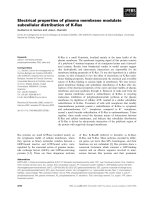
Fig. 1. Ocular hemorrhaging and retinal ectopias in mice with a
mutation in the laminin c1 chain (A). The red eyes of E18 homozy-
gous mutant mice indicate massive intraocular bleedings, but none
in the normally colored eyes from the heterozygous control (C)
embryos. A cross-section though a mutant eye (B) shows blood
vessel rupture in the anterior chamber of the eye as indicated by
the aggregated red blood cells next to the cornea (arrow). R, retina;
L, lens. A high power view (C) of the mutant retina (B, box) shows
ectopias of retinal cells (E) through ruptures in the retinal BM. The
smooth vitreal surface of a retina (R) from a control mouse is
shown in (D) for comparison. BV, hyaloid blood vessel. TEM micro-
graphs (E,F) of the hyaloid vasculature in the vitreous body (VB) and
around the lens (L) of the mutant mice showed herniation (H) of
endothelial cells through breaks (E, arrows) in the vascular BM
(BM) or ruptures of the entire vessel wall (F, arrow). LC, lens cap-
sule. Bar, (B) 150 lm; (C,D) 100 lm; (E) 100 nm; (F) 10 lm.
J. Candiello et al. Biomechanical properties of basement membranes
FEBS Journal 274 (2007) 2897–2908 ª 2007 The Authors Journal compilation ª 2007 FEBS 2899
of E4 and E9 ILM imaged at 40 lm · 40 lm. When the
ILM was imaged at 2 lm · 2 lm with higher imaging
force (low amplitude set point in intermittent-contact
mode), the images revealed a fibrillar structure
(Fig. 4B,D). These fibrillar networks are likely formed
by collagen 4 fibrils. The individual fibrils in the E9
ILM appeared to be thicker (Fig. 4D) than the E4 ILM
(Fig. 4B), suggesting modification of the BM during
development.
AFM measurement of ILM thickness
The thickness of ILM was obtained from AFM images
of the sharp edges of ILM where the underlining glass
substrate was exposed. To obtain sharp ILM ⁄ glass
edges, scratches in the ILM were made using plastic
pipette tips (Fig. 3C). Figure 5A shows a height mode
AFM image of a scratched edge of an E9 chick ILM
with the exposed glass surface on the left, and the
ILM on the right. Figure 5B shows the ILM height
profile as indicated by the dashed line in Fig. 5A along
with the height profile of an E4 ILM. At the scratch-
ing edge, the ILM height was elevated due to scratch-
induced folding and accumulation of the scratched
ILM debris. The thickness of the ILM was meas-
ured from the flat segment of the height profile
(dashed lines) with the glass surface serving as the zero
reference.
The ILM thickness measurements were made for
ILM preparations from four chick retinae at develop-
ment stages of E4, E9 and E15. For each ILM, ten
height measurements were made from different cross-
sections. The measured thicknesses from each ILM
sample are summarized in Table 1. The thickness
(mean ± SD) of the E4 chick ILM was 137 ± 22 nm
(n ¼ 40) and the thickness of E9 chick ILM was
402 ± 59 nm (n ¼ 40; Fig. 5C). There was a three-fold
change of ILM thickness (P<0.01) during develop-
ment between embryonic day 4 and embryonic day 9.
The ILM thickness for E15 retina was 406 ± 99 (n ¼
40); thus, ILM thickness did not change significantly
between E9 and E15 (Fig. 5C).
Elasticity of ILM
The Young’s modulus of chick embryonic ILM was
measured by AFM tip indention using pyramidal tips
(see Experimental procedures). The apparent Young’s
moduli of ILM samples from four different eyes were
each measured for E4, E9 and E15 chick embryos. On
each sample, 20 elasticity measurements were made
from randomly chosen points each separated by
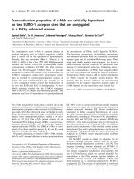
Fig. 2. Location (A) and protein composition (B) of the chick ILM. (A) Fluorescent micrograph of a cross-section of an E4 chick eye stained
for laminin-1 shows all BMs of the developing eye, including the lens capsule (L), the BM of the pigment epithelium (star) and the ILM
(arrow). The pial BM (P) of the adjacent diencephalon is labeled as well. Western blots (B) show that the ILM is comprised of the following
proteins: laminin-1 (LN) with bands at 200 and 400 kDa (lane 1). The band at 400 kDa is also labeled with an antibody to the C-terminal glo-
bular domains of laminin-1, and thus represents the a chain of laminin-1 (lane 2). Nidogen-1 (Ni) appeared as two bands, both of which were
confirmed to be nidogen-1 by MS (lane 3). The smear at 600, 400 and 700 kDa in lanes 4, 5 and 6 represents the proteoglycans agrin (AG),
collagen 18 (18) and perlecan (Per). The multiple bands of lane 7 represent monomeric and oligomeric forms of collagen 4. Degradation
bands of collagen 18 and 4 are indicated by stars (lanes 5 and 7). N-CAM, a cell membrane protein that is abundant in the retina (lane 9),
was not detectable in the ILM (lane 8). Likewise, collagen 9 (9), which is very abundant in the vitreous (lanes 11), is only present in traces in
the ILM matrix (lanes 10). The specific bands for each of the proteins are indicated by arrows. The samples for the collagen 4 and collagen 9
were run under nonreducing conditions. Bar ¼ 200 lm.
Biomechanical properties of basement membranes J. Candiello et al.
2900 FEBS Journal 274 (2007) 2897–2908 ª 2007 The Authors Journal compilation ª 2007 FEBS
1–5 lm. Figure 6A shows representative experimental
curves of AFM loading force versus z-piezo position
for E4 (black solid line), E9 (blue solid line) and E15
(pink solid line) ILM. The dotted line is a fitted curve
to the E4 ILM data using E (Young’s modulus) and
the initial contact point z
o
as fitting parameters. The
apparent Young’s modulus for the chick ILM of E4,
E9 and E15 embryos is summarized in Table 1 and
Fig. 6B. The apparent Young’s modulus of chick ILM
was 0.95 ± 0.54 MPa at E4, 3.34 ± 1.11 MPa at E9
and 3.57 ± 1.58 at E15. There was a significant
increase in the ILM stiffness (Young’s modulus) from
E4 to E9 (P<0.01), but no significant change of
ILM elasticity was observed from E9 to E15
(P > 0.05; Fig. 6B).
We also measured the elasticity of ILMs from post-
natal day (P)1 mice and adult mice. The elastic
(Young’s) modulus of the neonatal mouse ILM was
3.81 ± 1.07 MPa (mean ± SD, three different ILM
tissues, 16 measurements on each tissue). The apparent
Young’s modulus of the adult mouse ILM was
4.07 ± 2.25 MPa. There was no significant difference
between the apparent Young’s moduli of P1 and adult
mouse ILM. The ILM elasticity is very similar for the
neonatal mouse and the late embryonic chick
(3.81 ± 1.07 MPa versus 3.57 ± 1.58 MPa).
Discussion
The ILM as a model system for measuring BM
thickness and elasticity
To investigate the mechanical stability of BMs, we
chose the chick and mouse ILM as a model system.
The ILM is located at the vitreo-retinal border, and it
has the typical three-layered ultra structure of BMs that
includes the two laminae lucida interna and externa,
and the electron-dense lamina densa. Western blot
analysis and MS showed that the ILM consists of
extracellular matrix proteins that are also found in
other BMs. These included laminin-1, nidogen-1, colla-
gen 4 and the proteoglycans agrin, perlecan and colla-
gen 18, consistent wither previous studies [21,22]. Our
histological analysis of a mutant mouse showed that
the ILM is important to confine retinal cells because
breaks in the ILM lead to ectopic retinal cells in the
vitreous cavity (Fig. 1). Thus, the mechanical stability
provided by the ILM is critical for proper organogene-
sis of the eye. Likewise, endothelial herniation and fre-
quent breaks of ocular and cortical blood vessels in
this mutant mouse (Fig. 1) confirm that the mechanical
stability of BMs is one the essential functions of BMs
in situ.
To date, the mechanical properties of few BMs have
been characterized, in large part, due to difficulty of
isolating the delicate and thin BMs and the lack of
tools to make mechanical measurements on such sub-
micron thin membranes. A unique advantage of using
the ILM over other BMs is that it is readily separable
from the vitreous body, whereas most other BMs are
tightly connected to interstitial connective tissue.
Another unique advantage is that the ILM can be
prepared as large flat-mount preparations on solid
A
B
DC
F
E
G
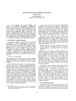
Fig. 3. Flat-mount preparations of ILMs from chick and mouse
retina. Large segments of chick (C) and mouse (G) ILM were pre-
pared on glass slides by mechanically splitting the retina. The isola-
ted ILM preparations were strongly labeled for laminin-1 (red in
C,G), identical to the strong labeling (red) of the ILM in cross-sec-
tions of chick (A) and mouse (F) retina (R). The white star in (A) is
next to the BM of the pigment epithelium. The sections were also
stained with the nuclear counter-stain Sytox-Green (Molecular
Probes, Eugene, OR, USA). The preparation shown in (C) was
scratched (white bar and Sc, scratch) for thickness measurements
with the AFM. TEM micrographs show that the ILM preparations
are clean BM sheets: a low power micrograph (D) shows a thin
sheet of ECM on the plastic support (P) that loops multiple times
at the margin. A high power view (E) from the area indicated in
panel D (arrow) showed the ILM as a 60 nm thin sheet, similar in
thickness and appearance as ILM in retinal cross-sections (B). The
measured thicknesses are indicated by the white bars. Bar, (A,F)
50 lm; (B,E) 100 nm; (C,G) 25 lm; (D) 2.5 lm.
J. Candiello et al. Biomechanical properties of basement membranes
FEBS Journal 274 (2007) 2897–2908 ª 2007 The Authors Journal compilation ª 2007 FEBS 2901
surfaces, such as glass or plastic, allowing reliable
thickness measurements. It is also of note that the
ILM isolation method uses firmly mounted retina as a
source; thus, the preparation procedure avoids the
chance for folding, stretching or compression of the
BM. Taken together, the ILM is a BM that shares all
typical features of most BMs and provides a series of
unique experimental advantages making it particularly
suitable for biomechanical measurements by AFM.
Thickness of BMs
AFM measurements showed that the chick ILM increa-
ses in thickness three-fold from 137 nm to 402 nm
between E4 and E9. However, attempts to measure the
thickness of mouse ILM were not successful because the
retinal surface of rat ILM was very uneven and
the measurements varied greatly, most likely due to the
firmly attached hyaloid blood vessels to the mouse
100 nm
0
Height
16 nm
0
Height
100 nm
0
Height
16 nm
0
Height
AB
C
D
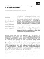
Fig. 4. AFM images of flat-mount chick E4
(A and B) and E9 (C,D) ILM samples. At
40 lm · 40 lm (A,C), the retinal side did
not exhibit distinct features. Zooming into a
2 lm · 2 lm region (B,D), the fibrillar net-
work of the ILM could be seen. The fibrils
in E9 ILM (B) appeared to be thicker than
that of E4 (D).
A
B
C
Fig. 5. (A) Showing a height mode image of an E9 ILM near a scratched edge. The area on the left of the image is the glass substrate,
whereas the area on the right is the ILM surface. A dashed line in (A) shows the location of a cross section profile, which is plotted as the
black trace in (B), and is used to determine the thickness of the E9 ILM. The height profile of an E4 ILM sample (red trace) is also plotted in
(B). (C) Summarizing the measured ILM thicknesses from four ILM samples for each of the E4, E9 and E16 development stages (ten meas-
urements per sample). There is a significant increase in ILM thickness from E4 to E9, but not between E9 and E15. Data in (C) are presen-
ted as the mean ± SD.
Biomechanical properties of basement membranes J. Candiello et al.
2902 FEBS Journal 274 (2007) 2897–2908 ª 2007 The Authors Journal compilation ª 2007 FEBS
ILM. Since the chick eye lacks a hyaloid vasculature,
this was not a problem for chick preparations.
Based on TEM images of retinal cross sections, the
thickness ILM from embryonic chick eyes has previ-
ously been estimated to be 50–70 nm [23] (Fig. 3B).
Similar values (40–120 nm) also have been reported for
most other BMs investigated using TEM, such as BMs
from muscle, blood vessels, the pia, lung and skin.
These BM thickness measurements are currently con-
sidered as textbook values for basal membranes [24].
However, sample preparation for TEM requires dehy-
dration, which will lead to shrinkage of the BMs.
Shrinkage following dehydration is probably much
greater for BMs than for other tissue structures because
at least three highly hydrated proteoglycans are major
BM constituents. To confirm our result that the hydra-
ted BM thickness is significantly greater than the previ-
ous TEM measurements, we measured the thicknesses
of two freshly isolated chick E8 ILM under hydrated
condition and then made measurements on the same
ILMs after tissues were dehydrated following standard
TEM drying procedures. The drying process reduced
the thicknesses of these ILM from 356 ± 25 nm and
404 ± 14 nm to 48 ± 7 nm and 52 ± 9 nm, a reduc-
tion of approximately 87% (Fig. 7). Therefore, we have
shown that the thickness of ILMs had been greatly
underestimated in previous TEM studies. This under-
estimation most likely applies to other BMs as well.
Elasticity of BMs
Although many connective tissues, such as cartilages
and BM, consist of similar extracellular matrix (ECM)
molecules, including various collagens and proteogly-
cans, their mechanical properties vary significantly due
to the different composition and crosslinks between
the ECM molecules. For example, the Young’s moduli
of cartilages have been measured in the range 0.95–
7.7 Mpa, depending on the location of the cartilage
and there are even variations of stiffness at different
regions of the same cartilage [25–30]. A recent AFM
indentation study of the highly flexible tectorial mem-
brane in the cochlea reported a Young’s modulus in
the range 37–135 kPa, with large spatial variations
within the membrane [31].
At present, very little data exists on the biomechani-
cal properties of the conventional, thin BMs. The most
extensively studied BM is the lens capsule, which can
be readily separated from the lens cortex and is the
thickest BM in the body (approximately 5–10 lm
for the aterior capsule and 20–30 lm for the posterior
capsule in humans) [32]. Under low strains, which
correspond to our AFM experimental conditions, the
Young’s modulus of lens capsules has been reported to
be approximately 0.6 MPa for rat, 0.82 MPa for cat,
and between 0.3 and 2.4 MPa in humans [33–36]. The
apparent Young’s modulus of the Bruch’s membrane
Table 1. The apparent Young’s modulus from each chick ILM at
E4, E9 and E15. Twenty measurements on randomly selected loca-
tion on each ILM sample were analyzed. The data are presented as
the mean ± SD.
Sample number
Apparent Young’s modulus (MPa)
E4 E9 E15
1 0.94 ± 0.35 3.28 ± 0.87 4.37 ± 1.74
2 0.79 ± 0.58 3.46 ± 1.00 2.73 ± 1.50
3 0.92 ± 0.39 3.54 ± 1.02 3.48 ± 1.36
4 1.19 ± 0.66 3.09 ± 1.43 3.84 ± 1.30
Average of four samples 0.95 ± 0.54 3.34 ± 1.11 3.57 ± 1.58
Fig. 6. Elasticity of the chick ILM. (A) Representative AFM force-displacement curves for indentation experiments on ILMs from E4 (black),
E9 (blue) and E15 (pink) chick retinae. The dotted line is a fitted curve to the E4 experiment data using the Sneddon model. (B) Summarizing
the average apparent Young’s modulus of chick ILM at E4, E9 and E15 (mean ± SD), the error bars represent standard deviations. Four ILM
tissues for each development stage were studied; 20 measurements from each ILM sample were analyzed. There was a significant increase
in the apparent Young’s modulus between E4 and E9, but not from E9 to E15.
J. Candiello et al. Biomechanical properties of basement membranes
FEBS Journal 274 (2007) 2897–2908 ª 2007 The Authors Journal compilation ª 2007 FEBS 2903
was approximately 1 MPa, measured on cryosections
of Bruch’s membrane by AFM indention [37; unpub-
lished observations]. The Bruch’s membrane is another
ocular BM (approximately 2 lm thick) located between
the retinal pigment epithelium and the choroid and is
composed of predominantly collagen 4 and elastin.
The present study showed that the changes in the
thickness and the bulk size of the chick ILM from E4
to E9 were accompanied by internal structural modifi-
cation during development. The apparent Young’s
modulus of ILM, an intrinsic measurement of the
material elasticity independent of the membrane thick-
ness, increased from 0.95 ± 0.54 MPa at E4 to
3.34 ± 1.11 MPa at E9 (Table 1), although there was
little change between E9 and E15 (3.34 ± 1.11 MPa
versus 3.57 ± 1.58 MPa). These ILM elasticity meas-
urements were made using sharp pyramidal tips (with a
tip diameter of approximately 20 nm). We also made
measurements using spherical tips (diameter 7 lm) to
confirm our results. In a previous AFM study of articu-
lar cartilage elasticity, the measured elasticity could dif-
fer up to 100-fold depending on whether a sharp tip or
a larger spherical tip was used, reflecting different tissue
properties at nanometer and micrometer scales [29]. We
did not detect significant discrepancy between measure-
ments made using sharp pyramidal tips versus larger
spherical tips. For E4 ILM, using the spherical tip, the
apparent Young’s modulus was 0.93 ± 0.19 MPa
(mean ± SD, two tissues and 16 measurements each),
which was very similar to results obtained using sharp
pyramidal tips.
The large increase of the apparent Young’s modulus
of ILM from E4 to E9 suggests significant remodeling
of internal ILM structure during this development per-
iod. Higher resolution AFM images of the ILM surface
show clear differences in the ILM fibrillar network at
E4 and E9 (Fig. 4B,D), consistent with structure modifi-
cations such as higher degrees of cross-link between
collagen fibrils. Inhibition of crosslinking in collagen
has been shown to significantly reduce the stiffness of
aorta [38]. The biomechanical properties of ECM also
depend on the variation in collagen and proteoglycan
content [39]. Increases in both the membrane thickness
and stiffness (Young’s modulus) would enhance BM
strength to resist stress induced during the growth of
the eye. The change in stiffness is biologically useful
because the embryos and its organs expand most
dramatically during early stages of development and a
more elastic BM is required, whereas at later stages
mechanical stability of organs becomes more important.
Biological significance
The phenotypic analysis of mice with mutations of
BM proteins strongly indicates that the integrity of the
BMs is a requirement for vascular stability and for
maintaining stable tissue borders. Vascular problems
were observed in mice with targeted deletions or muta-
tions of perlecan, nidogen and collagen 4 [5,14–16].
The vascular breaks occur predominantly in the brain
and eye, both organs in which the blood vessels are
not embedded in dense connective tissue and the
endothelial BMs are the sole stabilizer of the vessel
wall. Vascular breaks did not occur in the initial pro-
cess vasculogenesis, but rather in mid-embryogenesis,
when blood pressure rises. The ectopias that were pre-
sent in these mutant mice were prominent in the cortex
and retina, where the pial and retinal BMs have little
support from the adjacent connective tissue.
The fact that blood vessel breaks and CNS ectopias
occur in mice with mutations of different BM proteins
shows that these defects are linked to a general,
mechanical property of BMs rather than the lack of a
specific component. Furthermore, ectopias and blood
vessel ruptures were also observed in chick embryos in
which the ILM and the pial BM were enzymatically
disrupted [40–42], confirming that this phenotype is
not connected to the lack of a specific protein, but
rather the weakness of the ILM as a structure. We
propose that BMs have a critical role in stabilizing
the cortical and retinal tissue borders as well as blood
vessels in the CNS and the eye. In light of the rather
weak mechanical resistance provided by cells alone,
BMs are critical in the stability of tissues under stress,
Fig. 7. The cross section profiles of an E8 chick ILM under native,
hydrated state (red line) and after dehydration (black line). The ILM
thicknesses were measured from the elevation of the top mem-
brane surface (dashed lines). In this E8 chick ILM, the drying pro-
cess reduced the ILM thickness from 404 ± 14 nm to 52 ± 9 nm
(mean ± SD; ten measurements), which is a reduction of 87%.
Biomechanical properties of basement membranes J. Candiello et al.
2904 FEBS Journal 274 (2007) 2897–2908 ª 2007 The Authors Journal compilation ª 2007 FEBS
predominantly in the vascular system. In addition,
BMs maintain a smooth glial and neuron-impenetrable
border in the brain and retina that is essential for glial
cells attachment and cortical and retinal histogenesis.
Experimental procedures
Histology
To demonstrate the importance of BMs in the stability of
blood vessels and the maintenance of smooth tissue border
in brain and retina, the status of BMs in mice with a
targeted deletion of a 50 amino acid long segment in the
laminin a1 chain was investigated [8]. The eyes of E18 mice
were fixed in 2.5% glutaraldehyde overnight and embedded
in EPON
TM
(Hexion Speciality Chemicals, Columbus, OH,
USA) according to standard procedures. Eyes from embryos
that were homozygous or heterozygous for the mutation
were compared by light and electron microscopy in terms of
BM continuity, hemorrhages, blood vessel rupture and ret-
inal ectopias. Maintenance and killing of mice were carried
out under approved protocols and in accordance with NIH
and the European Community guidelines for animal care.
ILM preparation
For preparing BM flat-mounts for AFM, retinae from E4
to E15 chick embryos and from P1 and adult mice were
dissected and spread on membrane filters (Millipore, Bed-
ford, MA, USA). The filter ⁄ retinal sandwiches were placed,
vitreous surface down, on poly lysine-coated (Sigma,
St Louis, MO, USA) glass slides (Superfrost ⁄ plus, Fisher
Scientific, Pittsburgh, PA, USA). After a 5 min attachment
period, the filters with the retinae were lifted off from the
slides, a procedure that splits the retina at the vitreal sur-
face and leaves large segments of the retinal BMs on the
glass slides [20]. To remove adherent endfeet of the ventri-
cular cells from the BM sheets, the BMs were incubated
with 2% Triton-X-100 for 30 min and washed several times
in NaCl ⁄ Pi [20]. The preparations were always kept under
NaCl ⁄ Pi. To assist visualization of the transparent and
barely visible BM flat-mounts, some chick preparations
were stained with a monoclonal antibody to nidogen-1
(mAb 1G12) [43], for 1 h, followed by a Cy3-labeled goat
anti-mouse IgG (Jackson ImmunoResearch, West Grove,
PA, USA). For mouse ILM preparations, the flat-mounts
were labeled with an antibody to mouse laminin-1 (Invitro-
gen, Carlsbad, CA, USA). For thickness measurements, the
preparations were scratched with 100 lL pipette tips.
Western blot analysis
For western blot analysis, chick ILMs were isolated in bulk
as previously described [44], pelleted by centrifugation, and
dissolved in 8 m urea and SDS sample buffer. The proteins
were resolved by PAGE, transferred to nitrocellulose, and
the blots were labeled with antibodies to laminin-1 (mAb
3H11) [44], nidogen-1 (mAb 1G12) [43], agrin (mAb 6D2)
[45], collagen 18 (mAb 6C4) [46], perlecan (mAb 5C9) [47],
collagen 9 (mAb 2B9) [48] and NCAM (mAb 9H2) [45].
The monoclonal antibodies are available from the Develop-
mental Studies Hybridoma Bank (University of Iowa, IA,
USA). A rabbit antiserum against LG4-5 of laminin alpha
1 (E3 fragment, code no. 992+) was kindly provided by
Dr Takako Sasaki (Max Planck Institute of Biochemistry,
Munich, Germany).
The collagen 4 bands were detected by running the
PAGE under nonreducing conditions and using a poly-
clonal antiserum (Rockland, Gilbertsville, PA, USA) for
the western blots. The labeled proteins were detected by
alkaline phosphatase-labeled goat anti-mouse and goat
anti-rabbit IgG (Jackson ImmunoResearch) with Nitro
Blue tetrazolium and 5-bromo-4-chloroindol-2-yl phosphate
as chromogenes (Roche, Indianapolis, IN, USA).
MS
BM proteins were resolved by PAGE and reverse Zinc
stained. Eluted proteins from the stained bands were buf-
fered in 100 mm ammonium bicarbonate, denatured by
heating to 65 °C for 15 min after addition of 2% SDS,
reduced with 2.5 mm tris(2-carboxyethyl)phosphine, alkyl-
ated with 3.75 mm indole-3-acetic acid, followed by diges-
tion with porcine trypsin (Promega, Madison, WI, USA).
LC-MS experiments were performed on a Surveyor nano-
flow HPLC system interfaced with an ion trap mass spec-
trometer (LCQ Deca, Thermo Electron Corp., San Jose,
CA, USA). Data were acquired using the triple play
method and analyzed with Mascot Daemon, version 2.1.0
(Matrix Science, Boston, MA, USA) with the settings:
400–4000 mass range, scan grouping 1, precursor charge
state set to Auto, peptide error tolerance 1.5 Da, frag-
ment error tolerance 0.8 Da, one missed cleavage,
NCBI no. database (version 16 May 2006; 3284262
sequences, 112594017 residues), and variable modifications
were carbamidomethylation of cysteine and oxidation of
methionine.
AFM imaging and force indentation of ILM
All AFM imaging and force indentation experiments were
carried out using an MFP-3D Atomic Force Microscope
(Asylum Research, Santa Barbara, CA, USA), which was
placed on top of an Olympus IX-71 fluorescence micro-
scope (Olympus, Tokyo, Japan). Standard commercially
available, 100-lm long Si
3
N
4
cantilevers, with integrated
pyramidal tips (Veeco, Inc, Santa Barbara, CA, USA) and
a nominal spring constant, k, of 0.6 NÆm
)1
were used. The
J. Candiello et al. Biomechanical properties of basement membranes
FEBS Journal 274 (2007) 2897–2908 ª 2007 The Authors Journal compilation ª 2007 FEBS 2905
spring constant of each cantilever was measured by the
thermal fluctuation method [49] before each experiment.
For a few selected indentation experiments, we also
attached glass spherical beads of 7 lm diameter to the tips,
which did not alter the cantilever spring constants. The
topography of the ILM samples were imaged in inter-
mittent contact mode (AC, or tapping mode) with a scan
rate of approximately 1 lineÆs
)1
, in NaCl ⁄ Pi at room tem-
perature. The tissues were kept under NaCl ⁄ Pi solution
throughout the experiments. The elasticity of the ILM was
measured by nano-indentation with an AFM tip [50,51]. A
20 lm · 20 lmor10lm · 10 lm area of the ILM retinal
surface was first imaged with AFM, and indentions were
made over a 10 · 10 grid points evenly distributed over the
imaged area. The automated indentation was carried out
using the cFVol software program (Chad Ray, Duke Uni-
versity, Durham, NC, USA), at a rate of one load ⁄ unload
cycle per second. The speed of the AFM tip indenting the
tissue was between 2.0 and 10.0 lmÆs
)1
. Out of the 100
total indentations on each sample, 20 were randomly cho-
sen for quantitative analysis. To reduce the viscoelastic con-
tributions, the apparent Young’s modulus of the tissue at
each indentation point was calculated from only the retrac-
tion (unloading) portion of force-indentation curve using
the Sneddon model [50,52].
Calculation of ILM elasticity
The Sneddon model [53] was used to evaluate tissue elasticity
from the force-indentation measurements. Calculations
were made for both a conical indenter and a spherical
indenter. We used the conical geometric model for the sharp
AFM tips and the spherical model for the attached spherical
glass bead.
In the case of a conical indenter, the relationship between
the applied load ⁄ force f and the indentation d can be
expressed as:
f ¼
2
p
cot a
E
1 À m
2
d
2
where E is the Young’s modulus, m is the poisson’s ratio, a
is the half vertical angle of the AFM tip (a ¼ 35°). The
relationship between f and d for a spherical indenter can be
expressed as:
f ¼
4
3
E
1 Àm
2
ffiffiffi
R
p
d
3
2
where R is the radius of the spherical indenter.
The indentation d can be calculated from the AFM canti-
lever piezo position z and cantilever deflection d: d ¼
(z)z
o
))d , where z
o
is the initial indentation contact point.
The z–d relationship for conical and spherical indenters can
be similarly expressed, respectively, as:
z ¼ z
o
þ d þ
ffiffiffiffiffiffiffiffiffiffiffiffiffiffiffiffiffiffiffiffiffiffiffiffi
pkdð1 Àm
2
Þ
2E cot a
r
ð1Þ
and:
z ¼ z
o
þ d þ
2
3
ffiffiffiffiffiffiffiffiffiffiffiffiffiffiffiffiffiffiffiffiffiffiffi
3kdð1 Àm
2
Þ
4ER
1=2
r
ð2Þ
where k is the cantilever spring constant. Values of the
apparent Young’s modulus, E, were obtained from the
force-indentation data by curve fitting the experimentally
capture z–d curves with Eqns (1) and (2) using E and the
initial contact point z
o
as fitting parameters [50,54]. The
curve fitting was limited to the initial contact region of the
z–d curve, which corresponds region of small loading force
(f ¼ 6–8 nN) and small indentations (d ¼ 40–50 nm). The
poisson’s ratio was assumed to be m ¼ 0.47, a value meas-
ured on lens capsule, another retinal basal lamina [55].
Acknowledgements
This project was supported by grants from National
Institutes of Health (NIH EB004474) to JC and HL,
and the National Science Foundation (NSF,
IBN0240774) to WH.
References
1 Timpl R & Brown JC (1996) Supramolecular assembly
of basement membranes. Bioassays 18, 123–132.
2 Miner JH & Yurchenco PD (2004) Laminin functions
in tissue morphogenesis. Ann Rev Cell Dev Biol 20,
255–284.
3 Erickson AC & Couchman JR (2000) Still more com-
plexity in mammalian basement membranes. J Histo-
chem Cytochem 48, 1291–1306.
4 Smyth N, Vatansever NS, Murray P, Meyer M, Frie C,
Paulsson M & Edgar D (1999) Absence of basement
membranes after targeting the LAMC1 gene results in
embryonic lethality due to failure of endoderm differen-
tiation. J Cell Biol 144, 151–160.
5 Costell M, Gustafsson E, Aszodi A, Moergelin M,
Bloch W, Hunziger E, Addicks K, Timpl R & Faessler
R (1999) Perlecan maintains the integrity of cartilage
and some basement membranes. J Cell Biol 147,
1109–1122.
6 Arikawa-Hirasawa E, Watanabe H, Takami H,
Hassell JR & Yamada Y (1999) Perlecan is essential
for cartilage and cephalic development. Nat Genet 23,
354–358.
7 Poschl E, Schlotzer-Schrehardt U, Brachvogel B, Saito
K, Ninomiya Y & Mayer U (2004) Collagen IV is essen-
tial for basement membrane stability but dispensable for
initiation of its assembly during early development.
Development 131, 1619–1628.
Biomechanical properties of basement membranes J. Candiello et al.
2906 FEBS Journal 274 (2007) 2897–2908 ª 2007 The Authors Journal compilation ª 2007 FEBS
8 Willem M, Miosge N, Halfter W, Smyth N, Janetti I,
Burkhart A, Timpl R & Mayer U (2002) Specific abla-
tion of the nidogen-binding site in the laminin gamma1
chain interferes with kidney and lung development.
Development 129, 2711–2722.
9 Bader BL, Smyth N, Nedbal S, Miosge N, Baranosky
A, Mokkapati S & Murshed M (2005) Compound
genetic ablation of nidogen 1 and 2 causes basement
membrane defects and perinatal lethality in mice. Mol
Cell Biol 25 , 6846–6856.
10 Gautam M, Noakes PG, Moscoso L, Rupp F,
Scheller RH, Merlie JP & Sanes JR (1996) Defective
neuromuscular synaptogenesis in agrin-deficient mutant
mice. Cell 85, 525–535.
11 Fukai N, Eklund L, Marneros AG, Oh SP, Keene DR,
Tamarkin L, Niemela M, Ilves M, Li E, Pihlajaniemi T
et al. (2002) Lack of collagen XVIII ⁄ endostatin results
in eye abnormalities. EMBO J 21, 1535–1544.
12 Williams RS, Swisher CN, Jennings M, Ambler M &
Caviness VS (1984) Cerebro-ocular dysgenesis (Walker–
Warburg syndrome): neuropathologic and etiologic ana-
lysis. Neurology 34, 1531–1541.
13 Nakano I, Funahashi M, Takada K & Toda T (1996)
Are breaches in the glia limitans the primary cause of
the micropolygyria in Fukuyama-type congenital mus-
cular dystrophy (FCMD)? Pathological study of the
cerebral cortex of an FCMD fetus. Acta Neuropathol
91, 313–321.
14 Halfter W, Dong S, Yip YP, Willem M & Mayer U
(2002) A critical function of the pial basement mem-
brane in cortical histogenesis. J Neurosci 22, 6029–6040.
15 Halfter W, Willem M & Mayer U (2005) Basement
membrane-dependent survival of retinal ganglion cells.
Invest Ophthalmol Vis Sci 46, 1000–1009.
16 Gould DB, Phalan FC, Breedveld GJ, van Mil SE,
Smith RS, Schimenti JC, Aguglia U, van der Knaap
MS, Heutink P, John SW (2005) Mutations in Col4a1
cause perinatal cerebral hemorrhage and porencephaly.
Science 308, 1167–1140.
17 Georges-Labouesse E, Mark M, Messaddeq N &
Gansmuller A (1998) Essential role of alpha 6 integrins
in cortical and retinal lamination. Curr Biol 8, 983–986.
18 Graus-Porta D, Blaess S, Senften M, Littlewood-Evans
A, Damsky C, Huang Z, Orban P, Klein R, Schittny JC
& Muller U (2001) Beta1-class integrins regulate the
development of laminae and folia in the cerebral and
cerebral and cerebellar cortex. Neuron 16, 367–379.
19 Moore SA, Saito F, Chen J, Michele DE, Henry MD,
Messing A, Cohn RD, Ross-Barta SE, Westra S,
Williamson RA et al. (2002) Deletion of brain dystro-
glycan recapitulates aspects of congenital muscular dys-
trophy. Nature 418, 422–425.
20 Halfter W, Reckhaus W & Kroger S (1987) Nondirected
axonal growth on basal lamina from avian embryonic
neural retina. J Neurosci 7, 3712–3722.
21 Halfter W, Dong S, Schurer B, Osanger A, Schneider
WJ, Ruegg M & Cole GJ (2000) Composition, syn-
thesis, and assembly of the embryonic chick retinal
basal lamina. Dev Biol 220, 111–128.
22 Libby RT, Champliaud M-F, Claudepierre T, Xu Y,
Gibbons EP, Koch M, Burgeson R, Hunter DD &
Brunken WJ (2000) Laminin expression in adult and
developing retinae: evidence of two novel CNS laminins.
J Neurosci 20, 6517–6528.
23 Halfter W, Dong S, Schurer B, Ring C, Cole GJ & Eller
A (2005) Embryonic synthesis of the inner limiting
membrane and vitreous body. Invest Ophthalmol Vis Sci
46, 2202–2209.
24 Alberts B, Johnson A, Lewis J & Raff M (2002) Mole-
cular Biology of the Cell, 4th edn. Garland Science,
New York, NY.
25 Jurvelin JS, Buschmann MD & Hunziker EB (1997)
Optical and mechanical determination of Poisson’s ratio
of adult bovine humeral articular cartilage. J Biomech
30, 235–241.
26 Lee AJ, Grodzinsky H-P, Hsu SD, Martin M & Spector
M. (2000) Effects of harvest and cartilage repair proce-
dures on the physical and biochemical properties of
articular cartilage in the canine knee. J Orthop Res 18
,
790–799.
27 Gaon MD, Ho K-HK & and Wong BJF (2003) Mea-
surement of the elastic modulus of porcine septal carti-
lage specimens following Nd:YAG laser treatment.
Lasers Med Sci 18, 148–153.
28 Lambert RK, Baile EM, Moreno R, Bert J & Pare PD
(1991) A method for estimating the Young’s modulus
of complete tracheal cartilage rings. J Appl Physiol 70,
1991.
29 Stolz M, Raiteri R, Daniels AU, VanLandingham MR,
Baschong W & Aebi U (2004) Dynamic elastic modulus
of porcine articular cartilage determined at two different
levels of tissue organization by indentation-type atomic
force microscopy. Biophys J 86, 3269–3283.
30 Allen DM & Mao JJ (2004) Heterogeneous nanostruc-
tural and nanoelastic properties of pericellular and
interterritorial matrices of chondrocytes by atomic force
microscopy. J Struct Biol 145, 196–204.
31 Gueta R, Barlam D, Shneck RZ & Rousso I (2006)
Measurement of the mechanical properties of isolated
tectorial membrane using atomic force microscopy.
PNAS 103, 14790–14795.
32 Krag S & Andreassen TT (2003) Mechanical properties
of the human posterior lens capsule. Invest Ophthalmol
Vis Sci 44, 691–696.
33 Fisher RF & Wakely J (1976) The elastic constants
and ultrastructural organization of a basement membrane
(lens capsule). Proc R Soc Lond B Biol Sci 193, 335–358.
34 Fisher RF & Hayes BP (1982) The elastic constants and
ultrastructural organization of regenerated basement
membrane (lens capsule). Q J Exp Physiol 67, 213–224.
J. Candiello et al. Biomechanical properties of basement membranes
FEBS Journal 274 (2007) 2897–2908 ª 2007 The Authors Journal compilation ª 2007 FEBS 2907
35 Fisher RF & Hayes BP (1987) Macromolecular organi-
zation of collagen fibres in natural and tanned basement
membrane. J Mol Biol 198, 263–279.
36 Krag S, Olsen T & Andreassen TT (1997) Biomechanical
charachteristics of the human anterior lens capsule in
relation to age. Invest Ophthalmol Vis Sci 38, 357–363.
37 Candiello AJ, Feola A, Elyaderani T, Friberg R & Lin
H (2005) Nano indentation measurements of Bruch’s
membrane mechanical properties. Invest Ophthalmol Vis
Sci 46: ARVO abstract 1210.
38 Bruel A, Ortoft G & Oxlund H (1998) Inhibition of
cross-links in collagen is associated with reduced stiffness
of the aorta in young rats. Atherosclerosis 140, 135–145.
39 Kiviranta P, Rieppo J, Korhonen RK, Julkunen P,
Toyras J & Jurvelin JS (2006) Collagen network primar-
ily controls Poisson’s ratio of bovine articular cartilage
in compression. J Orthop Res 24, 690–699.
40 Halfter W (1998) Disruption of the retinal basal lamina
during early embryonic development leads to a retrac-
tion of vitreal end feet, an increased number of ganglion
cells, and aberrant axonal outgrowth. J Comp Neurol
397, 89–104.
41 Halfter W & Schurer B (1998) Disruption of the pial
basal lamina during early avian embryonic development
inhibits histogenesis and axonal pathfinding in the optic
tectrum. J Comp Neurol 397, 105–117.
42 Halfter W, Dong S, Balasubramani M & Bier M (2001)
Temporary disruption of the retinal basal lamina and its
effect on the retinal histogenesis. Dev Biol 238, 79–96.
43 Halfter W, Dong S, Schurer B, Osanger A, Schneider.
WJ, Ruegg M & Cole GJ (2000) Composition, synth-
esis, and assembly of the embryonic chick retinal basal
lamina. Dev Biol 220, 111–120.
44 Halfter W & Von Boxberg Y (1992) Axonal growth on
solubilized and reconstituted matrix from the embryonic
chicken retina inner limiting membrane. Eur J Neurosci
4, 840–852.
45 Halfter W, Schurer B, Yip J, Yip L, Tsen G, Lee JA &
Cole GJ (1997) Distribution and substrate properties of
agrin, a heparin sulfate proteoglycan of developing axo-
nal pathways. J Comp Neurol 383, 1–17.
46 Halfter W, Dong S, Schurer B & Cole GJ (1998) Col-
lagen XVIII is a basement membrane heparin sulfate
proteoglycan. J Biol Chem 273, 25404–25412.
47 Balasubramani M, Bier ME, Hummel S, Schneider WJ
& Halfter W (2004) Perlecan and its immunoglobulin
like domain IV are abundant in vitreous and serum of
the chick embryo. Matrix Biol 23, 143–152.
48 Ring C, Hassell J & Halfter W (1996) Expression pattern
of collagen IX and potential role in the segmentation of
the peripheral nervous system. Dev Biol 180, 41–53.
49 Hutter JL & Bechhoefer J (1993) Calibration of atomic-
force microscope tips. J Rev Sci Instrum 64, 1868–1873.
50 Radmacher M, Fritz M & Hansma PK (1995) Imaging
soft samples with the atomic force microscope: gelatin
in water and propanol. Biophys J 69, 264–270.
51 Rhee SK, Quist AP & Lal R (1998) Amyloid beta pro-
tein-(1-42) forms calcium-permeable, Zn
2+
-sensitive
channel. J Biol Chem 273, 13379–13382.
52 Quist AP, Rhee SK, Lin H & Lal R (2000) Physiologi-
cal role of gap-junctional hemichannels. Extracellular
calcium-dependent isosmotic Volume regulation. J Cell
Biol 148, 1063–1074.
53 Sneddon IN (1965) The relation between load and
penetration in the axisymmetric Boussinesq problem for
a punch of arbitrary profile. Int J Eng Sci 3, 47–57.
54 Almqvist N, Bhatia R, Primbs G, Desai N, Banerjee S
& Lal R (2004) Elasticity and adhesion force mapping
reveals real-time clustering of growth factor receptors
and associated changes in local cellular rheological
properties. Biophys J 86, 1753–1762.
55 Fisher RF (1969) The significance of the shape of the
lens and capsular energy changes in accommodation.
J Physiol 201, 1–19.
Biomechanical properties of basement membranes J. Candiello et al.
2908 FEBS Journal 274 (2007) 2897–2908 ª 2007 The Authors Journal compilation ª 2007 FEBS