Báo cáo khoa học: Direct detection of stereospecific soman hydrolysis by wild-type human serum paraoxonase potx
Bạn đang xem bản rút gọn của tài liệu. Xem và tải ngay bản đầy đủ của tài liệu tại đây (342.79 KB, 9 trang )
Direct detection of stereospecific soman hydrolysis by
wild-type human serum paraoxonase
David T. Yeung
1,2
, J. Richard Smith
3
, Richard E. Sweeney
4
, David E. Lenz
1
and Douglas M. Cerasoli
1
1 Physiology and Immunology Branch, Research Division, US Army Medical Research Institute of Chemical Defense, Aberdeen Proving
Ground, MD, USA
2 Department of Pharmacology and Experimental Therapeutics, University of Maryland at Baltimore, MD, USA
3 Medical Diagnostic and Chemical Branch, Analytical Toxicology Division, US Army Medical Research Institute of Chemical Defense,
Aberdeen Proving Ground, MD, USA
4 RESECO Research Engineering Consultants, Nottingham, PA, USA
Human serum paraoxonase 1 (HuPON1; EC 3.1.8.1) is
a human plasma enzyme previously shown to hydrolyze
insecticides and the highly toxic organophosphorus
(OP) nerve agents sarin (GB), O-ethyl S-(2-diisopropyl-
aminoethyl) methylphosphonothioate (VX), and soman
(GD; pinacolyl methylphosphonofluoridate) in vitro
and in vivo [1–3]. Although its catalytic efficacy against
GB, VX, and GD is low, it is the capacity to hydrolyze
these toxic nerve agents in vivo that makes HuPON1
attractive as a candidate bioscavenger of OP
compounds. It has been theorized that a genetically
engineered variant of HuPON1 with at least a 10-fold
increase in activity would be highly protective in vivo
against intoxication by OP compounds [4–7].
GD is a member of a class of highly toxic acetylcho-
linesterase inhibitors, all of which have their leaving
groups attached to a chiral phosphorus atom [8–11].
GD contains a second chiral center at one of the alkyl
side chain carbon atoms. Therefore, it exists as four
stereoisomers C+P+, C+P–, C–P+, and C–P–
(Fig. 1) [12–17]. Both of the P– isomers (C±P–) are
much more toxic in vivo and more readily inhibit
Keywords
diisopropylfluorophosphate; GC ⁄ MS;
paraoxonase 1; soman; stereoselectivity
Correspondence
D. Cerasoli, US Army Medical Research
Institute of Chemical Defense, 3100
Ricketts Point Road, Aberdeen Proving
Ground, MD 21010-5400, USA
Fax: +1 410 436 8377
Tel: +1 410 436 1338
E-mail:
(Received 19 October 2006, revised 5
December 2006, accepted 13 December
2006)
doi:10.1111/j.1742-4658.2006.05650.x
Human serum paraoxonase 1 (HuPON1; EC 3.1.8.1) is a calcium-depend-
ent six-fold b-propeller enzyme that has been shown to hydrolyze an array
of substrates, including organophosphorus (OP) chemical warfare nerve
agents. Although recent efforts utilizing site-directed mutagenesis have
demonstrated specific residues (such as Phe222 and His115) to be import-
ant in determining the specificity of OP substrate binding and hydrolysis,
little effort has focused on the substrate stereospecificity of the enzyme; dif-
ferent stereoisomers of OPs can differ in their toxicity by several orders of
magnitude. For example, the C±P– isomers of the chemical warfare agent
soman (GD) are known to be more toxic by three orders of magnitude. In
this study, the catalytic activity of HuPON1 towards each of the four chiral
isomers of GD was measured simultaneously via chiral GC ⁄ MS. The cata-
lytic efficiency (k
cat
⁄ K
m
) of the wild-type enzyme for the various stereoiso-
mers was determined by a simultaneous solution of hydrolysis kinetics for
each isomer. Derived k
cat
⁄ K
m
values ranged from 625 to 4130 mm
)1
Æmin
)1
,
with isomers being hydrolyzed in the order of preference C+P+ >
C–P+ > C+P– > C–P–. The results indicate that HuPON1 hydrolysis of
GD is stereoselective; substrate stereospecificity should be considered in
future efforts to enhance the OPase activity of this and other candidate
bioscavenger enzymes.
Abbreviations
DFP, diisopropylfluorophosphate; GB, sarin; GD, soman; HuPON1, human serum paraoxonase 1; OP, organophosphorus; PON1,
paraoxonase 1; VX, O-ethyl S-(2-diisopropylaminoethyl) methylphosphonothioate.
FEBS Journal 274 (2007) 1183–1191 ª 2007 FEBS No claim to original US government works 1183
acetylcholinesterase in vitro than the P+ isomers; the
bimolecular rate constants of acetylcholinesterase for
the C±P+ isomers are % 1000-fold lower than those
of the C±P– isomers, with assumed correspondingly
lower in vivo toxicity [12,13,15,18]. The hydrolytic clea-
vage of the phosphorus–fluorine (P–F) bond to form
P–OH renders GD nontoxic; this reaction is catalyzed
by OP hydrolases such as HuPON1 [3,9,18].
Although substantial efforts have focused on identify-
ing amino acid residues essential for HuPON1 enzymat-
ic activity [5,7,19–21], until very recently relatively little
attention has been paid to the more subtle question of
the substrate stereospecificity of the enzyme [22,23].
Knowledge of enzyme stereoselectivity is critical to
understanding substrate orientation and for the rational
design of mutants with enhanced activity towards the
more toxic isomers of specific substrates, such as GD.
We studied the kinetics of HuPON1-catalyzed
hydrolysis of the individual isomers of GD from a
racemic mixture of the nerve agent at concentrations
ranging from 0.2 to 3.0 mm, using a chiral GC ⁄ MS
approach. This allowed for simultaneous determination
of K
m
, k
cat
, and k
cat
⁄ K
m
values of HuPON1 for each
GD stereoisomer, resulting in unambiguous elucidation
of the extent of stereoselectivity of HuPON1-mediated
hydrolysis of GD.
Results
Analysis of GD stereoisomer hydrolysis using
GC
⁄
MS
The decrease in the concentration of each of the GD
isomers in the presence of HuPON1 over time was fol-
lowed using GC ⁄ MS analysis. All four stereoisomers
and the internal standard diisopropylfluorophosphate
(DFP) were quantitatively separated (Fig. 2) using a
Chiraldex c-cyclodextrin trifluoroacetyl column [24].
The elution order of individual GD stereoisomers from
a racemic sample was determined by examining the
retention times of individual purified stereoisomers
alone (data not shown).
The elution order detected was C–P–, C–P+, C+P–,
and then C+P+ at approximately 12.0, 12.8, 13.2,
and 13.6 min after injection, respectively (Fig. 2). Our
elution order differs from those previously reported
using different GC columns [9,16]. The DFP standard
eluted after all four GD stereoisomers, at % 17.3 min
post injection. The clear separation of peaks in the elu-
tion profile allowed for the simultaneous determination
of the fate of all four GD stereoisomers (Fig. 3)
[9,12,25].
Spontaneous hydrolysis of GD stereoisomers
Hydrolytic assays were carried out in the absence of
HuPON1 enzyme to define any effects of spontaneous
hydrolysis at pH 7.4 at room temperature. The ratios
of the areas under the curve for each stereoisomer
were determined at 0.5, 1.0, 3.0, 5.0, 15.0, and 240 min
following incubation of 2.0 mm racemic GD in super-
natant from cells transfected with empty plasmid vec-
tor. The ratios of C–P– ⁄ C–P+ ⁄ C+P– ⁄ C+P+ were
identified relative to the DFP internal standard and
were 23.4 ⁄ 26.7 ⁄ 26.6 ⁄ 23.2%, respectively, in good
agreement with previous reports [26,27]. The absolute
amount of GD and the relative percentages of each
stereoisomer were consistent across all sampling times,
differing by no more than 0.2% (data not shown),
indicating negligible spontaneous hydrolysis.
Effects of GD stereoisomer racemization
Spontaneous racemization of GD stereoisomers is
known to occur at the phosphorus atom in the pres-
ence of excess fluoride ion [26,27]. To determine if such
racemization was occurring in our experimental
system, studies were performed at room temperature in
50 mm glycine buffer (pH 7.4) with supernatant from
cells transfected with empty plasmid vector. The extent
Fig. 1. Stereoisomers of GD.
HuPON1 stereospecific hydrolysis of GD D. T. Yeung et al.
1184 FEBS Journal 274 (2007) 1183–1191 ª 2007 FEBS No claim to original US government works
of racemization was studied in reactions containing
semipurified 0.30 mm C–P– ⁄ C–P+ or C+P– ⁄ C+P+
mixtures of GD isomers in the presence of excess fluor-
ide ions (which varied from 0 to 2.0 mm NaF). In
addition, we incubated 1.0 mm racemic GD with
2.0 mm NaF under the same experimental conditions
to determine the extent of racemization under those
conditions. The results obtained from both sets of
experiments indicated that under the conditions used,
the presence of excess fluoride ions caused no appreci-
able racemization of either the C±P– or the C±P+
isomers. Furthermore, we did not observe any alter-
ation in the GC ⁄ MS isomer elution profile after incu-
bating 1.0 mm racemic GD with excess (2.0 mm NaF)
fluoride ions.
Characterization of wild-type HuPON1 activity
Initial rates of enzymatic hydrolysis of the individual
GD stereoisomers were estimated by plotting GD
concentration (for the individual stereoisomers) as a
function of time (Fig. 4). The concentration of each
11.00 11.50 12.00 12.50 13.00 13.50 14.00 1
0
200
400
600
800
1000
1200
1400
1600
1800
2000
2200
2400
2600
2800
3000
3200
3400
3600
3800
4000
Time (mins)
Abundance
C-P-
C-P+
C+P-
C+P+
Fig. 2. Gas chromatographic separation of
GD stereoisomers. Shown is a reconstruc-
ted ion chromatogram (m ⁄ z 126) of a
2.0 m
M racemic sample of GD (no enzyme)
analyzed by GC ⁄ MS after separation using a
Chiraldex c-cyclodextrin trifluoroacetyl col-
umn at 80 °C isothermal, with labels identi-
fying peaks corresponding to the individual
stereoisomers. The internal standard DFP
eluted at % 17.3 min (not shown).
Abundance
4000
5000
C-P-
C-P+
C+P-
C+P+
0 & 5 mins
0 & 5
mins
15 mins
15 mins
15 mins
5 mins
0 min
120 mins
120 mins
120
mins
3000
1000
2000
0
11.60 13.20 12.0 13.60 12.40 12.80
Time (minutes)
Fig. 3. Overlay of reconstructed ion chromatograms (m ⁄ z 126) of GD hydrolysis by HuPON1. Typical ion chromatograms indicating the relat-
ive abundance of the four GD stereoisomers (0.75 m
M racemic GD) after different incubation periods (i.e. 0, 5.0, 15.0, and 120 min, as indi-
cated) with wild-type HuPON1 enzyme. The various GD stereoisomers were eluted in the same order as shown in Fig. 2.
D. T. Yeung et al. HuPON1 stereospecific hydrolysis of GD
FEBS Journal 274 (2007) 1183–1191 ª 2007 FEBS No claim to original US government works 1185
specific stereoisomer was derived from a previously
determined GD standard curve and the area under the
curve for each stereoisomer was then normalized
against the DFP internal standard. The kinetic param-
eter K
m
of HuPON1 for each of the four stereoisomers
of GD was determined from the derived kinetic model
(Fig. 5, Table 1) as detailed in the Experimental proce-
dures, and ranged from 0.27 to 0.91 mm in the follow-
ing order: C–P– > C+P– > C–P+ > C+P+. The
k
cat
values for the hydrolysis of each stereoisomer were
also determin ed from the derived model (Table 1); the
values range from 501 to 1030 min
)1
, where
C+P+ > C–P+ > C+P– > C–P–. The bimolecular
rate constants derived from the model ranged from
4130 to 625 mm
)1
Æmin
)1
for C+P+ > C–P+ >
C+P– > C–P–, respectively. The average K
m
, k
cat
,
and k
cat
⁄ K
m
values for all four GD stereoisomers in
aggregate are 0.62 mm, 669 min
)1
, and 1739 mm
)1
Æ
min
)1
, which is in reasonable agreement with previ-
ously reported values obtained using a racemic mixture
of GD and plasma derived HuPON1 in a different
assay of enzymatic activity [1]. Finally, the kinetics of
HuPON1-mediated GD hydrolysis (2 mm) determined
in the presence of added NaF (1 mm) were indistin-
guishable from those measured in the absence of NaF;
these results indicate that under the experimental con-
ditions used, liberated fluoride ions do not enhance
racemization of GD or influence the stereospecificity
of HuPON1-mediated GD hydrolysis.
Discussion
It has recently been reported that a gene-shuffled,
bacterially expressed variant of PON1 exhibits
in vitro stereospecificity for the less toxic isomers of
both GD and cyclosarin [22]. In that study, enzy-
matic hydrolysis was determined by simultaneously
measuring the amount of OP and the inhibitory
capacity of the same OP after incubation with the
hybrid PON1 enzyme for different time intervals
[22]. Although that approach suggested preferential
degradation of the less toxic isomers, the results
could not distinguish between the C+ and C– iso-
mers. Attempts to obtain K
m
and k
cat
values for the
degradation of specific stereoisomers using this
approach were unsuccessful [22].
In this study, we have demonstrated that recombin-
ant wild-type HuPON1 exhibits modest, but distinct,
stereoselectivity in its catalytic hydrolysis of the four
GD stereoisomers. Whereas the C+P+ isomer was
preferentially hydrolyzed by HuPON1 (Figs 3,4;
Table 1), the k
cat
value for each of the C±P– isomers
was similar to that for C–P+ and was only half that
for the C+P+ isomer. Kinetic constants were deter-
mined directly for each stereoisomer after measuring
the individual stereoisomer concentrations as a func-
tion of time. A critical assumption in the analytical
model we developed to determine the kinetic constants
of each stereoisomer is that each isomer behaves as an
independent but competitive substrate in the reaction
(see Supplementary material for a more detailed des-
cription of the model used).
Although our chromatographic technique obtained
distinct baseline peak separation among the four GD
stereoisomers (Fig. 2), it must be appreciated that the
liberation of fluoride ions during hydrolysis has the
potential to racemize the phosphorus chiral center of
the unhydrolyzed GD in solution. Under conditions of
excess fluoride ions, neither the enantiomeric nor race-
mic GD mixtures displayed observable differences in
peak magnitude or elution order for the individual
stereoisomers. Furthermore, the presence of added
fluoride ions had no detectable effect on the stereose-
lectivity of HuPON1-mediated hydrolysis of GD, sug-
gesting that fluoride-induced racemization at the
phosphorus atom of GD does not contribute to the
decrease in concentration of any particular stereoisom-
er. Rather, the results support the premise that each
stereoisomer is behaving as an independent substrate
competing for the same active site, as stipulated by our
analytical model (Fig. 5). In addition, because HuP-
ON1 was not purified in our experimental approach,
the possibility also existed that other enzymes in the
0 50 100 150 200
0.00
0.05
0.10
0.15
0.20
Time (mins)
[GD] (m
M
)
Fig. 4. Representative time-course of hydrolysis of 0.75 mM race-
mic GD by HuPON1. Stereoisomers of GD were separated as
detailed in the Experimental procedures. Residual GD concentration
at each time point was derived by comparison with a standard con-
centration curve. C–P– (j), C–P+ (m), C+P– (.), and C+P+ (r).
The curves were fitted by one-phase exponential decay (r
2
¼ 0.97–
0.98). The plot shown is taken from one representative experiment.
HuPON1 stereospecific hydrolysis of GD D. T. Yeung et al.
1186 FEBS Journal 274 (2007) 1183–1191 ª 2007 FEBS No claim to original US government works
supernatant might be partially responsible for the
observed hydrolysis of GD. However, supernatant
collected from cells transfected with empty vector
plasmids showed negligible GD hydrolysis, thus
demonstrating that the observed hydrolysis of GD was
mediated by only the HuPON1 enzyme.
The stereospecificity of several different enzymes
for OP acetylcholinesterase inhibitors such as GD has
been studied for several decades. To date, the
enzymes examined have almost universally exhibited
considerable stereospecific preference for the less toxic
isomers of GD, including the recent results of Amitai
et al. with a recombinant gene-shuffled version of
PON1 [22,28]. Initial studies by Benschop et al.
[12,25] showed that acetylcholinesterase was selectively
inhibited by the C±P– GD stereoisomers by three
orders of magnitude more rapidly than by the
C±P+ isomers. Likewise, a bacterial phosphotriest-
erase [29] was found to hydrolyze the P+ GD analog
diastereomers 1000-fold faster than the more toxic
P– isomers. Benschop et al. [25] and De Jong et al.
[9] reported that for plasma and liver homogenates
from guinea pigs, mice and marmosets, binding
and ⁄ or hydrolysis of the C±P+ stereoisomers was
preferred. The only previous report of a lack of stere-
ospecificity in the enzyme-catalyzed hydrolysis of GD
was a study by Little et al. [18] who reported that an
enzyme with a molecular mass of 40 kDa, isolated as
a single peak by HPLC from a rat liver homogenate,
hydrolyzed all four GD stereoisomers at identical
rates. The fact that PON1 is a liver-expressed serum
enzyme with a molecular mass of 42 kDa and only
modest stereoselectivity for GD suggests that PON1
may have been responsible for the majority of the
enzymatic activity in that study. In this study, the
detection of stereoselectivity against GD by HuPON1
may be the result of different sources of the enzyme
(recombinant human versus rat plasma-derived)
and ⁄ or improved instrumental resolution.
Akin to many OP hydrolases, HuPON1 has broad
substrate specificity [3,7,19,20,22,30–34]. The recent
publication of the crystal structure of a gene-shuffled,
primarily rabbit PON1 variant [20] (the enzyme used
in the report of Amitai et al. [22]) and of a DFPase-
based HuPON1 homology model [5,7] have provided a
framework to support the efforts currently underway
to enhance PON1’s enzymatic activity against OP sub-
strates using rational design. This study demonstrates
that the catalytic efficiency (k
cat
⁄ K
m
) for hydrolysis of
each of the GD stereoisomers by wild-type HuPON1
differs by less than one order of magnitude (Table 1).
The k
cat
values of the individual isomers are quite sim-
ilar, with the turnover of the C+P+ isomer being
k
12
k
11
k
10
k
6
k
1
k
2
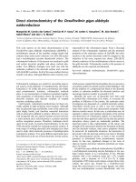
A
B
E
E
A
E
B
k
4
k
5
k
3
P
Q
C
E
C
R
k
9
k
7
E
D
D
S
k
8
Fig. 5. Reaction schematic of the racemic GD ⁄ HuPON1 system. A–D, various GD stereoisomers; E, PON1 enzyme; E
A
–E
D
, PON1–GD stere-
oisomer complexes; P-S, hydrolyzed products; k#, association ⁄ dissociation constants.
Table 1. Kinetic parameters for the enzymatic hydrolysis of the
various GD stereoisomers by recombinant wild-type HuPON1.
HuPON1 catalyzed GD hydrolysis was assayed in the presence of
at least 1.0 m
M CaCl
2
as described in Experimental procedures.
Kinetic results presented for each isomer were determined from at
least eight independent kinetic experiments (n ¼ 8).
GD isomer K
m
(mM) k
cat
(min
)1
) k
cat
⁄ K
m
(mM
)1
Æmin
)1
)
C–P– 0.91 ± 0.34 501 ± 45 625 ± 241
C–P+ 0.58 ± 0.23 593 ± 54 1160 ± 469
C+P– 0.71 ± 0.49 553 ± 163 1040 ± 465
C+P+ 0.27 ± 0.08 1030 ± 94 4130 ± 1090
D. T. Yeung et al. HuPON1 stereospecific hydrolysis of GD
FEBS Journal 274 (2007) 1183–1191 ª 2007 FEBS No claim to original US government works 1187
only twice that for the other three stereoisomers. The
K
m
values for the individual stereoisomers with wild-
type HuPON1 show a wider (almost fourfold)
variation, with the P– isomers exhibiting the highest
values. This suggests that either the P– isomers of GD
have a lower affinity for HuPON1 than the P+ iso-
mers, or that the P– isomers form more stable
enzyme–substrate complexes. Given the lack of infor-
mation about the rate of enzyme ⁄ substrate to
enzyme ⁄ product transitions in this system, it is not
currently possible to distinguish between these nonmu-
tually exclusive possibilities [35].
Data from HuPON1 presented in Table 1 suggest
that the observed variations in catalytic efficiency for
GD can be attributed largely to differences in the K
m
values of the enzyme for the various stereoisomers.
Although the stereochemistry of the substrates may be
important for binding, the results suggest that once
bound, the catalytic machinery is not overly sensitive
to the chirality of the groups around the phosphorus
atom. Therefore, small changes (via site-directed muta-
genesis) that reduce the K
m
for the more toxic isomers
might be singularly sufficient to make the enzyme a
viable bioscavenger for detoxification of OP anticholi-
nesterase poisons in vivo. For example, a reduction in
K
m
by 10-fold with no change in the V
max
value,
would enhance catalytic turnover of the more toxic
stereoisomers of GD such that they would be preferen-
tially hydrolyzed by several fold [4,5,7]. Such a mutant
would have considerable potential as a bioscavenger
capable of providing protection against nerve agent
poisoning.
Experimental procedures
Production of HuPON1
Wild-type recombinant HuPON1 enzymes were produced
as described previously [7]. Briefly, a pcDNA3 plasmid
(Invitrogen, Carlsbad, CA) encoding recombinant wild-type
HuPON1 was transiently transfected into human 293T
embryonic kidney cells, grown in DMEM (Cambrex Bio-
science, Walkersville, MD) supplemented with 5% fetal
bovine serum and 2% l-glutamine) at 70–90% confluency.
Secreted HuPON1 protein in cultured supernatant was har-
vested seven days after transfection. HuPON1 expression
was detected by immunblotting with mouse anti-HuPON1
mAb (kindly provided by R. James, University Hospital of
Geneva, Switzerland), probed with an alkaline-phosphatase
conjugated rabbit anti-mouse serum, and quantitated by
densitometry analysis (Un-Scan-It version 5.1, Silk Scienti-
fic Corp., Orem, UT) with a PON1 standard of known con-
centration (Randox Laboratories Ltd, Antrim, UK), and
verified by enzymatic assays for phenyl acetate and paraox-
on hydrolysis [36–38].
Determination of GD hydrolysis
Racemic GD (2.0 mgÆmL
)1
in saline), containing 2.5%
diisopropyl carbodiimide added as a stabilizer, was
obtained from the Research Development and Engineering
Command (Aberdeen Proving Ground, MD). Analysis
using nuclear magnetic resonance spectroscopy showed it to
be 96.7% pure. The pure individual GD stereoisomers were
previously prepared in ethyl acetate by the TNO Prins
Maurits Laboratory (Rijswijk, the Netherlands) [12].
Somanase activity was determined at room temperature as
detailed in Broomfield et al. [8] with minor variations. Specif-
ically, GD hydrolysis experiments were carried out using
1.50 mL of supernatant from cells transfected with either the
wild-type HuPON1 gene or empty vector. Supernatants were
incubated with the indicated concentrations of GD in 50 mm
glycine–NaOH buffer, pH 7.4 with 10 mm CaCl
2
. Total reac-
tion volume was 3.0 mL. At selected time intervals, 400 lL
aliquots were removed and inactivated through extraction
with an equal volume of GC-grade ethyl acetate (EM Sci-
ence, Cherry Hill, NJ) previously dried over a type 4A ⁄ grade
514 molecular sieve (Fisher Scientific, Fairlawn, NJ). The
organic layer (containing unhydrolyzed GD) was then
removed and dried over molecular sieve again. A 50-lL sam-
ple of this dried sample was collected and spiked with DFP
(Sigma-Aldrich, St Louis, MO) to a final concentration of
50 lm as the internal standard before injection into the gas
chromatograph [12]. The quantity of GD in each sample was
determined by comparison with both the DFP internal stand-
ard present in each sample and a standard GD calibration
curve. Calibration curves were obtained by using GD at five
different concentrations also spiked with a final concentra-
tion of 50 lm DFP in ethyl acetate as the internal standard.
Kinetic parameters of GD hydrolysis were determined using
at least eight different initial substrate concentrations that
ranged from 0.2 to 3.0 mm.
To determine the elution ⁄ retention time profile of the
four GD stereoisomers, samples of individual stereoisomers
were run under the same conditions as those used to deter-
mine the calibration curve.
Excess fluoride
⁄
racemization control experiments
To determine whether racemization occurs in our experi-
mental system, three independent control experiments were
performed under the same conditions as those used to
determine the calibration curve. First, 1.0 mm of racemic
GD was incubated with culture medium from cells trans-
fected with empty plasmid vector control in the presence of
excess fluoride ions (2.0 mm NaF). Second, semipurified
individual stereoisomers were also incubated with excessive
HuPON1 stereospecific hydrolysis of GD D. T. Yeung et al.
1188 FEBS Journal 274 (2007) 1183–1191 ª 2007 FEBS No claim to original US government works
fluoride ions. Finally, wild-type HuPON1 was reacted with
2mm GD as described above, but in the presence of 1 mm
NaF.
GC
⁄
MS analysis
GC separation of the GD stereoisomers was performed
using a modification of a previously developed method [24].
An Agilent 6890 gas chromatograph (Palo Alto, CA) was
fitted with a 20 m · 0.25 mm inside diameter Chiraldex c-
cyclodextrin trifluoroacetyl column, 0.125 lm film thickness
(Advanced Separation Technologies, Inc., Whippany, NJ).
A 2.5 m · 0.25 mm inside diameter cyano ⁄ phenyl ⁄ methyl
deactivated fused silica retention gap (Chrompack, Inc.,
Raritan, NJ) was installed at the injection end of the GC
and connected to the analytical column using a Chrompack
deactivated Quick-Seal glass connector. Helium was used as
the carrier gas at a linear velocity of 45 cmÆs
)1
. The oven
temperature was held initially at 80 °C for 14 min, pro-
grammed from 80 to 90 °Cat5°CÆmin
)1
, and held at
90 °C for 3 min. Split injections of 1 lL volume were made
using an Agilent 7683 autosampler. The injection port tem-
perature was 210 °C and the split ratio was % 1 : 100. The
GC was interfaced to an Agilent 5973 mass spectrometer
(MS) with an electron impact ion source. The MS operating
conditions were as follows: ion source pressure
% 1.0 · 10
)5
torr; source temperature, 230 °C; electron
energy, 70 eV; electron multiplier voltage +200 V relative
to the autotune setting; and transfer line temperature,
230 °C. The MS was operated using selected ion monitor-
ing (SIM). Four ions (m ⁄ z 69, 82, 99 and 126) were monit-
ored for the GD stereoisomers at a dwell time of 50 mÆs
)1
for each ion resulting in a scan rate of 3.77 cyclesÆs
)1
[39].
Three ions (m ⁄ z 69, 101 and 127) were monitored for DFP
[40]. A dwell time of 50 mÆs
)1
for each ion resulted in a
scan rate of 5 cyclesÆs
)1
. The m ⁄ z 126 and 127 ions were
used for quantitation of GD and DFP, respectively.
Calculation of kinetic constants
In the presence of a racemic mixture of GD, the catalyzed
reaction is analogous to simultaneously deriving the kin-
etic constants for the hydrolysis of four competitive sub-
strates. To do this, we used the model of GD–HuPON1
interaction shown in Fig. 5 and described in detail in the
supplementary material. The first-order rate equations of
the enzyme–substrate intermediates were set equal to zero
(the enzyme ‘steady-state’ assumption). The resulting set
of equations was solved to express the steady state
enzyme–substrate intermediate levels as functions of the
substrate concentrations and the kinetic parameters. A
conservation of enzyme assumption was employed to
obtain the free enzyme level in terms of the four enzyme–
substrate intermediates. Using these relationships, each
substrate rate equation was cast in terms of a single sub-
strate and integrated with respect to time to arrive at the
solutions. The derived solution for all four of the sub-
strates is shown below:
T
A
¼ðA
0
=V
maxA
Þð1 ÀðA=A
0
ÞðK
mA
=K
mA
ÞðV
maxA
=V
maxA
ÞÞ
þðB
0
=V
maxB
Þð1 ÀðA=A
0
ÞðK
mA
=K
mB
ÞðV
maxB
=V
maxA
ÞÞ
þðC
0
=V
maxC
Þð1 ÀðA=A
0
ÞðK
mA
=K
mC
ÞðV
maxC
=V
maxA
ÞÞ
þðD
0
=V
maxD
Þð1 ÀðA=A
0
ÞðK
mA
=K
mD
ÞðV
maxD
=V
maxA
ÞÞ
ðK
mA
=V
maxA
Þ Log
E
ðA=A
0
Þ
T
B
¼ðA
0
=V
maxA
Þð1 ÀðB=B
0
ÞðK
mB
=K
mA
ÞðV
maxA
=V
maxB
ÞÞ
þðB
0
=V
maxB
Þð1 ÀðB=B
0
ÞðK
mB
=K
mB
ÞðV
maxB
=V
maxB
ÞÞ
þðC
0
=V
maxC
Þð1 ÀðB=B
0
ÞðK
mB
=K
mC
ÞðV
maxC
=V
maxB
ÞÞ
þðD
0
=V
maxD
Þð1 ÀðB=B
0
ÞðK
mB
=K
mD
ÞðV
maxD
=V
maxB
ÞÞ
ðK
mB
=V
maxB
Þ LogðB=B
0
Þ
T
C
¼ðA
0
=V
maxA
Þð1 ÀðC=C
0
ÞðK
mC
=K
mA
ÞðV
maxA
=V
maxC
ÞÞ
þðB
0
=V
maxB
Þð1 ÀðC=C
0
ÞðK
mC
=K
mB
ÞðV
maxB
=V
maxC
ÞÞ
þðC
0
=V
maxC
Þð1 ÀðC=C
0
ÞðK
mC
=K
mC
ÞðV
maxC
=V
maxC
ÞÞ
þðD
0
=V
maxD
Þð1 ÀðC=C
0
ÞðK
mC
=K
mD
ÞðV
maxD
=V
maxC
ÞÞ
ðK
mC
=V
maxC
Þ LogðC=C
0
Þ
T
D
¼ðA
0
=V
maxA
Þð1 ÀðD=D
0
ÞðK
mD
=K
mA
ÞðV
maxA
=V
maxD
ÞÞ
þðB
0
=V
maxB
Þð1 ÀðD=D
0
ÞðK
mD
=K
mB
ÞðV
maxB
=V
maxD
ÞÞ
þðC
0
=V
maxC
Þð1 ÀðD=D
0
ÞðK
mD
=K
mC
ÞðV
maxC
=V
maxD
ÞÞ
þðD
0
=V
maxD
Þð1 ÀðD=D
0
ÞðK
mD
=K
mD
ÞðV
maxD
=V
maxD
ÞÞ
ðK
mD
=V
maxD
Þ LogðD=D
0
Þ:
Where K
mA
, K
mB
, K
mC
, and K
mD
are the Michaelis–Menten
constants for the four stereoisomers of GD; V
maxA
, V
maxB
,
V
maxC
, and V
maxD
are the corresponding maximum veloci-
ties; and A
0
, B
0
, C
0
, and D
0
are the initial concentrations of
each stereoisomer.
Although complex, the solutions give the time it would
take for each substrate (normalized to its initial level) to
fall to a particular level. As such, they were used to graph
curves of the substrate levels as functions of time. By
adjusting the kinetic parameters we were able to use a
Microsoft excel 2003 spreadsheet to fit these model curves
to the experimentally derived data (see Supplementary
material). The bimolecular rate constants (k
cat
⁄ K
m
) shown
in Table 1 are the average of eight independent experi-
ments
+
-standard deviation (n ¼ 8).
Acknowledgements
The work presented here by DTY is in partial fulfill-
ment of the requirements for the Doctorate of Philoso-
phy degree in Pharmacology from the University of
Maryland, Baltimore, MD. This research was suppor-
ted in part by an appointment to the Student Research
Participation Program at the US Army Medical
Research Institute of Chemical Defense administered
by the Oak Ridge Institute for Science and Education
through an interagency agreement between the US
D. T. Yeung et al. HuPON1 stereospecific hydrolysis of GD
FEBS Journal 274 (2007) 1183–1191 ª 2007 FEBS No claim to original US government works 1189
Department of Energy and USAMRMC. The opinions
or assertions contained herein are the private views of
the authors and are not to be construed as official or
as reflecting the views of the Army or the Department
of Defense.
References
1 Broomfield CA, Morris BC, Anderson R, Josse D &
Masson P (2000) Kinetics of nerve agent hydrolysis by
a human plasma enzyme. Proceedings of the CBMTS
III conference, 7–12 May 2000, Spiez, Switzerland.
2 Fu AL, Wang YX & Sun MJ (2005) Naked DNA pre-
vents soman intoxication. Biochem Biophys Res Com-
mun 328, 901–905.
3 Davies HG, Richter RJ, Keifer M, Broomfield CA, So-
walla J & Furlong CE (1996) The effect of the human
serum paraoxonase polymorphism is reversed with dia-
zoxon, soman and sarin. Nat Genet 14, 334–336.
4 Josse D, Lockridge O, Xie W, Bartels CF, Schopfer LM
& Masson P (2001) The active site of human paraoxo-
nase (PON1). J Appl Toxicol 21 (Suppl. 1), 7–11.
5 Josse D, Broomfield CA, Cerasoli D, Kirby S, Nichol-
son J, Bahnson B & Lenz DE (2002) Engineering of
HuPON1 for use as a catalytic bioscavenger in organo-
phosphate poisoning. Proceedings of the US Army
Medical Defense Bioscience Review. US Army Medical
Research Institute of Chemical Defense, Aberdeen Prov-
ing Ground, MD. DTIC no. pending.
6 Watkins LM, Mahoney HJ, McCulloch JK & Raushel
FM (1997) Augmented hydrolysis of diisopropyl fluoro-
phosphate in engineered mutants of phosphotriesterase.
J Biol Chem 272, 25596–25601.
7 Yeung DT, Josse D, Nicholson JD, Khanal A, McAn-
drew CW, Bahnson BJ, Lenz DE & Cerasoli DM
(2004) Structure ⁄ function analyses of human serum
paraoxonase (HuPON1) mutants designed from a
DFPase-like homology model. Biochim Biophys Acta
1702, 67–77.
8 Broomfield CA, Lenz DE & MacIver B (1986) The stabi-
lity of soman and its stereoisomers in aqueous solution:
toxicological considerations. Arch Toxicol 59, 261–265.
9 de Jong LP, van Dijk C & Benschop HP (1988) Hydro-
lysis of the four stereoisomers of soman catalyzed by
liver homogenate and plasma from rat, guinea pig and
marmoset, and by human plasma. Biochem Pharmacol
37, 2939–2948.
10 Sidell FR (1997) Nerve Agents. In Medical Aspects of
Chemical and Biological Warfare (Zajtchuk R, ed.), pp.
129–179. Office of the Surgeon General at TMM Publi-
cation, Washington, DC.
11 Benschop HP & de Jong LP (1998) Nerve agent stereoi-
somers: analysis, isolation, and toxicology. Accounts
Chem Res 21, 368–374.
12 Benschop HP, Konings CA, van Genderen J & de Jong
LP (1984) Isolation, anticholinesterase properties, and
acute toxicity in mice of the four stereoisomers of the
nerve agent soman. Toxicol Appl Pharmacol 72, 61–74.
13 Keijer JH & Wolring GZ (1969) Stereospecific aging of
phosphonylated cholinesterases. Biochim Biophys Acta
185, 465–468.
14 Benschop HP (1975) The absolute configuration of
chiral organophosphorus anticholinesterase poisoning.
Pesticide Biochem Physiol 5, 348–349.
15 Benschop HP, Berends F & de Jong LP (1981) GLC-
analysis and pharmacokinetics of the four stereoisomers
of Soman. Fundam Appl Toxicol 1, 177–182.
16 Lenz DE, Little JS, Broomfield CA & Ray R (1990)
Catalytic properties of nonspecific diisopropylfluoro-
phosphatases. In Chirality and Biological Activity
(Holmstedt B, Frank H & Testa B, eds), pp. 169–175.
Alan R. Liss, New York, NY.
17 Johnson JK, Cerasoli DM & Lenz DE (2005) Role of
immunogen design in induction of soman-specific mono-
clonal antibodies. Immunol Lett 96, 121–127.
18 Little JS, Broomfield CA, Fox-Talbot MK, Boucher LJ,
MacIver B & Lenz DE (1989) Partial characterization
of an enzyme that hydrolyzes sarin, soman. tabun, and
diisopropyl phosphorofluoridate (DFP). Biochem Phar-
macol 38, 23–29.
19 Aharoni A, Gaidukov L, Yagur S, Toker L, Silman I &
Tawfik DS (2004) Directed evolution of mammalian
paraoxonases PON1 and PON3 for bacterial expression
and catalytic specialization. Proc Natl Acad Sci USA
101, 482–487.
20 Harel M, Aharoni A, Gaidukov L, Brumshtein B,
Khersonsky O, Meged R, Dvir H, Ravelli RB, McCarthy
A, Toker L et al. (2004) Structure and evolution of the
serum paraoxonase family of detoxifying and anti-athero-
sclerotic enzymes. Nat Struct Mol Biol 11, 412–419.
21 Josse D, Xie W, Renault F, Rochu D, Schopfer LM,
Masson P & Lockridge O (1999) Identification of resi-
dues essential for human paraoxonase (PON1)
arylesterase ⁄ organophosphatase activities. Biochemistry
38, 2816–2825.
22 Amitai G, Gaidukov L, Adani R, Yishay S, Yacov G,
Kushnir M, Teitlboim S, Lindenbaum M, Bel P,
Khersonsky O et al. (2006) Enhanced stereoselective
hydrolysis of toxic organophosphates by directly
evolved variants of mammalian serum paraoxonase.
FEBS J 273, 1906–1919.
23 Khersonsky O & Tawfik DS (2006) The histidine 115–his-
tidine 134 dyad mediates the lactonase activity of mam-
malian serum paraoxonases. J Biol Chem 281, 7649–7656.
24 Smith JR & Schlager JJ (1996) Gas chromatographic
separation of the stereoisomers of organophosphorus
chemical warfare agents using cyclodextrin capillary col-
umns. J High Resolution Chromatogr 19, 151–154.
HuPON1 stereospecific hydrolysis of GD D. T. Yeung et al.
1190 FEBS Journal 274 (2007) 1183–1191 ª 2007 FEBS No claim to original US government works
25 Benschop HP, Konings CA, van Genderen J & de Jong
LP (1984) Isolation, in vitro activity, and acute toxicity
in mice of the four stereoisomers of soman. Fundam
Appl Toxicol 4, S84–S95.
26 Benschop HP, Bijleveld EC, Otto MF, Degenhardt CE,
Van Helden HP & de Jong LP (1985) Stabilization and
gas chromatographic analysis of the four stereoisomers
of 1,2,2-trimethylpropyl methylphosphonofluoridate
(soman) in rat blood. Anal Biochem 151, 242–253.
27 de Jong LP, Bijleveld EC, van Dijk C & Benschop HP
(1987) Assay of the chiral organophosphate, soman, in
biological samples. Int J Environ Anal Chem 29, 179–197.
28 Harvey SP, Kolakowski JE, Cheng TC, Rastogi VK,
Reiff LP, DeFrank JJ, Raushel FM & Hill C (2005)
Stereospecificity in the enzymatic hydrolysis of cyclo-
sarin (GF). Enzyme Microbial Technol 37, 547–555.
29 Li W, Lum KT, Chen-Goodspeed M, Sogorb MA &
Raushel FM (2001) Stereoselective detoxification of
chiral sarin and soman analogues by phosphotriesterase.
Bioorg Med Chem 9, 2083–2091.
30 Lacinski M, Skorupski W, Cieslinski A, Sokolowska J,
Trzeciak WH & Jakubowski H (2004) Determinants of
homocysteine-thiolactonase activity of the paraoxonase-
1 (PON1) protein in humans. Cell Mol Biol (Noisy-
le-Grand) 50, 885–893.
31 Primo-Parmo SL, Sorenson RC, Teiber J & Du La BN
(1996) The human serum paraoxonase ⁄ arylesterase gene
(PON1) is one member of a multigene family. Genomics
33, 498–507.
32 Sorenson RC, Primo-Parmo SL, Kuo CL, Adkins S,
Lockridge O & Du La BN (1995) Reconsideration of
the catalytic center and mechanism of mammalian
paraoxonase ⁄ arylesterase. Proc Natl Acad Sci USA 92,
7187–7191.
33 Rodrigo L, Mackness B, Durrington PN, Hernandez A &
Mackness MI (2001) Hydrolysis of platelet-activating
factor by human serum paraoxonase. Biochem J 354, 1–7.
34 Aharoni A, Gaidukov L, Khersonsky OMcQGS, Rood-
veldt C & Tawfik DS (2005) The ‘evolvability’ of pro-
miscuous protein functions. Nat Genet 37, 73–76.
35 Tipton KF (1973) Enzyme kinetics in relation to enzyme
inhibitors. Biochem Pharmacol 22, 2933–2941.
36 Gan KN, Smolen A, Eckerson HW & Du La BN
(1991) Purification of human serum paraoxonase ⁄
arylesterase. Evidence for one esterase catalyzing both
activities. Drug Metab Dispos 19, 100–106.
37 Yeung DT, Lenz DE & Cerasoli DM (2005) Analysis of
active-site amino-acid residues of human serum paraox-
onase using competitive substrates. FEBS J 272, 2225–
2230.
38 Aviram M, Billecke S, Sorenson R, Bisgaier C, Newton
R, Rosenblat M, Erogul J, Hsu C, Dunlop C & Du La
B (1998) Paraoxonase active site required for protection
against LDL oxidation involves its free sulfhydryl
group and is different from that required for its
arylesterase ⁄ paraoxonase activities: selective action of
human paraoxonase allozymes Q and R. Arterioscler
Thromb Vasc Biol 18, 1617–1624.
39 Wils ERJ (2005) Gas chromatography⁄ mass spectrome-
try in analysis of chemicals related to the chemical
weapons convention. In Chemical Weapons Convention
Chemical Analysis (Mesilaakso, M, ed.), pp. 249–279.
John Wiley, Hoboken.
40 Enqvist J, Manninen A, Ravio P, Kokko M, Kuronen
P, Hesso A, Savolahti P, Ali-Mattila E, Kenttamaa H,
Rautio M et al. (1983) Identification of precursors of
warfare agents, degradation products of non-phospho-
rus agents, and some potential agents. In Systematic
Identification of Chemical Warefare Agents (Miettinen
JK, Hase T, Hirsjarvi P, Paasivirta J, Pyysalo H &
Rahkamaa E, eds). The Ministry for Foreign Affairs of
Finland, Helsinki.
Supplementary material
The following supplementary material is available
online:
Fig. S1. Reaction schematic of the racemic GD
HuPON1 system.
Fig. S2. Hydrolysis of 0.37 mm racemic GD by
HuPON1.
Fig. S3. Comparison of theoretical and numerical solu-
tions.
Fig. S4. Comparison of assumed enzyme ‘steady-state’
levels and actual (numerically integrated) levels.
Fig. S5. Lineweaver–Burke plot of theoretical solutions
and measured data for hydrolysis of 1.67 mm racemic
GD by HuPON1.
Fig. S6. Hanes–Woolf plot of theoretical solutions and
measured data for hydrolysis of 1.67 mm racemic GD
by HuPON1.
Fig. S7. Eadie–Hofstee plot of theoretical solutions
and measured data for hydrolysis of 1.67 mm racemic
GD by HuPON1.
This material is available as part of the online article
from
Please note: Blackwell Publishing is not responsible
for the content or functionality of any supplementary
materials supplied by the authors. Any queries (other
than missing material) should be directed to the corres-
ponding author for the article.
D. T. Yeung et al. HuPON1 stereospecific hydrolysis of GD
FEBS Journal 274 (2007) 1183–1191 ª 2007 FEBS No claim to original US government works 1191