Báo cáo khoa học: Molecular basis of cerebral neurodegeneration in prion diseases pdf
Bạn đang xem bản rút gọn của tài liệu. Xem và tải ngay bản đầy đủ của tài liệu tại đây (531.91 KB, 6 trang )
MINIREVIEW
Molecular basis of cerebral neurodegeneration in prion
diseases
Jo
¨
rg Tatzelt
1
and Hermann M. Scha
¨
tzl
2
1 Department of Biochemistry, Neurobiochemistry, Ludwig-Maximilians-University Munich, Germany
2 Institute of Virology, Technical University of Munich, Germany
Prion diseases in their ‘classical’ and naturally occur-
ring forms are characterized by both neurodegenera-
tion, with clinical symptoms, and propagation of
infectious prions, the latter giving rise to the typical
transmissibility within and between species [1–5]. The
formation of the disease-associated isoform of prion
protein (PrP
Sc
) [i.e. the misfolded and partially protein-
ase K (PK)-resistant isoform of the cellular prion pro-
tein (PrP
C
)] is closely linked to the propagation of
infectious prions, but apparently is not sufficient to
induce neurodegeneration. Here, an important role in
mediating the neurodegeneration process is increasing
for PrP
C
. Evidence for this was found in neurografting
approaches [6], in conditional prion protein (PrP)
knockout studies [7] and in in vivo cross-linking experi-
ments of PrP
C
[8].
Some genetic forms of human prion disease appear
less transmissible, or even nontransmissible. With one
exception this is also true for the transgenic animal mod-
els established to mimic genetic prion diseases [1–5].
This nontransmissible character is reminiscent of ‘pro-
teinopathies’, sometimes linked to PrP overexpression,
rather than classical prion diseases, and is in line
with the concept of ‘nontransmissible prionopathies’
Keywords
amyloid; neurodegeneration; prion protein;
prion; trafficking; transmissibility
Correspondence
J. Tatzelt, Department of Biochemistry,
Ludwig-Maximilians-University Munich,
Schillerstrasse 44, 80336 Munich, Germany
Fax: +49 89 2180 75415
Tel: +49 89 2180 75442
E-mail:
H. M. Scha
¨
tzl, Institute of Virology,
Technical University Munich (TUM),
Trogerstraße 30, 81675 Munich, Germany
Fax: +49 89 4140 6823
Tel: +49 89 4140 6820
E-mail:
(Received 2 August 2006, revised 30
November 2006, accepted 4 December
2006)
doi:10.1111/j.1742-4658.2007.05633.x
The biochemical nature and the replication of infectious prions have been
intensively studied in recent years. Much less is known about the cellular
events underlying neuronal dysfunction and cell death. As the cellular func-
tion of the normal cellular isoform of prion protein is not exactly known,
the impact of gain of toxic function or loss of function, or a combination
of both, in prion pathology is still controversial. There is increasing evi-
dence that the normal cellular isoform of the prion protein is a key medi-
ator in prion pathology. Transgenic models were instrumental in dissecting
propagation of prions, disease-associated isoforms of prion protein and
amyloid production, and induction of neurodegeneration. Four experimen-
tal avenues will be discussed here which address scenarios of inappropriate
trafficking, folding, or targeting of the prion protein.
Abbreviations
Ctm
PrP, transmembrane form of PrP with the COOH-terminus in the endoplasmic reticulum lumen; cytoPrP, cytosolic PrP; Dpl, doppel
protein; ER, endoplasmic reticulum; HD, hydrophobic domain;
Ntm
PrP, transmembrane form of PrP with the NH
2
-terminus in the
endoplasmic reticulum lumen; PK, proteinase K; PrP, prion protein; PrP
C
, normal cellular isoform of PrP; PrP
Sc
, disease-associated isoform of
PrP; secPrP, secretory PrP.
606 FEBS Journal 274 (2007) 606–611 ª 2007 The Authors Journal compilation ª 2007 FEBS
introduced by C. Weissmann [3]. Two common phases
in prion diseases can be described. In a first phase, with
apparently obligate requirement for PrP
C
expression,
profound conformational changes give rise to PrP
aggregation and the formation of PrP
Sc
, resulting in
more or less pronounced amyloid formation. This is
paralleled by replication of infectious prions and trans-
missibility. In a second phase this is transduced into
physiological dysfunction in the central nervous system,
and neuronal damage.
On the other hand, transgenic mouse models have
been helpful in revealing that these features can occur
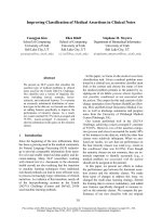
independently. There are models available which des-
cribe scenarios for neurodegeneration alone, prion pro-
pagation alone, or a combination of both (Fig. 1). In
the following, such in vivo models are described which
address certain aspects of membrane topology, folding,
intracellular targeting and trafficking of PrP. The term
‘toxicity’ is used by us here for induction of neuro-
degeneration, and PrP
Sc
is used synonymously for the
pathological form of PrP.
Toxicity of transmembrane isoforms
of PrP
The evidence that PrP
Sc
is directly neurotoxic is con-
troversial [6] and has fueled the search for other PrP
conformers involved in pathophysiological scenarios.
In the 1980s, it was shown, in cell-free translation-
translocation systems, that PrP can be found in more
than one topologic form [9]. The major form is the
fully translocated isoform giving rise to the known,
fully mature, PrP in the secretory pathway, located
finally at the outer leaflet of the plasma membrane by
its GPI-anchor (secPrP). In addition, the existence of
two different transmembrane forms of PrP was verified
[10]. One form, termed C-trans transmembrane
(
Ctm
PrP), has its COOH-terminus in the endoplasmic
reticulum (ER) lumen. The other form, termed N-trans
transmembrane (
Ntm
PrP), has its NH
2
-terminus in the
ER lumen. Both forms appear to span the membrane
at the same hydrophobic stretch in PrP [in general, res-
idues 110–135, previously termed TM1, now referred
to as the hydrophobic domain (HD)] (Fig. 2). Interest-
ingly, it was shown that during normal biogenesis of
PrP, only about two-thirds is expressed as the secre-
tory form (secPrP), less than 10% as the
Ctm
PrP and
the remainder as
Ntm
PrP. Naturally occurring and arti-
ficial mutations in the membrane-spanning segment
can lead to significantly increased generation of
Ctm
PrP. In addition, several pieces of evidence have
linked
Ctm
PrP to neurodegeneration in transgenic mice
PrP
cS
prion propagation
prion propagation
PrP
c
neurodegeneration
neurodegeneration
stress sensitive?
Fig. 1. Scheme illustrating putative scenarios in PrP pathology. In
the middle, the ‘classical’ pathway, resulting in neurodegeneration
and PrP propagation, is depicted. The other pathways are from
transgenic mouse models characterized by either PrP-induced neu-
rodegeneration or prion propagation. A loss of function of PrP
C
might result in sensitizing neurons to stress stimuli.
OHCOHC
SS-IPGSS-RE
lpD
OHCOHC
SS-IPG
β
1
α
1
β
2
α
2
α
3
β
1
α
1
β
2
α
2
α
3
α
1
β
2
α
2
α
3
RODHSS-RE
PrP
c
O
H
C
OHC
SS-IPGSS
-
RE
PrP F( 32-134)
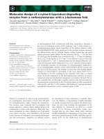
Fig. 2. Structure of PrP
c
, Dpl, and PrPDF(D32–134). Schematic presentation of the proteins mentioned in the text. a, alpha helix; b, beta
strand; ER-SS, endoplasmic reticulum signal sequence; GPI-SS, GPI anchor signal sequence; HD, hydrophobic domain (putative transmem-
brane domain of
Ctm
PrP); OR, octarepeat.
J. Tatzelt & H. M. Scha
¨
tzl Cerebral neurodegeneration in prion diseases
FEBS Journal 274 (2007) 606–611 ª 2007 The Authors Journal compilation ª 2007 FEBS 607
and to some heritable prion diseases (mutation A117V
in the Gerstmann–Stra
¨
ussler–Scheinker syndrome).
The
Ctm
PrP isoform has been hypothesized to repre-
sent an important intermediate in the pathway of prion-
induced neurodegeneration, by escaping ER resident
quality control mechanisms [10,11]. Of note, this takes
place in the absence of generation of ‘classical’ PK-
resistant PrP
Sc
and of infectious prions. On the other
hand, it was later shown that a prion infection appar-
ently can trigger the generation of ‘toxic’
Ctm
PrP [11].
This would link transmissible and genetic prion diseases
and provide a common pathway of neurodegeneration
in prion disease. Of note, another group has found, in
an additional transgenic model for
Ctm
PrP, that the
neurodegenerative phenotype is strongly dependent on
the co-expression of endogenous wild-type PrP [12].
Toxicity of PrP located in the cytosol
During the initial characterization of the biosynthesis
of PrP, in vitro studies revealed that PrP could, at least
in part, be localized in the cytosolic compartment. As
mentioned above, two different transmembrane topo-
logies were also found (
Ntm
PrP and
Ctm
PrP) and the
increased synthesis of
Ctm
PrP has been shown to coin-
cide with progressive neurodegeneration [10]. In these
isoforms, the internal HD (amino acids 112–135) of
PrP serves as a transmembrane domain [13]. In a yeast
model the HD interfered with the post-translational
import of PrP into the ER, and as a consequence yeast
growth was impaired and misfolded PrP accumulated
in the cytosol [14]. Interestingly, both
Ntm
PrP and
Ctm
PrP are partly cytosolic proteins. Nearly half of the
PrP molecule is exposed to the cytoplasm in the trans-
membrane configuration and could thereby facilitate
‘toxic’ signaling events residing in the cytoplasm.
Strong evidence that cytosolic PrP (cytoPrP) is neu-
rotoxic emerged from a transgenic mouse model. Mice
expressing a PrP mutant with a deleted N-terminal ER
targeting signal acquired severe ataxia owing to cere-
bellar degeneration and gliosis [15]. Cytotoxic effects
of cytoPrP were also observed in some cell culture
models [15–19], whereas in other studies the expression
of cytoPrP seemed not to interfere with cellular viabil-
ity [20,21].
Of interest, a small fraction of wild-type PrP can
also be found in the cytosol of cultured cells [22,23]
and neurons [24]. Moreover, some pathogenic muta-
tions linked to Gerstmann–Stra
¨
ussler–Scheinker syn-
drome in humans, such as Q160Stop and W145Stop,
significantly increase the fraction of cytosolically locali-
zed PrP [25,26]. These mutations do not change the
N-terminal ER signal sequence but delete parts of the
highly ordered C-terminal domain, revealing that
this region is necessary for the import of PrP
C
into the
ER [25].
What is the mechanism of cytoPrP-induced toxicity?
The first studies addressing this important issue were
recently described. By employing cytoPrP transgenic
mice [15], it was shown that toxicity correlates with
membrane localization of cytoPrP [19]. In a different
study, apoptotic effects were linked to the association of
cytoPrP with Bcl-2, an anti-apoptotic protein localized
at the cytosolic site of ER and mitochondria membranes
[17]. It also appeared that proteasomal activity and
cytosolic chaperones, such as Hsp70 and Hsp40, can
modulate the toxic potential of cytoPrP [17]. Of note, a
variety of previous reports indicated that PrP can inter-
act with chaperones, and that chaperones can modulate
the formation of misfolded PrP conformers [27].
Another important question involves the possible link
between the demise of scrapie-infected neurons and the
formation of cytosolically localized PrP. The first clues
from cell culture work show that aggresome formation
in scrapie-infected mouse neuroblastoma (ScN2a) cells
induces caspase-3 activation and apoptosis [28].
Toxicity of PrP located at the plasma membrane
Spontaneous cerebellar neurodegeneration in certain
strains of PRNP
0 ⁄ 0
mice [29] led to the discovery of
doppel (Dpl), a protein structurally related to PrP
C
[30]. Under physiological conditions, Dpl seems not to
be expressed in the brain; however, ectopic neuronal
expression of Dpl induces Purkinje cell degeneration
[31,32]. Dpl is complex glycosylated, harbors a
GPI-anchor and shows structural homology with the
C-terminal globular domain of PrP
C
, but lacks the
N-terminal octarepeats and the internal HD [33]
(Fig. 2). Interestingly, the expression of PrPDF, a
mutant devoid of the octarepeats and the HD (D32–
134), induces cerebellar degeneration similarly to Dpl
[32,34,35]. The neurotoxic potential of PrP variants
was found to correlate with the disruption of the HD,
indicating that the deletion of this domain, rather then
the absence of the octarepeat region, is linked to the
neurotoxic properties of PrPDF. The internal HD was
identified as an important domain for basolateral sort-
ing of PrP
C
. Moreover, Dpl, containing either the
whole N-terminal domain of PrP
C
or the HD only,
was sorted basolaterally, indicating that this domain
acts as a dominant sorting signal. Vice versa, Dpl or
PrP
C
lacking the HD were found mainly at the apical
surface of MDCK cells [36]. An interesting activity of
PrP
C
emerged from co-expression experiments in trans-
genic animals: full-length PrP
C
can antagonize both
Cerebral neurodegeneration in prion diseases J. Tatzelt & H. M. Scha
¨
tzl
608 FEBS Journal 274 (2007) 606–611 ª 2007 The Authors Journal compilation ª 2007 FEBS
Dpl- and PrPDF-induced neurodegeneration [32,35].
This effect is difficult to understand in the light of the
differential sorting of PrP
C
and Dpl. However, in
polarized cells expressing Dpl and PrP
C
, both proteins
are found at the same cellular locale, which could be a
prerequisite for a functional interaction [36].
Several studies have indicated that Dpl or PrPDF
can induce apoptotic cell death [37–39]. However, the
major question remains how these molecules, possibly
located at the plasma membrane, can activate pro-
apoptotic signaling pathways. In this context it might
be interesting to recall studies addressing the physiolo-
gical role of PrP
C
. They revealed that PrP
C
has neuro-
protective activity after an ischemic insult [40–43],
supports self-renewal of hematopoietic stem cells and
positively regulates neural precursor proliferation
[44,45]. This indicates that the deletion of the internal
HD could change a neuroprotective activity of wild-
type PrP to the pro-apoptotic activity of mutants, such
as PrPDF. The HD might directly mediate an interac-
tion of PrP
C
with accessory proteins, such as trans-
membrane proteins involved in PrP-induced signaling.
Alternatively, deletion of the HD could indirectly
affect intermolecular interactions by modulating the
PrP
C
tertiary or quaternary structure.
No central nervous system toxicity of
PrP missing the GPI-anchor
A leading role of neuronally expressed PrP
c
in medi-
ating neurodegeneration first emerged from neurograft-
ing studies [6] and later was reinforced by a
conditional PrP knockout analysis [7]. In line with
these findings, cross-linking studies of PrP
C
with
monoclonal antibodies in vivo demonstrated the neuro-
toxic signaling potential of PrP
C
[8]. An unexpected
twist came very recently by re-addressing an old obser-
vation. In prion-infected cultured mouse cells, it was
found that the absence of the GPI moiety of PrP redu-
ces the formation of PrP
Sc
[46,47]. Recently, two lines
of transgenic mice were produced which expressed a
PrP mutant devoid of the GPI-anchor. PrPDGPI
[named GPI(–)PrPsen in the mouse study] was
expressed in these mice and, similarly to the findings in
cultured cells, was efficiently secreted [48,49]. After
infection with three different prion strains, the trans-
genic mice did not develop clinical symptoms. Quite
unexpectedly, however, the brains of these mice con-
tained high prion titers, about 1 : 10 compared with
scrapie-infected wild-type mice. Moreover, the amount
of PrP
Sc
at 500 days post infection in the scrapie-infec-
ted PrPDGPI mice was higher than in scrapie-infected
wild-type mice. This was reflected by a high load of
amyloid plaques, which are less frequent in PrP wild-
type mice. Interestingly, the pathological features were
most pronounced along blood vessels [48].
In conclusion, although many more PrP plaques and
more PK-resistant PrP
Sc
were present than usual, the
mice harboured less prion infectivity in the brain and
showed no clinical signs. How does this all fit together?
First, the form of amyloid was different, reflected by a
different biophysical behaviour of nonglycosylated PrP
apparently highly prone to the formation of higher
aggregates. It seems to be a common underlying idea in
neurodegeneration that amyloid plaques are more an
end-product and that smaller units on the road of aggre-
gation (‘toxic folding intermediates’) are crucial players.
Second, the findings could indicate that neurotoxicity of
PrP
Sc
is linked to its propagation at the plasma mem-
brane or along the endocytic pathway. There might be
an ‘undesired and deadly’ interaction between PrP
Sc
and
PrP
C
, resulting in a ‘false’ or prolonged stimulation of
PrP
C
, thereby transducing a neurotoxic signal via PrP
C
.
Alternatively, PrP
Sc
or precursors thereof directly
interact with other cell-associated signaling molecules.
Regardless, the exact mechanism of the study clearly
emphasizes a critical role of the GPI anchor of PrP in
the pathogenesis of prion diseases.
Concluding remarks
The puzzle of how infectious prions, PrP
Sc
, and neuro-
degeneration are interconnected is still far from being
solved. Obviously, prion-induced neurodegeneration
may require membrane-anchored PrP in neurons,
whereas expression of secreted PrP DGPI or PrP
C
in
glia cells can promote the propagation of infectious
prions without clinical symptoms, or at least with a
significantly delayed onset. On the other hand, the des-
cribed transgenic mice models revealed neurodegenera-
tion induced by aberrant PrP conformers in the
absence of prion propagation. It will now be important
to show that neurotoxicity induced by alterations in
folding or trafficking of PrP
C
is indeed relevant to
neuronal cell death in a prion-diseased brain. How-
ever, they are valuable models to systematically study
pathways induced by neurotoxic protein conforma-
tions, a challenging question also in other neurodegen-
erative disorders, such as Alzheimer’s, polyglutamine
and Parkinson’s disease.
Acknowledgements
The work of the authors is supported by grants from the
‘Deutsche Forschungsgemeinschaft’, the ‘Bayerisches
Staatsministerium fu
¨
r Wissenschaft, Forschung und
J. Tatzelt & H. M. Scha
¨
tzl Cerebral neurodegeneration in prion diseases
FEBS Journal 274 (2007) 606–611 ª 2007 The Authors Journal compilation ª 2007 FEBS 609
Kunst’, the ‘Bayerisches Staatsministerium fu
¨
r Verbr-
aucherschutz’, the ‘Bundesministerium fu
¨
r Bildung und
Forschung’, and the European Union.
References
1 Prusiner SB (1998) Prions. Proc Natl Acad Sci USA 95,
13363–13383.
2 Aguzzi A & Polymenidou M (2004) Mammalian prion
biology: one century of evolving concepts. Cell 116,
313–327.
3 Weissmann C & Flechsig E (2003) PrP knock-out and
PrP transgenic mice in prion research. Br Med Bull 66,
43–60.
4 Collinge J (2001) Prion diseases of humans and animals:
their causes and molecular basis. Annu Rev Neurosci 24,
519–550.
5 Nunziante M, Gilch S & Scha
¨
tzl HM (2003) Prion dis-
eases: from molecular biology to intervention strategies.
Chembiochem 4, 1268–1284.
6 Brandner S, Isenmann S, Raeber A, Fischer M,
Sailer A, Kobayashi Y, Marino S, Weissmann C &
Aguzzi A (1996) Normal host prion protein necessary
for scrapie-induced neurotoxicity. Nature 379, 339–
343.
7 Mallucci G, Dickinson A, Linehan J, Klohn PC, Brand-
ner S & Collinge J (2003) Depleting neuronal PrP in
prion infection prevents disease and reverses spongiosis.
Science 302, 871–874.
8 Solforosi L, Criado JR, McGavern DB, Wirz S, San-
chez-Alavez M, Sugama S, DeGiorgio LA, Volpe BT,
Wiseman E, Abalos G et al. (2004) Cross-linking cellu-
lar prion protein triggers neuronal apoptosis in vivo.
Science 303, 1514–1516.
9 Hay B, Barry RA, Lieberburg I, Prusiner SB & Lin-
gappa VR (1987) Biogenesis and transmembrane orien-
tation of the cellular isoform of the scrapie prion
protein. Mol Cell Biol 7 , 914–920.
10 Hegde RS, Mastrianni JA, Scott MR, DeFea KA,
Tremblay P, Torchia M, DeArmond SJ, Prusiner SB &
Lingappa VR (1998) A transmembrane form of the
prion protein in neurodegenerative disease. Science 279,
827–834.
11 Hegde RS, Tremblay P, Groth D, DeArmond SJ, Prus-
iner SB & Lingappa VR (1999) Transmissible and
genetic prion diseases share a common pathway of neu-
rodegeneration. Nature 402, 822–826.
12 Stewart RS, Piccardo P, Ghetti B & Harris DA (2005)
Neurodegenerative illness in transgenic mice expressing
a transmembrane form of the prion protein. J Neurosci
25, 3469–3477.
13 Yost CS, Lopez CD, Prusiner SB, Myers RM & Lin-
gappa VR (1990) Non-hydrophobic extracytoplasmic
determinant of stop transfer in the prion protein. Nature
343, 669–672.
14 Heller U, Winklhofer KF, Heske J, Reintjes A & Tat-
zelt J (2003) Post-translational import of the prion pro-
tein into the endoplasmic reticulum interferes with cell
viability: a critical role for the putative transmembrane
domain. J Biol Chem 278, 36139–36147.
15 Ma J, Wollmann R & Lindquist S (2002) Neurotoxicity
and neurodegeneration when PrP accumulates in the
cytosol. Science 298, 1781–1785.
16 Grenier C, Bissonnette C, Volkov L & Roucou X
(2006) Molecular morphology and toxicity of cytoplas-
mic prion protein aggregates in neuronal and non-neu-
ronal cells. J Neurochem 97, 1456–1466.
17 Rambold AS, Miesbauer M, Rapaport D, Bartke T, Ba-
ier M, Winklhofer KF & Tatzelt J (2006) Association of
Bcl-2 with misfolded prion protein is linked to the toxic
potential of cytosolic PrP. Mol Biol Cell 17, 3356–3368.
18 Rane NS, Yonkovich JL & Hegde RS (2004) Protection
from cytosolic prion protein toxicity by modulation of
protein translocation. EMBO J
23, 4550–4559.
19 Wang X, Wang F, Arterburn L, Wollmann R & Ma J
(2006) The interaction between cytoplasmic prion pro-
tein and the hydrophobic lipid core of membrane corre-
lates with neurotoxicity. J Biol Chem 281, 13559–13565.
20 Fioriti L, Dossena S, Stewart LR, Stewart RS, Harris
DA, Forloni G & Chiesa R (2005) Cytosolic prion pro-
tein (PrP) is not toxic in N2a cells and primary neurons
expressing pathogenic PrP mutations. J Biol Chem 280,
11320–11328.
21 Roucou X, Guo Q, Zhang Y, Goodyer CG & LeBlanc
AC (2003) Cytosolic prion protein is not toxic and pro-
tects against Bax-mediated cell death in human primary
neurons. J Biol Chem 278, 40877–40881.
22 Ma J & Lindquist S (2001) Wild-type PrP and a mutant
associated with prion disease are subject to retrograde
transport and proteasome degradation. Proc Natl Acad
Sci USA 98, 14955–14960.
23 Yedidia Y, Horonchik L, Tzaban S, Yanai A & Tara-
boulos A (2001) Proteasomes and ubiquitin are involved
in the turnover of the wild-type prion protein. EMBO J
20, 5383–5391.
24 Mironov AJ, Latawiec D, Wille H, Bouzamondo-Bern-
stein E, Legname G, Williamson RA, Burton D, De-
Armond SJ, Prusiner SB & Peters PJ (2003) Cytosolic
prion protein in neurons. J Neurosci 23, 7183–7193.
25 Heske J, Heller U, Winklhofer KF & Tatzelt J (2004)
The C-terminal domain of the prion protein is necessary
and sufficient for import into the endoplasmic reticu-
lum. J Biol Chem 279, 5435–5443.
26 Zanusso G, Petersen RB, Jin T, Jing Y, Kanoush R,
Ferrari S, Gambetti P & Singh N (1999) Proteasomal
degradation and N-terminal protease resistance of the
codon 145 mutant prion protein. J Biol Chem 274,
23396–23404.
27 Winklhofer KF & Tatzelt J (2006) The role of chaper-
ones in Parkinson’s disease and prion diseases. In
Cerebral neurodegeneration in prion diseases J. Tatzelt & H. M. Scha
¨
tzl
610 FEBS Journal 274 (2007) 606–611 ª 2007 The Authors Journal compilation ª 2007 FEBS
Molecular Chaperones in Health and Diseases, Vol. 172
(Gaestel M, ed.), pp. 221–258. Springer-Verlag, Berlin ⁄
Heidelberg.
28 Kristiansen M, Messenger MJ, Klohn PC, Brandner S,
Wadsworth JD, Collinge J & Tabrizi SJ (2005) Disease-
related prion protein forms aggresomes in neuronal cells
leading to caspase-activation and apoptosis. J Biol
Chem 280, 38851–38861.
29 Sakaguchi S, Katamine S, Nishida N, Moriuchi R,
Shigematsu K, Sugimoto T, Nakatani A, Kataoka Y,
Houtani T, Shirabe S et al. (1996) Loss of cerebellar
Purkinje cells in aged mice homozygous for a disrupted
PrP gene. Nature 380, 528–531.
30 Moore RC, Lee IY, Silverman GL, Harrison PM,
Strome R, Heinrich C, Karunaratne A, Pasternak SH,
Chishti MA, Liang Y et al. (1999) Ataxia in prion pro-
tein (PrP)-deficient mice is associated with upregulation
of the novel PrP-like protein Doppel. J Mol Biol 292,
797–817.
31 Li A, Sakaguchi S, Shigematsu K, Atarashi R, Roy BC,
Nakaoke R, Arima K, Okimura N, Kopacek J & Kata-
mine S (2000) Physiological expression of the gene for
PrP-like protein, PrPLP ⁄ Dpl, by brain endothelial cells
and its ectopic expression in neurons of PrP-deficient
mice ataxic due to Purkinje cell degeneration. Am J
Pathol 157, 1447–1452.
32 Rossi D, Cozzio A, Flechsig E, Klein MA, Rulicke T,
Aguzzi A & Weissmann C (2001) Onset of ataxia and
Purkinje cell loss in PrP null mice inversely correlated
with Dpl level in brain. EMBO J 20, 694–702.
33 Mo H, Moore RC, Cohen FE, Westaway D, Prusiner
SB, Wright PE & Dyson HJ (2001) Two different neu-
rodegenerative diseases caused by proteins with similar
structures. Proc Natl Acad Sci USA 98, 2352–2357.
34 Flechsig E, Hegyi I, Leimeroth R, Zuniga A, Rossi D,
Cozzio A, Schwarz P, Rulicke T, Gotz J, Aguzzi A
et al. (2003) Expression of truncated PrP targeted to
Purkinje cells of PrP knockout mice causes Purkinje cell
death and ataxia. EMBO J 22, 3095–3101.
35 Shmerling D, Hegyi I, Fischer M, Bla
¨
ttler T, Brandner
S, Go
¨
tz J, Ru
¨
licke T, Flechsig E, Cozzio A, von Meh-
ring C et al. (1998) Expression of amino-terminally
truncated PrP in the mouse leading to ataxia and speci-
fic cerebellar lesions. Cell 93, 203–214.
36 Uelhoff A, Tatzelt J, Aguzzi A, Winklhofer KF &
Haass C (2005) A pathogenic PrP mutation and doppel
interfere with polarized sorting of the prion protein.
J Biol Chem 280, 5137–5140.
37 Drisaldi B, Coomaraswamy J, Mastrangelo P, Strome
B, Yang J, Watts JC, Chishti MA, Marvi M, Windl O,
Ahrens R et al. (2004) Genetic mapping of activity
determinants within cellular prion proteins: N-terminal
modules in PrPC offset pro-apoptotic activity of the
Doppel helix B ⁄ B¢ region. J Biol Chem 279, 55443–
55454.
38 Moore RC, Mastrangelo P, Bouzamondo E, Heinrich
C, Legname G, Prusiner SB, Hood L, Westaway D,
DeArmond SJ & Tremblay P (2001) Doppel-induced
cerebellar degeneration in transgenic mice. Proc Natl
Acad Sci USA 98, 15288–15293.
39 Qin K, Zhao L, Tang Y, Bhatta S, Simard JM & Zhao
RY (2006) Doppel-induced apoptosis and counteraction
by cellular prion protein in neuroblastoma and astro-
cytes. Neuroscience
141, 1375–1388.
40 McLennan NF, Brennan PM, McNeill A, Davies I,
Fotheringham A, Rennison KA, Ritchie D, Brannan F,
Head MW, Ironside JW et al. (2004) Prion protein
accumulation and neuroprotection in hypoxic brain
damage. Am J Pathol 165, 227–235.
41 Shyu WC, Lin SZ, Chiang MF, Ding DC, Li KW, Chen
SF, Yang HI & Li H (2005) Overexpression of PrPC by
adenovirus-mediated gene targeting reduces ischemic
injury in a stroke rat model. J Neurosci 25, 8967–8977.
42 Spudich A, Frigg R, Kilic E, Kilic U, Oesch B, Raeber
A, Bassetti CL & Hermann DM (2005) Aggravation of
ischemic brain injury by prion protein deficiency: role of
ERK-1 ⁄ -2 and STAT-1. Neurobiol Dis 20, 442–449.
43 Weise J, Sandau R, Schwarting S, Crome O, Wrede A,
Schulz-Schaeffer W, Zerr I & Bahr M (2006) Deletion
of cellular prion protein results in reduced Akt activa-
tion, enhanced postischemic caspase-3 activation, and
exacerbation of ischemic brain injury. Stroke 37,
1296–1300.
44 Steele AD, Emsley JG, Ozdinler PH, Lindquist S &
Macklis JD (2006) Prion protein (PrP
c
) positively regu-
lates neural precursor proliferation during developmen-
tal and adult mammalian neurogenesis. Proc Natl Acad
Sci USA 103, 3416–3421.
45 Zhang CC, Steele AD, Lindquist S & Lodish HF (2006)
Prion protein is expressed on long-term repopulating
hematopoietic stem cells and is important for their self-
renewal. Proc Natl Acad Sci USA 103, 2184–2189.
46 Caughey B & Raymond GJ (1991) The scrapie-asso-
ciated form of PrP is made from a cell surface precursor
that is both protease- and phospholipase-sensitive.
J Biol Chem 266, 18217–18223.
47 Rogers M, Yehiely F, Scott M & Prusiner SB (1993)
Conversion of truncated and elongated prion proteins
into the scrapie isoform in cultured cells. Proc Natl
Acad Sci USA 90, 3182–3186.
48 Chesebro B, Trifilo M, Race R, Meade-White K, Teng
C, LaCasse R, Raymond L, Favara C, Baron G, Priola
S et al. (2005) Anchorless prion protein results in infec-
tious amyloid disease without clinical scrapie. Science
308, 1435–1439.
49 Winklhofer KF, Heske J, Heller U, Reintjes A, Muranji
W, Moarefi I & Tatzelt J (2003) Determinants of the
in vivo-folding of the prion protein: a bipartite function
of helix 1 in folding and aggregation. J Biol Chem 278,
14961–14970.
J. Tatzelt & H. M. Scha
¨
tzl Cerebral neurodegeneration in prion diseases
FEBS Journal 274 (2007) 606–611 ª 2007 The Authors Journal compilation ª 2007 FEBS 611