Báo cáo khoa học: Characterization and expression analysis of the aspartic protease gene family of Cynara cardunculus L. docx
Bạn đang xem bản rút gọn của tài liệu. Xem và tải ngay bản đầy đủ của tài liệu tại đây (3.94 MB, 17 trang )
Characterization and expression analysis of the aspartic
protease gene family of Cynara cardunculus L.
Catarina Pimentel
1,2,3
, Dominique Van Der Straeten
3
, Euclides Pires
1,4
, Carlos Faro
1,4
and Claudina Rodrigues-Pousada
2
1 Departamento de Biologia Molecular e Biotecnologia do Centro de Neurocie
ˆ
ncias de Coimbra, Universidade de Coimbra, Portugal
2 Instituto de Tecnologia Quı
´
mica e Biolo
´
gica, Universidade Nova de Lisboa, Oeiras, Portugal
3 Unit Plant Hormone Signalling and Bio-imaging, Ghent University, Belgium
4 Departamento de Bioquı
´
mica, Faculdade de Cie
ˆ
ncias e Tecnologia, Universidade de Coimbra, Portugal
Aspartic proteases (APs) are widely distributed in nat-
ure, from simple organisms like the unicellular green
algae Chlamydomonas reinhardtii and the moss Physc-
omitrella patens [1], to the more complex gymnosperm
and angiosperm plants [2]. In contrast to those of their
animal counterparts, the biological functions of plant
APs are far from being deciphered. Nevertheless, plant
APs have been implicated in a plethora of biological
Keywords
aspartic proteases; cardosin; leader intron;
pistil; promoter
Correspondence
C. Rodrigues-Pousada, Instituto de
Tecnologia Quı
´
mica e Biolo
´
gica, Apt. 127,
2781-901 Oeiras, Portugal
Fax: +351 214433644
Tel: +351 214469624
E-mail:
C. Faro, Departamento de Bioquı
´
mica,
Faculdade de Cie
ˆ
ncias e Tecnologia,
Universidade de Coimbra, Apt. 3126, 3000
Coimbra, Portugal
Fax: +351 230480208
Tel: +351 239480210
E-mail:
Database
The nucleotide sequences of Cynara cardun-
culus L. aspartic protease genes have been
submitted to the EBI Data Bank under the
accession numbers AM286227 (cardosin B)
and AM286279 (cyprosin B)
(Received 26 December 2006, revised 13
February 2007, accepted 13 March 2007)
doi:10.1111/j.1742-4658.2007.05787.x
Cardosin A and cardosin B are two aspartic proteases mainly found in the
pistils of cardoon Cynara cardunculus L., whose flowers are traditionally
used in several Mediterranean countries in the manufacture of ewe’s cheese.
We have been characterizing cardosins at the biochemical, structural and
molecular levels. In this study, we show that the cardoon aspartic proteases
are encoded by a multigene family. The genes for cardosin A and cardo-
sin B, as well as those for two new cardoon aspartic proteases, designated
cardosin C and cardosin D, were characterized, and their expression in
C. cardunculus L. was analyzed by RT-PCR. Together with cardosins, a
partial clone of the cyprosin B gene was isolated, revealing that cardosin
and cyprosin genes coexist in the genome of the same plant. As a first
approach to understanding what dictates the flower-specific pattern of
cardosin genes, the respective gene 5¢ regulatory sequences were fused with
the reporter b-glucuronidase and introduced into Arabidopsis thaliana.A
subsequent deletion analysis of the promoter region of the cardosin A gene
allowed the identification of a region of approximately 500 bp essential for
gene expression in transgenic flowers. Additionally, the relevance of the lea-
der intron of the cardosin A and B genes for gene expression was evalu-
ated. Our data showed that the leader intron is essential for cardosin B
gene expression in A. thaliana. In silico analysis revealed the presence of
potential regulatory motifs that lay within the aforementioned regions and
therefore might be important in the regulation of cardosin expression.
Abbreviations
ACS, 1-aminocyclopropane-1-carboxylic acid synthase gene; AP, aspartic protease; GUS, b-glucuronidase; IME, intron mediated
enhancement; PR, pathogenesis-related protein; PSI, plant specific insert; SLG, S-locus glycoprotein gene; SLR, S-locus related gene;
UTR, untranslated region.
FEBS Journal 274 (2007) 2523–2539 ª 2007 The Authors Journal compilation ª 2007 FEBS 2523
functions, including the degradation and ⁄ or proteolytic
processing that occur during plant senescence, biotic
and abiotic stress responses, programmed cell death,
and reproduction [2].
Cardosin A and cardosin B are two floral APs, puri-
fied from Cynara cardunculus L. pistils, that have been
broadly studied and characterized [3–9]. To our know-
ledge, cardosins A and B represent the best character-
ized floral APs, together with cyprosins [10,11], two
other APs present in the pistils of C. cardunculus L.
Strikingly, cardosins and cyprosins have never been
copurified, and their coexistence in the plant remains
elusive.
Like many other plant APs, cardosins are synthes-
ized as inactive zymogens and undergo proteolytic pro-
cessing, leading to the activation of the enzyme [3,5,9].
Cardosins A and B exhibit distinct enzymatic proper-
ties [8], and diverge in terms of tissue localization [3,9].
Cardosin A was mainly found in the protein storage
vacuoles of the stigmatic papillae [6], whereas cardo-
sin B accumulates in the extracellular matrix of the
floral transmitting tissue [9]. Given that both enzymes
share a highly similar primary structure (73%), their
distinct biochemical behaviors could be due to the
slight differences observed between them [9]. Although
the biological functions of cardosins in the flowers of
C. cardunculus are not completely assigned, their pistil-
specific detection in all stages of flower development
[6,9] has suggested that they may participate in several
flower-specific events, such as flower senescence, defen-
sive mechanisms against insects and ⁄ or pathogens, and
reproduction [3,9].
Despite the large amount of information gathered in
the last decade on plant APs, little is known about AP
gene regulation. Indeed, all the data so far available
on AP gene expression regulation have been obtained
essentially from studies on proteases whose genes are
induced upon several environmental stimuli [12–15] or
specifically expressed in particular stages of the plant
life cycle [16–21].
In this study, the genomic sequences of the cardo-
sin A and B genes and of two new cardosin genes (those
encoding cardosins C and D) were isolated and charac-
terized. Our results showed that in cardoon as well as in
transgenic Arabidopsis plants, cardosin genes exhibit a
differential pattern of expression. To gain further
understanding of the mechanisms that dictate the
flower-specific expression pattern of cardosins, several
5¢-deletions of the cardosin A gene promoter region
were fused to the b-glucuronidase (GUS) reporter gene
and introduced into Arabidopsis thaliana plants. This
allowed us to delimit a region of 529 bp crucial for
cardosin A expression. We also evaluated the relevance
of the leader intron of the cardosin A and B genes on
gene expression in A. thaliana. Furthermore, the signi-
ficance of several putative cis elements found within the
identified regulatory regions of the genes is discussed.
Finally, an evolutionary relationship based on sequence
comparison of these proteases is presented.
Results
Isolation and characterization of cardosin genes
The previously cloned cardosin A full-length cDNA [3]
was used to screen a genomic library of C. cardunculus
Three phages ) k5, k6, and k18 ) were isolated and
subjected to restriction analysis and subcloning.
Phage k5 harbored the cardosin A gene, and the
remaining phages contained two new cardosin genes,
designated cardosins C ( k6) and D (k18). An additional
screen with a probe comprising a fragment of the cardo-
sin B gene, including its 3¢-UTR, yielded two positive
phages, k4.1 and k4.2. The former harbored the com-
plete sequence of the cardosin B gene, whereas the latter
enclosed a partial sequence of the cyprosin gene. Like
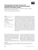
other plant AP genes, cardosin genes have their coding
region interrupted by 12 introns that occur in conserved
positions despite their variable sizes (Fig. 1). Both the
5¢- and 3¢-splice junctions are in good agreement with
the exon–intron consensus boundary sequences [22], and
the initiation codon is inserted in a well-conserved con-
text (AACATGGG) among plant genes [23].
Comparison of cardosin A, B and D genomic clones
with the respective cDNAs (Fig. 2) revealed the pres-
ence of an intron in the 5¢-UTR of the genes. The nuc-
leotide sequences of the cDNA and genomic clones of
cardosins diverge after a perfect match of six bases. At
the point of divergence, a consensus splicing acceptor
sequence, 5¢-AG-3¢, was found (Fig. 3). The remaining
bases of the leader sequence appear in the upstream
region of the genomic clone after an intervening
sequence of 966 bp (cardosin A), 953 bp (cardosin B)
or 1207 bp (cardosin D), with a consensus donor site
5¢-GT-3¢ at the 5¢-end, suggesting that this region rep-
resents an intron (Fig. 2). To map the transcription
initiation site of cardosin genes, primer extension ana-
lysis with an antisense oligonucleotide located in the
untranslated region determined by 5¢-RACE was car-
ried out (data not shown). The 5¢-end of cardosin
genes identified by primer extension analysis was lon-
ger than the one observed by 5¢-RACE (Fig. 2).
Although a leader intron seems to be a conserved
structural feature among AP genes [16,19], it does not
appear in the 5¢-UTR of the cardosin C gene. This
observation is based on the comparison of the genomic
Cardoon genes coding for aspartic proteases C. Pimentel et al.
2524 FEBS Journal 274 (2007) 2523–2539 ª 2007 The Authors Journal compilation ª 2007 FEBS
sequences of the cardosin A, C and D gene 5¢-flanking
regions (Fig. 3). Beyond an initial small match of
nucleotides immediately upstream from the initiation
codon, the homology among the three genes is inter-
rupted, but it is recovered several nucleotides upstream
from the 5¢-UTR of the cardosin A and D genes
(Fig. 3).
A TATA element [24], TATAAAA, is located 30 bp
upstream of the transcription start site of the cardo-
sin B gene, and two ‘CAAT’ box motifs are found at
positions ) 43 bp and ) 82 bp. The putative ‘TATA’
boxes of the cardosins A and D genes (TTTAAAA),
located ) 25 bp upstream of the transcription start site,
differ from the consensus sequence found in plant
genes (TATAWAWA) [24]. Identical sequences were,
however, identified in the rat tropomyosin gene [25]
and in the A. thaliana phenylalanine ammonia-lyase
gene (GenBank accession number X84728). ‘CAAT’
motifs are present in positions ) 71 bp and ) 74 bp of
the cardosin A and D genes, respectively.
The 5¢-flanking regions of the cardosin A, C and D
genes share a high degree of similarity (Fig. 3). How-
ever, the respective region of the cardosin B gene only
exhibits a stretch of 388 bp with significant homology
to the cardosin A and D genes (Fig. 3).
Predicted structural features of the new cardoon
APs
As expected, the deduced amino acid sequences of
cardosin C and cardosin D revealed that both
enzymes possess the typical structural domain organ-
ization of plant APs [2]. Cardosins and cyprosin B
share, in terms of primary structure, a high level of
similarity, with cardosins A, C and D exhibiting the
highest scores. Interestingly, the slight differences
Fig. 1. Schematic representation of the structure of the cardosin and cyprosin B genes. Filled boxes represent exons. Open triangles
symbolize introns. The size of each intron is indicated under the triangles in bp. The sequence of cyprosin B isolated was incomplete and
encompassed the last six exons of the gene.
Fig. 2. Determination of the transcription initiation site of the cardosin A, B and D genes. The alignment of the most extended 5¢-RACE prod-
ucts (CardA_cDNA, CardB_cDNA, and CardD_cDNA) against the corresponding genomic sequences (CardA_gDNA, CardB_gDNA, and
CardC_gDNA) revealed the presence of an intron within the 5¢-UTR of the genes. Primer extension analysis showed that the precise tran-
scription initiation site is located several nucleotides upstream of each gene’s longest 5¢-RACE product, at the nucleotide indicated by an
open arrow. The initiation codon is shaded in black. The leader intron consensus splicing donor and acceptor sequences are boxed. The size
of the intron is indicated in bp.
C. Pimentel et al. Cardoon genes coding for aspartic proteases
FEBS Journal 274 (2007) 2523–2539 ª 2007 The Authors Journal compilation ª 2007 FEBS 2525
among cardosins A, C and D comprise the RGD
and KGE motifs, which were demonstrated to be
important for the interaction of cardosin A with
phospholipase Da [7]. As depicted in Fig. 4, the
RGD ⁄ KGE motifs found in the primary structure of
cardosins A and C are replaced in cardosin D by
Fig. 3. Alignment of the 5¢-flanking regions of the cardosin A, C and D genes. Sequences sharing 100% similarity among the genes are sha-
ded in black. The sequences that are 100% identical between two of the genes are in gray. The leader introns of cardosins A and D are
boxed. The initiation codon is underlined. The A ⁄ T and G ⁄ A repeats are indicated by asterisks. The inverted repeat is indicated by arrows.
Horizontal lines indicate the absence of a nucleotide in the sequence. Lower-case letters represent unique sequences. The initiation of tran-
scription of the cardosin A and D genes is indicated by a bent arrow. The three dots represent omitted parts of the alignment. The cardo-
sin A sequence that is underlined (from ) 139 bp to + 232 bp) is the only region of the cardosin A 5¢-flanking region that shares significant
similarity with the corresponding region of the cardosin B gene (from ) 147 bp to + 238 bp).The 529 bp of the promoter region of the cardo-
sin A gene that is relevant for gene expression in Arabidopsis and the corresponding region of the cardosin C gene are double boxed.
Cardoon genes coding for aspartic proteases C. Pimentel et al.
2526 FEBS Journal 274 (2007) 2523–2539 ª 2007 The Authors Journal compilation ª 2007 FEBS
KGD ⁄ EGE motifs. These differences may have rele-
vant functional implications, as cardosin B harbors a
RGN ⁄ EGE motif and does not interact with phospho-
lipase Da [7].
Evolutionary relationships of cardosins and their
plant counterparts
The amino acid sequences of C. cardunculus APs were
compared with those of several other plant APs, by
means of the phylogenetic analysis program mega ver-
sion 3.0 [26], using the neighbor-joining method. On the
basis of the resulting phylogenetic tree, three distinct
groups within the typical plant AP family can be
defined (Fig. 5). Group I comprises the best studied
APs, and may further be divided into two smaller
groups. Group Ia includes the APs of the Brassicaceae
and Fabaceae families, as well as those found in mono-
cotyledonous plants. These APs have been implicated
in the proteolytic processing and ⁄ or degradation of
storage proteins (A. thaliana and Brassica napus APs,
orizasin and fitepsin), in leaf senescence (At4g0446,
BnU55032, and VuAP1), and in programmed cell
death events (SoyAP1 and fitepsin) [18,19,21,27–32].
Although the wheat AP (BAE20413) has not yet been
biochemically or molecularly characterized, its inclusion
A
B
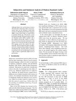
Fig. 4. Amino acid sequence alignment and
homology of cardosins A, B, C and D and
cyprosin A and B. (A) The amino acid
sequences were deduced from the genomic
sequences (this work), with the exception
of cyprosin A (X69193) and cyprosin B
(X81984). Identical sequences are indicated
by dots, and deleted amino acids by horizon-
tal lines. The signal peptide and prose-
quence are indicated by dashed and
continuous lines, respectively. The amino
acids forming the catalytic triads in the act-
ive site (DTG and DSG) are in bold italic.
The RGD and KGE motifs are boxed. Poten-
tial N-linked glycosylation sites are marked.
(B) Percentage amino acid identity and simi-
larity between C. cardunculus APs. The
upper and lower parts of the table corres-
pond to similarity and identity percentages,
respectively.
C. Pimentel et al. Cardoon genes coding for aspartic proteases
FEBS Journal 274 (2007) 2523–2539 ª 2007 The Authors Journal compilation ª 2007 FEBS 2527
in this group suggests that it might be involved in sim-
ilar biological functions. Within group I, the APs
At1g11910 and BnU55032, GmSoyAP1 and VuAP1, as
well as fitepsin and BAE20413, form a clade and appear
to be potential orthologs (Fig. 5).
Group Ib includes the APs from the Asteraceae fam-
ily, which have mostly been found in flowers and
therefore have been proposed to participate in flower-
specific events [3,9,33]. The topology of group Ib
suggests that, at some time during the evolution of
C. cardunculus, an AP ancestor gene has duplicated
and given rise to the branches comprising cyprosins
and cardosins. Subsequent duplications within both
branches should have occurred originating the group
actual configuration (Fig. 5).
The APs of group II have never been studied; how-
ever, as they are evolutionarily related, it is possible
that they share similar or complementary biological
functions. Interestingly no dicotyledonous plants were
found within this group (Fig. 5).
Finally, group III contains the tomato (L46681) and
potato (StAsp) APs, whose genes are induced upon
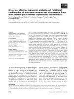
Fig. 5. Phylogenetic relationship between several APs. The phylogenetic analysis was carried out by the neighbor-joining method using MEGA
version 3.0. One thousand bootstrap replicates were calculated, and bootstrap values are shown at each node. Nodes were collapsed to a
single horizontal line whenever statistical support was less than 60%. On the basis of the AP family tree, it is possible to divide typical plant
APs into three groups (I, II, and III). Group I may be subdivided into two smaller groups: Ia and Ib. The two first letters of the sequence
name indicate the plant species. At, A. thaliana; Bn, B. napus; Cca, Ceutarea calcitrapa; Cc, C. cardunculus; Ch, Cy. humilis; Cr,
Ch. reiinhardtii; Gm, Glycine max (soy); Ha, Helianthus annuus (sunflower); Hs, Hemerocallis sp. (lily); Hv, Hordeum vulgare (barley); Ib, Ipo-
moea batatas (sweet potato); Le, Lycopersicon esculentum (tomato); Os, Oryza sativa (rice); St, Solanum tuberosum (potato); Ta, Triticum
aestivum (wheat); Vu, Vigna unguiculata (cowpea). The following characters indicate the sequence accession number (or the AGI code, in
the particular case of Arabidopsis APs) or the name of the enzyme: orizasin, D32165; fitepsin, X56136; cenprosin, Y09123; cyprosin A,
X69193; cyprosin B, X81984; VuAP1, AF287258; DSA4, AF082029; SoyAP1, AB069959; and SoyAP2, AB070857. Cardosin amino acid
sequences were deduced from the genomic sequences (this study).
Cardoon genes coding for aspartic proteases C. Pimentel et al.
2528 FEBS Journal 274 (2007) 2523–2539 ª 2007 The Authors Journal compilation ª 2007 FEBS
biotic stress challenge [14,15]. The group also includes
one of the soy Aps (SoyAP2), which is expressed in
several tissues and may be involved in seed germina-
tion [32], and the sweet potato AP (AF259982).
Cardosin genes exhibit distinct expression
patterns in C. cardunculus
Given the overall similarity among the cardosin A, C
and D genes, it became evident that our previous work
did not allow discrimination of these genes [3,6].
Within this context, we had designed primer pairs
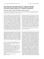
specific for each cardosin gene (Fig. 6A) and evalu-
ated gene expression by RT-PCR in three stages of pis-
til development and in several other organs of
C. cardunculus (Fig. 6B). Our results showed that:
(a) with the exception of stems, the cardosin A and D
genes share a similar pattern of expression, being ubiq-
uitously expressed; (b) cardosin B gene expression is
pistil-specific; and (c) cardosin C expression is flower-
specific and restricted to the pollen and to the pistils of
partially opened capitula (Fig. 6B).
Cardosin promoter regions are functional
in A. thaliana
To further investigate the spatial and temporal expres-
sion patterns of cardosin genes, each of their 5¢-flank-
ing regions (promoter and leader intron) was fused to
the GUS reporter gene in order to generate the con-
structs ) 2912pA::GUS (cardosin A), ) 3459pB::GUS
(cardosin B), ) 2040pC::GUS (cardosin C), and ) 1186
pD::GUS (cardosin D).
The ) 2912pA::GUS construct drives GUS expres-
sion in the pistils, petals and filaments in the early
stages of A. thaliana flower development in six of
the independent transformed plant lines analyzed
(Fig. 7A–C). The expression is mainly restricted to the
flowers, although staining can also be observed in
young stems. At the initial stages of pistil develop-
ment, intense staining is observed in the stigma, style
and ovary. However, the stigma staining tends to dis-
appear at the later stages of flower development
(Fig. 7A–C).
The 5¢-flanking region of the cardosin B gene
() 3459pB::GUS) induced GUS expression in the
anthers, at the initial stages of flower development
(Fig. 7M), and in the stigmatic papillae of mature flow-
ers (Fig. 7N,O). Within six independently transformed
Arabidopsis lines, GUS activity was not detected in
other plant organs, being confined to floral tissues.
In seven of eight plant lines transformed with the
cardosin C promoter region () 2040pC::GUS), the
transgene expression was confined to undifferentiated
flowers and styles (Fig. 7J–L), whereas construct
) 1186 pD::GUS (containing the cardosin D 5¢-flank-
ing region) was not able to drive GUS expression in
the eight independent plant lines analyzed (data not
shown). In addition, none of the negative controls
showed GUS staining (data not shown).
A
B
Fig. 6. Expression of cardosin genes during flower development and in several organs of C. cardunculus (A) Control analysis of the specifici-
ty of the PCR amplification of each cardosin gene. Phage DNA including each cardosin gene ) cardosin A (A), cardosin C (C), cardosin D (D),
and cardosin B (B) ) was used as template in these experiments. The gene-specific primers used were misAF1 ⁄ misR1117 (cardosin A), mis-
CF1 ⁄ misCR1 (cardosin C), and misDF1 ⁄ misDR1 (cardosin D). The primer pairs only amplified the corresponding gene, confirming their spe-
cificity. (B) RT-PCR analysis of cardosin genes, using the corresponding gene-specific primer pairs. The actin 2 gene of A. thaliana was used
as an amplification positive control. CC, pistils of closed capitulum; POC, pistils of partially open capitulum; OF, pistils of open capitulum; C,
negative control.
C. Pimentel et al. Cardoon genes coding for aspartic proteases
FEBS Journal 274 (2007) 2523–2539 ª 2007 The Authors Journal compilation ª 2007 FEBS 2529
A 529 bp region is crucial for cardosin A
expression in A. thaliana
As a first approach to the identification of cis regula-
tory elements involved in the control of cardosin gene
expression, we analyzed several 5¢-deletions of the
cardosin A promoter (Fig. 8) and examined their effect
on gene expression in transgenic plants. Our results
clearly show that the removal of 1 kb of the cardo-
sin A promoter region () 1792pA::GUS) did not
greatly affect the transgene expression (Fig. 7D–F) in
eight independent lines tested.
A subsequent 529 bp deletion of the promoter
region from position ) 1792 to position ) 1263 (Fig. 8)
completely abrogated transgene expression in all plant
lines (data not shown). As successive 500 bp deletions
to position ) 234 (Fig. 8) did not restore the transgene
expression, the presence of a negative regulator was
A
B
C
DE
F
G
H
I
J
K
L
M
N
O
P
Q
R
Fig. 7. Histochemical analysis of GUS activ-
ity in transgenic A. thaliana plants trans-
formed with cardosin A, B and D
constructs, containing the 5¢-flanking regions
of the genes fused to the reporter. Each
row of panels represents independent flow-
ers of plant lines transformed with the same
construct, at different stages of develop-
ment. The names of the constructs are
indicated on the right of each row of the
respective panels. o, ovary; p, petal; s, stig-
matic papillae; st, style; f, filament; a,
anther.
Cardoon genes coding for aspartic proteases C. Pimentel et al.
2530 FEBS Journal 274 (2007) 2523–2539 ª 2007 The Authors Journal compilation ª 2007 FEBS
ruled out, and we assumed that important regulatory
elements were present within the 529 bp region from
) 1792 bp to ) 1263 bp.
In silico analysis of this region (Fig. 3) revealed the
presence of three putative regulatory elements: a long
repetition (n ¼ 10) of the dinucleotide A⁄ T, followed
by a long repetition (n ¼ 12) of the dinucleotide G ⁄ A
and an inverted repeat. All of these sequences were
also found in the cardosin C promoter region, but
were absent from the corresponding region of the
cardosin D gene analyzed (Fig. 8).
The cardosin B but not the cardosin A leader
intron is essential for gene expression
It is known that introns may participate in gene regu-
lation, by modulating the level of expression and ⁄ or
determining the specific pattern of expression of a gene
[34–39]. To evaluate the relevance of the leader intron
in cardosin expression, we deleted it from the 5¢-flank-
ing region of the genes (Fig. 8). The deletion of the
cardosin A leader intron (construct pADi::GUS) did
not affect the staining pattern of GUS (Fig. 7G–I),
which was essentially similar to the one obtained when
A. thaliana plants were transformed with the cons-
truct ) 2913pA::GUS (Fig. 7A–C), in six of the eight
lines considered. Conversely, the deletion of the
respective region from the cardosin B gene (construct
pBDi::GUS; Fig. 8) completely abolished the transgene
expression (Fig. 7P–R) in all plant lines, highlighting
its important role in the regulation of cardosin B gene
expression.
Comparison of the leader intron of the cardosin B
gene with pistil-specific genes revealed the presence of
putative regulatory elements. A region of SLG
13
,a
gene involved in the prevention of self-pollination in
Brassica, encompassing three boxes (I, II, and III),
located 400 bp upstream of the initiation codon, is
required for pistil-specific gene expression in transgenic
tobacco [40]. A sequence sharing 77% similarity with
that mentioned above and spanning 34 bp was identi-
fied in the leader intron of the cardosin B gene
(Fig. 9). In addition, another element (motif III-rela-
ted) was identified 438 bp downstream of the SLG
13
-
like sequence [50] (Fig. 9). A similar motif is
potentially implicated in pistil-specific expression of a
pathogenesis-related protein gene from Pyrus serotina
in transgenic tobacco (Fig. 9) [41,42]. Moreover, a
motif III-related element also appears in the Arabidop-
sis AtS1 gene (a ‘Brassica-like’ S gene; Fig. 9) that is
expressed specifically in papillar cells and may function
in pollination [43].
We have made two extra constructs harboring only
the leader intron of the cardosin A and B genes. When
tested under physiologic conditions, these constructs
were not able to drive GUS expression in Arabidopsis
(data not shown), revealing that they cannot act as
alternative promoters.
Fig. 8. Structure of cardosin 5¢-flanking region–GUS fusion constructs. The striped boxes represent the leader intron. The stippled and gray
boxes indicate the (A ⁄ T) and (G ⁄ A) repeats, respectively. The inverted repeat found in the promoter regions of cardosins A and C is indicated
by opposing arrows. The initiation codon of each construct is indicated by a bent arrow.
C. Pimentel et al. Cardoon genes coding for aspartic proteases
FEBS Journal 274 (2007) 2523–2539 ª 2007 The Authors Journal compilation ª 2007 FEBS 2531
Discussion
Cloning by library screening of four full-length genes
encoding cardosins A, B, C and D precursors, together
with the cloning of a partial sequence of the cyprosin B
gene and the isolation of the cyprosin A cDNA [11],
indicates that C. cardunculus APs are encoded by a
multigene family composed of at least six members,
and reveal the coexistence of cardosins and cyprosins
within the same plant.
The gene structure of cardosins basically reflects
the same genomic organization as that of the few
other typical AP genes that have been analyzed
[16,19,29,30]. Given that monocotyledon and dicotyle-
don APs display the same pattern of exon–intron
arrangement, the insertion of introns within the cod-
ing region possibly occurred before the divergence of
both classes of plants [30]. Regarding the introns, the
loss or gain of sequences may have taken place after
monocotyledon and dicotyledon divergence, a fact
that may explain: (a) the variable length of introns
among different species and between gene family
members of the same plant; and (b) the absence of
one intron in the A. thaliana genes AtPaspA2 and At-
PaspA3 [29].
Cardosins and cyprosins share a similar structural
domain organization and display a high degree of
identity in terms of primary structure. Interestingly,
the slight differences among cardosins and between
cardosins and cyprosins comprise the motifs RGD and
KGE (Fig. 4). These motifs are known to mediate the
cardosin A–phospholipase Da interaction, which may
play an important physiologic role [7]. Cardosin B,
which harbors an EGE instead of a KGE motif, does
not bind to phospholipase Da [7]. Within C. carduncu-
lus APs, only cardosin A and cardosin C possess the
RGD and KGE motifs (Fig. 4), and therefore the for-
mation of a complex in planta with phospholipase Da
is possibly restricted to these proteases.
In contrast to cyprosins, cardosins do not contain
the residues Lys11 and Tyr13 (phytepsin amino acids
numbering) in the N-terminal domain. These residues
are well conserved among plant APs, and are involved
in the inactivation mechanism of the precursor form
of the enzymes [2,44]. Cardosins and the Cy. humilis
AP are the only plant APs known to date whose
Lys11 ⁄ Tyr13 residues are absent from the primary
structure, a feature that may explain the enzymatic
activity exhibited by recombinant procardosins (Vieira
et al., unpublished results). From the scenario of plant
AP evolution, it becomes evident that in C. carduncu-
lus, the loss of the inactivation mechanism of the pre-
cursor forms occurred after the duplication of an
ancestral gene common to cardosins and cyprosins
(Fig. 5).
Comparison of protein data [3,9] with the results of
gene expression studies (Fig. 6B) clearly indicates that
cardosins are specifically expressed in the flowers of
cardoon, although minor levels of cardosin A and D
transcripts could also be detected in other plant organs
(Fig. 6).
To further analyze the expression of cardosins, we
fused their promoter region with the reporter gene
GUS and assayed its activity in transgenic A. thaliana
(Fig. 7). A. thaliana possesses three AP genes whose
promoter regions do not exhibit any significant homol-
ogy with the corresponding regions of cardosin genes
(data not shown), which is in agreement with the dif-
ferent pattern of expression displayed by the APs of
both species [6,9,29]. Nevertheless, the lack of sequence
data on other plant AP promoter regions, in addition
to the evolutionary proximity of groups Ia and Ib
(Fig. 5), support our use of the model plant A. thaliana
in our studies.
A
B
Fig. 9. Conserved sequences among pistil-specific genes are also present in the leader intron of the cardosin B gene. (A) Sequences similar
to those identified by Dzelzkalns et al. (boxes I and III) within the promoter region of the SLG
13
gene [40] also appear in the leader intron of
the cardosin B gene. (B) A motif found in the S1, SLG, SLR1 and PR5 genes [41,42] is also present within the cardosin B leader intron, but
in an inverted position. Asterisks denote identical nucleotides. At, A. thaliana L.; Cc, C. cardunculus; Bo, B. oleracea; Ps, Py. serotina.
Cardoon genes coding for aspartic proteases C. Pimentel et al.
2532 FEBS Journal 274 (2007) 2523–2539 ª 2007 The Authors Journal compilation ª 2007 FEBS
In A. thaliana, cardosins exhibit a flower-specific
pattern of expression (Fig. 7), mirroring what was
observed in the cardoon. The tissue-specific pattern,
with some overlap of cardosins (cardosins A and C),
suggests that they may have specific and ⁄ or comple-
mentary functions within the flower. The presence of
cardosins A and C in the initial stages of flower devel-
opment is in agreement with the presence of cypros-
in B in flower meristems, as the antibody used in the
immunocytochemical study could not discriminate
among cardosins and cyprosins [45]. The presence of
cardosins in flower meristems, in addition to the lower
amount of processed protein detected in immature car-
doon flowers [3], led us to hypothesize that at least
cardosins A and C may play important physiologic
roles in the early stages of flower development, before
being processed into the two-chain active enzymes.
We did not detect GUS activity driven by the
5¢-flanking region of the cardosin A and C genes in
pollen grains. This observation is not in agreement
with the detection in pollen of cardosin A and C
transcripts in C. cardunculus L (Fig. 6). However, the
absence of other noncoding regions of the genes
(introns and 3¢-UTR), as well as the different sensitivi-
ties of the techniques (RT-PCR ⁄ GUS histochemical
detection), as previously observed by others [46], may
explain the discrepancies observed.
As a first attempt to determine the molecular basis
for the flower-specific pattern of cardosin expression,
several 5¢-deletions of cardosin A promoter regions
were generated, fused to the reporter GUS (Fig. 8),
and then analyzed by means of GUS histochemical
detection. Therefore, we were able to delimit a region
of 529 bp, located between positions ) 1792 bp and
)1263 bp, that is crucial for transgene expression in
A. thaliana.Anin silico search of this region against
several plant cis element databases revealed the pres-
ence of at least three motifs that may also be relevant
in cardosin A gene regulation. These motifs comprise
an A ⁄ T repeat, followed by a G ⁄ A repeat, preceded by
an inverted repeat (Figs 3 and 8). The A ⁄ T repeats
enhance gene expression [47–49] in a copy number-
dependent way [49]. Regions with five or more A ⁄ T
repeats are recognized by the high-mobility group pro-
tein in several plant species [50]. Repeats of the dinu-
cleotide G ⁄ A were reported to bind to the basic
pentacysteine protein, and may be involved in the
regulation of the expression of a diversity of genes
[51]. Moreover, several inverted repeats are known to
regulate gene expression [52]. Further studies should
be performed to establish whether the 529 bp region is
involved in the enhancement of gene expression or in
the determination of its flower-specific pattern. Muta-
tional analysis will determine the relevance of the iden-
tified motifs.
The high similarity among cardosins A, C and D,
which extends beyond the coding region (Fig. 3), and
the relevance of the region of 529 bp in cardosin A
expression may explain the ineffectiveness of the
cardosin D flanking region in driving GUS expression.
The cardosin C gene construct () 2040pC::GUS) also
contains a highly similar region (Fig. 3). However, the
sequence of the cardosin D promoter region included
in the analysis does not contain the equivalent 529 bp
region of cardosin A (Figs 3 and 8). It is therefore
possible that its absence in the cardosin D promoter
region could explain the lack of GUS activity in the
transgenic A. thaliana plants transformed with the con-
struct ) 1186 pD::GUS.
Although the presence of a leader intron is a well-con-
served feature in plant AP genes [16,19], its biological
role has not yet been clarified. To address this subject,
we deleted the corresponding regions from the cardo-
sin A and B genes (Fig. 8) and assayed GUS activity.
The removal of the cardosin A leader intron did not
affect GUS histochemical localization (Fig. 7G–I),
whereas its deletion from the cardosin B gene com-
pletely abrogated gene expression (Fig. 7P–R). How-
ever, we cannot rule out the hypothesis that GUS levels
varied when we deleted the cardosin A leader intron.
Two mechanisms may explain the lack of GUS
expression driven by the cardosin B construct depleted
of the leader intron: (a) positive regulatory elements lay
within the leader intron; or (b) the leader intron may be
increasing the steady state of mRNA levels, without
significantly affecting the rate of transcription (intron-
mediated enhancement [53]). Nuclear run-on transcrip-
tion assays, in addition to the design of constructs in
which the leader intron is inverted or partially deleted,
will give clues to indicate the precise mechanism.
The similarity between the leader introns of cardo-
sins A and B is restricted to the first 182 bp (Fig. 3). If
the leader intron of cardosin B harbors important reg-
ulatory elements, they should be confined to the
remaining sequence. We therefore compared this
region of the cardosin B gene with the promoter
regions of plant genes that are specifically expressed in
pistils, namely genes involved in self-pollination avoid-
ance (Fig. 9). The presence of a motif III-related
sequence [41] within the leader intron of the cardo-
sin B gene is particularly interesting, as its removal
from a SLG
13
promoter::GUS construct abolished
reporter expression in pistils of transgenic tobacco [40].
Furthermore, the deletion of an SLR1 gene region har-
boring one motif III-related sequence also eliminated
its expression in the stigma and styles of transgenic
C. Pimentel et al. Cardoon genes coding for aspartic proteases
FEBS Journal 274 (2007) 2523–2539 ª 2007 The Authors Journal compilation ª 2007 FEBS 2533
tobacco, and it was shown that as yet unidentified pis-
til transcription factor binds that region [54]. There-
fore, the motif III-related sequence is a good candidate
for a positive regulatory element of cardosin B gene
pistil expression. Future work on transgene expression
driven by a motif III-mutated leader intron of the
cardosin B gene fused with a minimal promoter will
certainly provide further insights into the relevance of
this sequence.
Experimental procedures
Plant material and growth conditions
C. cardunculus tissue samples were collected from field-
grown plants, frozen immediately in liquid nitrogen, and
kept at ) 80 ° C until use. A. thaliana (Columbia ecotype)
surface-sterilized seeds were sowed in Murashige and Skoog
medium (Duchefa Biochemie, Haarlem, the Netherlands),
pH 5.8, containing 0.7% w ⁄ v of plant agar (Duchefa
Biochemie). After 48 h of 4 °C stratification treatment in the
dark, the seeds were germinated at 22 °C under a 16 : 8 h
light ⁄ dark cycle. Two-week-old seedlings were subsequently
transferred to soil until completion of the plant life cycle.
Construction of a C. cardunculus library
and cardosin gene isolation
DNA was extracted from young leaves of C. cardunculus
subspecies flavescens, according to the method of Jofuku &
Goldberg [55] and partially digested with Sau3AI (New
England Biolabs, Beverley, MA, USA). The resulting
fragments, ranging from 15 to 25 kb, were cloned into the
kDash II vector (Stratagene, Beverley, MA, USA). Primary
recombinants were amplified to create a stable library. A
representative aliquot (380 000 plaque-forming units) of the
library was independently screened by plaque hybridization
with two different probes, CA-5¢ -and CB-3¢,
32
P-labeled
(Megaprime DNA Labelling System; Amersham, Uppsala,
Sweden), using standard protocols [56]. CA-5¢-consisted of
a fragment obtained by standard PCR amplification of the
cDNA of cardosin A (GeneBank accession number
AJ132884 [3], using the primer pair S#1 ⁄ R333 (Table 1).
CB-3¢ comprised a 300 bp DNA fragment including the 3¢-
UTR region of the cardosin B gene obtained by 3 ¢-RACE
and amplified by PCR with the primers CardBS and
FLCBR (Table 1). After three rounds of purification with
probe CA-5¢, three positive plaques, named k5, k6, and k18
were isolated. The library screening with the CB-3¢ probe
retrieved two positive plaques, k4.1 and k4.2. The phage
DNA was extracted [55], hydrolyzed with several restriction
enzymes, and analyzed by Southern blotting. Fragments of
phage DNA positively hybridizing with probes CA-5¢ or
CB-3¢ were cloned into the pZErO1 plasmid (Invitrogen,
Carlsbad, CA, USA) and sequenced using the Genom-
eLab DTCS-Quick Start Kit (Beckman Coulter, Krefeld,
Germany) and the automatic sequencer CEQ 8000 Genetic
Analysis System (Beckman Coulter).
As the cardosin A gene included in phage k5 was not
complete, we specifically amplified its 3¢-region by PCR.
Therefore, 300 ng of the nuclear DNA of C. cardunculus L
was isolated (Invisorb Spin Plant Genomic DNA purifi-
cation kit; Invitek, Berlin, Germany), and the region con-
taining the three last exons of the cardosin A gene was PCR
amplified with the primer pair Int10aF ⁄ R#2 (Table 1).
RACE experiments
Total RNA from the pistils of C. cardunculus flower buds
was isolated with the RNeasy Plant mini kit (Qiagen,
Valencia, CA, USA) and used to generate an adaptor-
ligated double-stranded cDNA RACE library with the
Marathon cDNA Amplification kit (Clontech, Palo Alto,
CA, USA). The 5¢-UTR regions of the cardosin genes
were amplified by PCR under standard conditions [56],
using primer R333 (Table 1) combined with the kit-provi-
ded adaptor primers. R333 hybridizes with exon I of
cardosin genes (Fig. 8) at a position 333 bp downstream
of the ATG. The 3¢-UTR of the cardosin B gene was
amplified by means of a similar strategy, but using the
CardBS primer as specific oligonucleotide (Table 1). The
PCR products were cloned with the TA cloning kit (Invi-
trogen), and sequenced by automated DNA sequencing as
described above.
RT-PCR analysis of cardosin gene expression
Total RNA from C. cardunculus was isolated, as described
above, from several tissues and from pistils of closed, parti-
ally opened and opened capitulum (the globular inflores-
cence grouped in a common receptacle surrounded by
bracts). The RNA integrity and possible nuclear DNA con-
tamination were evaluated in a 1% agarose gel, and equal
amounts of RNA (1 lg) were used for cDNA synthesis (1st
Strand cDNA Synthesis Kit for RT-PCR; Roche, Basel,
Switzerland). Four sets of primers, specific for each of the
cardosin genes, were used for RT-PCR amplification
(Table 1): misAF1 ⁄ misR1117 (63 °C, cardosin A); mis-
CF1 ⁄ misCR1 (65 °C, cardosin C); misDF1 ⁄ misDR1
(60 °C, cardosin D); and CardBS ⁄ CardBR (58 °C, cardo-
sin B) [9]. Owing to the high level of similarity among
cardosins A, C and D, specific amplification of each gene
was only possible after the introduction of an artificial mis-
match in the antepenultimate or penultimate base of the
specific primers (Table 1). As a negative control, the RNA
from seeds was amplified through a RT-PCR reaction, with
each set of primers, without the prior addition of avian
myeloblastosis virus-reverse transcriptase (AMV-RT). The
Cardoon genes coding for aspartic proteases C. Pimentel et al.
2534 FEBS Journal 274 (2007) 2523–2539 ª 2007 The Authors Journal compilation ª 2007 FEBS
A. thaliana actin 2 gene (AGI code: At3g18780) was ampli-
fied by RT-PCR with the specific primer pair ActF ⁄ ActR
(Table 1) and used as a positive control. At least two repli-
cas were carried out for each RT-PCR reaction, and the
products were sequenced to further confirm the specificity
of the amplification.
Chimeric plasmid constructs
The cardosin A, B, C and D 5¢-flanking regions were cloned,
using GATEWAY BP and LR reactions (Invitrogen), into
the binary vector pKGWFS7 [57]. In this vector, an in-frame
fusion between the regions coding for EgfpER and b-glucu-
Table 1. Sequences of the oligonucleotides used in this work.
Name Sequence (5¢–to3¢) Usage
S#1 ATGGGTACCTCAATCAAAGCAA Probe CA-5¢
R333 AGAACAGAACTTCCGGTATCG Probe CA-5¢-and
5¢-RACE
Int10aF GTGTGACACCGGTAATAAGCAG PCR
R#2 TCAAGCTGCTTCTGCAAATCC PCR
5¢PextA CACACCCTCCTTCATTGCTTCCATCAAATAACAC 5¢ Primer extension
misAF1 ATGGGACATTTGGCGCTAT
CCA
a
RT-PCR
(cardosin A)
misR1117 GGTGCACATCTCATCATGT
CTG
a
RT-PCR
(cardosin A)
misCF1 CCCCTGGTTGTCAAGCATAT
CTA
a
RT-PCR
(cardosin C)
misCR1 AGACTTGTCGTTATTCTTGT
GC
a
RT-PCR
(cardosin C)
misDF1 GGGTGCCTTCTTCAAAGTG
GTA
a
RT-PCR
(cardosin D)
misDR1 TGTATGCCACCAGAAGACTT
CA
a
RT-PCR
(cardosin D)
CardBS GATCTCGGCTGGGAAAGCG RT-PCR (cardosin B)
Probe CB-3¢
CardBR ATACCATTGCAGTCTACTATC RT-PCR
(cardosin B)
FLCBR TTTATTGGACCATTTTATTCCGG Probe CB-3¢
ActF GATATGGAAAAGATCTGGCATCAC RT-PCR
(Atactin 2)
ActR TCATACTCGGCCTTGGAGATCC RT-PCR
(Atactin 2)
) 2912AF
AAAAAGCAGGCTATGAATTGCTAGAGTTGGTTAATGC
b
) 2912pA::GUS and
pADi::GUS
) 1792AF
AAAAAGCAGGCTTGCTGTTCTAAGTGTACTAGCTGGA
b
) 1792pA::GUS
) 1263AF
AAAAAGCAGGCTCAAATTAAATCGACGGTTGAG
b
) 1263pA::GUS
) 764AF
AAAAAGCAGGCTCAATGTAGTACCAATTGGGGTACC
b
) 764pA::GUS
) 234AF
AAAAAGCAGGCTGAGAAATCTATGGAATAAATAAAAATTAGGG
b
) 234pA::GUS
PromAR
AGAAAGCTGGGTCGATGTTTCACTGAAACATTAATAGATATTC
b,c
All chimeric cardosin A
constructs except
pADi::GUS
PromARDi
AGAAAGCTGGGTCGATGTTTCATCACGTGTTATTTGATGGAAGCAATG
b,c,d
pADi::GUS
) 2040CF
AAAAAGCAGGCTTTAATAAGTTGTGCTTACACACAGTT
b
) 2040pC::GUS
) 2040CR
AGAAAGCTGGGTCGATGTTTCACTGCAACCATGAC
b,c
) 2040pC::GUS
) 1186DF
AAAAAGCAGGCTAACCACTATGATACCACTCACACCA
b
) 1186 pD::GUS
) 1186DR
AGAAAGCTGGGTCGATGTTTCACTGCAACCATGAC
b,c
) 1186 pD::GUS
) 3459BF
AAAAAGCAGGCTCTTAACGACTGCGTATATCCTC ) 3459pB::GUS and
pBDi::GUS
PromBR
AGAAAGCTGGGTCGATGTTTCACTGCAACCATGAC
b,c
) 3459pB::GUS
PromBRDi
AGAAAGCTGGGTCGATGTTTCACTCTTATTTGATGGAAGCAATGAAGG
b,c,d
pBDi::GUS
a
The artificial mismatch introduced in the primer is underlined.
b
The att sequences are underlined.
c
The ATG within the reverse primer,
mutated to ATC, is in Italic.
d
The six bases preceding the ATG were introduced in primers and are in Bold.
C. Pimentel et al. Cardoon genes coding for aspartic proteases
FEBS Journal 274 (2007) 2523–2539 ª 2007 The Authors Journal compilation ª 2007 FEBS 2535
ronidase (gus) is cloned downstream of the GATEWAY cas-
sette. The initiation codon is therefore provided by the vec-
tor, and is the same for all the constructs. The 5¢-flanking
region of each cardosin gene was amplified by PCR from the
respective genomic clone (k5, k6, k18 or k4.1), using the
Platinum Pfx DNA Polymerase (Invitrogen). At the 5¢-end,
the primers contained attB sites, allowing subsequent frag-
ment cloning by homologous recombination. The resulting
PCR products with terminal attB1 and attB2 sequences were
purified, and incubated with pDONR221 vector (Invitrogen)
containing the attP1 and attP2 recombination sites, and the
BP CLONASE enzyme (Invitrogen). This mixture was used
to transform DH5a-competent cells (Invitrogen), and the
recombinant clones were selected on kanamycin-containing
LB plates (50 lgÆmL
)1
; Fluka Biochemika, Buchs, Switzer-
land). Positive recombinant pDONR plasmids were incuba-
ted with the binary vector pKGWFS7 [57] in the presence of
LR CLONASE enzyme (Invitrogen). The pKGWFS7 vector
has attR1 and attR2 recombination sites positioned
upstream of the GUS reporter gene. After kanamycin selec-
tion, the resulting constructs were analyzed by restriction
enzyme hydrolysis and sequenced as described above. Four
plasmids containing the 5¢-flanking regions of cardosin A
() 2912pA::GUS), cardosin B () 3459pB::GUS), cardosin C
() 2040pC::GUS) and cardosin D () 1186 pD::GUS) were
produced in this way (Fig. 8). As we did not perform an
analysis of green fluorescent protein fluorescence, and for
simplicity, the constructs were named, for example,
2912pA::GUS and not 2912pA::GFP::GUS. The primer
pairs used in all chimeric constructs are listed in Table 1. To
avoid the use of cardosin ATG as the initiation codon, all
the reverse primers used to construct each GATEWAY cas-
sette harbor a mutation in ATG (mutated to ATC)
(Table 1). A similar strategy was used to make the con-
structs pADi::GUS and pBDi::GUS, which enclosed the
same 5¢-flanking region included in constructs ) 2912pA::
GUS and ) 3459pB::GUS, respectively, but without the
leader intron. The leader intron was deleted by PCR amplifi-
cation with the primer pairs ) 2912AF ⁄ PromARDi (cardo-
sin A gene) and ) 3459BF ⁄ PromBRDi (cardosin B gene)
(Table 1, Fig. 8). A nested set of 5¢ -deletions in the cardo-
sin A gene promoter region was also generated. Fragments
differing from 500 bp at their 5¢-end were amplified by
PCR and cloned into pKGWFS7, generating constructs
) 1792pA::GUS, ) 1263pA::GUS, ) 764pA::GUS and
) 234pA::GUS (Fig. 8). In addition, a fragment of the
3¢-UTR sequence of the rice Os-ACS5 gene (GenBank
accession no. X9706) [58,59] was cloned by homologous
recombination into vector pKGWFS7 and used as a nega-
tive control.
Plant transformation
Constructs were introduced into Agrobacterium tumefaciens
strain LBA4404 by electroporation [60]. Bacteria harboring
the plasmid with the desired cardosin gene 5¢-flanking
region were grown to saturation in LB medium, and used
to transform wild-type A. thaliana plants (T
0
plants) by the
floral dip method [61]. Transformants (T
1
plants) were
selected on Murashige and Skoog medium containing
50 mgÆL
)1
kanamycin and 0.7% w ⁄ v plant agar. Kanamy-
cin-resistant plants were grown to the next generation (T
2
)
and analyzed for GUS staining. A. thaliana infected with
untransformed Ag. tumefaciens LBA4404 was also used as
a negative control.
Histochemical analysis of GUS activity
Histochemical GUS staining was performed for T
2
vegetative
tissues (leaves, roots, stems, siliques, seeds, inflorescences)
and pollen with 5-bromo-4-chloro-3-indoxyl-b-d-glucuronic
acid (X-Gluc; ImmunoSource, Zoersel-Halle, Belgium) as
substrate [62]. Samples were stained for 16 or 24 h at 37 °C.
The stained organs were washed and incubated with 70%
ethanol for 2 h, and clarified by incubation with a CLP solu-
tion [50 g of chloral hydrate (Riedel ) de Hae
¨
n, Seelze, Ger-
many)] dissolved in 20 mL of lactic acid (Fluka Biochemika)
and 25 mL of melted phenol crystals (Merck, Darmstadt,
Germany)]. A stereo microscope (HQ Leica Microsystems,
Wetzlar, Germany) attached to an image acquisition system
was used to obtain the photographs. Eight independent
A. thaliana independently transformed lines were analyzed
per construct.
Sequence analysis
wise 2 ( and neural network
promoter prediction (itfly.org/seq_tools/
promoter.html) were used in gene structure and promoter
prediction, respectively. The multiple sequence alignments
were constructed using clustalw ( />clustalw), and edited and shaded in the program genedoc
version 2.6 ( />Phylogenetic analysis was conducted with mega version 3.0
[26], using the neighbor-joining method with Poisson
correction.
Acknowledgements
We gratefully acknowledge financial support provided
to C. P. by Fundac¸ a
˜
o para a Cieˆ ncia e Tecnologia
(PRAXIS XXI/BD/21655/99), FEBS (FEBS short-term
fellowship) and Fundac¸ a
˜
o Calouste Gulbenkian (short-
term fellowship).
References
1 Schaaf A, Reski R & Decker EL (2004) A novel aspar-
tic proteinase is targeted to the secretory pathway and
Cardoon genes coding for aspartic proteases C. Pimentel et al.
2536 FEBS Journal 274 (2007) 2523–2539 ª 2007 The Authors Journal compilation ª 2007 FEBS
to the vacuole in the moss Physcomitrella patens. Eur
J Cell Biol 83, 145–152.
2 Simo
˜
es I & Faro C (2004) Structure and function of plant
aspartic proteinases. Eur J Biochem 271, 2067–2075.
3 Faro C, Ramalho-Santos M, Vieira M, Mendes A,
Simoes I, Andrade R, Verı
´
ssimo P, Lin XL, Tang J &
Pires E (1999) Cloning and characterization of cDNA
encoding cardosin A, an RGD-containing plant aspartic
proteinase. J Biol Chem 274, 28724–28729.
4 Fraza
˜
o C, Bento I, Costa J, Soares CM, Verı
´
ssimo P,
Faro C, Pires E, Cooper J & Carrondo MA (1999)
Crystal structure of cardosin A, a glycosylated and
Arg-Gly-Asp-containing aspartic proteinase from the
flowers of Cynara cardunculus L. J Biol Chem 274,
27694–27701.
5 Ramalho-Santos M, Verı
´
ssimo P, Cortes L, Samyn B,
Van Beeumen J, Pires E & Faro C (1998) Identification
and proteolytic processing of procardosin A. Eur J Bio-
chem 255, 133–138.
6 Ramalho-Santos M, Pissarra J, Verı
´
ssimo P, Pereira S,
Salema R, Pires E & Faro CJ (1997) Cardosin A, an
abundant aspartic proteinase, accumulates in protein
storage vacuoles in the stigmatic papillae of Cynara
cardunculus L. Planta 203, 204–212.
7 Simo
˜
es I, Mueller EC, Otto A, Bur D, Cheung AY,
Faro C & Pires E (2005) Molecular analysis of the inter-
action between cardosin A and phospholipase Da.
FEBS J 272, 5786–5798.
8 Verı
´
ssimo P, Faro C, Moir AJG, Lin YZ, Tang J &
Pires E (1996) Purification, characterization and partial
amino acid sequencing of two new aspartic proteinases
from fresh flowers of Cynara cardunculus L. Eur J Bio-
chem 235, 762–768.
9 Vieira M, Pissarra J, Verı
´
ssimo P, Castanheira P, Costa
Y, Pires E & Faro C (2001) Molecular cloning and
characterization of cDNA encoding cardosin B, an
aspartic proteinase accumulating extracellularly in the
transmitting tissue of Cynara cardunculus L. Plant Mol
Biol 45, 529–539.
10 Brodelius PE, Cordeiro M, Mercke P, Domingos A,
Clemente A & Pais MS (1998) Molecular cloning of
aspartic proteinases from flowers of Cynara cardunculus
subsp. flavescens cv. cardoon and Centaurea calcitrapa.
Aspartic Proteinases 436, 435–439.
11 Cordeiro MC, Xue ZT, Pietrzak M, Pais MS &
Brodelius PE (1994) Isolation and characterization of a
cDNA from flowers of Cynara cardunculus encoding
cyprosin (an aspartic proteinase) and its use to study
the organ-specific expression of cyprosin. Plant Mol Biol
24, 733–741.
12 Cruz de Carvalho MH, d’Arcy-Lameta A, Roy-
Macauley H, Gareil M, El Maarouf H, Pham-Thi AT
& Zuily-Fodil Y (2001) Aspartic protease in leaves of
common bean (Phaseolus vulgaris L.) and cowpea
(Vigna unguiculata L. Walp): enzymatic activity, gene
expression and relation to drought susceptibility. FEBS
Lett 492, 242–246.
13 Guevara MG, Oliva CR, Machinandiarena M & Daleo
GR (1999) Purification and properties of an aspartic
protease from potato tuber that is inhibited by a basic
chitinase. Physiol Plant 106, 164–169.
14 Guevara MG, Almeida C, Mendieta JR, Faro CJ,
Verı
´
ssimo P, Pires EV & Daleo GR (2005) Molecular
cloning of a potato leaf cDNA encoding an aspartic
protease (StAsp) and its expression after P. infestans
infection. Plant Physiol Biochem 43, 882–889.
15 Schaller A & Ryan CA (1996) Molecular cloning of a
tomato leaf cDNA encoding an aspartic protease, a
systemic wound response protein. Plant Mol Biol 31,
1073–1077.
16 Asakura T, Watanabe H, Abe K & Arai S (1995) Rice
aspartic proteinase, oryzasin, expressed during seed
ripening and germination, has a gene organization dis-
tinct from those of animal and microbial aspartic pro-
teinases. Eur J Biochem 232, 77–83.
17 Bhalerao R, Keskitalo J, Sterky F, Erlandsson R,
Bjorkbacka H, Birve SJ, Karlsson J, Gardestrom P,
Gustafsson P, Lundeberg J et al. (2003) Gene expression
in autumn leaves. Plant Physiol 131, 430–442.
18 Buchanan-Wollaston V, Page T, Harrison E, Breeze E,
Lim PO, Nam HG, Lin JF, Wu SH, Swidzinski J,
Ishizaki K et al. (2005) Comparative transcriptome ana-
lysis reveals significant differences in gene expression
and signalling pathways between developmental and
dark ⁄ starvation-induced senescence in Arabidopsis.
Plant J 42, 567–585.
19 Cruz de Carvalho MH, Pham-Thi AT, Gareil M,
d’Arcy-Lameta A & Fodil YZ (2004) Isolation and
characterization of an aspartic proteinase gene from
cowpea (Vigna unguiculata L. Walp.). J Plant Physiol
161, 971–976.
20 Page T, Griffiths G & Buchanan-Wollaston V (2001)
Molecular and biochemical characterization of post-
harvest senescence in broccoli. Plant Physiol 125,
718–727.
21 Panavas T, Pikula A, Reid PD, Rubinstein B & Walker
EL (1999) Identification of senescence-associated genes
from daylily petals. Plant Mol Biol 40, 237–248.
22 Brown J & Simpson C (1998) Splice site selection plant
pre-mRNA splicing. Annu Rev Plant Physiol Plant Mol
Biol 49, 77–95.
23 Joshi CP, Zhou H, Huang X & Chiang VL (1997) Con-
text sequences of translation initiation codon in plants.
Plant Mol Biol 35, 993–1001.
24 Joshi CP (1987) An inspection of the domain between
putative TATA box and translation start site in 79 plant
genes. Nucleic Acids Res 15, 6643–6653.
25 Helfman DM, Cheley S, Kuismanen E, Finn LA &
Yamawaki-Kataoka Y (1986) Nonmuscle and muscle
tropomyosin isoforms are expressed from a single gene
C. Pimentel et al. Cardoon genes coding for aspartic proteases
FEBS Journal 274 (2007) 2523–2539 ª 2007 The Authors Journal compilation ª 2007 FEBS 2537
by alternative RNA splicing and polyadenylation. Mol
Cell Biol 6, 3582–3595.
26 Kumar S, Tamura K & Nei M (2004) MEGA3: inte-
grated software for molecular evolutionary genetics
analysis and sequence alignment. Brief Bioinform 5,
150–163.
27 Asakura T, Watanabe H, Abe K & Arai S (1997)
Oryzasin as an aspartic proteinase occurring in rice
seeds: purification, characterization, and application to
milk clotting. J Agric Food Chem 45, 1070–1075.
28 Buchanan-Wollaston V & Ainsworth C (1997) Leaf
senescence in Brassica napus: cloning of senescence
related genes by subtractive hybridisation. Plant Mol
Biol 33, 821–834.
29 Chen X, Pfeil JE & Gal S (2002) The three typical
aspartic proteinase genes of Arabidopsis thaliana are dif-
ferentially expressed. Eur J Biochem 269, 4675–4684.
30 D’Hondt K, Stack S, Gutteridge S, Vandekerckhove J,
Krebbers E & Gal S (1997) Aspartic proteinase genes in
the Brassicaceae Arabidopsis thaliana and Brassica
napus. Plant Mol Biol 33, 187–192.
31 Runeberg-Roos P & Saarma M (1998) Phytepsin, a
barley vacuolar aspartic proteinase, is highly expressed
during autolysis of developing tracheary elements and
sieve cells. Plant J 15, 139–145.
32 Terauchi K, Asakura T, Nishizawa NK, Matsumoto I
& Abe K (2004) Characterization of the genes for two
soybean aspartic proteinases and analysis of their differ-
ent tissue-dependent expression. Planta 218, 947–957.
33 Cordeiro MC, Lowther T, Dunn BM, Guruprasad K,
Blundell T, Pais MS & Brodelius PE (1998) Substrate
specificity and molecular modelling of aspartic protei-
nases (Cyprosins) from flowers of Cynara cardunculus
subsp. flavescens cv. cardoon. Aspartic Proteinases 436,
473–479.
34 Bolle C, Herrmann RG & Oelmuller R (1996) Intron
sequences are involved in the plastid- and light-depen-
dent expression of the spinach PsaD gene. Plant J 10,
919–924.
35 Casas-Mollano JA, Lao NT & Kavanagh TA (2006)
Intron-regulated expression of SUVH3, an Arabidopsis
Su (var) 3–9 homologue. J Exp Bot 57, 3301–3311.
36 Chung BY, Simons C, Firth AE, Brown CM & Hellens
RP (2006) Effect of 5¢UTR introns on gene expression
in Arabidopsis thaliana. BMC Genomics 7, 120–133.
37 Curi GC, Chan RL & Gonzalez DH (2005) The leader
intron of Arabidopsis thaliana genes encoding cyto-
chrome c oxidase subunit 5c promotes high-level expres-
sion by increasing transcript abundance and translation
efficiency. J Exp Bot 56, 2563–2571.
38 Rose AB (2004) The effect of intron location on intron-
mediated enhancement of gene expression in Arabidop-
sis. Plant J 40, 744–751.
39 Jeon JS, Lee S, Jung KH, Jun SH, Kim C & An G
(2000) Tissue-preferential expression of a rice alpha-
tubulin gene, OsTubA1, mediated by the first intron.
Plant Physiol 123
, 1005–1014.
40 Dzelzkalns VA, Thorsness MK, Dwyer KG, Baxter JS,
Balent MA, Nasrallah ME & Nasrallah JB (1993) Dis-
tinct cis-acting elements direct pistil-specific and pollen-
specific activity of the brassica-S locus glycoprotein gene
promoter. Plant Cell 5, 855–863.
41 Ficker M, Kirch HH, Eijlander R, Jacobsen E &
Thompson RD (1998) Multiple elements of the
S-2-RNase promoter from potato (Solanum tuberosum
L.) are required for cell type-specific expression in trans-
genic potato and tobacco. Mol Gen Genet 257,
132–142.
42 Sassa H, Ushijima K & Hirano H (2002) A pistil-specific
thaumatin ⁄ PR5-like protein gene of Japanese pear (Pyrus
serotina): sequence and promoter activity of the 5¢ region
in transgenic tobacco. Plant Mol Biol 50, 371–377.
43 Dwyer KG, Kandasamy MK, Mahosky DI, Acciai J,
Kudish BI, Miller JE, Nasrallah ME & Nasrallah JB
(1994) A superfamily of S locus-related sequences in
Arabidopsis: diverse structures and expression patterns.
Plant Cell 6, 1829–1843.
44 Kervinen J, Tobin GJ, Costa J, Waugh DS, Wlodawer
A & Zdanov A (1999) Crystal structure of plant aspar-
tic proteinase prophytepsin: inactivation and vacuolar
targeting. EMBO J 18, 3947–3955.
45 Brodelius M, Hiraiwa M, Marttila S, Al Karadaghi S,
Picaud S & Brodelius P (2005) Immunolocalization of
the saposin-like insert of plant aspartic proteinases exhi-
biting saposin C activity. Expression in young flower tis-
sues and in barley seeds. Physiol Plant 125, 405–418.
46 Takahashi N, Kuroda H, Kuromori T, Hirayama T,
Seki M, Shinozaki K, Shimada H & Matsui M (2004)
Expression and interaction analysis of Arabidopsis
Skp1-related genes. Plant Cell Physiol 45 , 83–91.
47 Bustos MM, Guiltinan MJ, Jordano J, Begum D,
Kalkan FA & Hall TC (1989) Regulation of beta-glu-
curonidase expression in transgenic tobacco plants by
an A ⁄ T-rich, cis-acting sequence found upstream of a
French bean beta-phaseolin gene. Plant Cell 1, 839–853.
48 Czarnecka E, Ingersoll JC & Gurley WB (1992) AT-rich
promoter elements of soybean heat shock gene
Gmhsp17.5E bind two distinct sets of nuclear proteins
in vitro. Plant Mol Biol 19, 985–1000.
49 Sandhu JS, Webster CI & Gray JC (1998) A ⁄ T-rich
sequences act as quantitative enhancers of gene expres-
sion in transgenic tobacco and potato plants. Plant Mol
Biol 37, 885–896.
50 Pedersen TJ, Arwood LJ, Spiker S, Guiltinan MJ &
Thompson WF (1991) High mobility group chromoso-
mal proteins bind to AT-rich tracts flanking plant genes.
Plant Mol Biol 16, 95–104.
51 Meister RJ, Williams LA, Monfared MM, Gallagher
TL, Kraft EA, Nelson CG & Gasser CS (2004) Defini-
tion and interactions of a positive regulatory element of
Cardoon genes coding for aspartic proteases C. Pimentel et al.
2538 FEBS Journal 274 (2007) 2523–2539 ª 2007 The Authors Journal compilation ª 2007 FEBS
the Arabidopsis INNER NO OUTER promoter. Plant
J 37, 426–438.
52 Sidorenko LV, Li X, Cocciolone SM, Chopra S,
Tagliani L, Bowen B, Daniels M & Peterson T (2000)
Complex structure of a maize Myb gene promoter:
functional analysis in transgenic plants. Plant J 22,
471–482.
53 Mascarenhas D, Mettler IJ, Pierce DA & Lowe HW
(1990) Intron-mediated enhancement of heterologous
gene expression in maize. Plant Mol Biol 15, 913–920.
54 Hackett RM, Cadwallader G & Franklin FC (1996)
Functional analysis of a Brassica oleracea SLR1 gene
promoter. Plant Physiol 112, 1601–1607.
55 Jofuku KD & Goldberg RB (1988) Analysis of plant
genome structure. In Plant Molecular Biology: a Practi-
cal Approach (Shaw CH, ed.), pp. 37–66. IRL Press,
Oxford.
56 Ausubel F, Brent R, Moore D, Seidman J, Smith J &
Struhl K (1995) Current Protocols in Molecular Biology.
Greene Publishing Associates and Wiley-Interscience,
New York.
57 Karimi M, Inze D & Depicker A (2002) GATEWAY
vectors for Agrobacterium-mediated plant transforma-
tion. Trends Plant Sci 7, 193–195.
58 Van Der Straeten D, Zhou Z, Prinsen E, Van Onckelen
HA & Van Montagu MC (2001) A comparative molecu-
lar-physiological study of submergence response in low-
land and deepwater rice. Plant Physiol 125, 955–968.
59 Zhou Z, de Almeida EJ, Rouan D, Michiels F, Van
Montagu M & Van Der SD (2002) Tissue localization
of a submergence-induced 1-aminocyclopropane-1-car-
boxylic acid synthase in rice. Plant Physiol 129, 72–84.
60 Wen-Jun S & Forde BG (1989) Efficient transformation
of Agrobacterium spp. by high voltage electroporation.
Nucleic Acids Res 17, 8385.
61 Clough SJ & Bent AF (1998) Floral dip: a simplified
method for Agrobacterium-mediated transformation of
Arabidopsis thaliana. Plant J 16, 735–743.
62 Jefferson RA, Kavanagh TA & Bevan MW (1987) GUS
fusions: beta-glucuronidase as a sensitive and versatile
gene fusion marker in higher plants. EMBO J 6,
3901–3907.
C. Pimentel et al. Cardoon genes coding for aspartic proteases
FEBS Journal 274 (2007) 2523–2539 ª 2007 The Authors Journal compilation ª 2007 FEBS 2539