Báo cáo khoa học: Caenorhabditis elegans has two genes encoding functional D-aspartate oxidases pot
Bạn đang xem bản rút gọn của tài liệu. Xem và tải ngay bản đầy đủ của tài liệu tại đây (1.33 MB, 13 trang )
Caenorhabditis elegans has two genes encoding functional
D-aspartate oxidases
Masumi Katane*, Yousuke Seida*, Masae Sekine, Takemitsu Furuchi and Hiroshi Homma
Laboratory of Biomolecular Science, School of Pharmaceutical Sciences, Kitasato University, Tokyo, Japan
As all amino acids except Gly have an asymmetric
a-carbon atom to which both amino and carboxyl
groups attach, they can exist as two kinds of stereo-
isomers, namely, the l-forms and d-forms. It has
long been believed that d-amino acids do not play a
significant role in the physiology of most organisms,
apart from bacteria, where they are essential as com-
ponents of cell wall peptidoglycans and antibiotic
peptides. However, advances in the methods used to
separate chiral amino acids have revealed that var-
ious living organisms contain several d-amino acids,
either in their free form or as protein components.
Studies to elucidate the physiological roles of such
in vivo d-amino acids have focused particularly on
free d-Ser and d-Asp. d-Ser was first found in the
early 1960s in lower animals such as earthworms [1]
and silkworms [2]. It was reported that the d-Ser
concentrations in the blood of the silkworm increased
at particular stages of metamorphosis [3], although
how d-Ser acts in metamorphosis remains unclear.
d-Ser was also found in the mammalian forebrain,
where it persists over the lifetime of the animal at
Keywords
Caenorhabditis elegans;
D-amino acid;
D-amino acid oxidase; D-aspartate oxidase;
flavoprotein
Correspondence
H. Homma, Laboratory of Biomolecular
Science, School of Pharmaceutical
Sciences, Kitasato University, 5-9-1
Shirokane, Minato-ku, Tokyo 108-8641,
Japan
Fax: +81 3 5791 6381
Tel: +81 3 5791 6229
E-mail:
*These authors contributed equally to this
work
Note
Nucleotide sequence data reported are avail-
able in the DDBJ ⁄ EMBL ⁄ GenBank databas-
es under the accession numbers AB275890,
AB275891, AB275892, and AB275893
(Received 17 August 2006, revised 31
October, accepted 6 November 2006)
doi:10.1111/j.1742-4658.2006.05571.x
Four cDNA clones that were annotated in the database as encoding
d-amino acid oxidase (DAAO) or d-aspartate oxidase (DASPO) were iso-
lated by RT-PCR from Caenorhabditis elegans RNA. The proteins
(Y69Ap, C47Ap, F18Ep, and F20Hp) encoded by the cloned cDNAs were
expressed in Escherichia coli as recombinant proteins with an N-terminal
His-tag. All proteins except F20Hp were recovered in the soluble fractions.
The recombinant Y69Ap has functional DAAO activity, as it can deami-
nate neutral and basic d-amino acids, whereas the recombinants C47Ap
and F18Ep have functional DASPO activities, as they can deaminate acidic
d-amino acids. Additional experiments using purified recombinant proteins
revealed that Y69Ap deaminates d-Arg more efficiently than d-Ala and d-
Met, and that C47Ap and F18Ep show distinct kinetic properties against
d-Asp, d-Glu, and N-methyl-d-Asp. This is the first time that cDNA clo-
ning of invertebrate DAAO and DASPO genes has been reported. In addi-
tion, our study reveals for the first time that C. elegans has at least two
genes encoding functional DASPOs and one gene encoding DAAO,
although it had previously been thought that organisms only bear one copy
each of these genes. The two C. elegans DASPOs differ in their substrate
specificities and possibly also in their subcellular localization.
Abbreviations
C47Ap, the C47A10.5 gene product; DAAO,
D-amino acid oxidase; DASPO, D-aspartate oxidase; F18Ep, the F18E3.7a gene product; F20Hp,
the F20H11.5 gene product; NMDA, N-methyl-
D-aspartate; PTS1, type 1 peroxisomal targeting signal; PTS2, type 2 peroxisomal targeting
signal; Y69Ap, the Y69A2AR.5 gene product.
FEBS Journal 274 (2007) 137–149 ª 2006 The Authors Journal compilation ª 2006 FEBS 137
high concentrations. d-Ser is also known to bind the
Gly-binding site of the N-methyl-d-Asp (NMDA)
subtype of the Glu receptor and to potentiate gluta-
matergic neurotransmission [4,5]. Consequently, it has
been proposed that d-Ser regulates the Glu-mediated
activation of the receptor by acting as a co-agonist.
With regard to free d-Asp, it differs from free d-Ser
in that it appears only transiently in various tissues
and cells of invertebrates and vertebrates. Several
lines of evidence have suggested that d-Asp plays
an important role in a wide variety of biological
activities in the nervous and endocrine systems, inclu-
ding hormonal secretion and steroidogenesis [6–17],
although the target molecule(s) of d-Asp remain to
be identified.
d-Amino acid oxidase (DAAO, EC 1.4.3.3) and
d-Asp oxidase (DASPO, EC 1.4.3.1) are FAD-containing
flavoproteins that catalyze the oxidative deamination of
d-amino acids with oxygen; this reaction produces
hydrogen peroxide, ammonia, and the corresponding
2-oxo acid. The two enzymes are similar in molecular
mass, primary structure, and the type of catalytic reac-
tion, but differ in their substrate specificity. DAAO
displays broad substrate specificity and generally pre-
fers nonpolar neutral d-amino acids such as d-Ala and
d-Met. In contrast, DASPO is highly specific for acidic
d-amino acids such as d-Asp and d-Glu, which are not
DAAO substrates. These enzymes have been found in
various eukaryotes and are reported to be localized in
the cellular peroxisomes [18–24]. Their biochemical
properties have been thoroughly investigated in vitro by
using proteins purified from fungus bodies [25–27] and
animal tissues [28–30]. In addition, the DAAO-enco-
ding genes of yeasts [31–33], fungus [34], fishes [35],
and mammals [36–42], and the DASPO-encoding genes
of yeast [43] and mammals [44–46] have been cloned
and overexpressed in heterologous organisms (e.g.
Escherichia coli and yeast) for functional characteriza-
tion of the resulting recombinant proteins. Notably,
neither DAAO nor DASPO activity has been reported
in prokaryotic cells, which lack peroxisomes. However,
recent genome analyses have revealed that eukaryotic
DAAO and DASPO gene homologues exist in the
genomes of eubacteria such as Streptomyces coelicolor
[47], Mycobacterium tuberculosis [48,49], and Pseudo-
monas aeruginosa [50].
The nematode Caenorhabditis elegans is a multicellu-
lar model animal, the genome sequencing of which was
completed in 1998 [51]. A large number of protein-cod-
ing regions have been detected in its genome, and the
functions of the protein products have been predicted
by gene annotation techniques. Although it has been
thought that organisms only bear one copy each of the
DAAO and DASPO genes, one of the C. elegans
databases, WormBase ( />has annotated four different genes (Y69A2AR.5,
C47A10.5, F18E3.7a, and F20H11.5 genes) as encoding
DAAO or DASPO. However, it has not been con-
firmed experimentally that the proteins encoded by
these genes (Y69Ap, C47Ap, F18Ep, and F20Hp,
respectively) are functional DAAOs or DASPOs. In
this report, we addressed this issue by first cloning the
cDNAs of these C. elegans genes and expressing the
recombinant proteins in E. coli. The enzymatic and
kinetic properties of the proteins were then determined.
This is the first time the cloning of invertebrate DAAO
and DASPO cDNAs has been reported. Moreover,
this study reveals that organisms can bear two genes
encoding functional DASPOs. In C. elegans, the two
DASPOs differ in their kinetic properties and perhaps
also in their subcellular distribution.
Results
Isolation and sequence analysis of the C. elegans
genes encoding DAAO and DASPO
cDNA fragments corresponding to the sequences of
each ORF of Y69A2AR.5, C47A10.5, F18E3.7a, and
F20H11.5 genes were cloned as described in Experi-
mental procedures. Sequence analysis of the cloned
cDNAs revealed that their sequences were identical
with the database sequences. Consequently, the
Y69Ap, C47Ap, F18Ep, and F20Hp cDNAs were pre-
dicted to encode proteins with 322, 334, 334, and 383
amino acids and calculated molecular masses of
36 117, 37 636, 37 607, and 42 501 Da, respectively.
C47Ap, F18Ep, and F20Hp shared relatively high
amino-acid sequence identity with each other (43.3–
69.0%) but were less homologous to Y69Ap (25.9–
30.2%). The four C. elegans proteins were only moder-
ately homologous to the DAAOs and DASPOs of
micro-organisms (19.8–28.7%) and vertebrates (27.0–
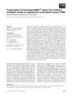
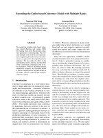
37.5%). Phylogenetic analysis also revealed that,
whereas C47Ap, F18Ep, and F20Hp clustered
together, the C. elegans proteins did not form a closed
cluster with any of the DAAOs or DASPOs of other
organisms (Fig. 1). These findings suggest that a pre-
cursor of the C. elegans proteins evolved from a pre-
cursor of the DAAOs and DASPOs found in other
organisms early during evolution.
Alignment of the deduced amino-acid sequences of
Y69Ap, C47Ap, F18Ep, and F20Hp with those of pig
DAAO [36], the yeast Rhodotorula gracilis DAAO [32],
bovine DASPO [46], and mouse DASPO [44] revealed
conservation of the FAD-binding consensus sequence
Caenorhabditis elegans D-aspartate oxidase M. Katane et al.
138 FEBS Journal 274 (2007) 137–149 ª 2006 The Authors Journal compilation ª 2006 FEBS
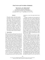
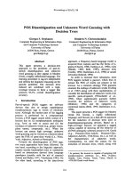
(GXGXXG) near the N-terminus (Fig. 2). Moreover,
the amino-acid sequences of Y69Ap and C47Ap
contain a C-terminal consensus sequence [(S ⁄ A ⁄ C)-
(K ⁄ R ⁄ H)-L] that is a type 1 peroxisomal targeting
signal (PTS1) [52]. The amino-acid sequences of
F18Ep and F20Hp lack this sequence. The Tyr224,
Tyr228, Arg283, and Gly313 residues in pig DAAO,
and the Met213, Tyr223, Tyr238, Arg285, and Ser335
residues in R. gracilis DAAO have been identified as
catalytically important residues by crystallographic
analyses [53–56] and mutagenesis experiments [57–61].
The corresponding residues in bovine and mouse
DASPOs have also been predicted to be catalytically
important by modeling of the 3D structures of the
DASPOs [44,61]. Moreover, a mutagenesis experiment
has revealed that the Arg216 and Arg237 residues of
mouse DASPO are catalytically important [44]. Our
alignment analysis suggests that the Tyr215, Arg270,
and Gly294 residues in Y69Ap, the Arg276 and
Gly308 residues in C47Ap, the Arg279 and Ser311 resi-
dues in F18Ep, and the Arg304 and Gly335 residues in
F20Hp correspond to the above-mentioned residues of
pig DAAO, R. gracilis DAAO, bovine DASPO, and
mouse DASPO (Fig. 2). However, other enzymatically
important residues mentioned are not conserved in the
C. elegans proteins. Thus, the primary structures of the
C. elegans proteins differ markedly from those of other
reported DAAOs and DASPOs. It is possible that
during evolution, the C. elegans proteins acquired dis-
tinctive properties.
Expression of the recombinant proteins in E. coli
and characterization of their enzymatic
properties
To confirm that the cloned cDNAs encode functional
DAAOs and DASPOs, expression plasmids for
N-terminally His-tagged Y69Ap, C47Ap, F18Ep, and
F20Hp were constructed. The molecular masses of the
N-terminally His-tagged Y69Ap, C47Ap, F18Ep, and
F20Hp were calculated to be 40 274, 41 794, 41 765,
and 46 658 Da, respectively. E. coli strain BL21(DE3)-
Fig. 1. Phylogenetic relationships of the
C. elegans cDNA products with the DAAOs
and DASPOs of other organisms. A data file
for the phylogenetic tree was created with
the
CLUSTALW Multiple Sequence Alignment
program (version 1.83) [75], and the phylo-
genetic tree was generated by using the
NJ
PLOT software [76]. The fruit fly, mos-
quito, and rat DASPOs are putative proteins.
The bacterial DAAOs are also putative pro-
teins and are used as an out-group. The
scale bar indicates a distance of 0.05 substi-
tutions per site. The UniProt accession num-
bers are: P. aeruginosa DAAO, P33642;
S. coelicolor DAAO, Q9X7P6; M. tuberculo-
sis DAAO, O07727; R. gracilis DAAO,
P80324; Trigonopsis variabilis DAAO,
Q99042; Fusarium solani DAAO, P24552;
Candida boidinii DAAO, Q9HGY3; Crypto-
coccus humicola DASPO, Q75WF1; fruit fly
DASPO, Q9VM80; mosquito DASPO,
Q7Q7G4; rabbit DAAO, P22942; pig DAAO,
P00371; guinea pig DAAO, Q9Z1M5; human
DAAO, P14920; rat DAAO, O35078; mouse
DAAO, P18894; hamster DAAO, Q9Z302;
carp DAAO, Q6TGN2; rat DASPO,
UPI000017E4D7; mouse DASPO, Q922Z0;
human DASPO-1, Q99489; bovine DASPO,
P31228.
M. Katane et al. Caenorhabditis elegans
D-aspartate oxidase
FEBS Journal 274 (2007) 137–149 ª 2006 The Authors Journal compilation ª 2006 FEBS 139
Fig. 2. Comparison of the amino acid sequences of C. elegans cDNAs with those of the DAAOs and DASPOs of other organisms. The
deduced amino-acid sequences of Y69Ap, C47Ap, F18Ep, F20Hp, pig DAAO [36], R. gracilis DAAO [32], bovine DASPO [46], and mouse
DASPO [44] were aligned by using the
CLUSTALW Multiple Sequence Alignment program (version 1.83) [75]. The amino-acid numbers of each
sequence are indicated on the right. Asterisks indicate amino-acid residues that are identical in all sequences. Conserved amino-acid substi-
tutions with low and high similarity are indicated by dots and double-dots, respectively. The FAD-binding motif (GXGXXG) is boxed. Amino-
acid residues that were experimentally proven to be catalytically important are shown as white letters on a black background. Amino-acid
residues presumed to be catalytically important are shaded in gray.
Caenorhabditis elegans
D-aspartate oxidase M. Katane et al.
140 FEBS Journal 274 (2007) 137–149 ª 2006 The Authors Journal compilation ª 2006 FEBS
pLysS cells were transformed with each expression
construct, then crude extracts and insoluble fractions
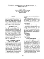
were subjected to western blot analysis. Recombinant
Y69Ap, C47Ap, and F18Ep were detected in the crude
extracts (Fig. 3A), and their apparent molecular mas-
ses were in good agreement with those calculated from
their deduced amino-acid sequences (Fig. 2). The insol-
uble fractions of the bacterial lysates exhibited intense
bands that had the same mobility as the bands in the
crude extracts (data not shown). Thus, the Y69Ap,
C47Ap, and F18Ep recombinant proteins were
expressed both as soluble and insoluble forms. In con-
trast, recombinant F20Hp was only detected in the
insoluble fraction (data not shown). We are now
searching for the optimal conditions that would allow
soluble recombinant F20Hp to be expressed. The crude
extract and insoluble fraction of cells transformed with
the parental plasmid did not have bands that were
recognized by the antibody to His-tag (Fig. 3A and
data not shown).
We subsequently examined the enzymatic activity of
the recombinant proteins against various amino acids.
Crude extracts of the transformed cells served as the
enzyme sources. The Y69Ap plasmid-transformed cell
extracts reproducibly showed enzymatic activity
against d-Ala. Three independent assays revealed that
this activity was 216.7 ± 23.9 mUÆ(mg protein)
)1
(mean ± SD). We repeatedly observed low levels of
activity against l-Ala, which is an enantiomer of d-Ala
(Table 1). This may be due to the fact that E. coli has
Ala racemase, which catalyzes the direct interconver-
sion between l-Ala and d-Ala [62] and thus may con-
vert l-Ala into d-Ala. Of the other neutral d-amino
acids examined, the Y69Ap extract was more active
against d-Met than against d-Ala but showed low
A B
Fig. 3. Analysis of the expression of recom-
binant proteins in E. coli and their purity. (A)
Cellular expression of recombinant Y69Ap,
C47Ap, and F18Ep was examined by west-
ern blotting of the crude extracts (20 lg)
using a His-tag antibody. (B) The proteins in
crude extracts (20 lg) and the purified
enzymes (0.5 lg) were separated on an
SDS ⁄ 12% polyacrylamide gel and stained
with Coomassie Brilliant Blue R-250. Crude
extracts from the parental plasmid pRSET-B
were also tested (Mock). MWM, molecular
mass marker proteins.
Table 1. Oxidase activities of the recombinant proteins against var-
ious amino acids. Appropriate amounts of crude extracts (10, 40,
and 15 lg of the Y69Ap, C47Ap, and F18Ep extracts, respectively)
were used as enzyme. Percentage activities relative to that detec-
ted with
D-Ala are shown for Y69Ap. Similarly, percentage activities
relative to those with
D-Asp are shown for C47Ap and F18Ep. A
445
corresponding to 100% values of Y69Ap, C47Ap, and F18Ep was
0.745, 0.396, and 1.072, respectively. Each value shown is the
mean ± SD from three independent assays. ND, Not determined;
NMLA, N-methyl-
L-aspartate.
Substrate
Relative activity (%)
Y69Ap C47Ap F18Ep
D-Ala 100 ± 11 4.5 ± 1.2 2.8 ± 0.2
D-Met 172 ± 19 3.8 ± 1.5 2.4 ± 1.1
D-Asn 19 ± 1.5 17 ± 3.2 6.1 ± 1.1
D-Arg 55 ± 6.4 < 0.1 < 0.1
D-Asp 2.2 ± 1.8 100 ± 2.1 100 ± 4.9
D-Glu < 0.1 247 ± 16 67 ± 2.0
NMDA < 0.1 313 ± 16 110 ± 2.6
L-Ala 34 ± 1.8 ND ND
L-Met 1.9 ± 1.7 ND ND
L-Arg < 0.1 ND ND
L-Asp ND 2.6 ± 0.3 < 0.1
L-Glu ND 1.3 ± 0.9 0.3 ± 0.3
NMLA ND 6.7 ± 0.3 1.1 ± 1.0
M. Katane et al. Caenorhabditis elegans
D-aspartate oxidase
FEBS Journal 274 (2007) 137–149 ª 2006 The Authors Journal compilation ª 2006 FEBS 141
activities against d-Asn. The Y69Ap extract was also
moderately active against the basic d-amino acid,
d-Arg, but had very low or undetectable activity
against the enantiomers, l-Met and l-Arg, and all
acidic d-amino acids examined. Thus, the protein
encoded by Y69Ap cDNA can catalyze the deamina-
tion of neutral and basic d-amino acids.
Three independent assays revealed that, unlike the
Y69Ap extract, the C47Ap and F18Ep extracts
had reproducible enzymatic activity against d-Asp
[28.8 ± 0.6 and 208.1 ± 10.1 mUÆ(mg protein)
)1
,
respectively]. These assays also revealed that both
extracts had activities against other acidic d-amino
acids, namely, d-Glu and NMDA (Table 1). Only very
low or undetectable activities were detected against the
enantiomers l-Asp, l-Glu, and N-methyl-l-Asp and all
neutral and basic d-amino acids examined. Thus, the
proteins encoded by the cloned C47Ap and F18Ep
cDNAs can catalyze the deamination of acidic
d-amino acids. The crude extract of cells transformed
with the parental plasmid lacked activity against every
d-amino acid and l-amino acid examined (data not
shown). Together, these observations confirm that the
Y69Ap cDNA encodes a functional DAAO that is
specific for a basic d-amino acid as well as for non-
polar neutral d-amino acids, whereas the C47Ap and
F18Ep cDNAs encode functional DASPOs that act on
acidic d-amino acids.
Purification of the recombinant proteins and their
kinetic characterization
To further characterize Y69Ap, C47Ap, and F18Ep,
the recombinant enzymes were purified to near-homo-
geneity by affinity chromatography using a chelating
column (Fig. 3B). The specific activity of purified
Y69Ap against d-Ala was 4.96 UÆ(mg protein)
)1
,
which is about 22.9 times higher than that of crude
extract. As expected, purified Y69Ap lacked enzymatic
activity against l-Ala (data not shown), which con-
firms that the activity of Y69Ap against d-Ala is stere-
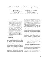
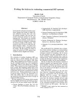
ospecific. We then obtained Hanes–Woolf plots to
determine the apparent kinetic parameters of the
deamination of d-Ala, d-Met and d-Arg catalyzed by
purified Y69Ap (Fig. 4A). The V
max
(maximal velocity)
values were 5.41, 7.43 and 2.52 UÆ(mg protein)
)1
for
d-Ala, d-Met and d-Arg, respectively. The k
cat
(molecular activity) values (calculated from the V
max
values and the estimated molecular mass of N-termin-
ally His-tagged Y69Ap) are listed in Table 2. Thus, the
highest V
max
and k
cat
values for Y69Ap were against
d-Met, followed by d-Ala and d-Arg. This matches
the hierarchy of Y69Ap activities revealed by the
experiments with the crude extract (Table 1). However,
the K
m
(Michaelis constant) value against d-Arg was
at least 10 times lower than those against d-Ala and
d-Met (Table 2). Therefore, the catalytic efficiency
(expressed as k
cat
⁄ K
m
) of Y69Ap against d-Arg was
7.3 and 3.8 times higher than against d-Ala and
d-Met, respectively. This indicates that Y69Ap prob-
ably prefers basic d-amino acid(s) to neutral d-amino
acids as its substrate.
The specific activities of purified C47Ap and F18Ep
against d-Asp were 4.99 and 4.33 UÆ(mg protein)
)1
,
–5 0 5 10 15 20 25 30 35 40
15
10
5
[S] / V (mM·mg·U
–1
)
[S] (mM)
A
D-Ala
D-Met
D-Arg
–5 0 5 10 15 20 25 30 35 40
15
10
5
[S] / V (mM·mg·U
–1
)
[S] (mM)
B
D-Asp
D-Glu
NMDA
–5 0 5 10 15 20 25 30 35 40
15
10
5
[S] / V (mM·mg·U
–1
)
[S] (mM)
C
D-Asp
D-Glu
NMDA
Fig. 4. Hanes–Woolf plots of the oxidase activity of the purified
enzymes. Enzymatic activities were assayed by using purified
Y69Ap (A), C47Ap (B), and F18Ep (C). The substrates used were
D-Ala (s), D-Met (h), and D-Arg (n) for Y69Ap, and D-Asp (d), D-Glu
(j), and NMDA (m) for C47Ap and F18Ep.
Caenorhabditis elegans
D-aspartate oxidase M. Katane et al.
142 FEBS Journal 274 (2007) 137–149 ª 2006 The Authors Journal compilation ª 2006 FEBS
which are 173 and 20.8 times higher than the specific
activities of the crude extracts, respectively. Hanes–
Woolf plots to determine the apparent kinetic parame-
ters of the deamination of d-Asp, d-Glu, and NMDA
catalyzed by these enzymes (Fig. 4B,C) revealed that
the V
max
values of purified C47Ap were 6.16, 7.62, and
8.80 UÆ(mg protein)
)1
, respectively, and the V
max
val-
ues of purified F18Ep were 4.40, 3.03, and 4.31 UÆ(mg
protein)
)1
, respectively. The k
cat
, K
m
, and k
cat
⁄ K
m
values of these enzymes against d-Asp, d-Glu, and
NMDA are listed in Table 2 and show that the cata-
lytic efficiency of C47Ap against d-Glu and NMDA
was about 10.9 and 3.4 times higher than that against
d-Asp, largely because of differences in the K
m
values
(substrate affinity). In contrast, the K
m
value of F18Ep
against NMDA was significantly higher than its K
m
value against d-Asp and d-Glu. Therefore, the cata-
lytic efficiency of F18Ep against NMDA was lower
than against d-Asp, whereas it was equally as efficient
against d-Glu and d-Asp. F18Ep was 3.8 times more
efficient against d -Asp than C47Ap, and C47Ap was
2.7 and 4.5 times more efficient against d-Glu and
NMDA than F18Ep, respectively. Thus, although
C47Ap and F18Ep both act on acidic d-amino acids,
they differ in their substrate specificity profiles.
Discussion
The deduced C-terminal amino acids of Y69Ap and
C47Ap are SKL (Fig. 2), which corresponds to the
PTS1 consensus sequence [52]. These enzymes are thus
predicted to localize to peroxisomes, like the DAAOs
and DASPOs of other organisms [18–24]. In contrast,
the three deduced C-terminal amino acids of F18Ep
and F20Hp were LGL and LDD, respectively, which
do not correspond to the PTS1 consensus sequence.
The sequences of F18Ep and F20Hp also lacked
the bipartite consensus sequence -(R ⁄ K)-(L ⁄ V ⁄ I)-X5-
(H ⁄ Q)-(L ⁄ A)-, a type 2 peroxisomal targeting signal
(PTS2) [52]. In C. elegans, the PTS2-dependent path-
way was reported to be absent [63]. Representative sig-
nal sequences that prompt the localization of proteins
to organelles other than peroxisome were also not
found in the F18Ep and F20Hp sequences. Hence,
F18Ep and F20Hp probably localize to the cytoplasm.
However, another possibility is that these proteins
localize to peroxisomes via a PTS1-independent import
pathway. The existence of such a novel pathway is sug-
gested by the report that the peroxisomal importation
of acyl-CoA oxidase of the yeast, Saccharomyces
cerevisiae, was not disturbed in cells that lacked
PTS1-dependent or PTS2-dependent importation [64].
Alternatively, it is possible that F18Ep and F20Hp are
imported into peroxisomes by forming a complex(es)
with PTS1-bearing proteins such as Y69Ap and
C47Ap.
This study demonstrates that the DAAO (Y69Ap)
and DASPOs (C47Ap and F18Ep) in C. elegans can be
expressed in E. coli as functional recombinant proteins.
E. coli has often been used as a host organism to pre-
pare recombinant DAAOs and DASPOs of various
organisms. However, it can be difficult to overexpress
these enzymes in active and soluble forms in E. coli for
the following reasons. First, DAAO and DASPO cata-
lyze the deamination of d-Ala and d-Glu, respectively,
which are essential components of the peptidoglycans
in bacterial cell walls. Secondly, the enzymatic reactions
catalyzed by DAAO and DASPO produce hydrogen
peroxide, which is highly toxic for E. coli. Conse-
quently, overexpression of these enzymes may inhibit
E. coli cell growth. Indeed, successful expression of
mammalian DAAOs, apart from those from pigs, mice,
and humans, in E. coli has not been reported. More-
over, in the case of porcine DAAO, it was reported that
only 25 mg purified enzyme was obtained from 40 g
wet cell paste [59]. With regard to our own observa-
tions, we found that recombinant Y69Ap was readily
overexpressed in E. coli, recovered in the soluble frac-
tion, and purified to near-homogeneity (Fig. 3). About
4 mg purified Y69Ap was obtained from 2 g wet cell
paste. In contrast, recombinant F20Hp was not recov-
ered in the soluble fraction. This may be related to the
Table 2. Apparent steady-state kinetic parameters of the purified recombinant proteins against several D-amino acids. ND, Not determined.
Substrate
Y69Ap C47Ap F18Ep
k
cat
(s
)1
)
K
m
(mM)
k
cat
⁄ K
m
(s
)1
ÆM
)1
)
k
cat
(s
)1
)
K
m
(mM)
k
cat
⁄ K
m
(s
)1
ÆM
)1
)
k
cat
(s
)1
)
K
m
(mM)
k
cat
⁄ K
m
(s
)1
ÆM
)1
)
D-Ala 3.63 1.72 2113 ND ND ND ND ND ND
D-Met 4.98 1.22 4082 ND ND ND ND ND ND
D-Arg 1.69 0.11 15 394 ND ND ND ND ND ND
D-Asp ND ND ND 4.29 2.02 2125 3.07 0.38 8066
D-Glu ND ND ND 5.31 0.23 23 066 2.11 0.25 8427
NMDA ND ND ND 6.13 0.84 7300 3.00 1.84 1629
M. Katane et al. Caenorhabditis elegans
D-aspartate oxidase
FEBS Journal 274 (2007) 137–149 ª 2006 The Authors Journal compilation ª 2006 FEBS 143
potentially adverse effects of overexpressing DAAOs
and DASPOs in E. coli mentioned above. It will be
necessary to improve the expression system and the
purification procedure before we can functionally char-
acterize recombinant F20Hp.
A number of reports have characterized the enzymat-
ic properties of the DAAOs of various organisms by
analyzing recombinant proteins expressed in E. coli or
yeast. These reports reveal that these DAAOs differ
markedly in their activities and substrate specificities.
For example, the k
cat
values of the porcine, human,
R. gracilis, and carp DAAOs against d-Ala are repor-
ted to be 6.4, 5.2, 350, and 190 s
)1
, respectively [58,65–
67]. The k
cat
value of Y69Ap against d-Ala (3.63 s
)1
)is
similar to those of the pig and human DAAOs. More-
over, like pig DAAO [59], Y69Ap was more active
against d-Met than against d-Ala (Tables 1 and 2). In
contrast, the R. gracilis and carp DAAOs are more act-
ive against d-Ala than against any of the other amino-
acid substrates examined [61,67]. This suggests that
Y69Ap may be more similar in terms of its enzymatic
properties to mammalian DAAOs than to microbial
and fish DAAOs. However, Y69Ap was also active
with d-Arg, which is a poor substrate for pig DAAO.
This disparity may relate to structural difference(s)
between the active sites of Y69Ap and pig DAAO. This
is supported by the fact that, whereas the catalytically
important Tyr228, Arg283, and Gly313 residues in pig
DAAO [54,55,59] are all conserved in Y69Ap, Tyr224
is not (Fig. 2). Moreover, comparison of the experi-
mentally determined crystal structure of pig DAAO [54]
with the predicted 3D structure of Y69Ap generated
with the SWISS-MODEL server [68] suggests that the
Y69Ap residue Phe229 is in the same structural posi-
tion as Tyr224 of pig DAAO (data not shown). Muta-
genesis experiments and crystallographic analyses will
be necessary to elucidate the role of Phe229 in the enzy-
matic activity of Y69Ap against basic d-amino acid(s).
In this study, we have demonstrated that, although
C47Ap and F18Ep both act on acidic d-amino acids,
they differ in their kinetics and preferences for partic-
ular substrates. C47Ap functioned more efficiently
against d-Glu and NMDA than against d-Asp,
whereas F18Ep acted more efficiently against d-Asp
and d-Glu than against NMDA (Table 2). Examina-
tion of the reported kinetic properties of recombinant
mammalian and micro-organism DASPOs suggests
that C47Ap and F18Ep are unusual with regard to
their specificity for d-Glu. The catalytic efficiency of
C47Ap against d-Glu is 23 066 s
)1
Æm
)1
, which is 108
and 923 times higher than the respective catalytic effi-
ciencies of bovine and Cryptococcus humicola DASPOs
against d-Glu (213 and 25.0 s
)1
Æm
)1
, respectively)
[43,69]. Similarly, the catalytic efficiency of F18Ep
against d-Glu is 8427 s
)1
Æm
)1
, which is 39.6 and 337
times higher than the catalytic efficiencies of bovine
and C. humicola DASPOs, respectively. As C. elegans
lives in soil and eats micro-organisms that are prob-
ably rich in d-Glu, it is possible that diet-derived
d-Glu is incorporated into the body of C. elegans.
Although little is currently understood about the
amounts and physiological functions of d-amino acids
in C. elegans, it was recently reported that injection of
d-Glu into a silkworm, which is a multicellular model
insect, induced muscle contraction [70]. It is possible
that excess amounts of d-Glu are as toxic for C. ele-
gans as they are for the silkworm, and that C. elegans
may need C47Ap and F18Ep to deaminate d-Glu and
thereby neutralize the toxicity of diet-derived d-Glu.
To our knowledge, this is the first report of the clo-
ning of invertebrate DAAO and DASPO cDNAs. In
addition, we have demonstrated for the first time that
an organism ( C. elegans) can have multiple active
DASPO (C47Ap and F18Ep) genes. The tissue
localization of C47Ap and F18Ep within the body of
C. elegans remains to be elucidated. As C47Ap and
F18Ep are encoded by distinct genes in different loci
in the C. elegans genome, their transcriptional regula-
tions are possibly independent. Thus, it is possible
that these two enzymes are tissue-specific isoforms in
C. elegans. Green fluorescent protein (GFP)-based or
b-galactosidase-based gene expression and in situ
hybridization analyses may reveal the localization
of C47Ap and F18Ep within the whole body of
C. elegans. If these two proteins are expressed in the
same cell, they may localize to distinct regions within
the cell. It is likely that C47Ap is localized in the per-
oxisome in a PTS1-dependent manner, whereas F18Ep
remains in the cytoplasm. However, the biological
significance of the multiple DASPOs in C. elegans is
currently unclear. That other organisms may also have
more than one DASPO is suggested by a study show-
ing that two proteins, DASPO-1 and DASPO-2, are
translated from a single human DASPO mRNA by
alternative splicing [45], although the function of
DASPO-2 remains to be clarified. Further studies will
be needed to determine the tissue and cellular distri-
butions of C47Ap and F18Ep and their expression
during the development of C. elegans.
Experimental procedures
Animals and chemicals
Caenorhabditis elegans Bristol strain N2 and E. coli strain
OP50 were kindly provided by Y. Nakagawa (Laboratory
Caenorhabditis elegans D-aspartate oxidase M. Katane et al.
144 FEBS Journal 274 (2007) 137–149 ª 2006 The Authors Journal compilation ª 2006 FEBS
of Hygienic Chemistry, School of Pharmaceutical Sciences,
Kitasato University, Japan). The C. elegans worms were
maintained at 20 °C on NGM agar plates seeded with
E. coli as described by Brenner [71].
d- and l-amino acids, ampicillin, BSA, and Aspergil-
lus niger catalase were purchased from Sigma-Aldrich Inc.
(St Louis, MO, USA). FAD, isopropyl b-d-thiogalactopyr-
anoside, and imidazole were purchased from Wako Pure
Chemical Industries (Osaka, Japan). Other chemicals were
of the highest grade available and purchased from commer-
cial sources.
Isolation of cDNA clones from C. elegans
Cultured C. elegans were collected by the standard method
[71]. Their total RNAs were extracted by using ISOGEN
reagent (Nippon Gene, Tokyo, Japan), according to
the manufacturer’s instructions. For first-strand cDNA
synthesis, the RNA samples (5 lg) were reverse-transcribed
for 50 min at 42 °C in a 20-l L reaction volume with
200 U SuperScript II Reverse Transcriptase and 0.5 lg
oligo(dT)
12)18
primer (Invitrogen, Carlsbad, CA, USA).
The cDNAs of the Y69A2AR.5 (WormBase gene ID:
WBGene00022076; genomic location: 2 614 199–2 617 030
on chromosome IV), C47A10.5 (WormBase gene ID:
WBGene00008127; genomic location: 17 777 009–17 774
090 on chromosome V), F18E3.7a (WormBase gene ID:
WBGene00017565; genomic location: 7 433 290–7 434 835
on chromosome V), and F20H11.5 (WormBase gene ID:
WBGene00017648; genomic location: 6 587 836–6 589 212
on chromosome III) genes were amplified by PCR using
the first-strand cDNA as a template and the following
primers: for Y69A2AR.5,5¢-AGATCTATGCCTAAA
ATTGCTGTACTAGGCGCAGG-3¢ (forward primer) and
5¢-GAATTCTCACAACTTCGACTTTTTCATTTTCAGC-
3¢ (reverse primer); for C47A10.5 ,5¢-AGATCTATGACT
CCAAAAATCGCAATAATCGGCG-3¢ (forward primer)
and 5¢-GGTACCTCACAGTTTCGAAGAATTTAGAGC
GG-3¢ (reverse primer); for F18E3.7a,5¢-AGATCTAT
GGCAAACATAATTCCGAAGATTGC-3¢ (forward pri-
mer) and 5¢-GAATTCTTATAATCCTAGTGCAGTCTT
AACAAG-3¢ (reverse primer); and for F20H11.5,5¢-AG
ATCTATGCTGTATGCTCTTCTTCTCCTC-3¢ (forward
primer) and 5¢-GGTACCCTAATCATCAAGATATTTA
ACCCATTCGG-3¢ (reverse primer). These primer sets were
designed so that (a) additional BglII sites were created at
the 5¢ ends of the forward primers for all genes, (b) addi-
tional EcoRI sites were created at the 5¢ ends of the reverse
primers for the Y69A2AR.5 and F18E3.7a genes, and (c)
additional KpnI sites were created at the 5¢ ends of the
reverse primers for the C47A10.5 and F20H11.5 genes. The
PCR products were cloned into pT7Blue (Novagen, Madi-
son, WI, USA), thus generating pT7-Y69Ap, pT7-C47Ap,
pT7-F18Ep, and pT7-F20Hp, and were then sequenced.
Construction of recombinant protein expression
plasmids
To construct the Y69Ap expression plasmid, the 1.0-kb
BglII–EcoRI fragment containing the entire Y69Ap-coding
sequence of pT7-Y69Ap was subcloned into pRSET-B
(Invitrogen), resulting in the N-terminally His-tagged
Y69Ap expression plasmid pRSET-His-Y69Ap. Similarly,
the 1.0-kb BglII–KpnI fragment containing the entire
C47Ap-coding sequence of pT7-C47Ap, the 1.0-kb BglII–
EcoRI fragment containing the entire F18Ep-coding
sequence of pT7-F18Ep, and the 1.2-kb BglII–KpnI frag-
ment containing the entire F20Hp-coding sequence of
pT7-F20Hp were subcloned into pRSET-B, resulting in
the N-terminally His-tagged C47Ap, F18Ep, and F20Hp
expression plasmids (pRSET-His-C47Ap, pRSET-His-
F18Ep, and pRSET-His-F20Hp), respectively.
Expression and purification of recombinant
proteins
Escherichia coli strain BL21(DE3)pLysS cells transformed
with the expression plasmids were grown at 37 °C with sha-
king in LB medium containing ampicillin (100 lgÆmL
)1
).
When A
620
reached 0.5, isopropyl b-d-thiogalactopyrano-
side was added to a final concentration of 0.01 mm, and
the cells were grown at 30 °C for an additional 20 h. The
cells were pelleted by centrifugation at 10 000 g for 10 min
at 4 °C in a Kubota RA-200J rotor using a model 1920
Kubota centrifuge (Kubota corporation, Tokyo, Japan).
The pellets were then resuspended in lysis buffer consisting
of BugBuster Protein Extraction Reagent (Novagen), 50 lm
FAD, and protease inhibitors (Roche Applied Science,
Mannheim, Germany) (5 mL lysis buffer per g wet cell
paste was used). The cell suspension was incubated for
20 min at room temperature with gentle shaking. The
resulting lysates were centrifuged at 12 000 g for 20 min at
4 °C (Kubota model 1920 centrifuge with RA-200J rotor)
to pellet the insoluble cell debris. The supernatant (crude
extract) was filtered through a 0.45 lm membrane filter
(Asahi Techno Glass Corporation, Tokyo, Japan) and used
immediately for further purification or stored frozen at
)80 °C until use. To prepare the insoluble fraction, the pel-
leted cell debris was resuspended in 10 mm phosphate-
buffered saline (pH 7.4), mixed with an equal volume of
4% SDS solution, and boiled immediately.
The N-terminally His-tagged recombinant proteins were
purified by affinity chromatography using a chelating col-
umn. Crude extracts, prepared as described above, were
applied to a HiTrap Chelating HP column (1 mL; Amer-
sham Biosciences, Piscataway, NJ, USA) equilibrated with
20 mm sodium dihydrogen phosphate buffer (pH 7.4) con-
taining 0.5 m NaCl and 10 mm imidazole. The column was
washed with the same buffer, and the bound proteins were
M. Katane et al. Caenorhabditis elegans D-aspartate oxidase
FEBS Journal 274 (2007) 137–149 ª 2006 The Authors Journal compilation ª 2006 FEBS 145
eluted with a linear gradient of 10–500 mm imidazole. Each
fraction (2 mL) containing the recombinant proteins was
mixed with 50 lL2mm FAD and dialyzed for 3 h twice
against 1 L 10 mm sodium pyrophosphate buffer (pH 8.3)
containing 2 mm EDTA, 5 mm 2-mercaptoethanol, and
10% glycerol. The dialyzed fractions were pooled as the
purified enzyme and used immediately for enzyme assays or
stored frozen at )80 °C until use.
Detection of recombinant proteins
The protein concentrations in the crude extracts, insoluble
fractions, and purified enzymes were determined by the
method of Bradford [72] using BSA as a standard. Crude
extracts (20 lg) and insoluble fractions (20 lg) were subjec-
ted to SDS ⁄ PAGE (12% gel) and western blotting using
anti-(His-tag) serum (His-probe; Santa Cruz Biotechnology,
Santa Cruz, CA, USA) (1 : 1000 dilution) as the primary
antibody and horseradish peroxidase-conjugated anti-rabbit
IgG (Jackson ImmunoResearch Laboratories, West Grove,
PA, USA) (1 : 5000 dilution) as the secondary reagent. The
protein bands were visualized with an enhanced chemilumi-
nescence reagent (Amersham Biosciences) and by exposure
to Lumi-Film Chemiluminescent Detection Film (Roche
Applied Science). To analyze the protein purity, the pro-
teins in the crude extracts (20 lg) and the purified enzymes
(0.5 lg) were separated on an SDS ⁄ 12% polyacrylamide gel
and stained with Coomassie Brilliant Blue R-250. Broad-
range molecular mass standards (Bio-Rad, Hercules, CA,
USA) served as molecular mass marker proteins.
Assays of enzymatic activity
Oxidase activities were determined by colorimetric measure-
ment of the corresponding 2-oxo acids produced from the
amino acids used, as previously described by Nagata et al.
[73] with following modifications. Appropriate amounts of
enzyme (10–40 and 0.5–1.0 lg crude extracts and purified
enzymes, respectively) were mixed with a reaction mixture
consisting of 40 mm sodium pyrophosphate buffer
(pH 8.3), 50 lm FAD, and 20 mm amino acid in a final
volume of 150 lL, and incubated at 37 °C. The incubation
times with crude extracts and purified enzymes were 30 and
15 min, respectively. Subsequently, 10 lL 100% (w ⁄ v) tri-
chloroacetic acid was added to stop the reactions, and pro-
teins were pelleted by centrifugation at 20 000 g for 10 min
at 4 °C (Kubota model 1920 centrifuge with RA-48J rotor).
The supernatant (150 lL) was mixed with 100 lL 0.1%
(w ⁄ v) 2,4-dinitrophenylhydrazine in 2 m HCl, and incuba-
ted at 37 °C for 15 min, then 750 lL of 3.75 m NaOH was
added, and the solution was cleared by centrifugation at
20 000 g for 10 min at 4 °C (Kubota model 1920 centrifuge
with RA-48J rotor). The absorbance of the supernatant at
445 nm was measured against the blank prepared from a
reaction mixture lacking the amino acid. One unit of
enzyme activity was defined as the production of 1 lmol of
the corresponding 2-oxo acid per min under the above
assay conditions. For kinetic analyses, different final con-
centrations (0, 0.5, 1, 2, 5, 10, 20, and 40 mm) of several
d-amino acids were used as substrates. In some cases,
A. niger catalase (5 l g) was added to the reaction mixture
to prevent the decarboxylation of the 2-oxo acid by the
hydrogen peroxide that was produced by the reaction [74].
Acknowledgements
We thank Professor Y. Nakagawa (School of Pharma-
ceutical Sciences, Kitasato University, Japan) for pro-
viding C. elegans Bristol strain N2 and E. coli strain
OP50.
References
1 Rosenberg H & Ennor AH (1960) Occurrence of free
d-serine in the earthworm. Nature 187, 617–618.
2 Srinivasan NG, Corrigan JJ & Meister A (1962)
d-Serine in the blood of the silkworm Bombyx mori and
other lepidoptera. J Biol Chem 237 , 3844–3845.
3 Corrigan JJ & Srinivasan NG (1966) The occurrence
of certain d-amino acids in insects. Biochemistry 5,
1185–1190.
4 Matsui T, Sekiguchi M, Hashimoto A, Tomita U,
Nishikawa T & Wada K (1995) Functional comparison
of d-serine and glycine in rodents: the effect on cloned
NMDA receptors and the extracellular concentration.
J Neurochem 65, 454–458.
5 Mothet JP, Parent AT, Wolosker H, Brady RO Jr,
Linden DJ, Ferris CD, Rogawski MA & Snyder SH
(2000) d-Serine is an endogenous ligand for the glycine
site of the N-methyl-d-aspartate receptor. Proc Natl
Acad Sci USA 97, 4926–4931.
6 D’Aniello A, Di Cosmo A, Di Cristo C, Annunziato L,
Petrucelli L & Fisher G (1996) Involvement of d-aspar-
tic acid in the synthesis of testosterone in rat testes. Life
Sci 59, 97–104.
7 D’Aniello G, Tolino A, D’Aniello A, Errico F,
Fisher GH & Di Fiore MM (2000) The role of
d-aspartic acid and N-methyl- d-aspartic acid in the
regulation of prolactin release. Endocrinology 141,
3862–3870.
8 D’Aniello A, Spinelli P, De Simone A, D’Aniello S,
Branno M, Aniello F, Fisher GH, Di Fiore MM &
Rastogi RK (2003) Occurrence and neuroendocrine role
of d-aspartic acid and N-methyl-d-aspartic acid in
Ciona intestinalis. FEBS Lett 552, 193–198.
9 Lamanna C, Assisi L, Botte V & Di Fiore MM (2006)
Involvement of D-Asp in P450 aromatase activity and
estrogen receptors in boar testis. Amino Acids,
doi:10.1007 ⁄ s00726-006-0351-9.
Caenorhabditis elegans D-aspartate oxidase M. Katane et al.
146 FEBS Journal 274 (2007) 137–149 ª 2006 The Authors Journal compilation ª 2006 FEBS
10 Long Z, Lee JA, Okamoto T, Nimura N, Imai K &
Homma H (2000) d-Aspartate in a prolactin-secreting
clonal strain of rat pituitary tumor cells (GH
3
). Biochem
Biophys Res Commun 276, 1143–1147.
11 Nagata Y, Homma H, Lee JA & Imai K (1999)
d-Aspartate stimulation of testosterone synthesis in rat
Leydig cells. FEBS Lett 444, 160–164.
12 Pampillo M, del Carmen Diaz M, Duvilanski BH,
Rettori V, Seilicovich A & Lasaga M (2001) Differential
effects of glutamate agonists and d-aspartate on
oxytocin release from hypothalamus and posterior
pituitary of male rats. Endocrine 15, 309–315.
13 Raucci F, Assisi L, D’Aniello S, Spinelli P, Botte V &
Di Fiore MM (2004) Testicular endocrine activity is
upregulated by d-aspartic acid in the green frog, Rana
esculenta. J Endocrinol 182, 365–376.
14 Raucci F, D’Aniello S & Di Fiore MM (2005)
Endocrine roles of d-aspartic acid in the testis of lizard
Podarcis s. sicula. J Endocrinol 187, 347–359.
15 Spinelli P, Brown ER, Ferrandino G, Branno M,
Montarolo PG, D’Aniello E, Rastogi RK, D’Aniello B,
Baccari GC, Fisher G, et al. (2006) d-Aspartic acid in
the nervous system of Aplysia limacina: possible role in
neurotransmission. J Cell Physiol 206, 672–681.
16 Takigawa Y, Homma H, Lee JA, Fukushima T, Santa T,
Iwatsubo T & Imai K (1998) d-Aspartate uptake into
cultured rat pinealocytes and the concomitant effect on
l-aspartate levels and melatonin secretion. Biochem
Biophys Res Commun 248, 641–647.
17 Wang H, Wolosker H, Pevsner J, Snyder SH & Selkoe
DJ (2000) Regulation of rat magnocellular neurosecre-
tory system by d-aspartate: evidence for biological role
(s) of a naturally occurring free d-amino acid in
mammals. J Endocrinol 167, 247–252.
18 Amery L, Brees C, Baes M, Setoyama C, Miura R,
Mannaerts GP & Van Veldhoven PP (1998) C-Terminal
tripeptide Ser-Asn-Leu (SNL) of human d-aspartate
oxidase is a functional peroxisome-targeting signal.
Biochem J 336, 367–371.
19 Beard ME (1990) d-Aspartate oxidation by rat and
bovine renal peroxisomes: an electron microscopic
cytochemical study. J Histochem Cytochem 38, 1377–
1381.
20 Kera Y, Niino A, Ikeda T, Okada H & Yamada R
(1998) Peroxisomal localization of d-aspartate oxidase
and development of peroxisomes in the yeast Crypto-
coccus humicolus UJ1 grown on d-aspartate. Biochim
Biophys Acta 1379
, 399–405.
21 Parveen Z, Large A, Grewal N, Lata N, Cancio I,
Cajaraville MP, Perry CJ & Connock MJ (2001)
D-Aspartate oxidase and d-amino acid oxidase are
localised in the peroxisomes of terrestrial gastropods.
Eur J Cell Biol 80, 651–660.
22 Van Veldhoven PP, Brees C & Mannaerts GP
(1991) d-Aspartate oxidase, a peroxisomal enzyme in
liver of rat and man. Biochim Biophys Acta 1073,
203–208.
23 Zaar K, Vo
¨
lkl A & Fahimi HD (1989) d-Aspartate
oxidase in rat, bovine and sheep kidney cortex is loca-
lized in peroxisomes. Biochem J 261, 233–238.
24 Zaar K, Ko
¨
st HP, Schad A, Vo
¨
lkl A, Baumgart E &
Fahimi HD (2002) Cellular and subcellular distribution
of d-aspartate oxidase in human and rat brain. J Comp
Neurol 450, 272–282.
25 Pilone MS, Pollegioni L, Casalin P, Curti B & Ronchi S
(1989) Properties of d-amino-acid oxidase from Rhodo-
torula gracilis. Eur J Biochem 180, 199–204.
26 Pollegioni L, Langkau B, Tischer W, Ghisla S & Pilone
MS (1993) Kinetic mechanism of d -amino acid oxidases
from Rhodotorula gracilis and Trigonopsis variabilis.
J Biol Chem 268, 13850–13857.
27 Yamada R, Ujiie H, Kera Y, Nakase T, Kitagawa K,
Imasaka T, Arimoto K, Takahashi M & Matsumura Y
(1996) Purification and properties of d-aspartate oxidase
from Cryptococcus humicolus UJ1. Biochim Biophys
Acta 1294, 153–158.
28 Curti B, Ronchi S, Branzoli U, Ferri G & Williams CH
Jr (1973) Improved purification, amino acid analysis
and molecular weight of homogenous d-amino acid oxi-
dase from pig kidney. Biochim Biophys Acta 327, 266–
273.
29 Negri A, Massey V & Williams CH Jr (1987) d-Aspar-
tate oxidase from beef kidney. Purification and proper-
ties. J Biol Chem 262, 10026–10034.
30 Tedeschi G, Negri A, Ceciliani F, Ronchi S, Vetere A,
D’Aniello G & D’Aniello A (1994) Properties of the
flavoenzyme d-aspartate oxidase from Octopus vulgaris.
Biochim Biophys Acta 1207, 217–222.
31 Gonza
´
lez FJ, Montes J, Martin F, Lo
´
pez MC, Fermi-
n
˜
a
´
n E, Catala
´
n J, Gala
´
n MA & Domı
´
nguez A (1997)
Molecular cloning of TvDAO1, a gene encoding a
d-amino acid oxidase from Trigonopsis variabilis and its
expression in Saccharomyces cerevisiae and Kluyvero-
myces lactis. Yeast 13, 1399–1398.
32 Pollegioni L, Molla G, Campaner S, Martegani E &
Pilone MS (1997) Cloning, sequencing and expression in
E. coli of a d-amino acid oxidase cDNA from Rhodotor-
ula gracilis active on cephalosporin C. J Biotechnol 58,
115–123.
33 Yurimoto H, Hasegawa T, Sakai Y & Kato N (2000)
Physiological role of the d-amino acid oxidase gene,
DAO1, in carbon and nitrogen metabolism in the
methylotrophic yeast Candida boidinii. Yeast 16, 1217–
1227.
34 Isogai T, Ono H, Ishitani Y, Kojo H, Ueda Y & Koh-
saka M (1990) Structure and expression of cDNA for d-
amino acid oxidase active against cephalosporin C from
Fusarium solani. J Biochem (Tokyo) 108, 1063–1069.
35 Sarower MG, Okada S & Abe H (2003) Molecular
characterization of d-amino acid oxidase from common
M. Katane et al. Caenorhabditis elegans D-aspartate oxidase
FEBS Journal 274 (2007) 137–149 ª 2006 The Authors Journal compilation ª 2006 FEBS 147
carp Cyprinus carpio and its induction with exogenous
free d-alanine. Arch Biochem Biophys 420, 121–129.
36 Fukui K, Watanabe F, Shibata T & Miyake Y (1987)
Molecular cloning and sequence analysis of cDNAs
encoding porcine kidney d-amino acid oxidase. Bio-
chemistry 26, 3612–3618.
37 Fukui K & Miyake Y (1992) Molecular cloning
and chromosomal localization of a human gene
encoding d-amino-acid oxidase. J Biol Chem 267,
18631–18638.
38 Konno R (1998) Rat d-amino-acid oxidase cDNA: rat
d-amino-acid oxidase as an intermediate form between
mouse and other mammalian d-amino-acid oxidases.
Biochim Biophys Acta 1395, 165–170.
39 Konno R, Kurabayashi A, Tsuchiya M & Niwa A
(1999) Guinea pig d-amino-acid oxidase cDNA and
phylogenetic position. DNA Seq 10, 85–91.
40 Momoi K, Fukui K, Tada M & Miyake Y (1990) Gene
expression of d-amino acid oxidase in rabbit kidney.
J Biochem (Tokyo) 108, 406–413.
41 Tada M, Fukui K, Momoi K & Miyake Y (1990) Clon-
ing and expression of a cDNA encoding mouse kidney
d-amino acid oxidase. Gene 90, 293–297.
42 Tsuchiya M, Kurabayashi A & Konno R (2003) Ham-
ster d-amino-acid oxidase cDNA: rodents lack the 27th
amino acid residue in d-amino-acid oxidase. Amino
Acids 24, 223–226.
43 Takahashi S, Takahashi T, Kera Y, Matsunaga R, Shi-
buya H & Yamada RH (2004) Cloning and expression
in Escherichia coli of the d-aspartate oxidase gene from
the yeast Cryptococcus humicola and characterization of
the recombinant enzyme. J Biochem (Tokyo) 135, 533–
540.
44 Katane M, Furuchi T, Sekine M & Homma H (2006)
Molecular cloning of a cDNA encoding mouse d-aspar-
tate oxidase and functional characterization of its
recombinant proteins by site-directed mutagenesis.
Amino Acids, doi:10.1007 ⁄ s00726-006-0350-x.
45 Setoyama C & Miura R (1997) Structural and func-
tional characterization of the human brain d-aspartate
oxidase. J Biochem (Tokyo) 121, 798–803.
46 Simonic T, Duga S, Negri A, Tedeschi G, Malcovati M,
Tenchini ML & Ronchi S (1997) cDNA cloning and
expression of the flavoprotein d-aspartate oxidase from
bovine kidney cortex. Biochem J 322 (3), 729–735.
47 Bentley SD, Chater KF, Cerdeno-Tarraga AM, Challis
GL, Thomson NR, James KD, Harris DE, Quail MA,
Kieser H, Harper D, et al. (2002) Complete genome
sequence of the model actinomycete Streptomyces coeli-
color A3 (2). Nature 417, 141–147.
48 Cole ST, Brosch R, Parkhill J, Garnier T, Churcher C,
Harris D, Gordon SV, Eiglmeier K, Gas S, Barry CE,
et al. (1998) Deciphering the biology of Mycobacterium
tuberculosis from the complete genome sequence. Nature
393, 537–544.
49 Camus JC, Pryor MJ, Medigue C & Cole ST (2002)
Re-annotation of the genome sequence of Myco-
bacterium tuberculosis H37Rv. Microbiology 148, 2967–
2973.
50 Stover CK, Pham XQ, Erwin AL, Mizoguchi SD, Warr-
ener P, Hickey MJ, Brinkman FS, Hufnagle WO,
Kowalik DJ, Lagrou M, et al. (2000) Complete genome
sequence of Pseudomonas aeruginosa PA01, an oppor-
tunistic pathogen. Nature 406, 959–964.
51 C. elegans sequencing consortium (1998) Genome
sequence of the nematode C. elegans: a platform for
investigating biology. Science 282, 2012–2018.
52 Hettema EH, Distel B & Tabak HF (1999) Import of
proteins into peroxisomes. Biochim Biophys Acta 1451,
17–34.
53 Mattevi A, Vanoni MA, Todone F, Rizzi M, Teplyakov
A, Coda A, Bolognesi M & Curti B (1996) Crystal
structure of d-amino acid oxidase: a case of active site
mirror-image convergent evolution with flavocyto-
chrome b
2
. Proc Natl Acad Sci USA 93, 7496–7501.
54 Mizutani H, Miyahara I, Hirotsu K, Nishina Y, Shiga
K, Setoyama C & Miura R (2000) Three-dimensional
structure of the purple intermediate of porcine kidney
d-amino acid oxidase. Optimization of the oxidative
half-reaction through alignment of the product with
reduced flavin. J Biochem (Tokyo) 128, 73–81.
55 Todone F, Vanoni MA, Mozzarelli A, Bolognesi M,
Coda A, Curti B & Mattevi A (1997) Active site plasti-
city in d-amino acid oxidase: a crystallographic analysis.
Biochemistry 36 , 5853–5860.
56 Umhau S, Pollegioni L, Molla G, Diederichs K, Welte
W, Pilone MS & Ghisla S (2000) The x-ray structure of
d-amino acid oxidase at very high resolution identifies
the chemical mechanism of flavin-dependent substrate
dehydrogenation. Proc Natl Acad Sci USA 97, 12463–
12468.
57 Boselli A, Sacchi S, Job V, Pilone MS & Pollegioni L
(2002) Role of tyrosine 238 in the active site of Rhodo-
torula gracilis d-amino acid oxidase. A site-directed
mutagenesis study. Eur J Biochem 269, 4762–4771.
58 Harris CM, Molla G, Pilone MS & Pollegioni L (1999)
Studies on the reaction mechanism of Rhodotorula graci-
lis d-amino-acid oxidase. Role of the highly conserved
Tyr-223 on substrate binding and catalysis. J Biol Chem
274, 36233–36240.
59 Pollegioni L, Fukui K & Massey V (1994) Studies on
the kinetic mechanism of pig kidney d-amino acid oxi-
dase by site-directed mutagenesis of tyrosine 224 and
tyrosine 228. J Biol Chem 269 , 31666–31673.
60 Pollegioni L, Diederichs K, Molla G, Umhau S, Welte
W, Ghisla S & Pilone MS (2002) Yeast d-amino acid
oxidase: structural basis of its catalytic properties.
J Mol Biol 324, 535–546.
61 Sacchi S, Lorenzi S, Molla G, Pilone MS, Rossetti C &
Pollegioni L (2002) Engineering the substrate specificity
Caenorhabditis elegans D-aspartate oxidase M. Katane et al.
148 FEBS Journal 274 (2007) 137–149 ª 2006 The Authors Journal compilation ª 2006 FEBS
of d-amino-acid oxidase. J Biol Chem 277, 27510–
27516.
62 Lilley PE, Stamford NP, Vasudevan SG & Dixon NE
(1993) The 92-min region of the Escherichia coli chro-
mosome: location and cloning of the ubiA and alr genes.
Gene 129, 9–16.
63 Motley AM, Hettema EH, Ketting R, Plasterk R &
Tabak FH (2000) Caenorhabditis elegans has a single
pathway to target matrix proteins to peroxisomes.
EMBO Rep 1, 40–46.
64 Zhang JW, Han Y & Lazarow PB (1993) Novel peroxi-
some clustering mutants and peroxisome biogenesis
mutants of Saccharomyces cerevisiae. J Cell Biol 123,
1133–1147.
65 Dixon M & Kleppe K (1965) d-Amino acid oxidase. II.
Specificity, competitive inhibition and reaction sequence.
Biochim Biophys Acta 96, 368–382.
66 Molla G, Sacchi S, Bernasconi M, Pilone MS, Fukui K
& Polegioni L (2006) Characterization of human
d-amino acid oxidase. FEBS Lett 580 , 2358–2364.
67 Sarower MG, Okada S & Abe H (2005) Catalytic and
structural characteristics of carp hepatopancreas
d-amino acid oxidase expressed in Escherichia coli. Comp
Biochem Physiol B Biochem Mol Biol 140, 417–425.
68 Schwede T, Kopp J, Guex N & Peitsch MC (2003)
SWISS-MODEL: an automated protein homology-
modeling server. Nucleic Acids Res 31, 3381–3385.
69 Negri A, Tedeschi G, Ceciliani F & Ronchi S (1999)
Purification of beef kidney d-aspartate oxidase overex-
pressed in Escherichia coli and characterization of its
redox potentials and oxidative activity towards agonists
and antagonists of excitatory amino acid receptors.
Biochim Biophys Acta 1431, 212–222.
70 Sekimizu K, Larranaga J, Hamamoto H, Sekine M,
Furuchi T, Katane M, Homma H & Matsuki N (2005)
D-Glutamic acid-induced muscle contraction in the
silkworm, Bombyx mori. J Biochem (Tokyo) 137, 199–
203.
71 Brenner S (1974) The genetics of Caenorhabditis elegans.
Genetics 77, 71–94.
72 Bradford MM (1976) A rapid and sensitive method for
the quantitation of microgram quantities of protein uti-
lizing the principle of protein-dye binding. Anal Biochem
72, 248–254.
73 Nagata Y, Shimojo T & Akino T (1988) Two spectro-
photometric assays for d-amino acid oxidase: for the
study of distribution patterns. Int J Biochem 20, 1235–
1238.
74 Hamase K, Nagayasu R, Morikawa A, Konno R &
Zaitsu K (2006) Sensitive high-performance liquid chro-
matographic assay for d-amino-acid oxidase activity in
mammalian tissues using a fluorescent non-natural sub-
strate, 5-fluoro-d-tryptophan. J Chromatogr A 1106,
159–164.
75 Higgins DG, Thompson JD & Gibson TJ (1996) Using
CLUSTAL for multiple sequence alignments. Methods
Enzymol 266, 383–402.
76 Perriere G & Gouy M (1996) WWW-query: an on-line
retrieval system for biological sequence banks. Biochimie
78, 364–369.
M. Katane et al. Caenorhabditis elegans D-aspartate oxidase
FEBS Journal 274 (2007) 137–149 ª 2006 The Authors Journal compilation ª 2006 FEBS 149